CfDNA Sequencing: Technological Approaches and Bioinformatic Issues - MDPI
←
→
Page content transcription
If your browser does not render page correctly, please read the page content below
pharmaceuticals

Review
cfDNA Sequencing: Technological Approaches and
Bioinformatic Issues
Elodie Bohers * , Pierre-Julien Viailly                   and Fabrice Jardin

                                          INSERM U1245, Henri Becquerel Center, IRIB, Normandy University, 76000 Rouen, France;
                                          pierre-julien.viailly@chb.unicancer.fr (P.-J.V.); fabrice.jardin@chb.unicancer.fr (F.J.)
                                          * Correspondence: elodie.bohers@chb.unicancer.fr

                                          Abstract: In the era of precision medicine, it is crucial to identify molecular alterations that will guide
                                          the therapeutic management of patients. In this context, circulating tumoral DNA (ctDNA) released
                                          by the tumor in body fluids, like blood, and carrying its molecular characteristics is becoming a
                                          powerful biomarker for non-invasive detection and monitoring of cancer. Major recent technological
                                          advances, especially in terms of sequencing, have made possible its analysis, the challenge still
                                          being its reliable early detection. Different parameters, from the pre-analytical phase to the choice of
                                          sequencing technology and bioinformatic tools can influence the sensitivity of ctDNA detection.

                                          Keywords: cell-free DNA; circulating tumoral DNA; sequencing technologies; bioinformatics

                                          1. Introduction
                                      Cell free circulating DNA (cfDNA) refers to DNA fragments present outside of cells

                                          in body fluids such as plasma, urine, and cerebrospinal fluid (CSF). CfDNA was first
Citation: Bohers, E.; Viailly, P.-J.;     identified in 1948 from plasma of healthy individuals [1]. Afterward, studies showed that
Jardin, F. cfDNA Sequencing:              the quantity of this cfDNA in the blood was increased under pathological conditions such
Technological Approaches and              as auto-immune diseases [2] but also cancers [3]. In 1989, Philippe Anker and Maurice
Bioinformatic Issues. Pharmaceuticals
                                          Stroun, from the University of Geneva, demonstrated that this cfDNA from cancer patients
2021, 14, 596. https://doi.org/
                                          carries the characteristics of the DNA from tumoral cells [4]. Next, using the recently
10.3390/ph14060596
                                          developed technique of PCR, David Sidransky and his team found the same mutations of
                                          TP53 in bladder tumoral samples and urine pellets from patients [5]. Then, the research
Academic Editor: Gerald Reischl
                                          and identification of genomic anomalies specific of a cancer type in the circulating DNA,
Received: 14 May 2021
                                          such as NRAS and KRAS mutations or HER-2 amplifications [6–8], started to expand, and
Accepted: 18 June 2021
                                          for the first time, the term of circulating tumor DNA (ctDNA) appeared.
Published: 21 June 2021
                                                Since the highlighting of this circulating DNA of tumoral origin, technological de-
                                          velopments in molecular biology, from quantitative and digital PCR to Next Generation
Publisher’s Note: MDPI stays neutral
                                          Sequencing, turned it into a powerful liquid biopsy tool. At the era of precision medicine,
with regard to jurisdictional claims in   it seems crucial to identify molecular alterations that will be able to guide the therapeutic
published maps and institutional affil-   management of patients. As tumors release DNA in the blood or other body fluids such
iations.                                  as urine, this circulating tumoral DNA, containing the molecular characteristics of the
                                          tumor, can be collected with a simple body fluid sample. Since it is minimally invasive,
                                          this liquid biopsy is easily repeatable during follow up and in case of relapse. It is also
                                          of major interest in some particular cancers where a tumoral biopsy is difficult to obtain
Copyright: © 2021 by the authors.
                                          such as primary central nervous system lymphoma [9] or cancer subtypes with tissue
Licensee MDPI, Basel, Switzerland.
                                          biopsy containing very little tumoral cells such as Hodgkin lymphoma (HL) for which
This article is an open access article    Reed–Sternbeg cells represent only 0.1 to 2% of the tumoral mass [10,11]. In these particular
distributed under the terms and           conditions and malignancies, the sequencing of ctDNA in body fluids could serve as a
conditions of the Creative Commons        surrogate for a tumor biopsy. Other body fluids than blood are often used according to
Attribution (CC BY) license (https://     the localization of the tumor, such as urine for bladder cancers or cerebrospinal fluid for
creativecommons.org/licenses/by/          cerebral tumors [9,12] but blood is the body fluid most often used in studies.
4.0/).

Pharmaceuticals 2021, 14, 596. https://doi.org/10.3390/ph14060596                                  https://www.mdpi.com/journal/pharmaceuticals
Pharmaceuticals 2021, 14, 596                                                                                           2 of 20

                                      In blood, average cfDNA concentration in healthy individuals can range between 0
                                and 100 ng/mL of plasma with an average of 30 ng/mL of plasma and is significantly
                                higher in blood of cancer patients, varying between 0 and 1000 ng/mL, with an average of
                                180 ng/mL [13]. This concentration is correlated with the stage of the cancer, increasing
                                with higher stages, and the size of the tumor. Circulating DNA of tumoral origin represents
                                from 0.01 to more than 90% of the total cell free DNA found in blood [14]. In different
                                types of cancers, a large scale ctDNA sequencing study has shown an association between
                                ctDNA levels and mutational tumor burden [15]. Moreover, given the spatial heterogeneity
                                observed in tumor tissue, ctDNA analysis can determine the complete molecular landscape
                                of a patient’s tumor and give supplementary information on drug targetable alterations and
                                resistant variants [16]. ctDNA kinetics during follow up is correlated with prognosis, as a
                                drastic reduction in its level after treatment is associated with better prognosis, whereas an
                                increase usually means the evolution of drug resistant clones and an ultimate therapeutic
                                failure [17–20].
                                      Detection of ctDNA during MRD follow up to predict early relapse and at diagnosis
                                in early stages of cancer continues to be a challenge, as the fraction of tumoral DNA
                                contents in total circulating DNA may be
Pharmaceuticals2021,
Pharmaceuticals 2021,14,
14,596
596 3 of
of20
21
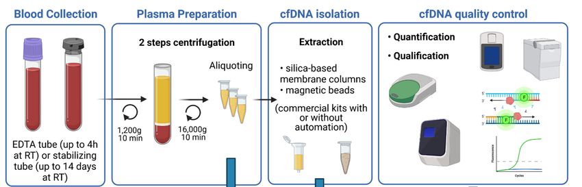
Figure 1. Schematic overview of the main steps for blood sample processing and cfDNA extraction. Blood, collected in
Figure 1. Schematic overview of the main steps for blood sample processing and cfDNA extraction. Blood, collected in
EDTA
EDTAororstabilizing
stabilizingtubes,
tubes,goes
goesthrough
throughtwo
tworounds
roundsofofcentrifugation
centrifugationtotoobtain
obtainplasma
plasmasamples.
samples.CfDNA
CfDNAisisisolated
isolatedfrom
from
plasma using commercial kits and is quantified and qualified for further analysis.
plasma using commercial kits and is quantified and qualified for further analysis.
Once sampling,
For optimal blood collection
extraction of cfDNA tubes
from have to besamples,
plasma centrifugedit isfor plasma separation.
recommended to use
This step can also affect cfDNA concentration and several studies set out to determine
blood collected into sample collection tubes that provide efficient stabilization of plasma.
the best centrifugation protocol. The two-step centrifugation protocols turned out to be
Several studies have compared different blood collection tubes (BCTs), especially conven-
the suitable ones to prevent unwanted release of genomic DNA. Blood cells first have to
tional anticoagulant EDTA tubes and as well as long-term storage BCTs from four differ-
be removed by slow centrifugation (1200–2000× g for 10 min at +4 ◦ C or RT) in order to
ent manufacturers (such as Streck (cfDNA BCT), Roche Diagnostics (Cell-Free DNA Col-
avoid cell lysis. Whereas afterwards, cellular debris and fragments will be removed by
lection Tube), Qiagen (PAXgene Blood ccfDNA Tube), and Norgen Biotek Corp. (cf-
short-term high-speed microcentrifugation of the plasma supernatant (12,000–16,000× g
DNA/cf-RNA Preservative tubes)). These last BCTs are pre-coated with preservatives to
for 10 min at +4 ◦ C or RT), either before or after a freeze–thaw cycle [28,29]. The most
prevent cell lysis and, therefore, reduce the release of RNA and DNA from hematopoietic
crucial step is to not disturb the buffy coat while collecting the plasma after the first spin.
cells. All these studies concluded that time between sampling and first centrifugation is a
Plasma samples should then be aliquoted to avoid repeated freeze–thaw cycles and kept at
major point when using EDTA collection tubes, and that this time should not exceed 4 h.
−80 ◦ C for long-term storage.
Whereas, the other BCTs, containing the stabilizing agent, can be stored at room temper-
Yield of cfDNA can also differ according to the extraction kit. Various commercial
ature several
purification days
kits havewithout affecting
been tested, in the further kits
particular analytical performances
from Qiagen (QIAamp (upcirculating
to 14 days
recommended but until 3 days for better results). However, despite
nucleic acid kit and QIAamp min Elute ccfDNA mini kit), Promega (Maxwell RSC ccfDNA the presence of stabi-
lizing agents,
plasma the ambient
kit), Applied temperature
Biosystems (Mag MAXmust cell-free
be respected
DNAtoisolation
avoid contamination
kit), and Norgen with
normal genomic DNA [24–27]. Therefore, EDTA tubes are suitable
Biotek (plasma/serum cell-free circulating DNA Purification midi kit). These kits workfor internal analysis or
monocentric studies, but if blood has to be shipped for external analysis,
either with columns or magnetic beads and some are or can be automated [30,31]. All specialized long-
term BCTs
studies are to
agreed more convenient.
conclude that Qiagen kits, with or without automation, give the best
Once sampling, blood collection
performances. Once extracted, cfDNA tubes
should have to be centrifuged
be stored at −80 ◦ C. for plasma separation.
ThisSeveral
step canstudies
also affect cfDNA concentration
emphasized the importance andto several
performstudies set outquality
consistent to determine the
controls
best on
(QC) centrifugation
the isolated protocol.
circulating The two-step
DNA. cfDNA centrifugation protocols
is released through turned and
apoptosis out necrosis
to be the
suitable
of normalonesandtomalignant
prevent unwanted
cells and isrelease
highly offragmented
genomic DNA. [32].Blood cells
Its size first have
ranged to be
between
removed by slow centrifugation (1200–2000× g for 10 min at +4
20 and 220 bp with a maximum peak at 167 bp, corresponding to the length of DNA °C or RT) in order to avoid
cell lysis.around
wrapped Whereas afterwards,
a single cellular[33].
nucleosome debris
Theandusefragments will be
of fluorimetric removed
methods is by
notshort-
suit-
termtohigh-speed
able accurately microcentrifugation
quantify cfDNA as itofwill thenot
plasma supernatant
discriminate cfDNA (12,000–16,000×
from contaminatingg for 10
min at +4DNA
genomic °C or (gDNA).
RT), either before or after
Contrariwise, a freeze–thaw
capillary cycle [28,29].
electrophoresis can The most crucial
measure the sizestep
of
is to not
DNA disturb the
fragments andbuffy
give coat while collecting
an estimation of the the plasma
absolute after the firstof
concentration spin. Plasma
cfDNA sam-
[34,35],
pleswill
but should then be aliquoted
not evaluate the presenceto avoid repeatedthat
of impurities freeze–thaw cycles
could inhibit and kept atenzymatic
downstream −80 °C for
long-term storage.
reactions. QPCR-based and ddPCR methods can evaluate amplificability of cfDNA, as
well as concentration and integrity, but are negatively impacted by gDNA contamination,
through distorting the ratio between short and long amplicons [36]. Recently, Alcaide andPharmaceuticals 2021, 14, 596 4 of 20
colleagues developed a promising multiplex ddPCR single-well assay, which can evaluate
the quantity, quality, and fragment size distribution of cfDNA samples using low inputs
and without the need of reference samples and calibration curves. This assay targets at the
same time several olfactory receptor genes, representing three fragment size ranges, and a
customizable control diploid locus. Unfortunately, the determination of cfDNA yields can
still be affected by gDNA contamination and by copy number alterations [37].
Despite recent promising progresses, the pre-analytical process of blood samples still
need standardization and further investigations to improve quality controls of the cfDNA
that will be used to detect circulating DNA of tumoral origin.
3. Detection of ctDNA by Sequencing Technologies
Sequencing technologies for detection and analysis of the ctDNA range from point
mutations analyses using PCR-based methods to analyses of whole genome using NGS
based methods. The choice of the method employed depends on the application and the
sensitivity intended (see Table 1 for comparison of some selected techniques).
Table 1. Comparison of some sequencing technologies for ctDNA detection.
Sensitivity
Analysis Type Technique Targets Applications Advantages Limitations
(LoD)
ARMS-PCR Cancer detection and High specificity and
monitoring, No multiplexing,
sensitivity, cost
qPCR 0.01–0.1% Hotspot mutation targetable alterations, limited to detection of
PNA-LNA effective, rapid, ease
some assays known mutations
Clamp PCR of use
approved for
COLD PCR clinical use
PCR based
methods ddPCR Cancer detection and Up to 5 targets, high Limited multiplexing
monitoring, sensitivity and (number of
Hotspot mutations, targetable alterations, specificity, absolute fluorescent colors),
digital PCR 0.01–0.1% gene fusions, CNV some assays quantification, single limited to detection of
approved for molecule analysis, known mutations
clinical use cost effective, rapid,
BEAMing ease of use
PCR SERS Cancer detection and
PCR based coupled to monitoring, Multiplexing capacity Limited to detection
0.1–1% Known mutations
methods spectrome- targetable alterations, of known mutations
UltraSEEK
try for research use
Amplicon methods
by multiplex PCR
Tam-Seq 2% High specificity (depend on fragment
size), no error
Known and
Cancer detection and correction
unknown
monitoring, Amplicon methods
eTam-Seq 0.02% mutations, indels, Error correction
classification, by multiplex PCR
targeted CNV, chromosomal
targetable alterations, Error correction by Amplicon methods
Safe-SeqS 0.01–0.05% rearrangements
for research use SSCS by multiplex PCR
(capture)
Duplex Error correction by Amplicon methods
0.0001–0.1%
sequencing DSCS by multiplex PCR
Error correction by
SSCS, Hybrid capture
Less comprehensive
TEC-Seq 0.05–0.1% method (not
than WGS or WES
dependent on
NGS based fragment size)
methods Amplicon methods
single
by SPE (not
primer Less comprehensive
0.5–1% dependent on
extension than WGS or WES
fragment size), error
(SPE)
correction by SSCS
SPE-duplex Error correction by Less comprehensive
0.1–0.2%
UMI DSCS than WGS or WES
Need large input,
Hybrid capture allelic bias (capture),
method (not stereotypical errors
CAPP-Seq 0.02%
dependent on (hybridization step),
fragment size) less comprehensive
than WGS or WES
Error correction by
iDES 0.00025– Less comprehensive
DSCS and correction
eCAPP-Seq 0.004% than WGS or WES
of stereotypical errors
Non-invasive
VDJ monitoring,
Ig-HTS 0.001% Very high sensitivity Tissue biopsy needed
rearrangements approved for
clinical usePharmaceuticals 2021, 14, 596 5 of 20
Table 1. Cont.
Sensitivity
Analysis Type Technique Targets Applications Advantages Limitations
(LoD)
Mutation discovery
Coding regions,
and signatures,
intron-exon
Cancer detection, detection of CNV, Low sensitivity
junctions,
monitoring of fusion genes, (increasing depth
promoters,
WES 5% resistant clones in rearrangements, lead to high cost),
untranslated
metastasis, for predicted need bioinformatic
regions,
Untargeted research use neoantigens and expertise
non-coding DNA
Tumor Mutational
of miRNA genes
Burden
Structural variants Expensive, variable
Cancer localization Shallow sequencing,
(fragmentation sensitivity (low) and
and origin, early genome wide
pattern, specificity, need
WGS 5–10% detection (early and profiling,
genome-wide CNV, bioinformatic
late stage), for identification of
methylation expertise, lots of data
research use cancer signatures
profile) generated
Abbreviations: PCR—polymerase chain reaction; ARMS—amplification refractory mutation system; qPCR—quantitative real-time
PCR; ddPCR—droplet digital PCR; BEAMing—beads, emulsion, amplification, magnetics; SERS—surface-enhanced Raman spec-
troscopy; PNA/LNA—peptide nucleic acid/locked nucleic acids; NGS—next-generation sequencing; Tam-Seq—Tagged-amplicon
deep sequencing; TEC—targeted error correction; CAPP-Seq—Cancer Personalized Profiling by Deep Sequencing; iDES—Integrated
Digital Error Suppression; Ig-HTS—Immunoglobulin high-throughput sequencing; WES—whole exome sequencing; WGS—whole
genome sequencing; LoD—Limit of Detection; CNV—Copy Number Variation; indels—insertions/deletions; SSCS—single-stranded
consensus sequence; DSCS—double-stranded consensus sequence.
Targeted approaches can detect, with high sensitivity, specificity and at a fast and
cost-effective rate, already known recurrent mutations. These hotspot mutations frequently
occur in a specific type of tumor and can be, most of the time, targeted by a therapy. Thus,
targeted approaches can be very useful for the follow up of minimal residual disease to
early detect relapse or track resistant mutations. Contrariwise, untargeted approaches are
less sensitive but are useful for the discovery of new DNA mutations and genome wide
alterations such as copy number variations (CNV, or copy number alterations, CNA).
Several parameters in the sequencing processing can affect the sensitivity of detection.
One of them, also depending in part on the pre analytical process, is to put enough genome
equivalent of cfDNA in the sequencing reaction to have enough altered molecule to detect.
For example, as around 3000 copies of haploid genome are present in 10 ng of DNA,
approximatively 60 ng of cfDNA will be required for a sensitivity of 0.01% (one rare event
in 10,000 molecules), which is often challenging, even more if we consider that more
than one observation is necessary to determine a true variant. Amplification steps cannot
replace low input of cfDNA because the polymerase will introduce errors, increasing the
risk to have false positive variants. Another parameter that may improve sensitivity is to
monitor multiple alterations simultaneously in order to increase the chances of detecting
ctDNA. With a binomial simulation, Van der Pol and Mouliere showed that, in theory and
at a given concentration of cfDNA, increasing the number of mutations analyzed could
improve the detection of low fraction ctDNA [21]. This kind of analysis was made possible
with the advent of next-generation sequencing technologies, by increasing the possibility
of multiplexing.
3.1. PCR-Based Methods
PCR-based methods, such as the derivatives of qPCR and digital PCR, are fast, cost-
effective, and relatively simple to carry out and analyze. They allow detection of single or
few mutations at low variants allele frequency, up to 0.1% and less, with high specificity.
3.1.1. Quantitative PCR
At first, the quantitative PCR (qPCR) method, by measuring the fluorescence emitted
by a labeled probe during amplification of a targeted gene, was used to estimate the
concentration of cfDNA in plasma of patients with cancer [38]. Later, qPCR assays were
developed to detect mutations in tumoral cfDNA and the sensitivity of detection was
improved by promoting the specific amplification of the mutant allele. Among the most
used techniques, we can find ARMS-PCR, PNA-LNA Clamp PCR, or COLD PCR.Pharmaceuticals 2021, 14, 596 6 of 20
ARMS-PCR (amplification-refractory mutation system) is a simple method for de-
tecting point mutations or small deletions, in which DNA is amplified by allele specific
primers. In this technique, the lack of 30 to 50 exonuclease proofreading activity of the
Taq polymerase reduces dramatically the annealing and hence the amplification in case of
mismatch at the 30 end of the primer. The limit of detection for this technique seems very
variable according to the studies published, depending on the method, the samples used to
determine this threshold or the mutations themselves. Although there are some improve-
ments of the method, the false positive rate is still high with a limit of detection around
0.5 to 1% in plasma samples [39,40]. This limit can go down to 0.015% with ARMS-plus
that includes a “Wild-type blocker” and in which amplicons were shortened to 50–80 bp,
prohibiting the non-specific amplification and thus increasing the detection specificity [41].
PNA-LNA (peptide nucleic acid-locked nucleic acid) Clamp PCR uses a blocking syn-
thetic nucleic acid analog complementary to wild type sequence to favor the amplification
of the mutant allele. This method is particularly used in non-small cell lung cancer (NSCLC)
to detect EGFR mutations, especially T790M mutations in tumor resistant to EGFR-TKIs
(tyrosine kinase inhibitors), where cfDNA could be an alternative to the re-biopsy. This
technique shows a high sensitivity with the detection of 0.1% mutant allele and a specificity
of 79%. Using smaller PCR products and by increasing the number of cycles, Watanabe
and colleagues reached less than 0.1% detection rate [42]. More recently, a dual PNA
clamping-mediated LNA-PNA PCR clamp (LNA-dPNA PCR clamp) assay with two PCR
rounds of PNA clamping succeeded in achieving a limit of detection of 0.01% [43].
COLD PCR (co-amplification at lower denaturation temperature-PCR) is an ampli-
fication method that selectively enriches low-abundance variant alleles from a mixture
of wild-type and variation-containing DNA, irrespective of mutation type and position,
by exploiting the critical denaturation temperature. The use of a lower denaturation
temperature results in selective denaturation of molecules containing wild-type mutant
heteroduplexes, which is followed by amplification. COLD-PCR has been used to improve
the reliability of a number of different assays that traditionally use conventional PCR, such
as Sanger sequencing, pyrosequencing or qPCR, greatly increasing their sensitivity. Thus,
this method can detect mutant allele fraction down to 0.1% [44,45].
3.1.2. Digital PCR
As an example in lymphoma, this technique has a potential clinical use in diffuse large
B cell lymphoma (DLBCL), as co-occurring mutations in MYD88 and CD79B can predict
response to Ibrutinib treatment, thus providing a predictive molecular tool for patient and
therapy selection [46]. As well, in primary central nervous system lymphoma (PCNSL),
mutation MYD88 L265P was identified by ddPCR in cerebrospinal fluid or vitreous fluid
with a superior sensitivity when compared with qPCR [47,48]. Since this mutation is found
in up to 85% of PCNSL cases and not in non-hematological brain tumors, this ddPCR assay
may be a promising technique for minimally invasive confirmation of PCNSL diagnosis.
BEAMing (beads, emulsion, amplification, magnetics) is a highly sensitive digital
PCR method that combines emulsion PCR and flow cytometry to identify and quantify
specific somatic mutations present in DNA [49]. Diehl and coworkers used a BEAMing
approach to detect mutations in cfDNA from patients with colorectal cancer, showing that
ctDNA dynamics reflects tumor responses and progression, and that ctDNA detection
after surgery represented a marker of residual disease [50]. This method, mainly used so
far in solid tumors, such as colorectal [51], breast [52], and lung cancers [53], has a highly
sensitive detection rate with variant allele fraction as low as 0.01%.
Although ddPCR allows for quantitative assessment of mutant frequencies in cfDNA,
it is limited by the number of fluorescent probes that can be used in one assay (up to
five) [54,55].
Copy number variations have also been investigating in cfDNA using ddPCR. Even if
the number of targets is limited, it can be a useful tool for detecting, simply and rapidly,Pharmaceuticals 2021, 14, 596 7 of 20
some gains or losses, which are associated with poor prognosis at diagnosis or during
follow-up [56,57].
DdPCR can also be suitable to detect chromosomal rearrangements, especially in
hematological malignancies. Among others, assays have been developed for transloca-
tion t(11;14) deregulating the CCND1 gene and translocation t(14;18) deregulating the
BCL2 gene, which are frequently observed in Mantle cell lymphoma (MCL) and follicular
lymphoma (FL), respectively [58,59]. The sensitivity of these techniques can go down
to 0.01%.
3.1.3. PCR Coupled with Mass Spectrometry
The major limitation of the previous PCR-based approaches is their very limited
multiplexing ability. Mass spectrometry-based methods such as surface-enhanced Raman
spectroscopy (SERS) and UltraSEEK are adaptation of the conventional PCR method with
a unique advantage in multiplexing to detect ctDNA mutations at low frequency with low
input amount of cfDNA and fast turnaround time.
SERS is a surface-sensitive technique that enhances Raman scattering by molecules
adsorbed on rough metal surfaces or by nanostructures such as plasmonic-magnetic silica
nanotubes [60]. The detection of target specific DNA is based on the use of labeled
nanotags (Raman reporters) and the measurement of the Shift in the spectrum of Raman
reporter that can provide information about low-frequency transitions in molecules. The
status of mutations is then analyzed with SERS spectrum where unique spectral peaks
demonstrated the presence of targeted mutations. Multiplex PCR/SERS identifying three
hotspot mutations has been developed in melanoma and colorectal cancer with a limit of
detection as few as 0.1% [61,62].
The UltraSEEK chemistry is able to interrogate multiple informative variants within
a single reaction. In this method, the mutant allele is specifically targeted by a primer
extension step that omits the wild type allele. Reaction products are subsequently captured
to a solid support, washed and released. Eluted products are then submitted to MALDI-
TOF Mass Spectrometry. The use of a 68 mutations panel on cfDNA from melanoma
patients showed the same sensitivity as ddPCR [63]. In NSCLC, the limit of detection of
the UltraSEEK Lung Panel, consists of 73 variants, was 0.125–1% with low input of specific
tumoral cfDNA fragments beforehand measured with the LiquidIQ Panel [64]. Of note, this
study showed the importance of preanalytical cfDNA quality control and input amount for
the accuracy of liquid biopsy testing. The comparison between UltraSEEK and a real-time
PCR test (cobas EGFR Mutation test v2) showed a concordance of 100% with more than
10 ng of cfDNA, whereas it fell to 73–84% when less than 8 ng were used, implying a loss
of sensitivity.
Overall, these PCR-based assays are very effective tools for detecting mutations at
a relatively low-cost, which make them feasible in routine clinical practices. The main
limitation is the limited multiplexing ability, which restricts the possibility of targets and
can lead to a greater consumption of material. Furthermore, the alterations detected must
be previously known such as hotspot mutations, which is more suitable for a minimal
residual disease but less as a diagnostic tool.
3.2. Targeted NGS-Based Methods
Targeted deep sequencing techniques are still limited to a certain number of regions
but can cover entire genes or entire coding regions of genes. Thus, they are suitable for
genes without hotspot mutations, which is often the case for loss of function mutations in
tumor suppressor genes.
Targeted enrichment in library construction can be achieved by direct amplification
(amplicon or multiplex PCR) or hybridization capture (hybrid capture) of the DNA regions
of interest. Techniques using multiplex PCR-based methods are more dependent on
the length of the fragments and may require several simultaneous reactions for target
enrichment to cover a large region of a gene, consuming more DNA. Hybrid capturePharmaceuticals 2021, 14, 596 8 of 20
methods employ custom RNA probes complementary to targeted regions and are able to
detect both single nucleotide variants (SNV) and structural variants [65]. In this method
of enrichment, the fragmentation of cfDNA can lead to a heterogeneous coverage across
targeted exons with a lower fragment depth in the edge regions of exons, which must be
taken into consideration when designing the panels for ctDNA sequencing [66].
The main issue of going down in sensitivity is the reliability of interpretation in the
discrimination between the true and the false variants. Although they have high sensitivity
and specificity, NGS platforms show a random error rate between 0.1 and 1.5% per base
call, but library preparation protocols have been upgraded to improve the detection of rare
variants [67,68]. In targeted DNA sequencing, the use of few DNA molecules combined
with ultra-deep sequencing increases the risk to read several times the same molecule
where polymerase errors are introduced at any step during the NGS process, leading to the
inability to confidently call rare variants. One of the major recent technological advances
is the use of molecular barcodes, which are random sequences introduced before any
amplification step. They allow the counting of original DNA molecules instead of PCR
duplicates, thereby enabling digital sequencing and resulting in unbiased and accurate
mutation profiles with an increased sensitivity [69–72].
• Tagged-amplicon deep sequencing (Tam-Seq)
Tam-Seq is an amplicon method using a target enrichment array with barcoded
primers to prepare the amplicon library for NGS. First, an initial targeted preamplification
step is carried out, followed by a selective amplification of the regions of interest in single-
plex reactions. Then, sequencing adaptors and sample-specific barcodes are attached to the
amplicons in a further PCR. It was first able to detect mutations in circulating DNA with
high sensitivity and specificity (>97%) at allele frequencies as low as 2% [73]. The technique
has been recently improved (enhanced Tam-Seq, eTam-Seq) with a primer design strategy,
allowing for amplification of highly fragmented DNA, a workflow reducing the background
error rate, and a more efficient calling algorithm with better detection of SNV and indels
(insertions/deletions), and also CNV [74]. This assay, using an optimal amount of DNA,
detected 94% mutations at 0.25–0.33% allele fraction (AF) with a limit of detection down to
0.02% AF with high per-base specificity (99.9997%). In this study comparison of eTam-Seq
with dPCR showed a good concordance between the two techniques, demonstrating the
quantitative accuracy of eTAm-Seq technology for reliable detection of mutations at low
allele frequency [74].
• Safe-Sequencing System (Safe-SeqS)
This amplicon method was originally described by the group of Bert Vogelstein [69]. It
was the first approach using molecular barcodes in DNA sequencing, to increase sensitivity
of massively parallel sequencing. In this technique, a unique identifier (UID) is assigned
to each template molecule before any amplification. Thereby, PCR fragments with the
same UID are considered mutant if more than 95% of them contain the identical mutation.
Thus, this method allows a correction of amplification and sequencing errors and can
quantify rare mutations with a sensitivity of 0.05% of allele fraction. Safe-SeqS showed
high performance in detecting mutations in cfDNA from patients with solid tumors, for
molecular profiling as well as real-time monitoring of minimal residual disease [75]. A
recent study on three independent cohort of nonmetastatic colorectal cancer, showed a
median mutant allele frequency of 0.046% with a minimum of 0.01% [76].
• Duplex sequencing
Duplex sequencing is an improvement of the Safe-SeqS technique [77,78]. In this
method, a semi-degenerated double stranded unique barcoded adapter is ligated to a
target double stranded DNA. After sequencing, molecules with the duplex adaptors are
compared and mutations are retained only if there is a consensus between both strands.
Thus, in addition to get rid of PCR and sequencing errors, the advantage of this technique
is to identify artifacts due to sample alterations [79] because it can examine both strands
individually and the damage to them is usually not identical (error correction by double-Pharmaceuticals 2021, 14, 596 9 of 20
stranded consensus sequence). The theoretically sensitivity of this approach to discovering
mutants is one molecule among 10ˆ7 which is much higher in accuracy than conventional
next-generation sequencing methods [77,78].
Several studies, in various types of cancers, applied this method on plasma cfDNA. In
combination with target enrichment using hybrid capture, this approach allowed detection
of tumoral fraction at 0.1% and below with high sensitivity and specificity, providing a
powerful tool for diagnosis as well as longitudinal monitoring of disease [80–82].
• Targeted error correction sequencing (TEC-Seq)
In this technique, molecular barcoding is also used to facilitate the discrimination
between true mutations and false positive variants. DNA fragments are tagged each one
with a different “exogenous” DNA barcode before any amplification, as for Safe-SeqS, but
not only. The start and end genome mapping positions of paired-end sequenced fragments
were also used as “endogenous barcodes” to distinguish between individual molecules.
This combination of barcodes allows keeping track of each fragment as they are sequenced
around 30,000 times [70]. This approach was applied to several type of solid cancers and
demonstrated ability for early stage detection. The analytical sensitivity was 100% and 89%
for detecting mutations present at 0.2% and 0.1%, respectively, using minimum thresholds
of 0.05% in hot-spot positions and 0.1% at all other locations, resulting in a sensitivity of
97.4% overall, and without detection of false positives (less than one error in three million
bases sequenced).
• Single primer extension (SPE) with unique molecular barcode
SPE is an amplicon-based method used by QIAGEN in their QIASeq targeted DNA
panel kits. This approach uses only one gene specific primer (GSP) for amplification of each
genomic region, which makes it less dependent on the size of DNA fragments than PCR
using two primers and offers a uniform coverage. As for capture, the first step is a fragmen-
tation step in which the buffer used inhibits fragmentation of the high length fragments
of DNA such as contaminating gDNA. The following steps are reparation and ligation of
adapters. These adapters will be used for amplification of targeted region (together with
GSP) and contain the degenerated molecular barcodes (UMI, Unique Molecular Index).
Moreover, given this UMI contains 12 base pairs, it allows a large number of combinations
and a very little risk for redundancy [71]. Theorical sensitivity threshold of this technique
is 0.5–1% with over 90% sensitivity and a very few number of false positive. Recently,
improvement by using duplex UMI adapters lowered the sensitivity up to 0.1–0.2% allele
fractions [83].
This technique of deep sequencing, using molecular barcodes to improve accuracy in
variant detection, has been used at diagnosis in order to identify actionable genetic alter-
ation with targeted therapies available for treatment or hotspot mutations to be tracked with
ddPCR during follow up, with a detection of variant allele frequency down to 1–5% [84,85].
Further investigations are needed to find the real limit of detection of this technology,
which may be below 1% as other techniques using molecular barcoding.
This approach also allowed detection of CNV. In PCR-based library construction,
amplification introduces biases in further reads count because the amplification factor
is dependent on many parameters such as library size, GC content, region length or
competition between primers overlapping the same locus. Thus, the use of UMI via the
mCNA tool allows the direct count of targeted DNA molecules before any amplification
and the detection of CNV in a robust and sensitive way [86].
• Cancer Personalized Profiling by Deep Sequencing (CAPP-Seq)
CAPP-Seq is an ultra-sensitive assay consisting of a hybrid capture-based NGS method
developed for ctDNA detection. In this technique, the first important step is to query cancer
databases to identify known recurrent mutations for a particular cancer type. Then, bi-
otinylated oligonucleotide probes, named “Selector”, are designed to target large segments
of the concerned regions. The protocol is optimized for low DNA levels and sensitivity is
increased using deep sequencing [87,88]. The sensitivity is also improved by its ability toPharmaceuticals 2021, 14, 596 10 of 20
detect simultaneously various types of alterations: single nucleotide variants, rearrange-
ments, insertions/deletions, and copy number alterations. It was originally described to
detect and monitor lung cancer but was successfully adapted to a broad range of cancers,
including different types of solid tumors as well as hematological malignancies such as
DLBCL, LF, and HL [10,20,89–91].
With this method, ctDNA was detected in blood of NSCLC patients with 96% speci-
ficity for mutant allele fraction down to 0.02%. It was improved in 2016, with the use of
iDES (Integrated Digital Error Suppression). This iDES-enhanced CAPP-Seq combines
CAPP-Seq with duplex barcoding sequencing technology and with a computational algo-
rithm that removes stereotypical errors associated with the CAPP-Seq hybridization step.
This improved version of CAPP-Seq has shown a high sensitivity in the detection of EGFR
mutations in cfDNA of NSCLC patients, with variant allele frequency as low as 0.004%
with >99.99% specificity. Moreover, using duplex sequencing and covering a large number
of mutations (≥200), the authors outperformed iDES and managed to detect ctDNA down
to 0.00025%, with an input of only 32 ng of cfDNA [92].
• Immunoglobulin high-throughput sequencing (Ig-HTS)
This test was specifically developed for MRD in hematological malignancies. In this
method, ultra-deep sequencing of genomic DNA, with a set of locus-specific multiplex PCR
covering all possible rearranged IgH, IgK, and IgL receptor gene sequences, firstly identifies
the tumor-specific clonotype. Then, this clonotype can be tracked as a specific fingerprint
to quantify ctDNA in lymphoma disease monitoring with a sensitivity of approximatively
10-6 [93–95]. This technique presents some technical limitations, including the need of
tissue biopsy to identify clonotype and difficulties to identify clonotype sequences in
some lymphoma types such as DLBCL of the germinal center type and FL because of
somatic hypermutation (SHM). Nevertheless, this method has shown high performance
in surveillance ctDNA, after complete remission, to identify risk of recurrence before any
clinical evidence of disease in most patients (with a median of 3.5 months) [93,94].
This approach was also used for MRD monitoring in DLBCL patients after CAR-T
cell therapy, showing correlation with clinical and radiologic outcomes for all the patients
tested [96].
3.3. Untargeted NGS-Based Methods
As mentioned previously, untargeted approaches, namely whole exome and whole
genome sequencing (WES, WGS), are less sensitive than targeted approaches. The sensitiv-
ity of these techniques on cfDNA is estimated around 5–10%, as compared to less than 0.1%
for a targeted sequencing approach [97], making it difficult to detect rare events, especially
in situations of early detection or minimal residual disease. Moreover, these technologies
are more expensive and require both very high throughput sequencing equipment and
expertise to analyze the large amount of data generated, which makes its implementation
in routine practices challenging. However, these approaches may be necessary for the
discovery of new alterations in the context of initial profiling at diagnosis, to provide infor-
mation for the use of more sensitive targeted techniques during disease monitoring. Even
if they are not suitable to detect subclonal events, they may be useful, considering intra
tumoral heterogeneity, to highlight new drug targets or to track drug resistance clones [98].
WES is, most of the time, limited to coding regions and splicing sites of genes but
it is a good compromise for exploration of unknown mutations at a reasonable cost. It
can identify both driver and passenger mutations and also can be extended to promoters,
untranslated regions, and non-coding DNA of miRNA genes. Even if protein-coding genes
constitute only approximately 1.5% of the human genome, they contain a great majority of
the disease-causing mutations [99]. The technical feasibility of whole-exome sequencing
(WES) on cfDNA has been demonstrated in various solid tumors and some hematological
malignancies [98]. Low coverage and sensitivity, compared to targeted NGS technologies
does not allow for the detection of rare variants but WES of cfDNA is suitable for mutational
analysis of patients with advanced tumors and increased ctDNA fractions (>5% mutantPharmaceuticals 2021, 14, 596 11 of 20
allele fraction). The first exome-wide sequencing analysis of ctDNA was performed to
analyze serial plasma samples (before initiating treatment and at disease recurrence), in
order to track genomic evolution and response to therapy in patients with metastatic
cancer (breast, ovarian, and lung cancer) receiving systemic therapy [100]. These samples
contained high percentages of ctDNA (between 5% and 55%) and the average depth of
sequencing coverage ranged from 31- to 160-fold. This study showed the possibility to
identify candidate genetic alterations driving treatment resistance using cfDNA analysis.
These findings largely agreed with additional studies demonstrating that whole-exome
sequencing of cfDNA in metastatic patients could serve as a surrogate for tumor genome
analysis, considering the difficulties of doing multiple biopsies and the high ctDNA allele
frequencies making WES possible [101–104].
Additionally, given intra tumoral heterogeneity, analysis comparing mutational profile
between tumor and cfDNA mostly identified more mutations in cfDNA with a high
prevalence of targetable genes. Beyond SNV detection, WES of cfDNA also allowed
analysis of mutational signatures, copy number variations, fusion genes, rearrangements,
predicted neoantigens, and tumor mutational burden [98].
Contrariwise to WES, WGS technologies is more suitable to detect ctDNA by identify-
ing structural and non-coding variations such as genome-wide copy number aberrations,
methylation profiles, and fragmentation patterns.
To override the cost and analysis time limitations caused by WGS, Heitzer and col-
leagues developed a shallow genome-wide sequencing approach called Plasma-Seq [105].
This method uses an Illumina MiSeq instrument, which is a benchtop high-throughput
sequencing platform often available in routine laboratories. This technique does not have
a sufficient sequencing resolution to identify SNV but is able to detect CNV in cfDNA
at a depth of 0.1×, with a specificity >80% when ctDNA fraction is ≥10%. Recently,
this approach of shallow WGS has been successfully used in cfDNA of DLBCL and HL
patients to identify copy number patterns that can differentiate the two diseases at diag-
nosis [106]. These copy number aberrations were also correlated with clinical parameters,
and longitudinal analyses showed correlation with disease status. Moreover, the sen-
sitivity and informativity for HL was better in cfDNA than in tumor, as for mutation
detection [10,11,106].
Aneuploidy has also been explored with WGS derived techniques such as Fast-SeqS
(Fast Aneuploidy Screening Test-Sequencing System) and WALDO (Within Sample Aneu-
ploidy Detection), using a single specific primer pair to amplify dispersed retrotransposon
regions throughout the genome (long interspersed nuclear elements (LINEs)) [107,108]. By
simulations with synthetic DNA, the bioinformatic tool WALDO showed high performance
to detect individual chromosome arm gain or loss with a fraction of ctDNA >5%, and up
to 1% of tumoral fraction with a sensitivity of 78%. However, due to their mechanism of
detection, these techniques are limited to cancers presenting aneuploidy.
In order to detect genomic rearrangements, Leary et al. developed a technique called
PARE (personalized analysis of rearranged ends), which uses WGS mate-paired analysis of
the tumoral DNA to identify patient specific genomic rearrangement. This assay is highly
sensitive with detection of ctDNA lower than 0.001% of total cfDNA [109]. Analyses, in
breast and colorectal cancers, suggest that ctDNA concentrations at levels >0.75% could
be detected in the cfDNA of patients with a sensitivity >90% and a specificity >99%,
and that even a single copy of rearrangement from ctDNA can be detected without false
positives [110]. In a recent study, PARE was employed to detect rearrangements in gastric
tumor, which were used to design a quantitative PCR assay targeting rearranged loci
for quantitative monitoring in cfDNA. Thus, the authors were able to predict relapse as
the presence of postoperative ctDNA was significantly correlated with cancer recurrence
within 12 months of surgery [111].
WGS, combined with artificial intelligence, can also identify genome-wide fragmenta-
tion patterns in cfDNA. Several studies in different cancer types have shown that these
patterns can be used to detect ctDNA in body fluids and with very low plasma ctDNAPharmaceuticals 2021, 14, 596 12 of 20
fraction [112,113]. Indeed, ctDNA fragments are generally shorter and more variable in
their length than those found in controls are. Moreover, beyond this difference of size of
fragments in cfDNA between healthy individuals and patients with cancer, their location
in the genome can be informative of the epigenetic profile of the origin cells. Indeed, the
cfDNA fragmentation landscape represents a nucleosome footprint reflecting the cell and
tissue of origin, potentially enabling non-invasive diagnosis of cancer type [112]. Recently,
Cristiano et al. used this approach for the early detection of ctDNA from 236 patients
with various cancers and reported sensitivities ranging from 57 to 99% with a specificity of
98% [114]. This nucleosome footprinting firstly identified by WGS represents nucleosome
depletion at transcription start sites of highly expressed genes and the capture of this
chromatin accessibility profile was used by CAPP-Seq technology to define gene expression
differences and thus determine the cell-of-origin in DLBCL subjects from cfDNA [115].
Among epigenetic alterations, aberrant DNA methylation events can also represent
an ideal biomarker for detection and classification of early stage cancer, as they occur
early in cancer development, sometimes before the acquisition of SNVs. Multiple liquid
biopsy studies have been performed utilizing DNA methylation markers in various cancer
types [21]. As whole genome bisulfite sequencing is inefficient due to low recovery and
degradation of DNA after bisulfite conversion [116], high cost and limited information
recovery given the low genome-wide abundance of CpGs, techniques has been developed
to pre-enrich methylated DNA fragments with or without bisulfite treatment. These strate-
gies are either very targeted, as methylation events of interest occur at known, stereotyped
positions [117], or larger to identify methylation patterns, which have been shown to enable
accurate determination of cell-of-origin from cfDNA and non-invasive cancer classification.
For example, a technique for cell-free methylated DNA immunoprecipitation followed by
high throughput sequencing (cfMe-DIP) has been developed for genome-wide methylation
exploration of bisulfite-free plasma DNA, on low input cfDNA and with enough sensitivity
for early detection of cancer [118]. More recently, a semi targeted assay of 595 genomic
regions covering 11,787 CpG sites, named PanSeer assay, allowed the detection of five
types of cancer in 88% of post-diagnosis patients with a specificity of 96% [119]. Even if
the result needs confirmation, the authors also detected cancer in 95% of asymptomatic
individuals who were later diagnosed, demonstrating that cancer can be non-invasively
detected up to four years before diagnosis. In lymphoma, aberrant promoter methylation
patterns detected in cfDNA have been shown to be an independent and significant poor
prognostic factor for 5-year overall survival in DLBCL, outperforming existing clinical risk
parameters an independent [120,121].
Moreover, as healthy cells also participate to epigenetic changes, it may need to be
distinguished from these of cancers cells [21]. Thus, it could be of major interest to combine
epigenetic analysis of the entire cfDNA pool with mutational analysis of ctDNA molecules.
4. Bioinformatical Methods
While cfDNA seems to be a promising screening tool, it still remains a real challenge
for bioinformatics. While common bioinformatics strategies allow variant identification
down to 2–5% allele frequency, in most cases, ctDNA accounts for a small fraction of total
cfDNA since most of cfDNA is derived from non-cancer cells and especially blood cells.
ctDNA fraction can be lower than 0.1%, leading to the detection of somatic mutations at the
same level as the sequencing noise. It implies the use of in silico strategies to distinguish
true positive variant calls from sequencing noise.
It has been reported from healthy controls that under an allele fraction of 0.02%, more
than 50% of sequenced genomic positions had sequencing artifacts [92]. These errors
are particularly due to library preparation, the error rates of NGS technologies, and the
physical characteristic of the cfDNA fragments.
In addition, there are many tools and therefore many bioinformatics parameters that
need to be optimized when analyzing cfDNA samples. While major progress has been made
in the harmonization of tumor analyzes with the GATK4 Best Practices Workflows [122],Pharmaceuticals 2021, 14, 596 13 of 20
there is not yet an international consensus for bioinformatic cfDNA analysis and research
in this area remains very active.
4.1. Adapter Contamination
The quality of cfDNA analysis is particularly impacted by adapter contamination.
cfDNA fragments could be shorter than usual which may result in the sequencing of
adapters due to too many sequencing cycles compared to their lengths. Consequently,
these reads could be either unmappable to the reference genome or could have a lower
alignment score. These alignment scores are considered by a large number of bioinformatic
tools and could finally affect the results of variant caller algorithms. Adapter contamination
could be found both in 50 and in 30 of sequenced reads.
Many softwares were developed to find and trim adapters, like Cutadapt [123], Tag-
Cleaner [124], Trim Galore [125], or Trimmomatic [126]. In general, these algorithms also
integrate the trimming of low quality nucleotides and the extraction of molecular barcodes.
4.2. Library Biases and Molecular Barcoding
The amplification of the libraries by PCR includes many biases for counting mutated
reads because the number of aligned reads is no longer directly proportional to the number
of initial unique targeted DNA fragments. The amplification factor of each region is
unknown and depends on many parameters such as library size, GC content, or fragment
length. This bias is particularly present for samples with low DNA concentration at
extraction. Recent advances in library preparation allow the addition of Unique Molecular
Identifiers (UMI) to each read. UMI are especially useful to correct library amplification
biases by making each DNA molecule in a population of reads distinct.
There are two main bioinformatic approaches to use UMI for cfDNA analysis.
The first one consists in grouping PCR duplicates prior to any downstream analysis
by merging sequences harboring the same UMI tag. To perform this task, the most popular
tool is UMI-tools [127]. The advantage of this approach is that it allows the use of classic
bioinformatic pipelines after deduplicating the reads. It erases amplification biases due to
cfDNA characteristics. However, it no longer provides access to essential information such
as the amplification factor of each UMI or the discordant mutation calls of reads having the
same UMI.
More recent approaches consist in using new bioinformatic algorithms for variant and
CNV calling which are able to take into account the information carried by the UMIs after
alignment, i.e., at the end of data processing.
For example, the UMI-VarCal algorithm [128] tries to quantify the number of concor-
dant and discordant UMIs for each candidate variant during the variant calling process.
Concordant UMIs were defined as number of unique UMIs for which all the reads carrying
these UMIs validate the presence of the variant. Conversely, discordant UMIs quantify
the number of abnormal substitutions like sequencing or PCR errors. Another example
of barcode-aware variant caller is SmCounter [71]. SmCounter uses a barcode level allele
probability and UMI counts to reject candidate mutations lacking enough barcodes with
good read evidence. These approaches make it possible to distinguish true mutations at
low frequency from sequencing noise and is particularly useful for cfDNA analysis.
Many biases due to the amplification step while preparing sequencing libraries prevent
the direct quantification of loci copy-number [129]. cfDNA fragments are often shorter than
DNA extracted from tissue and make it impossible to use conventional approaches for the
detection of CNV such as read-depth algorithms. Recent approaches, like mCNA [86], use
the UMI counts instead of read counts to improve high-resolution copy number variation
of genes.
4.3. Bioinformatics Processing
There is not yet an international consensus for bioinformatic cfDNA analysis pipeline.
The bioinformatics tools and parameters must be adapted to the nature of the sequencedPharmaceuticals 2021, 14, 596 14 of 20
samples (quantity of DNA, quality of extraction, integrity of the cfDNA, etc.), to the kits
used to prepare libraries, to the presence of UMI in library construction or not, and finally
to the sequencing depth. In addition, sequencing biases are often sample specific which
requires an objective assessment of sequencing noise at sample level.
However, this variability, specific to each sample, is not incompatible with an objective
evaluation of the performance of bioinformatic algorithms. Some first tools, like UMI-
Gen [130], allow to create in silico alignment datasets to evaluate the performance of variant
calling and filtration tools. UMI-Gen is a UMI-based read simulator, which reproduces
targeted sequencing paired-end alignment files (BAM) by estimating sequencing noise
from a set of reference BAM files. It is particularly useful for evaluating the performance of
variant calling tools because it allows to vary many parameters (sequencing depth, number
of initial UMI, etc.) and to insert variants at frequencies of interest during the simulation. It
thus makes it possible to optimize bioinformatic pipelines according to the targeted panels
or the sequencing technology.
5. Conclusions
Many studies have demonstrated that analysis of ctDNA, as a liquid biopsy, is a
powerful tool for non-invasive genotyping across various cancer types, in solid tumors,
as well as in hematological malignancies. Investigations have shown the possibility to
use ctDNA both at diagnosis, for prognosis or targeted therapies, and during longitudinal
monitoring of the disease, as a dynamic biomarker of tumor burden during treatment and
to detect relapse after treatment. Moreover, liquid biopsy could be a surrogate for tissue
biopsy in some particular cases of tumors not accessible for surgery or spread of tumoral
mass and metastasis, given the intra tumoral heterogeneity.
Nowadays, the main issue in ctDNA analysis is to go down in sensitivity without
generating false positive results, especially for early detection of cancer at diagnosis and
relapse. Due to its short fragment length, low quantity, and small fraction in cfDNA [131],
reliable detection of ctDNA can still be a major technical challenge. That is why the
most suitable ctDNA assay for a specific application has to be chosen according to its
analytical performance characteristics [66]. However, recent optimizations in techniques,
from standardization of preanalytical processing to the development of high sensitive
sequencing technologies with the help of bioinformatical algorithms for error correction,
has the potential to detect ctDNA at the molecular level with a great accuracy.
The next step in the near future could be the integration of ctDNA detection assays
into prospective multicentric studies and clinical trials to establish its true clinical utility.
Author Contributions: Writing—original draft preparation, E.B. and P.-J.V.; writing—review and
editing, E.B., P.-J.V. and F.J. All authors have read and agreed to the published version of the manuscript.
Funding: This research received no external funding.
Institutional Review Board Statement: Not applicable.
Informed Consent Statement: Not applicable.
Data Availability Statement: Not applicable.
Acknowledgments: Figure 1 was created by E.B. with BioRender.com (21 June 2021, Agreement
number: LU22M8WBV5).
Conflicts of Interest: The authors declare no conflict of interest.
References
1. Mandel, P.; Metais, P. Nuclear acids in human blood plasma. C. R. Seances Soc. Biol. Fil. 1948, 142, 241–243.
2. Koffler, D.; Agnello, V.; Winchester, R.; Kunkel, H.G. The Occurrence of Single-Stranded DNA in the Serum of Patients with
Systemic Lupus Erythematosus and Other Diseases. J. Clin. Investig. 1973, 52, 198–204. [CrossRef]
3. Leon, S.A.; Shapiro, B.; Sklaroff, D.M.; Yaros, M.J. Free DNA in the serum of cancer patients and the effect of therapy. Cancer Res.
1977, 37, 646–650. [PubMed]You can also read