Obesity and Thyroid Axis - Review - MDPI
←
→
Page content transcription
If your browser does not render page correctly, please read the page content below
International Journal of
               Environmental Research
               and Public Health

Review
Obesity and Thyroid Axis
Krzysztof Walczak 1 and Lucyna Sieminska 2, *

                                          1   Department of Thoracic Surgery, Faculty of Medical Sciences in Zabrze, Medical University of Silesia in
                                              Katowice, 41-800 Zabrze, Poland; krzysiekmed@gmail.com
                                          2   Department of Pathophysiology and Endocrinology, Faculty of Medical Sciences in Zabrze,
                                              Medical University of Silesia in Katowice, 41-800 Zabrze, Poland
                                          *   Correspondence: LSieminska@sum.edu.pl

                                          Abstract: Development of obesity is primarily the result of imbalance between energy intake and
                                          energy expenditure. Thyroid hormones influence energy expenditure by regulating cellular respi-
                                          ration and thermogenesis and by determining resting metabolic rate. Triiodothyronine influences
                                          lipid turnover in adipocytes and impacts appetite regulation through the central nervous system,
                                          mainly the hypothalamus. Thyroid-stimulating hormone may also influence thermogenesis, suppress
                                          appetite and regulate lipid storage through lipolysis and lipogenesis control. Subclinical hypothy-
                                          roidism may induce changes in basal metabolic rate with subsequent increase in BMI, but obesity can
                                          also affect thyroid function via several mechanisms such as lipotoxicity and changes in adipokines
                                          and inflammatory cytokine secretion. The present study investigated the complex and mutual
                                          relationships between the thyroid axis and adiposity.

                                          Keywords: adipose tissue; obesity; thyroid; TSH; adipokines


Citation: Walczak, K.; Sieminska, L.
                                          1. Introduction
Obesity and Thyroid Axis. Int. J.
Environ. Res. Public Health 2021, 18,          Thyroid hormonal tests are routinely ordered when seeking obesity origins [1]. Often
9434. https://doi.org/10.3390/            higher levels of serum thyrotropin (thyroid-stimulating hormone, TSH) within the reference
ijerph18189434                            range or slightly elevated levels are found in the obese state [2–4]. In the past decades, a
                                          number of reports tried to explain whether thyroid dysfunction is the cause or rather the
Academic Editors: Zbigniew                consequence of excess adipose tissue; the answer, however, remains unclear.
Adamczewski and Magdalena Stasiak              The relations between the hypothalamic–pituitary–thyroid axis (HPT) and obesity are
                                          complex and include different interactions. Thyroid hormones (THs) and TSH indepen-
Received: 31 May 2021                     dently regulate the mass and function of adipose tissue; however, adipose tissue, through
Accepted: 3 September 2021                the production of adipokines (adipocytokines), also affects the activity of the HPT system.
Published: 7 September 2021               The objectives of this review were (1) the presentation of fundamental mechanisms linking
                                          the HPT axis with energy balance, (2) characterization of factors modifying primary rela-
Publisher’s Note: MDPI stays neutral      tionships and (3) presentation of complicated relationships in a manner helping a physician
with regard to jurisdictional claims in   evaluating the thyroid axis of obese patients to conclude if optimizing the thyroid axis
published maps and institutional affil-   is desirable.
iations.
                                               THs regulate the function of many tissues and organs—heart, liver, brain, skeletal
                                          muscles, pancreas and adipose tissue. They control the energy balance, appetite, basal
                                          metabolic rate (BMR), thermogenesis, free fatty acid (FFA) oxidation, glucose and lipid
                                          metabolism [5–7]. The actions of THs in tissues are determined by transmembrane trans-
Copyright: © 2021 by the authors.         port, intracellular deiodinase (DIO) activity and expression of TH receptors (TRs). Free THs
Licensee MDPI, Basel, Switzerland.        can bind to membrane-bound receptors or can be transported from the extracellular space
This article is an open access article    into the cytosol through specific transporters: monocarboxylate transporter 8 (MCT8) and
distributed under the terms and           organic anion-transportingpolypeptide 1c1 (OATP1c1). Adrenergic receptors and signaling
conditions of the Creative Commons
                                          also play an important role. The direct evidence for the influence of THs on adipose tissue is
Attribution (CC BY) license (https://
                                          the expression of TRs in human adipocytes. Triiodothyronine (T3) acts through TRs: TRα-1,
creativecommons.org/licenses/by/
                                          TRα-2, TRβ-1 and TRβ-2. The receptors are encoded by two genes: TRα and TRβ [8,9].
4.0/).

Int. J. Environ. Res. Public Health 2021, 18, 9434. https://doi.org/10.3390/ijerph18189434                      https://www.mdpi.com/journal/ijerph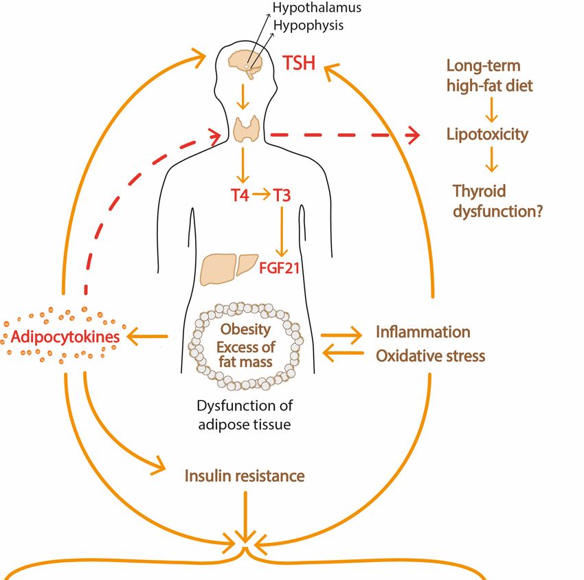
Int. J. Environ. Res. Public Health 2021, 18, 9434                                                                             2 of 23

                                        T3 is considered to be the active form of the hormone, and T4 is mainly its precursor. As
                                        a specific endogenous ligand, T3 modulates the activity of nuclear and mitochondrial
                                        TRs. It was demonstrated that THs exert both direct action on transcription (canonical
                                        regulation of gene transcription, called genomic) and non-genomic effects, mediated by cell
                                        membrane and mitochondrial binding sites (non-genomic activity). Recently, it was shown
                                        that 3,5-diiodo-L-thyronine (3,5-T2) derived from T3 by deiodination has a positive effect
                                        on energy metabolism and lipid metabolism. 3,5-T2 increases the rate of consumption by
                                        mitochondria, the BMR and prevents high-fat-diet-induced obesity [10].
                                              The main site of thyrotropin action is the thyroid gland, and TSH works by binding to
                                        its receptor G-protein-coupled TSH receptor (TSHR). In addition to thyroid tissues, the pres-
                                        ence of TSHR was reported in some non-thyroidal tissues including liver, ovary, adipocytes,
                                        immune cells, ocular muscles and erythrocytes; however, its role in extra-thyroid tissues
                                        remains unclear. It was assumed that TSH/TSHR signaling plays a role in the maintenance
                                        of the proper function of adipose tissue and TSH could affect thermogenesis, adipoge-
                                        nesis and lipolysis/lipogenesis balance. Several in vitro experiments demonstrated the
                                        presence of TSHR in mouse and rat preadipocytes and that TSHR expression increased
                                        with preadipocyte differentiation [11,12]. Functional TSHRs were expressed in rat brown
                                        adipose tissue (BAT), and TSH increased the expression of DIO2 and uncoupling protein
                                        type 1 (UCP-1) [13]. Studies have revealed the presence of TSHRs in human white and
                                        brown adipocytes [14–21].
                                              Bell and coworkers found the expression of TSHR mRNA in subcutaneous abdom-
                                        inal tissue [16]. They also described the presence of functional TSHR protein in human
                                        preadipocytes isolated from abdominal subcutaneous and omental tissue. TSH treatment
                                        of preadipocytes induced activation of the p70 S6 kinase, a downstream target of TSHR,
                                        independently of cAMP-mediated TSH signaling [16]. TSHR transcripts in human adult
                                        abdominal adipose tissue were also detected by Crisp et al. [14]. In addition, TSHR protein
                                        expression in the human subcutaneous adipose tissue was determined in 120 patients with
                                        different BMIs by Lu et al. [15]. The TSHR expression tends to increase with increasing
                                        BMI. In the study by Janson et al. [20], TSHR RNA expression in adipose tissue and isolated
                                        adipocytes obtained from infants, children and adults was demonstrated. The expression
                                        seemed to be age-dependent as it was higher in infants than adult adipose tissue. In the
                                        study by Nannipieri et al. [21], TSHR expression was shown to decrease in obesity.
                                              The generation of new adipocytes by differentiation of fibroblast-like preadipocytes
                                        into mature adipocytes, under the influence of various transcription factors, is called
                                        adipogenesis. TSH-TSHR signaling plays a regulatory role in this process. TSHR mRNA
                                        levels in mature adipocytes were 100-fold greater than in preadipocytes in the in vitro
                                        experiment [22]. It is important to realize that, in certain situations, adipogenesis slows
                                        down. Various factors reduce or enhance expression of TSHR. Some mechanisms are
                                        understood, and in the following sections, we present complex functions regulating TSH-
                                        TSHR signaling.
                                              TSHR activation occurs not only at elevated TSH but also in Graves’ disease when
                                        thyroid-stimulating autoantibodies (TSAb) activate TSHR on orbital fibroblasts and T cells.
                                        In Graves’ disease, TSHR enhances adipogenesis not only in orbital adipose tissue but also
                                        in white adipose tissue [18]. Some researchers speculate that TSHR activation by TSAb
                                        affects body fat composition in Graves’ disease patients; however, the final effect depends
                                        on thyroid status. In hyperthyroid patients, excess of THs and stimulation of TSHR by
                                        TSAb promotes adipogenesis and brown adipose tissue formation, and therefore energy
                                        can be dissipated as heat and patients lose weight. Persisted elevated TSAb in patients
                                        with Graves’ disease and posttreatment euthyroidism favors adipogenesis in white adipose
                                        tissue and leads to weight gain.
                                              TSH consists of two subunits: a common α subunit and a unique β subunit. Recently,
                                        TSH β (TSHB) gene mRNA and protein expression in both visceral and subcutaneous
                                        human adipose tissue were described; however, TSH alpha subunit mRNA and protein
                                        were not demonstrated [22,23]. TSHB mRNA was positively correlated with the expres-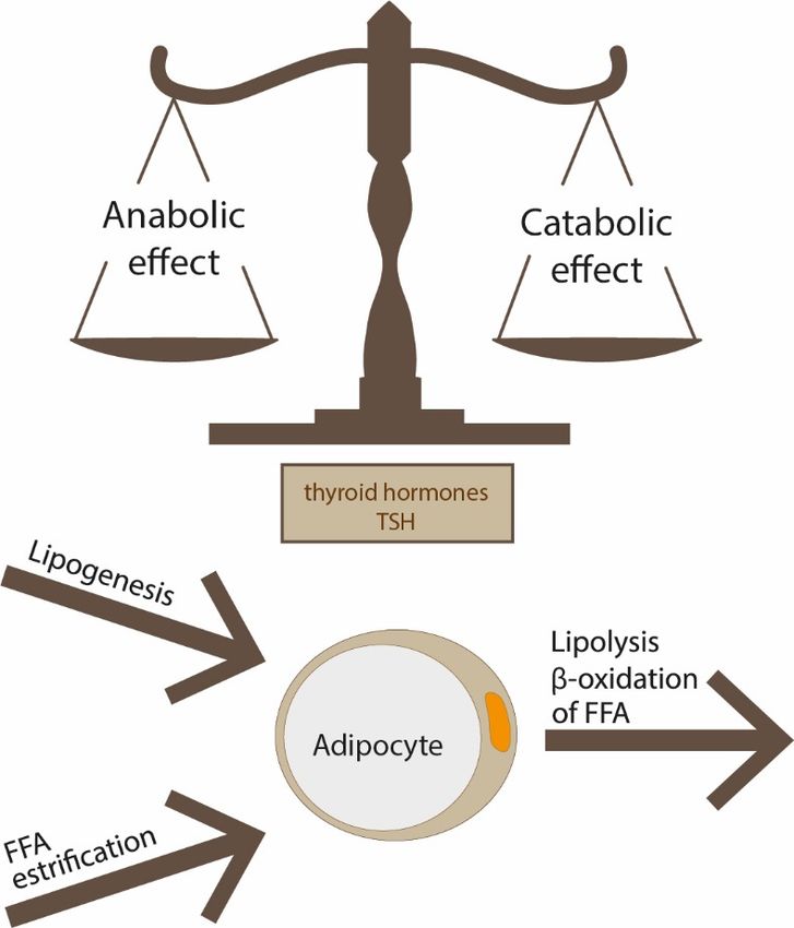
Int. J. Environ. Res. Public Health 2021, 18, 9434                                                                              3 of 23

                                        sion of mitochondrial function and fatty-acid-mobilization-related genes which suggests
                                        involvement in energy expenditure [23]. Adipose-tissue TSHB protein level was positively
                                        correlated with mitochondrial respiratory capacity only in the visceral depot. Interest-
                                        ingly, no association was found between TSH protein and TSHB gene expression [23].
                                        Moreover, adipose-tissue TSHB mRNA was significantly correlated with serum total and
                                        LDL-cholesterol levels in three independent groups of patients (cohort 1 = 38 patients with
                                        mean BMI 45.1 ± 6.4 kg/m2 , cohort 2 = 73 subjects with BMI between 19.5–85.2 kg/m2 ,
                                        cohort 3 = 20 participants with mean BMI 42.8 ± 5.5 kg/m2 ). The results provide evidence
                                        that TSHB is implicated with cholesterol metabolism in human adipose tissue. Both visceral
                                        and subcutaneous adipose tissue TSHB correlated positively with mRNA of 3-hydroxy-3-
                                        methylglutaryl-CoA reductase (HMGCR), the key enzyme in cholesterol biosynthesis. No
                                        associations were found between serum total and LDL cholesterol and TSHR expression
                                        in adipose tissue [22]. Administration of recombinant human TSH resulted in increased
                                        HMGCR mRNA levels in fully differentiated adipocytes but not in preadipocytes [22].
                                        Treatment of human mature adipocytes with recombinant TSH enhanced mitochondrial
                                        respiratory capacity and ATP production and led to increased adipogenesis-related genes,
                                        but no effects were observed in human preadipocytes with low expression of TSHR [23].
                                        As an inverse correlation between thyroxine and TSHB mRNA in adipose tissue was found,
                                        Moreno-Navarrete et al. suggested that local TSH production may be related to THs [22].
                                        Moreover, researchers demonstrated that the main source of TSHB was stromal vascular
                                        cell fraction. All the observations make the detailed role of TSHB in adipose tissue not fully
                                        understood, but it seems likely that TSHB acts as a paracrine factor that is implicated in
                                        lipid metabolism and energy homeostasis. In order to explore specific mechanisms linking
                                        TSHB and adipocyte function, further studies are needed.
                                              Reports suggest that thyrotropin-releasing hormone (TRH) and TSH play a role in the
                                        regulation of appetite. Central administration of TRH and TSH in experimental animals
                                        caused a reduction in food intake, and similar effects were seen following peripheral
                                        administration of TRH (Table 1) [24].

                                        Table 1. Effect of TRH, TSH and T3 on appetite.

                                                           Hormone                                      Food Intake
                                                              TRH                                            ↓
                                                              TSH                                            ↓
                                                               T3                                            ↑

                                        2. Different Depots of Adipose Tissue
                                              There are two main types of fat tissue—white adipose tissue (WAT) and BAT—which
                                        serve opposite functions in energy balance regulation. WAT includes subcutaneous and
                                        visceral deposits, and the latter can grow as mesenteric, omental, retroperitoneal, epicardial
                                        and perivascular fat. WAT is composed mainly of white adipocytes and preadipocytes, but
                                        it also contains several other cells such as macrophages, lymphocytes, fibroblasts, stromal
                                        cells and extracellular matrix. WAT plays a role as an energy reservoir in the form of
                                        triglycerides and as an important endocrine organ that produces adipocytokines. White
                                        adipocytes specialize in lipid storage in response to energy status and can mobilize energy
                                        as FFAs if necessary. WAT has the ability to change volume and can expand or contract. It
                                        grows by two mechanisms: hyperplasia (increased number of fat cells) and hypertrophy
                                        (increased adipocyte volume). The size of adipocytes depends on adipocyte triglyceride
                                        storage which is the final result of the balance between lipogenesis, lipolysis and fatty acid
                                        oxidation. These processes are tightly regulated by insulin and catecholamines; however,
                                        TSH and THs also play a role (Figure 1). This phenomenon may evolve with age, and it
                                        may be affected by other factors.
                                              Under the influence of transcription factors, such as peroxisome proliferator-activated
                                        receptor γ (PPARγ) and CCAAT/enhancer-binding proteins (C/EBPs), generation of new
                                        adipocytes through differentiation of progenitor cells occurs (adipogenesis). This process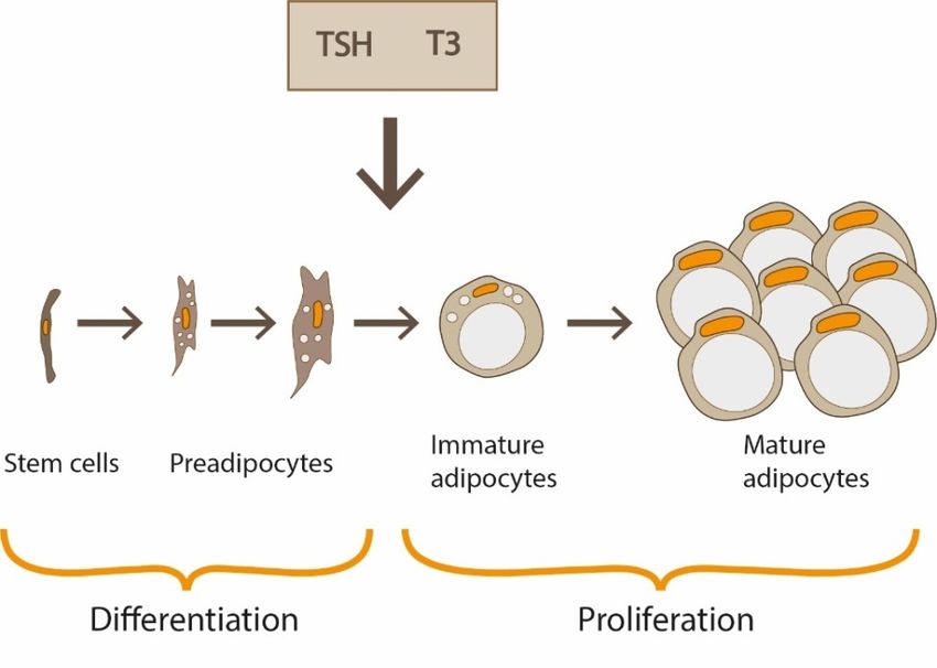
Int. J. Environ. Res. Public Health 2021, 18, x 4 of 24
Int. J. Environ. Res. Public Health 2021, 18, x 4 of 24
Int. J. Environ. Res. Public Health 2021, 18, 9434 4 of 23
Under the influence of transcription factors, such as peroxisome proliferator-acti-
vatedUnder the γinfluence
receptor (PPARγ)ofandtranscription factors, such as proteins
CCAAT/enhancer-binding peroxisome proliferator-acti-
(C/EBPs), generation
vated receptor γ (PPARγ) and CCAAT/enhancer-binding proteins (C/EBPs),
of new adipocytes through differentiation of progenitor cells occurs (adipogenesis). generation
This
isof newunder
also
process adipocytes
TSH/THs
is also underthrough differentiation
control
TSH/THs [5] [5] of
(Figure
control 2).progenitor
Increased
(Figure cells occurs
adipocyte
2). Increased (adipogenesis).
size
adipocytecorrelates This
with
size correlates
process
impaired isadipogenesis
also under
with impaired TSH/THs
[5]. [5].control [5] (Figure 2). Increased adipocyte size correlates
adipogenesis
with impaired adipogenesis [5].
Figure1.1.The
Figure Thesize
sizeofofadipocytes
adipocytesisisthe
theresult
resultofofthe
thebalance
balancebetween
betweenlipogenesis,
lipogenesis,lipolysis
lipolysisand
andfatty
fatty
Figure 1. The size
acid oxidation; of adipocytes
these is the
processes are result of
regulated bythe
THsbalance between lipogenesis, lipolysis and fatty
and TSH.
acid
acidoxidation;
oxidation;these
theseprocesses
processesare
areregulated
regulatedby byTHs
THsand
andTSH.
TSH.
Figure2.2. Adipose
Figure Adipose tissue grows
grows by
bytwo
twomechanisms:
mechanisms:hyperplasia
hyperplasia(increased
(increasednumber
number of fat cells)
of fat and
cells)
Figure 2. Adipose
hypertrophy tissue grows
(increased by two
adipocyte mechanisms:
volume). hyperplasia of
The differentiation (increased
progenitor number of fat cells)
cells through and
preadi-
and hypertrophy (increased adipocyte volume). The differentiation of progenitor cells through
hypertrophy (increased adipocyte
pocytes into adipocytes, as well as volume).
the The differentiation
proliferation of of of progenitor
adipocytes, cells throughcontrol.
preadi-
preadipocytes into adipocytes, as well as the proliferation adipocytes,isisalso
alsounder
underTSH/THs
TSH/THs control.
pocytes into adipocytes, as well as the proliferation of adipocytes, is also under TSH/THs control.
It has long been thought that BAT occurs only in newborns and then disappears.
However, recently, it was shown that BAT is present also in adults, and its high activity
Int. J. Environ. Res. Public Health 2021, 18, 9434                                                                             5 of 23

                                        is associated with a favorable metabolic profile [5–7]. The basic function of BAT is heat
                                        production in a process called thermogenesis.

                                        3. Thermogenesis
                                             The heat generated during vital functions sufficient to maintain body temperature is
                                        called obligatory thermogenesis. THs may increase obligatory thermogenesis by increasing
                                        the BMR. THs also control adaptive (facultative) thermogenesis mediated by BAT. Isolated
                                        signaling of THs in dissipating energy is insufficient, and to provide an adequate effect
                                        synergy of both THs and adrenergic signaling is necessary. THs regulate non-shivering
                                        thermogenesis by targeting BAT, WAT, skeletal muscle and skin blood flow.
                                             Unlike white adipocytes, which store lipids as a single large droplet, brown adipocytes
                                        contain many smaller droplets. They are characterized by numerous mitochondria that
                                        express the specific UCP1 and are richly innervated by sympathetic nerves. The UCP1 is
                                        responsible for energy dissipation as heat, and its activation is controlled by adrenergic
                                        activity and by the concentration of T3 in adipocytes [5,6]. Under sympathetic stimulation,
                                        β3-adrenergic receptors are activated, and FFA-releasing lipolysis begins. Recently it
                                        was reported that thermogenesis in human BAT is mediated by the stimulation of β1-AR
                                        and β2-AR [25]. Cold stress via stimulation of the sympathetic nervous system and by
                                        increasing norepinephrine (NE) in BAT acutely stimulates BAT-specific T3 production and
                                        mitochondrial heat generation. Adrenergic stimulation increases the activity of DIO2 and
                                        the intracellular conversion of thyroxine (T4) to T3 that directly stimulates UCP1 expression
                                        via TH-response elements (TREs) located in the 50 -flanking region of UCP1 [26]. This
                                        phenomenon occurs in BAT within hours after acute cold stress; however, during persistent
                                        cold exposure, the remodeling of WAT takes place, during which mature adipocytes begin
                                        to acquire the thermogenic capability.
                                             T3 acts synergistically with NE released from sympathetic nerve endings to stimulate
                                        heat production. Increased local production of T3 from T4 in BAT via sympathetic activation
                                        and DIO2 expression has been well documented. Both DIO2 and UCP-1 are the indexes of
                                        thermogenic activation of BAT [5,6].
                                             Not only THs but also TRs are essential for adequate thermogenesis in BAT. Brown
                                        adipocytes possess both TRα1 and TRβ nuclear receptors. While TRα1 is necessary for
                                        the proper adrenergic stimulation of brown adipocytes, TRβ isoform is responsible for
                                        T3-regulated UCP1 activation that was confirmed in TRβ gene knockout mice [27,28].
                                        Locally generated T3 by conversion of T4 by DIO2 is required for saturation of TRs [29].
                                             The combined synergistic actions of both T3 and NE can efficiently induce UCP1
                                        expression; therefore, in different pathological conditions with deficits or excess of THs,
                                        this mechanism could be disturbed. Whereas cold-induced adaptive thermogenesis plays
                                        a role when thyroid function is normal, in hypothyroid rats, UCP1 expression is lower and
                                        could not be upregulated during cold exposure [30]. In humans, it is well known that BAT
                                        thermogenesis is reduced in hypothyroidism and patients often report cold intolerance. It is
                                        worth mentioning that mechanisms partially compensating for the reduction of thermoge-
                                        nesis exist. Experimental models revealed that animals with mild hypothyroidism display
                                        elevated BAT adrenergic sensitivity [30,31]. Increased sympathetic tone in a compensatory
                                        manner can activate BAT and increase heat production. Thus, sympathetic activity seems
                                        to partially compensate for diminished THs signaling. However, when hypothyroidism
                                        reaches a significant level, the above mechanism collapses, which can be explained by the
                                        central regulation of this phenomenon [28].
                                             Weiner et al. described another compensatory response [32]. The decreased metabolic
                                        activity of BAT in hypothyroidism was associated with a strong increase of thermogenesis
                                        in BAT and with significant upregulation of the adrenergic system. The authors also
                                        detected an increased expression of genes typical for BAT within WAT. They speculated
                                        that browning of WAT could be the compensatory mechanism preventing a further decrease
                                        in body temperature in hypothyroidism. Besides WAT browning, adipogenesis also occurs
Int. J. Environ. Res. Public Health 2021, 18, 9434 6 of 23
in hypothyroid mice. Endo et al. documented that not only THs but also TSH stimulates
UCP1 expression in BAT of mice [19].
The results derived from animal study are in line with the findings in human clinical
study of hypothyroid patients. Lapa et al. demonstrated BAT activity by visualization
of 18F-FDG uptake in some subjects with thyroid cancer after total thyroidectomy [33].
Interestingly, BAT-positive patients with increased glucose utilization in BAT had higher
TSH concentrations, were younger and had a lower BMI. Similar findings were presented
by Broeders et al. who demonstrated 18F-FDG uptake in PET-CT in hypothyroid patients
after cold exposure [34]. However, a major limitation of those studies is the small number
of subjects enrolled (6 cases with BAT activation in the study by Lapa et al. and 10 patients
in the study by Broeders et al.); therefore the interpretation of data needs to be drawn with
caution. In a larger group of patients with subclinical or overt hypothyroidism (n = 42),
it was demonstrated that cold-induced thermogenesis was decreased in hypothyroid
patients and that sufficient replacement of THs restores adaptive thermogenesis [35]. In
a recent study by Maushart et al., BAT activity assessed by 18F-FDG uptake in PET-CT
after cold exposure was related to levels of free T4 (fT4). It was shown that cold-induced
thermogenesis was approximately fourfold higher in subjects in the highest tertile of fT4
when compared to the lowest tertile of fT4 [36].
Another compensatory mechanism partially compensating for the reduction of ther-
mogenesis could be thyroid-driven cutaneous vasoconstriction [34].
In addition to direct actions of THs on BAT, T3 may indirectly regulate thermogenesis
in BAT through a central mechanism [37]. López et al. found that intracerebroventricular
infusion of T3 diminished the AMPK activity in the ventromedial hypothalamus (VMH),
which promotes sympathetic tone and stimulates BAT thermogenesis [38]. Central action
of T3 plays a role in the upregulation of thermogenic markers, such as UCP1, hormone-
sensitive lipase (HSL) and DIO2, and increases heat generation in the electron transport
chain of brown adipocytes [5–7]. The control of BAT thermogenesis was found to be
dependent on TRα1, whereas contribution of TRβ remains unknown [28]. Central actions
of THs influencing BAT thermogenesis make regulatory mechanisms more complicated. A
mouse model showed that THs via TRα1 signaling regulate heat dissipation through tail
artery function [28].
Not only THs but also thyrotropin may be involved in the regulation of thermoge-
nesis. TSHRs are present on brown adipocytes, and some studies suggest that TSH, by
binding to TSHR, increases basal and T3-stimulated UCP1 expression [19,39]. In brown
adipocytes, TSH increases DIO2 activity, lipolysis and O2 consumption, which confirm
the role of TSH in thermogenesis [39]. It is proposed that TSH/TSHR signaling in BAT
might be responsible for dissipating energy as heat in the process mediated by UCP1 in the
hypothyroid state [5,6,19].
Another central mechanism regulating the heat production in BAT includes hypophys-
iotropic TRH neurons within the PVN that release TRH from axon terminals into median
eminence. Once released from the hypothalamus, TRH is delivered via the hypothalamic–
pituitary portal vessel system into the anterior pituitary where it stimulates secretion of
TSH by thyrotrophs via TRH receptor-1. Thyrotropin stimulates T4 and T3 secretion from
the thyroid. The increase of circulating THs levels after the activation of hypothalamic
TRH neurons within the PVN is very likely to contribute to initiation of the thermogenic
response in BAT. There is a negative correlation between serum THs concentrations and
TRH expression in the PVN. The negative feedback of TRH neurons depends on THs
entering through MCT8 and OATP1c1 transporters and on the interaction of β1-TR and
β2-TR with T3. Tanycytes, specialized ependymal cells, play an important role in feedback
control through regulating local T3 availability and by controlling TRH transport in the
portal vessels [40].
Cold exposure stimulates TRH neurons in the PVN, leading to activation of the
sympathetic nervous system, NE release, increased THs release and elevated activity of
T3 in BAT [41,42]. Cold response within TRH neurons is transient because TRH mRNAInt. J. Environ. Res. Public Health 2021, 18, 9434 7 of 23
expression is increased in the PVN within 1 h after exposure to cold and returns to original
levels after 2 h. In the same experiment, T3 levels peaked after 2 h and started to return to
control values [43]. The results suggest that circulating THs do not play an essential role.
The fact that TRH knockout mice show cold intolerance, which is not corrected with THs
supplementation, also confirms the role of TRH neurons independently of THs.
It has been shown that lean people express higher amounts of UCP1 in BAT than
obese subjects [5,7]. The impaired function of BAT and the lower activity of brown fat
predispose to weight gain and promote obesity. Negative relationships between BMI and
the mass, as well as activity of BAT, are observed [37]. Stimulation of thermogenesis in
BAT is associated with weight reduction and with numerous metabolic benefits such as
increased insulin sensitivity and lipid uptake. It has been shown that white adipocytes,
under specific conditions, may undergo transformation into a thermogenic phenotype. The
process called “browning” or “beiging” occurs mainly in subcutaneous depots of WAT and
in inguinal fat. Browning is characterized by the appearance of beige adipocytes that pos-
sess UCP1 but appear to be molecularly and functionally different from brown adipocytes.
Beige adipocytes that increase in WAT are also identified as brite. It is not fully understood
how beige adipocytes are formed, and the process of transdifferentiation of white to beige
adipocytes is under intensive research. It has been shown that large white adipocytes
can differentiate into beige adipocytes in response to cold or β3-adrenergic agonists [37].
Moreover, the beige adipocytes may lose UCP1 expression when the experimental animals
are transferred back to warmer conditions, indicating that the process is reversible. It
has been shown that simultaneous action of insulin and leptin on proopiomelanocortin
(POMC) neurons increases the browning of white fat [44]. In addition to central mecha-
nisms, leptin promotes browning also through peripheral mechanisms. In skeletal muscle,
leptin regulates irisin expression, which can promote the browning of WAT [45]. Irisin,
a myokine discovered in 2012, cleaved from its parent FNDC5 in response to exercise,
seemed to be an interesting and promising factor with the browning effect. Controversy
regarding irisin, its formation and function, especially in humans, remains [46]. Browning
of WAT was documented after intensive exercise in mice and was attenuated in FNDC5
knockout mice [47]. WAT developed characteristics of BAT after irisin exposure in mice,
and adipocytes browned when incubated with recombinant FNDC5 [48]. However, there
is little knowledge about the role of irisin in humans, and the results of different studies do
not confirm irisin’s response to exercise [49,50]. Currently, there is a discussion whether
irisin could be a link between thyroid function and adiposity [51]. Since reliability of
commercial enzyme-linked immunosorbent assays (ELISAs) for irisin measurements has
been questioned [46], conclusions of such studies need to be carefully evaluated.
Another factor that may contribute to the conversion of white to brown fat tissue is
fibroblast growth factor 21 (FGF21), which is produced mainly by the liver but at lower
temperatures and after stimulation of β3-adrenergic receptors also by BAT [52]. FGF21
analogs and FGF21 receptor agonists constitute the group of molecules known as the FGF21
class that has beneficial metabolic effects and promotes weight and adiposity loss. FGF21
induces WAT browning; however, the mechanisms stimulating thermogenesis are not fully
recognized. Several studies demonstrated that in UCP1 knockout mice FGF21 stimulates
BAT thermogenesis which was UCP1-independent [53]. Those observations suggest that
FGF21 is likely to stimulate BAT thermogenesis through a mechanism other than UCP1.
It is considered that some Chinese medicine agents such as resveratrol, berberine and
curcumin can induce browning of WAT [54], but their detailed role in the physiology of
adipose tissue needs further study.
There is evidence that THs play a role in the browning of adipose tissue by central
and peripheral mechanisms (Figure 3). Alvarez-Crespo et al. showed in their mouse
study that T3, acting centrally on the ventromedial nucleus of the hypothalamus (VMH),
inhibits the activity of AMP-activated protein kinase (AMPK) and thus leads to increased
thermogenesis in both BAT and WAT depots [55]. It has been documented that WAT
browning can be blocked in a mitogen protein kinase MKK6 depending fashion andInt. J. Environ. Res. Public Health 2021, 18, 9434 8 of 23
Int. J. Environ. Res. Public Health 2021, 18, x 8 of 24
T3 participates in this phenomenon. Matesanz et al. [56] found that MKK6 deletion in
inmice
this increases
phenomenon. Matesanzbrowning,
T3-mediated et al. [56] which
found that MKK6
results deletion in
in MKK6-/- mice
mice increases
being T3-
protected
mediated browning,
from obesity induced which
by a results
high-fat indiet.
MKK6-/-
Theymice
also being
foundprotected fromthyroid
that blocking obesity hormone
induced
by a high-fat
synthesis diet.
with They also foundeliminates
propylthiouracil that blocking thyroidUCP1
increased hormone synthesis
expression in with
WAT,propylthi-
observed
ouracil
in miceeliminates
lacking MKK6,increased UCP1
while expression
addition in WAT, observed
of exogenous T3 restores in the
miceincreased
lacking MKK6,
energy
while addition of exogenous T3 restores the increased energy expenditure
expenditure in mice MKKP6-/-. Moreover, the researchers observed that treatment of T3 in mice MKKP6-
/-. Moreover,
induces the researchers
AMPK/p38 activationobserved that treatment
that correlates of T3levels
with higher induces AMPK/p38
of UCP1.To studyactivation
whether
that
the correlates with higher
effects observed levelsare
in animals of UCP1.To
present instudy whether
humans, MKKP6 the effects observed
expression in animals
in visceral fat of
are present
obese in humans,
and lean subjects MKKP6 expression
was investigated, andinthe
visceral fat revealed
analysis of obese higher
and lean subjects
levels was
of MKKP6
investigated, and the
protein in obesity. In analysis revealedmodel
an experimental higherconducted
levels of MKKP6 protein inmice
in TRβ knockout obesity.
and In
UCP1an
experimental
knockout mice, model conducted
Johann in TRβ
et al. [57] knockout amice
documented and
direct UCP1
effect of knockout mice, Johann
T3 on browning throughet
al. [57]regardless
TRβ, documented a direct
of the effect of system.
sympathetic T3 on browning throughwith
In mice treated TRβ,T3,regardless
induction ofof
the sym-
hepatic
expression
pathetic of FGF21
system. In miceviatreated
TRβ and proliferator-activated
with receptor
T3, induction of hepatic α (PPARα)
expression activation
of FGF21 was
via TRβ
observed
and [58].
proliferator-activated receptor α (PPARα) activation was observed [58].
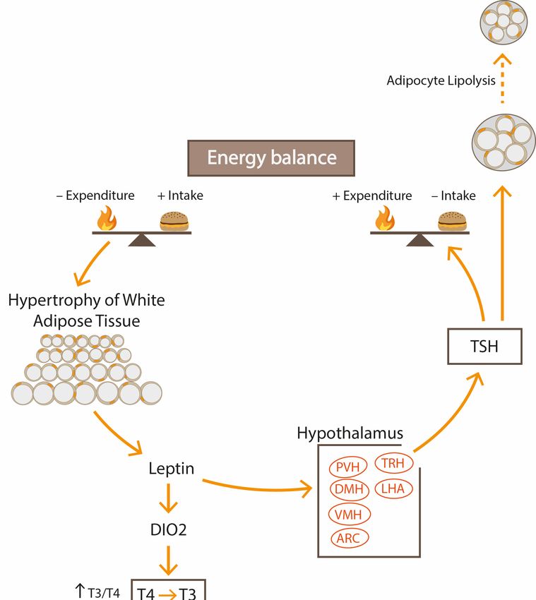
In conditions
Figure3.3.In
Figure conditions ofof excessive
excessivecaloric
caloricsupply,
supply,leptin
leptinactivates TRH
activates expression
TRH andand
expression thenthen
synthesis
syn-
of TSH
thesis of and
TSHTHs. It suppresses
and THs. appetite
It suppresses and promotes
appetite energyenergy
and promotes expenditure by increasing
expenditure lipolysis
by increasing and
lipol-
thermogenesis.
ysis In obeseInpeople,
and thermogenesis. obese leptin
people,can activate
leptin DIO expression
can activate and can and
DIO expression increase conversion
can increase of
con-
version of THs
T4 to T3. T4 toplay
T3. THs
a roleplay a role
in the in the browning
browning of adiposeoftissue.
adipose tissue.Int. J. Environ. Res. Public Health 2021, 18, 9434 9 of 23
In patients with obesity, the functions of WAT and BAT are significantly impaired.
Proinflammatory M1 macrophages and CD4+ Th1 cells are recruited, and after infiltrating
WAT and BAT depots, they block thermogenic activity. It was documented that M1-
derived inflammatory cytokines inhibit UCP1 expression in BAT and WAT. Moreover, the
noradrenergic signaling and catecholamine-induced lipolysis are reduced [59]. Chung et al.
found that direct contact of inflammatory cells and adipocytes inhibits the recruitment of
beige adipocytes [60].
4. Energy Expenditure and THS/Thyroid Axis
THs modulate the BMR and are important determinants for heat production during
shivering and non-shivering thermogenesis [61]. THs stimulate resting energy expenditure
(REE) mainly through increasing ATP production in muscles and by generating Na+/K+
as well as Ca2+ gradients. Total energy expenditure includes BMR and adaptive thermoge-
nesis. Normal thyroid function is essential for both obligatory and adaptive thermogenesis.
In humans, about 30% of energy expenditure is TH-dependent and even slight thyroid
dysfunction can lead to reduction in thermogenesis and may slow down a BMR. Al-Adsani
et al. [62] found that a small change of 5–10% in REE (reduction of only 75–150 kcal/day),
which corresponded with a small change in TSH concentration within the normal range,
could generate weight gain of several kilos in the period of 10 years. Thus, changes in body
weight correlate with TSH levels. Moreover, positive associations between free T3 (fT3),
fT3/fT4 ratio and visceral fat area quantified by magnetic resonance imaging (MRI) were
also observed [63].
However, it is not fully known whether changes in TSH and THs levels are the cause
or the consequence of differences in body weight.
5. Stimulation of Hypothalamus–Pituitary–Thyroid Axis in Obesity
Multiple studies on the relationship between obesity and thyroid function indicate
positive correlations between BMI and TSH as well as between BMI and free or total T3.
Slightly elevated TSH and T3 or fT3 concentrations are the most common abnormalities in
obese children [64,65]. Some researchers consider fT4 rather than TSH to be the optimal
indicator of thyroid status [66]. In obese subjects with primary subclinical hypothyroidism
and with increased TSH, elevated T3 may be the result of augmented peripheral deiodina-
tion and preferential secretion of T3. It is possible that polymorphisms in DIOs converting
T4 to T3 influence the proportion of THs. Moreover, leptin acts as an important regulator
of peripheral and central DIOs, thereby affecting the level of THs. These mechanisms, in
part, may underlie the elevated fT3 concentrations and fT3/fT4 ratios [21,63,67].
In obese individuals, elevated TSH levels may be the result of the stimulation of
the HPT axis. In the animal model, a diet high in fat and simple carbohydrates led to
a significant increase in T3 and TSH already in the first month of observation, and this
phenomenon persisted for the next five months. At the same time, leptin receptors were
significantly downregulated, and thus leptin resistance developed [68].
The stimulation of the HPT axis observed in obesity is mainly due to centrally acting
leptin, which regulates the activity of neurons in the hypothalamus and has both direct
and indirect effects on TRH–TSH secretion (Figure 3). TRH is a tripeptide amide (pGlu-
His-ProNH2) synthesized by TRH neurons within the PVN, and then it is transported to
the median eminence in the third ventricle and through the portal system to the anterior
pituitary where it stimulates the secretion of TSH from thyrotrophs. TRH neurons influence
energy homeostasis by central regulation of thermogenesis and, through the action of THs,
stimulate mitochondrial oxygen consumption and increase thermogenesis.
There are three main groups of neurons that have synaptic connections with the
hypophysiotropic TRH neurons: the arcuate nucleus (ARC) of the hypothalamus, the
dorsomedial nucleus of the hypothalamus (DMN) and catecholamine-producing neurons
in the brainstem [5]. The ARC contains two populations of appetite-control neurons: medial
localized orexigenic neurons that synthesize neuropeptide Y (NPY) and agouti-relatedInt. J. Environ. Res. Public Health 2021, 18, 9434 10 of 23
peptide (AGRP) and lateral anorexigenic neurons that produce POMC, α-MSH and CART.
Both neuronal groups respond to peripheral nutritional signals such as changes in leptin,
ghrelin, insulin and glucose.
DMN neurons also receive signals related to energy homeostasis, including leptin
signaling, but these signals interact with the DMN indirectly through the ARC [69]. How
DMN regulates the hypophysiotropic TRH neurons is not fully understood, but it seems
to exert an inhibitory effect. Bilateral destruction of the DMN has been shown to increase
the 24h release of T3, which confirms the effect on the activity of the HPT axis [70]. DMN
appears to be involved in the circadian regulation of TRH hypophysiotropic neurons.
The activity of TRH neurons is also regulated by the catecholaminergic innervation.
NE has been shown to stimulate TRH gene transcription. Both adrenergic and noradrener-
gic axons activate TRH neurons [69].
Overnutrition, through hyperleptinemia, activates TRH expression and then synthesis
of thyrotropin and THs. It promotes energy expenditure by increasing BMR, thermoge-
nesis, lipolysis and glycolysis. During fasting, the synthesis and release of TRH in the
hypothalamus as well as the concentration of THs in the serum decrease.
The direct interaction of leptin on the activity of the HPT axis includes the effect
on TRH-ergic neurons within the PVN through leptin receptors. The indirect effect of
leptin includes regulating mechanisms of the neuronal hypothalamic network involved
in the energy regulation balance. In the ARC, the critical place of the hypothalamus
regulating feeding and metabolism, leptin increases the synthesis of anorexic peptides,
POMC, α-melanocyte-stimulating hormone (α-MSH) and cocaine- and amphetamine-
regulated transcript (CART) that are projected from the ARC to the PVN and activate
pro-TRH gene expression. Moreover, leptin downregulates orexigenic peptides, NPY
and AGRP, which antagonizes the α-MSH stimulatory effects on pro-TRH expression.
In conditions of excessive caloric supply, leptin simultaneously suppresses appetite by
increasing the expression of POMC and by inhibiting the expression of NPY and AGRP in
the ARC of the hypothalamus.
However, in obesity, hyperleptinemia does not reduce appetite and does not increase
energy expenditure. Those adaptive mechanisms may be disturbed if individuals were
being overfed in early life stages, resulting in overeating and overweight in adults. In
studies run on animals, it was found that perinatal overfeeding induced leptin resistance in
the regulating neuropeptide systems of the hypothalamus. In adulthood, leptin-controlled
arcuate neurons were unresponsive to signals from adipose tissue and insulin which
disrupted the energy homeostasis and food intake regulation [71].
In adult Lep ob/Lep ob mice, the lack of leptin led to serious disturbances in the
functioning of both AGRP/NPY and αMSH pathways [72]. However, the treatment of
those mice with leptin for 20 days was unsuccessful, as it did not restore normal PVH
signaling. Thus, modulation of leptin activity in the hypothalamic pathways appears to be
limited to the neonatal period.
During fasting, the ARC pathway is dominant and mediates the action of leptin on
TRH neurons. This thesis is based on the observation that destruction of the ARC prevents
the influence of leptin on the expression of the TRH gene in the PVN [73]. Moreover, while
central administration of leptin activates TRH neurons in the PVN, administration of a
melanocortin antagonist completely prevents the stimulating effect of centrally admin-
istered leptin on TRH release and TSH secretion [74]. This indicates that leptin affects
the hypophysiotropic TRH neurons mainly through the melanocortin system. Moreover,
in NPY/MC4R double KO mice lacking signaling for both NPY and melanocortin, the
fasting-induced suppression of the HPT axis is completely prevented [75].
Long-lasting excessive caloric intake and increased weight gain change the adaptive
abilities of the HPT axis, and in obesity, leptin resistance appears primarily at the ARC
level, but the direct effect of leptin on the PVN remains unchanged. In obese animals on a
high-fat diet, POMC and MSH-α expression in the ARC remains unchanged, suggestingInt. J. Environ. Res. Public Health 2021, 18, 9434 11 of 23
that increased caloric intake reduces the sensitivity of anorexigenic neurons in this nucleus
to leptin [76].
6. Thyroid Hormones and TSH Regulate the Balance between Lipogenesis and
Lipolysis and Control Adipogenesis
6.1. Thyroid Hormones
All TR isoforms—TRα-1, TRα-2 and TRβ-1—are present in white and brown adipocytes,
and TRα-1 is more abundant than others [7]. Biological effects of THs depend on intra-
cellular levels of T3 which is generated from T4 by the action of DIO type 1 (DIO1) and
DIO2. DIO3 deactivates THs by production of reverse T3 and diiodothyronine. DIOs
are selenoenzymes that are regulated by thyroid status, local selenium availability and
proinflammatory cytokines [77].
DIOs are present in different tissues. DIO1 is abundantly expressed in the thyroid
gland, pituitary, liver, kidneys and WAT, whereas DIO2 is present in the pituitary, muscles,
placenta and BAT. Under physiological conditions, DIO3 is expressed in the brain, placenta
and pancreas, while under various pathophysiological conditions, its expression begins to
be present in other tissues. Little is known about DIO activity in WAT, and only DIO1 was
found to be an important source of intracellular concentrations of T3. It was demonstrated
that the level of DIO1 mRNA was higher in subcutaneous and visceral fat of obese when
compared with non-obese subjects [78]. Moreover, DIO1 activity was elevated in obese
patients, in contrast to DIO2 and DIO3 that were close to the detection limits. Moreover,
there were no differences in their activities between obese vs. non-obese subjects and
between different fat depots. As leptin expression was upregulated in obesity and was
positively correlated with DIO1, Ortega et al. [78] speculated that leptin can modulate
DIO1 activity, thereby affecting the local conversion of T4 to active T3. This phenomenon
could underlie the elevated fT3 levels and higher fT3/fT4 ratios observed in obese subjects.
However, dozens of controversial findings have been published in papers addressing DIO1,
DIO2 and DIO3 activities in human and animal experiments and results of those studies
are non-conclusive [78–80]. Pihlajamaki et al. analyzed transcriptomes from liver samples
of obese patients, and they found that THs signaling could be suppressed, as DIO2 and
DIO3 gene expression was decreased, whereas expression of DIO1 was not changed [80].
As leptin receptors were also decreased in obese individuals when compared with lean
controls, it could be assumed that downregulation of leptin signaling impairs THs action
in the liver and may be the cause of fat accumulation in the liver [80].
In BAT, locally produced T3 is required for thermogenic function. T3 enhances NE-
induced lipolysis and heat generation. However, during cold exposure, T3 is responsible
for lipogenesis, and the balance between lipolysis and lipogenesis depends on D2-mediated
conversion of T4 to T3 as well as on sympathetic stimulation [81].
In WAT, T3 regulates the activity of the entire enzyme machinery responsible for
lipogenesis and lipolysis. In healthy conditions, lipolysis balances lipogenesis, even if an
excessive supply of energy prevails (Figure 1). T3 enhances lipolysis by increasing the
number of lipolytic β2-adrenergic receptors and through postreceptor signaling, mainly
c-AMP-dependent. Local production of T3, mediated by DIO2 in response to leptin, could
directly affect the activity of lipolytic enzymes such as carnitine palmitoyltransferase 1α,
adiponutrin, lipase of triacylglycerol, desnutrin and lipoprotein lipase. Lipolytic effect is
also due to the augmentation of hormone-sensitive lipase activity, which is responsible for
triacylglycerol hydrolysis in adipose tissue. The lipolytic effect of T3 is pronounced when
NE is binding to adrenergic receptors [82]. In hyperthyroidism, catecholamine-induced
lipolysis is enhanced and leads to a reduction of fat mass.
THs signaling is also essential for lipogenesis in both subcutaneous WAT and BAT, and
β3-adrenergic stimulation plays an essential role. In vitro studies showed that insulin and
T3 act synergistically on intracellular signaling pathways and induce enzymatic activity.
Different studies reported that T3 stimulates the expression of lipogenic enzymes in both
liver and adipose tissue: acety-CoA-carboxylase, fatty acid synthase, malic enzyme and
glucose-6-phosphate dehydrogenase, especially in a feeding state high in carbohydratesInt. J. Environ. Res. Public Health 2021, 18, 9434 12 of 23
and protein [83,84]. In animals on a high-carbohydrate diet, THs are known to induce
lipogenesis [85].
The BAT and subcutaneous WAT are under sympathetic nervous system control, and
the adrenergic stimulation plays a key role in thermogenesis, lipolysis and lipogenesis. An
association between sympathetic signaling in subcutaneous WAT via the β3-AR-cAMP-
PKA pathway and T3/TR signaling promoting both thermogenesis and lipogenesis was
demonstrated [82]. The β3-AR agonists cannot stimulate either WAT lipogenic gene
expression or UCP1 expression in the absence of THs, while T3 administration triggers these
responses. In denervated adipose tissue in hypothyroid rats, lipogenesis and thermogenesis
are impaired [31].
In vivo and in vitro studies have shown that T3 promotes adipogenesis in WAT
through adipogenic signal pathways: C/EBPs and PPARγ (Figure 2). Impaired adipogene-
sis was observed in transgenic mice with mutant TRα1 [86]. Synthesis of fatty acids and
simultaneously proliferation and differentiation of immature adipocytes are critical for
lipid accumulation and lead to hypertrophy and hyperplasia of adipose tissue.
In addition to affecting lipid metabolism, there is evidence that THs are involved
in appetite regulation. It is well established that T3 can directly increase food intake via
central hypothalamic regulations (Table 1) [24].
6.2. TSH
TSH stimulates adipocyte lipolysis, as was proved in various in vitro experiments [20,87–90].
Lipolysis is the process of hydrolysis of triglycerides into a glycerol and FFAs that are
released into circulation. The essential regulators stimulating lipolysis in humans are cate-
cholamines (via β1, β2 and β3-AR) and natriuretic peptides, whereas the antilipolytic effect
is mediated by insulin and catecholamines (through α2-AR). Several other factors such as
TSH, NPY, growth hormone, glucocorticoids and tumor necrosis factor (TNF) can regulate
lipolysis, either directly by specific receptors or indirectly by modulating the lipolytic
cascade [90]. Stimulation of TSHR increases intracellular cAMP levels through activation
of adenylyl cyclase. Perilipin and HSL are required for lipolysis and FFA release from
adipocytes. It was found that thyrotropin activates phosphorylation of perilipin and HSL
in a protein-kinase-A-dependent manner in mouse 3T3-L1 adipocytes as well as in primary
human adipocytes [87]. Vizek et al. documented in vitro that TSH stimulated glycerol
release from fat cells isolated from the subcutaneous adipose tissue of newborns but had
no effect on fat cells from adults [88]. Marcus et al. found that in neonatal adipocytes, TSH
could induce a significant lipolytic effect [91]. It is believed that high TSH levels occurring
in infants stimulate lipolysis and FFA release that are a source of energy shortly after birth
and before lactation [20,91]. This effect is reduced in the next years of life and in adults
is not as significant as in newborn/pediatric age. To determine whether TSH induces
lipolysis in vivo, serum FFAs were measured after administration of recombinant human
TSH in patients with thyroid cancer after thyroidectomy. The lipolysis stimulation effect
was evident only in subjects with BMI < 30 [87].
In vivo studies indicate a role of TSHR in the lipolytic effect of TSH. In the model
of TSHR knockout mice, reduced responsiveness to the lipolytic effect of TSH and a
hypertrophy of mice adipocytes were observed [92]. An increased basal lipolysis found
in mice lacking functional TSHR is thought to be related to an increase in adipocyte size.
Furthermore, in the study by Lundbäck et al., inactivation of TSHR resulted in the decreased
expression of genes responsible for adipogenesis such as PPARγ and C/EBPs and in
reduction of lipolytic adrenergic receptors including β1-AR and β3-AR [93]. Those results
suggest that TSH/TSHR signaling is responsible for adrenergic activity in adipocytes.
Mice lacking functional TSHR within adipose tissue exhibited the decreased expression
of adiponectin gene and downregulated UCP1 in BAT. TSHR knockout animals were
predisposed to obesity development.
However, the results of the above experiment are not consistent with other obser-
vations. The model of global TSHR knockout mice, adipose-specific TSHR knockoutInt. J. Environ. Res. Public Health 2021, 18, 9434 13 of 23
mice, thyroid-specific TSHR knockout mice and hypothyroid mice was created by Zhang
et al. [94]. These researchers indicated that elevated TSH reduces energy expenditure and
promotes adiposity, while mice lacking TSHR showed higher metabolic rates and were resis-
tant to obesity. Furthermore, the browning of WAT was observed in TSHR knockout mice.
In the in vitro part of the experiment, it was documented that the AMPK/PRDM16/PGC1α
pathway is responsible for the thermogenic effect of TSH [94].
It is suggested that, under certain conditions, TSH may have a pro-lipogenic effect. In
primary human differentiated adipocytes, it was found that insulin reduced the ability of
TSH to stimulate lipolysis [89]. In the same experiment, TSH inhibited insulin signaling,
and therefore it can be assumed that interactions between TSH and insulin are bilateral.
It was demonstrated that lipogenesis/lipolysis balance in adipocytes depends on BMI,
insulin resistance and the time of exposure to the elevated TSH concentration. At lower
BMI values, the inhibitory effect of TSH on insulin-stimulated lipogenesis is dominant.
However, higher BMI appeared to cause enhancement in lipogenesis, especially over a
longer period of time. It seems that in adipocytes of obese subjects, when TSH and insulin
act together for a long time, TSH signaling interferes with insulin signaling which leads to
increased lipogenesis and reduced lipolysis [89]. In mature adipocytes, TSH can decrease
expression of triglyceride lipase, which catalyzes the hydrolysis of triglycerides [95].
Thyrotropin can also increase glycerol 3-phosphate acyltransferase 3 activity. The
enzyme is responsible for triglyceride synthesis and lipid storage as well as for the adipo-
genesis process. Ma et al. [96] found that TSH promoted fat accumulation in vivo, both
by augmenting adipocytes hypertrophy and by hyperplasia of adipose tissue. In TSHR
knockdown mice, the expression of genes responsible for fatty acid synthesis and triglyc-
eride storage was downregulated and animals exhibited resistance to high-fat-diet-induced
obesity. TSH stimulated lipogenesis through AMPκ/PPARγ signaling.
Different studies showed that not only THs but also TSH provides an important
contribution to adipogenesis (Figure 2). Valyasevi et al. reported that orbital preadipocyte
fibroblasts increased their TSHR expression with differentiation into adipocytes [17]. TSHR
expression was increased at both mRNA and protein levels when 3T3-L1 preadipocytes
were induced to differentiate, while knocking down TSHR blocked this stimulating ef-
fect [15]. It was showed that TSH administration in human preadipocytes with low TSHR
levels did not result in significant expression of adipogenic genes and lipid accumula-
tion whereas TSH administration in mature adipocytes with high TSHR levels increased
adipogenesis-related gene expression (FASN, ADIPQ, SLC2A4) [23]. Lu et al. reported
that TSHR mRNA and protein levels significantly increased during the differentiation of
murine 3T3-L1 preadipocytes, and knocking down TSHR resulted in delayed cell differen-
tiation [15]. The TSHR transcript levels in visceral fat from mice fed with a high-fat diet
increased in obese mice compared to control mice fed with an ordinary diet [15]. These
findings are consistent with the study by Ma et al. [96]. The ablation of TSHR in mice
resulted in decreased adipogenesis and caused the resistance to high-fat-diet-induced
obesity. Lu et al. [15] also analyzed the effect of obesity on TSHR transcript levels in human
subcutaneous adipose tissue and they reported that it was higher in subjects with BMI > 25
than in people with BMI < 25. However, the effect of obesity on TSHR is not obvious.
Nannipieri et al. [21] detected TSHR and beta-actin protein expression in subcutaneous
and visceral adipose tissue obtained during abdominal surgery in 107 severely obese pa-
tients. In both adipose depots, TSHR expression was lower in obese than in 12 lean control
individuals. The protein expression of TSHR in subcutaneous adipose tissue obtained from
obese patients was also depressed. The findings are concordant with the observations in the
animal model by Lundbäck and coworkers [93]. They found that mice with reduced TSHR
expression gained weight faster than control mice. In keeping with this observation, Comas
et al. [23] suggested that TSH can promote mitochondrial function in human adipocytes,
thus potentially being a link between energy and lipid metabolism.
TSHR stimulation by species-specific TSH remains a topic of current investigations.
In most experiments, bovine TSH is used because it effectively stimulates TSHR, especiallyInt. J. Environ. Res. Public Health 2021, 18, 9434 14 of 23
human TSHR. It is well known that bovine TSH has a higher affinity to the human TSHR
and higher signaling activity than human TSH [97]. TSH exists as a heterodimer consisting
of α and β subunits, bound together to produce the active form of the hormone. The
human TSH subunit β gene is located on chromosome 1, the gene coding for the α subunit
is located on chromosome 6. The human TSHB gene consists of three exons, of which exon
1 is noncoding. The mouse TSH β subunit consists of five exons, and only exons 4 and 5 are
the coding exons [98]. Although recombinant human TSH has a slower metabolic clearance
rate and longer duration of action than does native human TSH, it is less effective. On the
other hand, rat or mouse TSH is rarely available for in vivo and in vitro experiments. What
is worth noting is that the observed diversity of results may be the consequence of different
animal and cell models used in laboratories. Moreover, differences between species can
make the interpretation of experiments difficult and confusing. Consequently, many of the
reported observations of TSHR signaling and effects on extrathyroidal tissues and cells
may be “non-physiological”.
6.3. TSH and Chronic Inflammation
Low-grade chronic inflammation, characterized by secretion of proinflammatory
adipokines, leads to endothelial dysfunction, oxidative stress, atherogenesis and insulin
resistance. Some authors speculate that persistently elevated thyrotropin stimulates in-
flammatory state [99–108], which contributes to adipose tissue dysfunction and metabolic
syndrome and then may facilitate weight gain. In vitro studies revealed that TSH en-
hances the expression of leptin mRNA in adipose tissue and adipokine release during
TSH-stimulated lipolysis [102]. In adipocytes that express TSHRs, TSH stimulates secretion
not only of the leptin but also of other inflammatory cytokines, such as interleukin-6 (IL-6),
TNF-α and monocyte chemoattractant protein-1 (MCP-1), which in turn lead to adipose
tissue dysfunction [100,101]. In vivo and in vitro studies showed that IL-6 release from
differentiated adipocytes is stimulated by TSH and the involvement of cAMP-protein
kinase A pathway was suggested [99]. Significantly increased concentrations of IL-6, CRP
and MCP-1 were detected in subclinical hypothyroid subjects [95–98]. In our previous
study, higher TSH and IL-6 concentrations were related to metabolic syndrome [104].
It was documented that thyrotropin upregulates mRNA expression of MCP-1 and
protein release from human abdominal differentiated adipocytes. Signaling through the
inhibitor of the nuclear factor kappa-β kinase (IKKβ) pathway is responsible for this ef-
fect [103]. The result of another experiment documented a stimulating effect of thyrotropin
on macrophages via the IκBkinase (IKKB)/nuclear factor κB pathway [100]. In the study
involving autoimmune thyroiditis patients, elevated TSH levels were accompanied by
increased serum concentrations of MCP-1 [107]. In patients with extreme obesity, ele-
vated TSH concentration significantly correlated with proinflammatory cytokines such
leptin, IL6, ICAM-1 and E-selectin [108]. In humans, recombinant TSH administered to pa-
tients with differentiated thyroid cancer during postoperative diagnosis stimulated leptin
secretion [109].
Resistin is another adipocytokine with adverse effects on metabolism and the vascular
system. In the study conducted by Eke Koyuncu et al. [110], the authors found that hy-
pothyroid patients had significantly higher resistin serum concentrations when compared
to controls. Low-grade inflammatory state is implicated with diseases such as coronary
heart disease, myocardial infarction, heart failure, chronic renal disease, obstructive sleep
apnea, rheumatoid arthritis, depression, colorectal, gastric and lung cancers as well as
metastatic progression [111].
Not only hyperthyrotropinemia stimulates proinflammatory states, but also elevation
of inflammatory cytokines may affect thyroid function [112].
6.4. Lipotoxicity
Obese subjects often exhibit hypertriglyceridemia which leads to accumulation of
lipids in tissues other than adipose tissue. This phenomenon is known as lipotoxicity.You can also read