Establishing and maintaining fertility: the importance of cell cycle arrest
←
→
Page content transcription
If your browser does not render page correctly, please read the page content below
Downloaded from genesdev.cshlp.org on August 29, 2021 - Published by Cold Spring Harbor Laboratory Press

REVIEW

Establishing and maintaining fertility: the
importance of cell cycle arrest
Emily R. Frost,1,2,3 Güneş Taylor,3 Mark A. Baker,1,2 Robin Lovell-Badge,3 and Jessie M. Sutherland1,2
1
 Priority Research Centre for Reproductive Science, School of Biomedical Science and Pharmacy, School of Environmental and Life
Sciences, University of Newcastle, Callaghan, New South Wales 2308, Australia; 2Hunter Medical Research Institute, New
Lambton Heights, New South Wales 2305, Australia; 3Stem Cell Biology and Developmental Genetics Laboratory, The Francis
Crick Institute, London NW1 1AT, United Kingdom

Development of the ovary or testis is required to establish                 hibitors (CKIs) halt proliferation by binding to CDKs and
reproductive competence. Gonad development relies on                        initiating cell cycle arrest.
key cell fate decisions that occur early in embryonic de-                      INK4 and Cip/Kip are two main families of cyclin-
velopment and are actively maintained. During gonad de-                     dependent kinase inhibitors. The INK4 family acts in a re-
velopment, both germ cells and somatic cells proliferate                    stricted manner as INK4 family members p16INK4a,
extensively, a process facilitated by cell cycle regulation.                p15INK4b, p18INK4c, and p19INK4d prevent CDK4 and
This review focuses on the Cip/Kip family of cyclin-de-                     CDK6 binding to the D-type cyclins only (Sherr and Roberts
pendent kinase inhibitors (CKIs) in mouse gonad develop-                    1999). Thus, after removing repressive INK4 proteins, cells
ment. We particularly highlight recent single-cell RNA                      immediately enter G1 phase by the action of Cyclin D. In
sequencing studies that show the heterogeneity of cy-                       contrast, the Cip/Kip family of cyclin-dependent kinase in-
clin-dependent kinase inhibitors. This diversity high-                      hibitors bind all cyclins and CDKs and therefore have much
lights new roles for cell cycle inhibitors in controlling                   more diverse functions (Nakayama and Nakayama 1998).
and maintaining female fertility.                                           The Cip/Kip family members—p21Cip1, encoded by the cy-
                                                                            clin-dependent kinase inhibitor 1A (Cdkn1a) gene; p27Kip1,
                                                                            encoded by the cyclin dependent kinase inhibitor 1B
                                                                            (Cdkn1b) gene; and p57Kip2, encoded by the cyclin depen-
                                                                            dent kinase inhibitor 1C (Cdkn1c) gene—are traditionally
Every organism relies on the cell division cycle for devel-                 associated with controlling cell divisions (Fig. 2; Li et al.
opment. Maintaining the balance between cell differenti-                    2012; Pippa et al. 2012; Jeannot et al. 2015; Orlando et al.
ation and cell growth is essential in order for cells to                    2015; Bicer et al. 2017). However, recent studies show that
access nutrients (Ruijtenberg and van den Heuvel 2016).                     p21Cip1, p27kip1, and p57kip2 also regulate other cellular pro-
Broadly, this balance shifts with age; during early develop-                cesses, including apoptosis, cell migration, cell fate deci-
ment, rapid proliferation is required to create vital organs,               sions, and transcriptional regulation (for extensive
but a reduction in proliferation is observed in many tis-                   reviews, see Sherr and Roberts 1999; Besson et al. 2008;
sues with increasing age (Khosla et al. 2020). Driven by                    Bachs et al. 2018). Specifically, in the mammalian gonads,
proteins such as cyclin-dependent kinases (CDKs) and cy-                    recent technological advances are giving more insight into
clins, cells progress through the three phases of the cell cy-              the importance of the cell cycle.
cle prior to mitosis and proliferation in M phase (Fig. 1).                    Mammalian gonads are unique organs as they contain
These phases, G1, S, G2, and M, are separated by tightly                    not only somatic cells but also germ cells; hence, mitosis
regulated checkpoints that must be passed for the cell to                   and meiosis occur in parallel within the same organ. This
progress into the next phase. For example, at the G1 check-                 dynamic process must be tightly regulated to ensure suc-
point, CDK proteins bind to cyclin D, which allows for the                  cessful transmission of genetic material to the next gener-
release of transcription factors to push the cell through to                ation. Interestingly, changes in proliferation are one of the
S phase (Sherr and Roberts 1999; Kanatsu-Shinohara et al.                   earliest differences between the ovaries and testes
2010; Granados-Aparici et al. 2019). For cells to exit the                  (Schmahl et al. 2000; Schmahl and Capel 2003). These dif-
cell cycle and stop proliferating, CDKs need to be restrict-                ferences persist into adulthood, when the testes are con-
ed. At the G1/S checkpoint, cyclin-dependent kinase in-                     tinually producing new sperm, but the ovaries are
                                                                            unable to generate new oocytes. Regulation of the cell cy-
                                                                            cle by CDKs and cyclins has been reviewed extensively in
[Keywords: Cip/Kip; gonad; granulosa; oocyte]
Corresponding author: robin.lovell-badge@crick.ac.uk
Article published online ahead of print. Article and publication date are
online at http://www.genesdev.org/cgi/doi/10.1101/gad.348151.120. Free-     © 2021 Frost et al. This article, published in Genes & Development, is
ly available online through the Genes & Development Open Access             available under a Creative Commons License (Attribution 4.0 Internation-
option.                                                                     al), as described at http://creativecommons.org/licenses/by/4.0/.

                  GENES & DEVELOPMENT 35:1–16 Published by Cold Spring Harbor Laboratory Press; ISSN 0890-9369/21; www.genesdev.org               1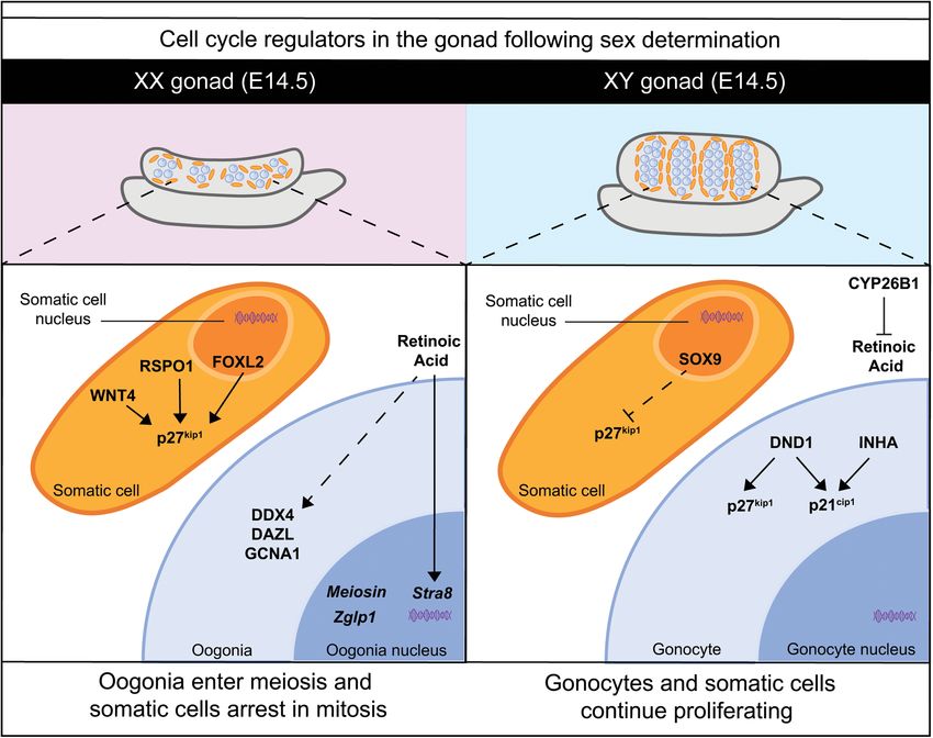
Downloaded from genesdev.cshlp.org on August 29, 2021 - Published by Cold Spring Harbor Laboratory Press

Frost et al.

                                                                                Figure 1. Phases of the cell cycle and the role of
                                                                                cell cycle inhibitors. The four phases of the cell cy-
                                                                                cle are G1, S, G2, and M phase. Cells must pass
                                                                                through the G1, S, and G2 phases to enter mitosis.
                                                                                At the G1 stage, cells can exit the cell cycle by enter-
                                                                                ing the G0 phase. Cyclins are required to progress
                                                                                through the four cell cycle stages. Cyclins A, B, D,
                                                                                and E vary in expression level depending on the
                                                                                cell cycle stage. Cell cycle inhibitors are important
                                                                                regulators of cell cycle progression and prolifera-
                                                                                tion. There are two main families of cyclin-depen-
                                                                                dent kinase inhibitors: the Cip/Kip family and the
                                                                                INK4 family. The Cip/Kip family consists of three
                                                                                proteins (p21cip1, p27kip1, and p57kip2), and the
                                                                                INK4 family consists of four proteins (p16INK4a,
                                                                                p15INK4b, p18INK4c, and p19INK4d). The Cip/Kip fam-
                                                                                ily is capable of inhibiting Cyclin A, Cyclin D, and
                                                                                Cyclin E, whereas the INK4 family can only inhibit
                                                                                Cyclin D. With the cyclin protein inhibited, the cell
                                                                                is unable to progress to the next stage of the cell
                                                                                cycle.

the testes (Wolgemuth and Roberts 2010; Lim and Kaldis             formation of sex-specific gonads occurs later in embryonic
2013); however, recent studies reveal precise control of           development. This means that the mammalian gonads are
the cell cycle during ovarian development, highlighting            a remarkable system to study cell fate decisions as, unlike
the need for a new review of this field. Cip/Kip proteins          other organs, the gonad develops as a bipotential organ
are abundantly expressed in the early gonad, and impor-            with the ability to become either an ovary or a testis. In
tantly, deletion of the Cip/Kip family members in mice             mice, gonads are specified from the coelomic epithelium
leads to unexpected gonad-specific fertility phenotypes
in both sexes (Fero et al. 1996; Holsberger et al. 2005;
Rajareddy et al. 2007; Lin et al. 2015). The primary focus
of this review is on data obtained in mice as it is the model
used in most recent reports pertaining to primary sex de-
termination and early ovarian development. Details about
the gene networks involved in ovarian somatic cell differ-
entiation, mitotic and meiotic arrest in germ cells, and the
changes in Cip/Kip protein expression during key ovarian
transitions, e.g., from establishing the oocyte reserve to
selective oocyte maturation, are presented (Fig. 3). Sin-
gle-cell RNA sequencing permits the gathering of large
amounts of information, both on gene expression and
cell populations, in a single experiment. In particular,
this technology has unveiled new cell types or subpopula-
tions in nearly all tissues explored, expanding our under-
standing in its wake. By exploring these data sets with a
cell cycle-centric view, it is evident there is still much
to learn about cell cycle control in specifying cell fate in
                                                                   Figure 2. Canonical Cip/Kip protein action to regulate cell pro-
the developing gonad. By investigating the dynamic                 liferation. The Cip/Kip family of proteins regulate cellular prolif-
changes in Cip/Kip expression across mouse ovarian de-             eration by inhibiting the action of CDKs. All three Cip/Kip
velopment, this review illustrates and emphasizes the im-          proteins (p21cip1, p27kip1, and p57kip2) bind to CDK2 and Cyclin
portance of Cip/Kip proteins in establishing and                   E to keep cells in the G0 phase of the cell cycle and maintain dor-
maintaining the gonad, and ultimately ensuring fertility.          mancy. When a cell receives signals to proliferate, Cip/Kip inhibi-
                                                                   tion can be removed in multiple ways. Cip/Kip proteins become
                                                                   phosphorylated, which allows them to bind to Cyclin D and
Key genes drive primary sex determination in the somatic           CDK4, which drives the cell to enter into G1 phase. Phosphoryla-
cells of XX and XY gonads                                          tion or acetylation of Cip/Kip proteins marks it for ubiquitina-
                                                                   tion, and thus the protein is degraded by the proteasome. These
While mammalian sex is determined by XY male/XX fe-                post-translational modifications control Cip/Kip action to regu-
male sex chromosomes, respectively, at fertilization, the          late cellular proliferation.

2       GENES & DEVELOPMENT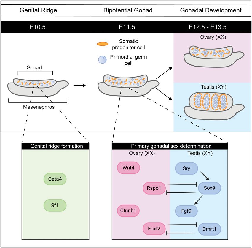
Downloaded from genesdev.cshlp.org on August 29, 2021 - Published by Cold Spring Harbor Laboratory Press

                                                                                                          Cell cycle arrest in the gonad

                                                                                        Figure 3. Key genes involved in the
                                                                                        somatic cells and the germ cells during the
                                                                                        development of the mouse ovary. The top
                                                                                        panel shows diagrams of the mouse ovary
                                                                                        during embryonic and early postnatal devel-
                                                                                        opment, with the embryonic and postnatal
                                                                                        day indicated in the black bar above. From
                                                                                        E10.5, the gonad contains both the primor-
                                                                                        dial germ cells (blue) and somatic supporting
                                                                                        cell precursors (orange). A range of genes
                                                                                        contribute to and participate in the gene reg-
                                                                                        ulatory networks controlling mouse ovarian
                                                                                        development, which are shown in the bot-
                                                                                        tom panel. Only a selection of genes shown
                                                                                        to be critical for each developmental process
                                                                                        are shown.

around embryonic day (E) 9.5, where proliferating cells            and steroidogenic cells, originate from a single progenitor
form a layer on the mesonephros (the embryonic kidney)             cell lineage within the genital ridge (Stévant et al. 2018).
(Hu et al. 2013). Less than 24 h later, at E10.3, this layer       Primary sex determination is the process by which cells
of cells on the mesonephros then thickens, forming the             of the supporting cell precursor lineage differentiate into
genital ridge (Fig. 4; Hu et al. 2013). Primordial germ cells      Sertoli cells typical of the testis or pregranulosa cells of
concurrently migrate from the mesoderm into the genital            the ovary (Zhao et al. 2018; Stévant et al. 2019). These trig-
ridge as it thickens (Richardson and Lehmann 2010). Gen-           ger other cell types, including the steroidogenic cells,
ital ridge formation is dependent on the expression of             germ cells, and connective tissue cells, to follow the tes-
GATA binding protein 4 (GATA4), which activates other              ticular or ovarian pathway, thereby shaping the gonad
early gonadal factors like LIM homeobox 9 (LHX9) and               and laying the foundations for future reproductive capac-
steroidogenic factor 1 (SF1) that sustain the continued            ity. A failure of the gonads to develop as either XX ovaries
growth of the genital ridge (Richardson and Lehmann                or XY testes leads to infertility and disorders/differences
2010; Hu et al. 2013). Once the genital ridge thickens,            of sex development (DSDs), often associated with infertil-
this structure is known as a gonad, which contains both            ity (Eozenou et al. 2020; Estermann and Smith 2020). The
somatic cells and germ cells. The two critical somatic             gene regulatory networks relevant to sex determination in
cell populations in XX and XY gonads, the supporting cells         mice are detailed in several previous reviews (Jakob and

                                                                                                    GENES & DEVELOPMENT               3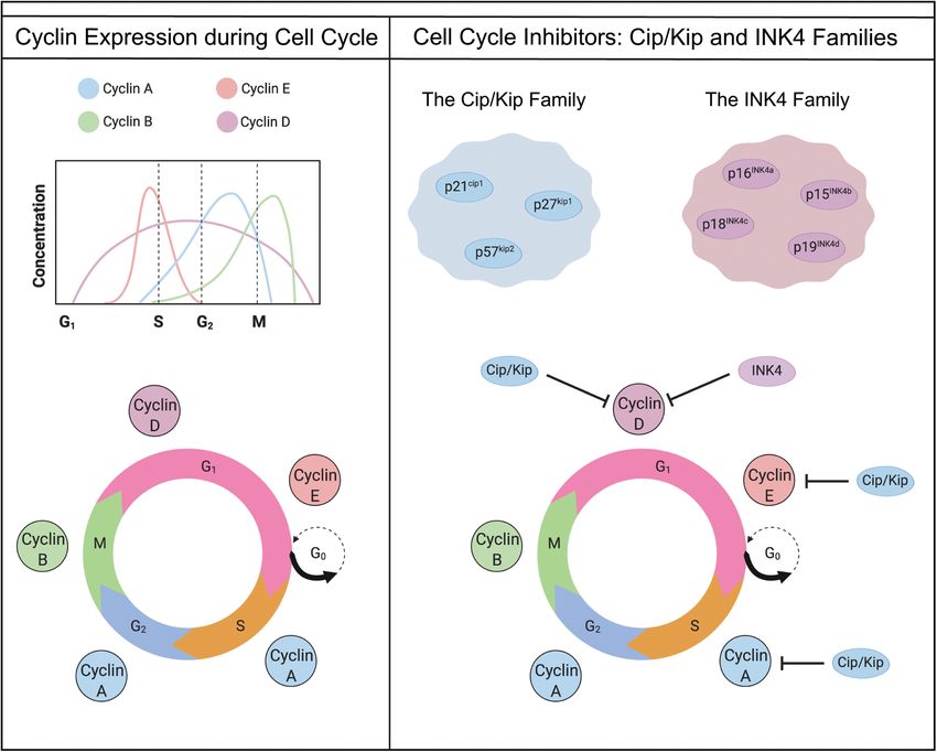
Downloaded from genesdev.cshlp.org on August 29, 2021 - Published by Cold Spring Harbor Laboratory Press
Frost et al.
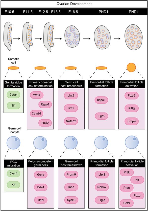
Figure 4. Factors controlling primary gonadal
sex determination in mice. Around embryonic
day 10.5 (E10.5) in mice, the genital ridge forms
where the coelomic epithelium thickens. The
transcription factors Gata4 and Sf1 are critical
for the formation of the genital ridge and its ex-
pansion into the early gonad. By E11.5, the go-
nad contains both the primordial germ cells
(blue) and somatic supporting cell precursors
(orange), as well as precursors of steroidogenic
cells and some connective tissue cells. At
this time point, primary gonadal sex determi-
nation is initiated, driven by factors expressed
in the supporting cell precursors. While testis
determination is known to be initiated by
one factor, Sry, and then orchestrated by
Sox9, female gonadal sex determination is ini-
tiated by multiple factors. Wnt4, Rspo1,
Ctnnb1, Runx1, and Foxl2, and perhaps others,
all work to specify the ovarian fate (Ottolenghi
et al. 2007; Gonen and Lovell-Badge 2019;
Nicol et al. 2019). In the male, Sry stimulates
expression of Sox9, which then up-regulates
male-specific genes, including Fgf9 and
Dmrt1 (Koopman et al. 1991; Kim et al.
2006). In both sexes, active repression of sex-
specific factors maintains the gonadal identity
(Uhlenhaut et al. 2009; Lindeman et al. 2015).
Lovell-Badge 2011; Capel 2017; Stévant and Nef 2019). noprecipitation sequencing (ChIP-seq), and assay for
Importantly for sex determination, the Y-encoded (male- transposase-accessible chromatin sequencing (ATAC-
specific) gene Sry is expressed in XY gonads and leads to seq) has accelerated our knowledge of the fundamental
the up-regulation of its critical target gene, SRY-related genes controlling many developmental processes (Stévant
high-mobility group (HMG) box 9 (Sox9) (Koopman et al. and Nef 2018, 2019; Estermann and Smith 2020; Tam and
1991). High levels of SOX9 then stimulate the expression Ho 2020). These techniques have been applied, primarily
of additional genes required for testis development and for in mice, to unravel the initiation of sex-determining path-
other aspects of the male phenotype, including fibroblast ways in mammalian gonads (Jameson et al. 2012; Garcia-
growth factor 9 (Fgf9), doublesex and Mab-3-related tran- Moreno et al. 2018; Stévant et al. 2018, 2019). Time-course
scription factor 1(Dmrt1), and anti-Müllerian hormone analyses conducted before, during, and after sex determi-
(Amh) (Kim et al. 2006; Sekido and Lovell-Badge 2009; Ja- nation, from E10.5 to E13.5, in both XX and XY gonads of
kob and Lovell-Badge 2011). In XX gonads, in the absence mice (Stévant et al. 2018, 2019; Zhao et al. 2018) provide
of Sry, there are multiple female-determining factors that a wealth of temporal information. In this review, we inte-
work together, including Wnt family member 4 (Wnt4), grate these publications to reveal novel facets of cell cycle
β-catenin 1 (Ctnnb1), R-spondin precursor 1 (Rspo-1), regulation in the gonad and provide an in-depth character-
and Forkhead box L2 (Foxl2) (Vainio et al. 1999; Ottolen- ization of Cip/Kip transcriptional expression (Fig. 5).
ghi et al. 2007; Chassot et al. 2014). Starting from the bipo- Interestingly, unlike the majority of genes expressed in
tential gonad, these key genes must work together to the gonads, which are characteristically up-regulated at
activate or repress genes to establish and then maintain sex determination, transcriptomic studies show a baseline
gonad identity (Fig. 4). These early steps, between E11.0 level of cyclin-dependent kinase inhibitor 1A (Cdkn1a),
and E11.5, are accompanied by hundreds of genes being cyclin-dependent kinase inhibitor 1B (Cdkn1b), and cy-
up-regulated or down-regulated in XX and in XY gonads clin-dependent kinase inhibitor 1C (Cdkn1c) transcript
(Zhao et al. 2018; Stévant et al. 2019). expression prior to sex determination and expression of
Sry at E10.5 (Stévant et al. 2018, 2019; Zhao et al. 2018).
These data suggest a baseline level of cyclin-dependent ki-
Sex differences in cell cycle regulatory genes during nase inhibitors is required for supporting early gonad-spe-
gonadal sex determination cific factors during the growth of the genital ridges (Ikeda
et al. 1994; Miyamoto et al. 2008; Hu et al. 2013). By E11.5,
The advent of novel sequencing technologies such as sin- all three CKIs—Cdkn1a, Cdkn1b, and Cdkn1c—are ex-
gle-cell RNA sequencing (scRNA-seq), chromatin immu- pressed during sex determination; however, following
4 GENES & DEVELOPMENT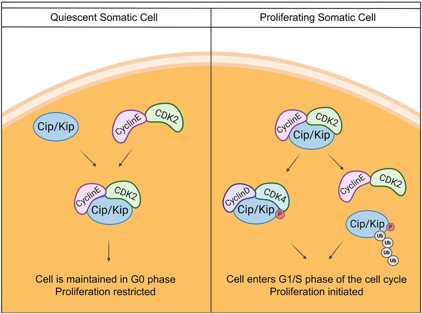
Downloaded from genesdev.cshlp.org on August 29, 2021 - Published by Cold Spring Harbor Laboratory Press

                                                                                                          Cell cycle arrest in the gonad

                                                                      eage, positive for the somatic cell marker steroidogenic
                                                                      factor 1 Nr5a1 (Stévant et al. 2019). These data imply
                                                                      that expression of the Cip/Kip family, and thus cell cycle
                                                                      arrest, is functionally important for ovarian fate.
                                                                         XX and XY gonads show substantial differences in cell
                                                                      proliferation immediately following sex determination
                                                                      (Schmahl et al. 2000), which are most likely attributed
                                                                      to transcriptional regulation. ChIP-seq experiments in re-
                                                                      cent years have identified novel interactions involving
                                                                      transcription factors that may control cell cycle arrest. Re-
                                                                      cent work by Nicol et al. (2018) performing ChIP-seq for
                                                                      the forkhead box L2 (FOXL2) protein in wild-type E14.5
                                                                      mouse ovaries revealed that FOXL2 binds to an enhancer
                                                                      of Cdkn1b. Importantly, this FOXL2 peak overlaps with a
                                                                      RUNX1 ChIP-seq peak, illustrating co-operation, or at
                                                                      least redundancy, between ovarian factors to ensure the
                                                                      activation or repression of key target genes (Nicol et al.
Figure 5. Expression of Cip/Kip transcripts across mouse gonad-       2019). The Cdkn1b enhancer region is also bound by the
al sex determination. Around E10.5 when the genital ridge thick-
                                                                      protestis factors SOX9 and DMRT1 (Rahmoun et al.
ens, Cdkn1a, Cdkn1b, and Cdkn1c transcripts are all expressed in
the somatic supporting cells (Zhao et al. 2018; Stévant et al.
                                                                      2017; Nicol et al. 2018). These original findings suggest
2019). The genital ridge increases in size, forming the bipotential   a dual approach, where p27kip1 expression is promoted
gonad. At E11.5, sex determination occurs, and the somatic sup-       by ovarian factors and repressed by testicular factors. Oth-
porting cells begin to differentiate into the XX or XY somatic cell   er experiments, where ectopic expression of FOXL2 is
lineages. In the testes, Cdkn1c is down-regulated in the somatic      driven by the Nr5a1 locus in the testis, result in the up-
cells, and low levels of Cdkn1a and Cdkn1b are maintained, to al-     regulation of Cdkn1b transcript (Nicol et al. 2018). It is
low the testes to increase in size to form the testis cords. In the   important to note that Cdkn1b is unlikely to be the
ovary, Cdkn1a and Cdkn1b are up-regulated in the somatic sup-         only factor restricting proliferation in the ovary, and
porting cells to maintain cells in dormancy. Germ cell expression     FOXL2 and SOX9 are involved in gene regulatory net-
of Cdkn1a, Cdkn1b, and Cdkn1c is currently unknown during sex
                                                                      works that must involve additional genes controlling
determination. Gene expression graphs of Cdkn1a, Cdkn1b, and
Cdkn1c transcripts during gonadal development are adapted
                                                                      and promoting proliferation. More interrogation of the
from Zhao et al. (2018).                                              CHIP-seq data sets is needed to map this network of cell
                                                                      cycle control in the gonad. Taken together, this indicates
                                                                      the male program represses Cdkn1b in the embryonic tes-
supporting cell specification, their relative expression              tis, but more importantly suggests that the key female fac-
changes individually (Nef et al. 2005; Zhao et al. 2018).             tor FOXL2 is capable of promoting Cdkn1b expression.
Each Cip/Kip CKI contains a CDK binding domain; how-                  This information, when combined with the RNA se-
ever, large variability exists in the remainder of each pro-          quencing data sets (Stévant et al. 2018, 2019), illustrates
tein’s molecular structure (Nakayama and Nakayama                     that expression of Cip/Kip family members in the sup-
1998). Cdkn1a encodes the shortest protein (p21cip1),                 porting cell lineage contributes to the mechanisms of pri-
and p21cip1 interacts with proliferating cell nuclear anti-           mary gonadal sex determination while they are in turn
gen (PCNA), showing a direct inhibition of the cell cycle             regulated by known players in this process.
(Besson et al. 2008). p27kip1 and p57kip1 are longer proteins            Additional evidence supporting the regulation of Cip/
and have distinct domains, including the cyclin binding               Kip transcripts by ovarian-specific factors, including
domain and CDK binding domain (Bachs et al. 2018).                    FOXL2, comes from genetic mouse models. When
These two proteins also have linker domains and pro-                  FOXL2 and WNT4 are deleted in mice, Cdkn1b tran-
line-rich domains with unknown functions, which may                   scripts and p27kip1 protein are down-regulated in the em-
be implicated in the unique roles that these proteins are             bryonic ovary (Garcia-Ortiz et al. 2009; Maatouk et al.
involved with during development (Nakayama and                        2013; Gustin et al. 2016). Conversely, when FOXL2 and
Nakayama 1998; Bachs et al. 2018). The expression of                  RUNX1 (factors specific to pregranulosa cells) are geneti-
Cdkn1a and Cdkn1b transcripts increases fourfold and                  cally deleted, Cdkn1a expression increases (Nicol et al.
1.4-fold, respectively, in the E11.5 developing ovary, so             2018, 2019). This implies that cell cycle arrest is particu-
that these transcripts are elevated specifically in the XX            larly important in the ovary following sex determination,
gonad (Fig. 5; Zhao et al. 2018). Cdkn1a expression pla-              and multiple factors act redundantly to ensure activation
teaus at E12.5; however, Cdkn1b transcript levels contin-             of cyclin-dependent kinase inhibitors. Whether the regu-
ue to rise as gestation continues (Zhao et al. 2018). The             lation of Cdkn1a and Cdkn1b by ovary-specific factors
opposite trend is observed with Cdkn1c (p57kip2) tran-                persists in the days following sex determination is cur-
script levels, where, by E12.5, Cdkn1c transcript is                  rently unknown. Do ovarian factors continue to stimulate
down-regulated in the XY gonad but maintained in the                  the transcription of Cdkn1a and Cdkn1b, or is their ex-
XX gonad (Stévant et al. 2018, 2019; Zhao et al. 2018).               pression maintained through other downstream canonical
This is specifically restricted to the pregranulosa cell lin-         cell cycle regulators? FOXL2, in particular, is known to

                                                                                                     GENES & DEVELOPMENT              5
Downloaded from genesdev.cshlp.org on August 29, 2021 - Published by Cold Spring Harbor Laboratory Press
Frost et al.
become essential to maintain the ovary, because its post- by data that show the role of p27kip1 in the nucleus is not
natal deletion leads to gonadal sex reversal (Uhlenhaut crucial to cell cycle arrest, because prospermatogonia are
et al. 2009). Uncovering when FOXL2 becomes the sole still able to arrest at the appropriate stage in p27kip1-null
factor necessary for ovarian identity, without the help of mice (Beumer et al. 1999). The factors responsible for the
other pathways, will lead to insights into what genes re- activation of p27kip1 in the arrested prospermatogonia
quire consistent regulation throughout development. have yet to be determined. Making use of recent single-
cell sequencing studies performed in the embryonic testis,
Sexual dimorphism is maintained in cell cycle regulatory both in the CD1 strain (Stévant et al. 2018, 2019) and in the
genes following gonad differentiation C57BL/6 strain (Tan et al. 2020), may allow for the identi-
fication of genes that are expressed at the same time as
Once gonadal sex is specified around E11.5, the ovaries or p21cip1 and p27kip1 in the germ cells. In-depth analyses of
testes must undergo many genetic and morphological these data sets together could yield interesting informa-
changes to make them capable of maturing and support- tion of the signal that initiates Cip/Kip expression in the
ing germ cells. Both the primordial germ cells, which germ cells and where this signal originates from. Do
have migrated into the gonad, and the somatic cells alter intrinsic germ cell factors like DND1 or INHA induce
their cell cycle status in a sex-dependent manner. One prospermatogonia expression of p27kip1, or is this due to
of the earliest differences distinguishing ovary and testis extrinsic factors from the Sertoli cell lineage activating
development is the resumption of proliferation in the tes- pathways in the prospermatogonia nucleus (Nicol et al.
tes (Schmahl et al. 2000; Schmahl and Capel 2003). In 2018)? Manipulating genetic networks active in germ cells
mice, the change in cell cycle status presents as a clear often proves challenging, because the mice become infer-
morphological difference, where the testes are already tile. Alternative approaches, such as the conditional dele-
double the size of the ovaries at E13.5 (Schmahl et al. tion of SOX9 at different time points in the Sertoli cells,
2000; Schmahl and Capel 2003; Nel-Themaat et al. might reveal whether the XY somatic cell environment is
2009). This rapid proliferation is essential for forming em- critical for the induction of p27kip1 in the gonocyte. Howev-
bryonic testis cords, which eventually give rise to the er, this type of experiment may be difficult to interpret,
seminiferous tubules of the adult testis. Pioneering work either because functional redundancy between Sox9 and
by Schmahl et al. (2000). showed that Sry activity (pre- Sox8 may not lead to a sufficient change in the Sertoli cells,
sumably via Sox9) is essential for promoting cell prolifer- or (and particularly if both genes are mutated) a sudden
ation in the testis. Furthermore, when proliferation of the change to a granulosa cell fate would be too disruptive. Al-
somatic cells is inhibited, just prior to sex determination ternatively, examination of p27kip1 nuclear expression in
(between E10.8 and E11.2), the testis is unable to correctly the XY testis with ectopic FOXL2 expression (Nicol et al.
form testis cords and thus cannot support fertility in the 2018) might indicate that the somatic cells are inducing
adult male (Schmahl and Capel 2003). It is now widely ac- gene changes in the gonocyte. Conversely, making use of
cepted that Sertoli cells up-regulate the expression of key conditional deletion tools to delete DND1 and INHA spe-
cell cycle genes to induce their proliferation (Rotgers et al. cifically in the gonocyte would uncover whether these fac-
2018; Stévant et al. 2018, 2019; Zhao et al. 2018). tors, in combination with p27kip1, are critical for inducing
Cip/Kip members were originally considered essential key genetic changes that promote the production and devel-
factors in the testis after sex determination, given that gon- opment of a mature spermatozoon. This may give insight
ocytes, the germ cells at this stage of development, are held into novel genetic factors that disrupt the development of
in mitotic arrest. However, there is now evidence that, at healthy spermatozoa and contribute to male infertility.
least in mice, Cip/Kip protein expression is variable- and In the developing ovary, initial studies proposed that fol-
strain-dependent. In the CD1 strain, p21cip1, p27kip1, and lowing sex determination, the decreased growth rate of XX
p57kip2 are all present in fetal germ cells in the testis at gonads compared with XY gonads is due to the high expres-
E14.5 (Western et al. 2008). In this strain, the RNA bind- sion of Cip/Kip cell cycle inhibitors (Nef et al. 2005). How-
ing-protein dead end 1 (DND1) and inhibin α (INHA) are ever, no embryonic ovarian defects are found in mouse
able to induce p21cip1 and p27kip1 in prospermatogonia to models where p21cip1, p27kip1, or p57kip2 are genetically ab-
maintain dormancy (Western 2009; Cook et al. 2011; Men- lated (Nakayama and Nakayama 1998; Rajareddy et al.
dis et al. 2011). In contrast, the investigators note that 2007). In addition, the ovaries in these three global dele-
p21cip1 and p27kip1 proteins are absent in the prospermato- tion models do not match the fast growth rate or the size
gonia in the C57BL/6 strain at E14.5, which is corroborated of the male testis, and moreover, p21cip1-null mice and
by other studies (Beumer et al. 1999; Western et al. 2008). p57kip2-null female mice are fertile (Nakayama and
Interestingly, p27kip1 is localized to the prospermatogonia Nakayama 1998). These data support the concepts of re-
nucleus just prior to birth in C57BL/6 mice (Beumer et al. dundancy between multiple dormancy factors, and/or
1999). One explanation for this strain-specific difference in the presence of a gene regulatory network with cofactors
Cip/Kip expression may be that strain-specific inbred mu- that regulate each other. These ideas are highly plausible
tations trigger cell cycle checkpoints that change the tim- in the context of the ovary, where several factors deter-
ing of p21cip1 and p27kip1 expression in the germ cells. This mine ovarian fate cooperatively, compared with the acti-
delay, in C57BL/6 mice in this case, can be overcome and vation of single genes (Sry and then Sox9) in the testis
does not impact the progression of the prospermatogonia (Fig. 4; Mork et al. 2012; Maatouk et al. 2013; Pannetier
throughout development. This description is supported et al. 2016; Stévant and Nef 2019).
6 GENES & DEVELOPMENTDownloaded from genesdev.cshlp.org on August 29, 2021 - Published by Cold Spring Harbor Laboratory Press
Cell cycle arrest in the gonad
Within the embryonic ovary, p27kip1 is restricted initial- fertility in adulthood (Fig. 6). Although there can be germ-
ly to the somatic cells that become the granulosa cells post- line stem cells in lower vertebrates, all of the oocytes that
natally, and p27kip1 expression is only found in the oocytes a female mammal will ever ovulate originate from the res-
around postnatal day (PND) 4 (Rajareddy et al. 2007). The ervoir of oogonia established by the end of gestation (Sarraj
expression of p27kip1 in somatic cells is maintained by and Drummond 2012). These are all arrested in meiosis I,
the female-determining factors, including WNT4, and at present there is limited evidence, none of which is
RSPO1, and FOXL2, in the E14.5 mouse ovary (Fig. 6; compelling, to suggest that new oocytes are able to be gen-
Fero et al. 1996; Maatouk et al. 2013; Gustin et al. 2016; erated postnatally in vivo (McGee and Hsueh 2000; Woods
Richardson et al. 2020). Interestingly, in WNT4-null and Tilly 2013; Navaroli et al. 2016; Zarate-Garcia et al.
mice, p27kip1 is specifically down-regulated at the anterior 2016; Wagner et al. 2020). This ultimately means that
pole of the ovary at E15.5 (Maatouk et al. 2013). This area of the female germ cell pool must rapidly proliferate during
p27kip1 down-regulation shows an up-regulation of the gestation, such that by the end of gestation there remains
male factor anti-Müllerian hormone (AMH), which is tes- a sufficient number of oocytes (∼8000 oocytes in mice)
tis-specific before birth but is expressed by granulosa cells (Kerr et al. 2006) to establish and maintain this reserve
in the postnatal ovary (Maatouk et al. 2013). In the days fol- for the entirety of the female reproductive life span
lowing sex determination, up-regulation of p27kip1 expres- (McGee and Hsueh 2000; Skinner 2005; Adhikari and
sion in the pregranulosa cells coincides with the entry of Liu 2009; McLaughlin and McIver 2009). Given that the
oogonia into meiosis, thus establishing the germ cell female germ cell pool must proliferate rapidly over a short
pool. Conditional deletion of p27kip1 from the pregranulosa gestational time, it is assumed that the cyclin-dependent
cell lineage would confirm whether an interaction exists kinase inhibitors are down-regulated. Indeed, p27kip1 is
between p27kip1 in the pregranulosa cells and the initiation not detected by immunofluorescence in the female germ
of meiosis in the germ cells, thus linking pregranulosa cell cells between E12.5 and E14.5 in the developing ovaries
signaling to cell cycle control in the female germ cells. (Rajareddy et al. 2007; Mork et al. 2012). It would be valu-
able to determine whether down-regulation of cyclin-de-
pendent kinase inhibitors in the oogonia is necessary to
Oogonia enter meiosis shortly after sex determination facilitate the switch to meiosis. Following E12.5, after
sex determination, there are two major differences be-
The establishment of the germ cell pool during ovarian de- tween germ cells in the ovary and in the testis. In the
velopment in mammals is necessary to ensure continued male, germ cell cysts must break down in a manner to
Figure 6. Differential control is exerted
over cell cycle regulators in the gonad fol-
lowing sex determination. By E14.5 in XX
gonads, the pregranulosa cells stop dividing.
Factors involved in ovary development,
such as FOXL2, WNT4, and RSPO1, have
all been shown to activate p27kip1 to main-
tain the somatic cells in quiescence (Maa-
touk et al. 2013; Nicol et al. 2018;
Richardson et al. 2020). In the XX gonad,
enter and then arrest in meiosis, thereby es-
tablishing the ovarian reserve, although
many oogonia are lost through atresia be-
fore birth. Retinoic acid, and perhaps other
factors, stimulates the expression of Stra8
in the nucleus, which then results in the
up-regulation of several critical meiotic reg-
ulators, including DDX4, DAZL, and
GCNA1 (Bowles and Koopman 2010).
Meiosin and Zglp1 are two factors recently
shown to be important for activating the
oogenic fate in the oogonia nucleus (Ishi-
guro et al. 2020; Nagaoka et al. 2020). In
contrast, in the XY gonad there is continued
proliferation of somatic cells (including Ser-
toli cells) and gonocytes, although the latter
undergo cell cycle arrest (G0/G1) at about
E13.5, as they become prospermatogonia, resuming mitotic cell divisions after birth (Jiang et al. 2019; Ruthig et al. 2019; Yan et al.
2020). With SOX9 expression in Sertoli cells, p27kip1 expression is repressed, allowing for cell cycle divisions (Nicol et al. 2018).
CYP26B1, also made by Sertoli cells, inhibits the action of retinoic acid to prevent the gonocytes in the developing testis from entering
meiosis (Bowles et al. 2006). Within the gonocyte, DND1 and INHA activate p27kip1 and p21cip1 to limit the proliferation of the germ cell
pool to prevent the formation of teratomas (Mendis et al. 2011; Cook et al. 2011; Ruthig et al. 2019).
GENES & DEVELOPMENT 7Downloaded from genesdev.cshlp.org on August 29, 2021 - Published by Cold Spring Harbor Laboratory Press Frost et al. retain a high proportion of germ cells, which go on to be- tional gamete, germ cells must also turn off pluripotency come germline stem cells (Lei and Spradling 2013b). This factors, including NANOG and OCT4 (Feng et al. 2014; requires the male germ cells to arrest in mitosis, which Jørgensen and Rajpert-De Meyts 2014; Arora et al. 2016). may be facilitated by cell cycle inhibitors. The second ma- Over a 4-d period, between E12.5 and E16.5, the oogonia jor difference between germ cells in the ovary and in the switch off OCT4 and up-regulate SYCP3 in an anterior- testis is the decision to enter meiosis early or to arrest in to-posterior wave (Niu and Spradling 2020). These subtle mitosis (Bowles and Koopman 2010). transcriptional changes cannot be detected using tradi- Germ cell sex is not controlled by the presence of XX or tional global RNA sequencing methods. However, recent XY chromosomes within the germ cells; instead, they rely single-cell sequencing approaches have been successfully on the surrounding somatic environment for their com- used to uncover other discreet spatial and temporal regu- mitment to oogenesis or spermatogenesis (Adams and lators of meiosis (Niu and Spradling 2020; Zhao et al. McLaren 2002). Watershed coculture studies, where 2020). Both studies performed fine-scale analyses of the E11.5 LacZ-labeled XX germ cells were cultured with germ cell meiotic transcriptomes covering the E12.5 and E12.5 XY genital ridges, showed that XX germ cells would, E14.5 (Niu and Spradling 2020) and E12.5, E14.5, and rather than switch to meiosis, continue in mitosis in the E16.5 (Zhao et al. 2020) time points, respectively. Another XY genital ridge (Adams and McLaren 2002). There are unreviewed preprint also analyzed germ cell transcrip- also studies in chimeras and in sex-reversed mice, both tomes between E10.5 and E16.5 (Mayère et al. 2021). All XX male and XY female, that germ cell sex is dependent the studies conclude that XX and XY germ cells diverge on the type of gonad, i.e., ovaries or testes, rather than around E12.5 and that thousands of genes varied during on their own sex chromosome status (Ottolenghi et al. this period of development. By analyzing these extensive 2007; Vernet et al. 2014). These results showed that sig- data sets, multiple pathways and gene regulatory net- nals from the surrounding somatic environment stimu- works will be identified that are important for initiating late and drive the switch from mitosis to meiosis. and progressing meiosis. Multiple signals emanate from the somatic environ- Together with the information obtained about the initi- ment to promote meiosis. The classical pathway is the ret- ation of meiosis, these data sets have also provided a inoic acid pathway, where retinoic acid (RA) released wealth of information about Cip/Kip family expression from the mesonephros into the gonad is responsible for between E12.5 and E16.5 in mice (Niu and Spradling stimulating meiosis within the ovary (Bowles et al. 2020; Zhao et al. 2020). Both studies showed that Cdkn1a 2006). Expression of the premeiotic marker Stra8 and expression peaks in the preleptotene stage of germ cell de- up-regulation of the meiotic markers Sycp3 and Dmc1 velopment and decreases as the germ cells enter the lepto- are stimulated by RA signaling (Fig. 6; Bowles et al. tene stage of meiosis (Niu and Spradling 2020; Zhao et al. 2006). New studies suggest that it may not be retinoic 2020). In contrast, Cdkn1c expression peaks at the lepto- acid, with meiosis occurring normally in mice without tene stage, before decreasing as the germ cells continue any retinoic acid receptors (Vernet et al. 2020). Addition- through meiosis (Niu and Spradling 2020). Interestingly, ally, new pathways responsible for stimulating the oogo- Cdkn1b transcript is expressed stably throughout the nia to enter meiosis are emerging. For example, a recent stages of meiosis, despite not being detected by immuno- study identified MEIOSIN as a cofactor of STRA8, where fluorescence (Rajareddy et al. 2007; Niu and Spradling MEIOSIN and STRA8 bind to the transcriptional start 2020). Alternatively, both Cdkn1a and Cdkn1c decrease sites of target genes, allowing for the rapid transcription as the germ cells enter meiosis; however, it is unknown that is required of germ cells entering meiosis (Ishiguro whether this is a contributing factor to meiosis entry or et al. 2020). Another factor, ZGLP1, precedes the expres- is a consequence of the change in the cell division process. sion of Stra8, and Zglp1−/− mice have atrophied ovaries with no oocytes as early as E17.5 (Nagaoka et al. 2020). Promising data show that ZGLP1 is downstream from Communication between the oogonia and somatic cells BMP2 in primordial germ cell-like cells (PGCLCs), linking establishes germ cell nests in the ovary the in vitro model to the in vivo mouse model (Nagaoka et al. 2020). These data follow on from recent work that As the ovary continues to develop, the oogonia rely pri- described a mechanism for meiosis entry where BMP is marily on the ovarian environment, established by the also required alongside RA for germ cell sex specification pregranulosa cells. The somatic pregranulosa cells con- (Miyauchi et al. 2017). Both MEIOSIN and ZGLP1 are es- tribute to the structure, known as a germ cell nest, which sential for stimulating the oogenic program, but it is un- contains the oogonia (Fig. 7; Pepling and Spradling 1998, clear what signals are activating these factors in vivo. 2001). Nest formation is a lengthened process, where cysts Further work to examine how BMP and RA coordinate begin to form around E10.5 and E11.5, and finish assembl- meiosis entry will provide more detailed networks that ing around E13.5–E14.5, through five rounds of divisions promote the activation of meiosis at specific temporal of the oogonia (Lei and Spradling 2016). Much like the on- windows. set of meiosis, germ cell nest formation is asynchronous A major challenge in studying the mitosis-to-meiosis (Lei and Spradling 2016; Wang et al. 2017). Due to this switch in mice is that their germ cells enter meiosis asyn- asynchronous nature of germ cell nest formation, little chronously, in an anterior-to-posterior pattern (Bullejos is known about the pathways responsible that guide the and Koopman 2004). To enter meiosis and become a func- formation of the nests. However, communication 8 GENES & DEVELOPMENT
Downloaded from genesdev.cshlp.org on August 29, 2021 - Published by Cold Spring Harbor Laboratory Press

                                                                                                          Cell cycle arrest in the gonad

                                                                                  Figure 7. Germ cell nest breakdown and primor-
                                                                                  dial follicle formation are asynchronous events.
                                                                                  During embryonic development, XX primordial
                                                                                  germ cells arrest in the diplotene stage of prophase
                                                                                  and are termed oogonia. By E18.5, these oogonia
                                                                                  are now termed oocytes and are surrounded by in-
                                                                                  tercellular bridges (purple) and somatic pregranu-
                                                                                  losa cells (orange). This structure is termed a
                                                                                  germ cell nest. The process of germ cell nest
                                                                                  breakdown is asynchronous, with medullary nests
                                                                                  broken down first. Germ cell nests begin to be bro-
                                                                                  ken down before birth, and the somatic pregranu-
                                                                                  losa cells (orange) infiltrate the nest and surround
                                                                                  the oocyte, through a process known as primordial
                                                                                  follicle formation. A primordial follicle is an oo-
                                                                                  cyte surrounded by one layer of flattened pregra-
                                                                                  nulosa cells. Primordial follicles remain dormant
                                                                                  until the process of primordial follicle activation,
                                                                                  where follicles are selected to develop. This pro-
                                                                                  cess also begins first in the medullary region of
                                                                                  the ovary around PND4, and cortical follicles re-
                                                                                  main quiescent.

between JAGGED1 in the oocytes and NOTCH2 in the                   rez-Sanz et al. 2013). However, this phenotype is
pregranulosa cells is considered important in regulating           observed in many gene deletion studies and through the
nest formation (Xu and Gridley 2013; Vanorny et al.                use of microtubule inhibitors, both in vivo and in vitro,
2014). Each nest holds several somatic cells, as well as           and therefore it may not be specific to cell cycle inter-
germ cells connected by intercellular bridges, which join          ferences. This highlights that the mechanisms in germ
the cytoplasm of individual germ cells within a cyst (Lei          cell nest formation and breakdown that drive these
and Spradling 2013b, 2016). In the germ cells at least, ca-        ovarian phenotypes are unknown and require further
nonical cell cycle machinery must be silenced to block cy-         examination.
tokinesis and allow intercellular bridge formation (Lei and
Spradling 2013b). How the germ cells are programmed to
cease cycling and generate germ cell cyst structures war-          Germ cell nest breakdown leads to the formation
rants further study but may be due to the broad action of          of primordial follicles
STRA8 (Kojima et al. 2019). Large cysts separate into
smaller cysts such that, by E17.5, cysts contain between           A large body of work has contributed to a detailed under-
three and four oogonia, and at birth cysts only contain            standing of the process of germ cell nest breakdown (for re-
two oogonia (Lei and Spradling 2013a). It is thought               views on this topic, see Tingen et al. 2009; Wear et al.
that cytokinesis remains incomplete to form the intercel-          2016; Ikami et al. 2017). This process underpins another
lular bridges within each cyst (Lei and Spradling 2013b).          key stage of ovarian development, as nest breakdown is
However, the direct mechanisms that drive the break-               the first step in the formation of primordial follicles (Tin-
down of these cysts are not known in detail. Pioneering            gen et al. 2009; Suzuki et al. 2015; Rosario et al. 2019). Pri-
studies from Lei and Spradling (2016) have contributed             mordial follicles are the structures that maintain oocytes
the most to this question, showing that cyst breakdown             in dormancy after birth and into adulthood (Skinner 2005).
is dependent on a number of events, including the fusion           A primordial follicle is composed of an oocyte surrounded
of germ cell membranes, the transfer of cytoplasmic ma-            by one layer of flattened pregranulosa cells (Fig. 7;
terials between germ cells, and the apoptosis of remnant           McLaughlin and McIver 2009; Tingen et al. 2009; Reddy
materials and nuclei. As the mechanisms governing germ             et al. 2010; Sánchez and Smitz 2012; Sarraj and Drum-
cell nest breakdown are still being elucidated, little is          mond 2012). Proper formation of the primordial follicles
known about the expression of the Cip/Kip family during            relies on the specific temporal initiation of germ cell
germ cell nest formation and breakdown. One observa-               nest breakdown (Hardy et al. 2018; Ford et al. 2019; Chak-
tion in p27kip1-null mice is an accelerated assembly of            ravarthi et al. 2020). Germ cell nest breakdown is initiated
primordial follicles, where at PND1, oogonia are already           around E17.5 in the medullary region of the ovary. Nest
separated from the nest structure and pregranulosa cells           breakdown then continues over a period of ∼5 d, with
are surrounding the oogonia, much earlier than in ovaries          medullary cysts the first to form primordial follicles. Cor-
from wild-type littermates (Rajareddy et al. 2007). These          tical cysts break down after birth, ∼3 d postpartum (Wang
mice also have higher rates of multi-oocyte follicles, indi-       et al. 2017). Over this time, pregranulosa cells invade the
cating a defect in the formation of primordial follicles (Pe-      germ cell nests, and the intercellular bridges between

                                                                                                    GENES & DEVELOPMENT               9
Downloaded from genesdev.cshlp.org on August 29, 2021 - Published by Cold Spring Harbor Laboratory Press
Frost et al.
germ cells are broken down (Pepling and Spradling 2001; form early in development and validate the independent
Lei and Spradling 2013b). origins of several distinct populations of pregranulosa
Many of the canonical pathways in intercellular bridge cells (Niu and Spradling 2020). One of the key contribu-
breakdown are common to those involved in follicle for- tions of this work to our knowledge is that two distinct
mation, including NOTCH signaling, E-cadherin, KITL, classes of primordial follicles are formed during germ
and TGF-β signaling (Chen et al. 2007; Jones and Pepling cell nest breakdown and that these primordial follicles
2013; Xu and Gridley 2013; Wang et al. 2015). These path- contain pregranulosa cells with different gene expression
ways have ligands and receptors in both granulosa cells signatures (Niu and Spradling 2020). These two classes
and oocytes that signal to each other to coordinate germ of primordial follicles also vary in their cell cycle status,
cell nest breakdown. Recently, novel transcription factors where medullary wave 1 follicles immediately activate
have been found to stimulate and facilitate germ cell nest and the granulosa cells begin proliferating, whereas corti-
breakdown. SP1, IRX3, and IRX5 are all factors expressed cal wave 2 follicles remain arrested (Niu and Spradling
in pregranulosa cells that promote germ cell nest break- 2020). It would be interesting to examine the regulatory
down (Fu et al. 2018; Cai et al. 2019). When SP1 is lost, mechanisms controlling the resumption of the cell cycle
germ cell nests persist and primordial follicle formation in wave 1 follicles. Importantly, in the Niu and Spradling
does not occur, leading to an infertility phenotype after (2020) data set, all three Cip/Kip family members Cdkn1a,
birth (Cai et al. 2019). Irx3 is specifically expressed in Cdkn1b, and Cdkn1c are all expressed in both pregranu-
the intercellular bridges of germ cell nests, and when losa populations, whether the cells are of a bipotential
both Irx3 and Irx5 are deleted, defective granulosa cells cell origin or an epithelial cell origin. A subsequent study
fail to connect with the oocyte, causing aberrant follicle from Wang et al. (2020) performed a similar study at E16.5,
formation and apoptosis of the developing follicles (Fu PND0, and PND3 to better understand the dynamics of
et al. 2018). Other factors are specific to the signals origi- primordial follicle assembly. This study confirmed the
nating from the oocyte, including factors like ELAV-like presence of multiple populations of pregranulosa cells
protein 2 (ELAVL2) and DEAD-box helicase 6 (DDX6) and went one step further to examine the transcriptional
(Kato et al. 2019). Loss of ELAVL2 or DDX6 in isolation networks that are involved in the interactions between
both lead to an infertility phenotype in mice, with no oo- oocytes and pregranulosa cells (Wang et al. 2020). In agree-
cytes remaining in the ovary 2 wk after birth due to a ment with the Niu and Spradling (2020) study, Wang et al.
defect in primordial follicle formation (Kato et al. 2019). (2020) comment on the expression of Cdkn1a and
Together these studies show that the maintenance of Cdkn1b in the pregranulosa cells and hypothesize an in-
granulosa–oocyte communication is essential for germ teraction with the FOXO pathways in oocytes (Wang
cell nest breakdown and follicle formation. Importantly, et al. 2020).
if this communication is disrupted or fails, there are major The identification of multiple populations of pregranu-
consequences for future fertility. losa cells has been reported before, although single-cell se-
quencing allows the interrogation of these cell types in
great detail at the transcriptional level. Over the past 10
Single-cell sequencing data sets provide new insights yr, studies have shown that two classes of primordial fol-
into germ cell nest breakdown and primordial licles are formed during early postnatal life. Mounting ev-
follicle formation idence shows heterogeneity within the pregranulosa cell
population, indicating that the SF1-positive lineage from
Due to the intricate communication networks that exist the bipotential gonad is not the only source of pregranu-
between oocytes and the granulosa cells, it has proven dif- losa cells (Mork et al. 2012; Rastetter et al. 2014; Nicol
ficult to isolate the group of cells responsible for the initi- et al. 2018; Stévant et al. 2019). For example, cells from
ation of germ cell nest breakdown and follicle formation. the ovarian surface epithelium migrate into the ovarian
Pioneering single-cell sequencing experiments provide cortex and differentiate into pregranulosa cells (Mork
alternative methods to lineage-tracing studies. Using sin- et al. 2012). The epithelial-derived pregranulosa cells re-
gle-cell sequencing, individual cells are clustered together main near the epithelium in the cortex of the ovary and
into related populations based on transcriptomic data. In express leucine-rich repeat-containing G protein-coupled
the ovary, the pregranulosa cell lineages and subclusters receptor 5 (LGR5) (Ng et al. 2014; Rastetter et al. 2014).
have been reconstructed using single-cell data sets (Niu These two populations of pregranulosa cells remain re-
and Spradling 2020; Wang et al. 2020). Comprehensive stricted to distinct domains of the ovary to contribute to
data sets covering the stages of germ cell nest breakdown, two classes of primordial follicles. The inner ovarian med-
primordial follicle formation, and primordial follicle acti- ullary region contains germ cells surrounded by pregranu-
vation were generated using single-cell sequencing on losa cells positive for FOXL2 expression, and the outer
whole ovaries from E18.5, PND1, and PND5 (Niu and ovarian cortical region contains migrating pregranulosa
Spradling 2020). Unlike the single-cell sequencing data cells from the epithelium positive for LGR5 expression,
sets from Stévant et al. (2018, 2019), which isolated cells and later express FOXL2 (Rastetter et al. 2014). The dis-
using the expression of SF1, this study allows the simulta- tinction between these populations of primordial follicles
neous comparison of germ cells and somatic cells. Prelim- is very important, as medullary primordial follicles acti-
inary findings from this study reveal key insights into vate for development just after birth (Mork et al. 2012;
ovarian development. Namely, that germ cells are uni- Maatouk et al. 2013). In contrast, cortical primordial
10 GENES & DEVELOPMENTDownloaded from genesdev.cshlp.org on August 29, 2021 - Published by Cold Spring Harbor Laboratory Press
Cell cycle arrest in the gonad
follicles maintain dormancy in the ovary and provide the ian phenotypes. First, accelerated activation of all
oocytes for the reproductive life span of mice (Mork et al. primordial follicles occurs immediately after birth (Rajar-
2012). To maintain fertility, these follicles must remain eddy et al. 2007). As no primordial follicles remain in the
dormant, until temporally selected for the process of pri- ovary by 12 wk of age, the mouse is infertile (Rajareddy
mordial follicle activation. et al. 2007). Second, as the ovary contains a larger propor-
tion of multi-oocyte follicles, germ cell nest breakdown is
perturbed (Perez-Sanz et al. 2013). This is attributed to a
Numerous pathways lead to a wave of primordial follicle defect in pregranulosa cell migration into the cyst neces-
activation postnatally sary to encircle and separate individual oocytes (Perez-
Sanz et al. 2013). siRNA knockdown of p27kip1 in 3-d
Primordial follicle activation is the process by which dor-
mouse ovaries also presents with a precocious activation
mant follicles are selectively chosen for growth and devel-
of all the primordial follicles (Hirashima et al. 2011). Com-
opment in preparation for ovulation. This overarching
bined, these experiments describe an essential role for cell
developmental process is known as folliculogenesis, and
cycle inhibitors in postnatal ovary development. It is im-
a number of current and comprehensive reviews detail
portant to note that these studies are performed in mice
this process (Kerr et al. 2013; Hsueh et al. 2015; Kallen
with complete Cip/Kip deletions that are present from
et al. 2018; Ford et al. 2019). While new sperm are contin-
birth, rather than targeted conditional cell type-specific
uously produced during the reproductive life span of
deletions or inducible deletions. As a result, the initiation
males, there is no generation of new oocytes in the ovary,
of the defect is still unclear; for example, it is conceivable
and the primordial follicles that are assembled in the peri-
that the adult ovarian fertility phenotype persists from a
natal period are responsible for the entire reproductive life
germ cell defect at the time of cyst breakdown. Precise ge-
span of the adult female (McLaughlin and McIver 2009).
netic experiments, such as a Cip/Kip-inducible deletion
Consequently, tight regulation of follicle activation is im-
mouse line or a Cip/Kip deleted only in the germ cells (us-
perative to ensuring fertility. Activation of all primordial
ing a germ cell Cre line), would add valuable information
follicles at once or an inability to coordinate temporal fol-
to the field about the function of Cip/Kip during follicle
licle activation results in premature infertility. Numerous
formation and development. Using the single-cell se-
studies in mice have shown that, similar to germ cell nest
quencing techniques in mouse models where follicle acti-
breakdown, cellular cross-talk between oocytes and
vation is dysregulated, as in the p27-null mouse model,
pregranulosa cells is essential for coordinating primordial
will also highlight the changes at the gene level and give
follicle activation (Adhikari and Liu 2009). Well-charac-
an insight into how p27kip1 might control primordial folli-
terized signaling pathways in the activation of primordial
cle activation. Another question worth addressing is
follicles include the phosphoinositide 3-kinase (PI3K)
whether p27kip1 can also act as a transcription factor dur-
pathway, phosphatase and tensin homolog (PTEN) path-
ing follicle development to stimulate other dormancy fac-
way, kit ligand (KIT/L) pathway, forkhead box L2
tors to sustain oocytes for future fertility. The role of Cip/
(FOXL2) pathway, and JAK/STAT pathway (Uda et al.
Kip members in follicle activation may be also an indirect
2004; Hutt et al. 2006; Reddy et al. 2008; Jagarlamudi
effect of the follicle activation process. Loss of p27kip1
et al. 2009; Sutherland et al. 2012, 2018; Hall et al. 2017;
leaves several cyclins and cyclin-dependent kinases free
Grosbois and Demeestere 2018). These studies illustrate
to promote cell cycle progression, thus encouraging gran-
that it is unlikely for a singular pathway to stimulate or re-
ulosa cell proliferation. Cyclin D is known to be directly
press the activation of primordial follicles. Moreover, it is
regulated by SMAD3 (Granados-Aparici et al. 2019),
the coordinated interplay of numerous signaling pathways
which is a well-known inhibitor of follicle activation, im-
that regulates this pivotal developmental process.
plicating other cell cycle genes in primordial follicle acti-
vation. Further functional studies into the networks with
Cell cycle inhibitors maintain primordial follicles which the cyclin-dependent kinase inhibitors interact
in quiescence will elucidate whether these factors are involved in novel
pathways or in combination with well-established follicle
Factors that inhibit primordial follicle activation also play activation pathways.
a critical role in maintaining fertility, and cell cycle inhib-
itors maintain pregranulosa cell identity in the dormant
state by preventing cellular proliferation. Both p21cip1 Conclusions
and p27kip1 have been implicated in germ cell nest break-
down and primordial follicle formation (Fig. 7; Bayrak and Until recently, our knowledge about ovarian development
Oktay 2003; Rajareddy et al. 2007). p21cip1 is regulated by has largely been limited to information derived from ana-
the proliferation pathway member JAK2, which is known lyzing the expression and function of single genes. The ad-
to facilitate germ cell nest breakdown (Huang et al. 2018). vent of new sequencing technologies like single-cell RNA
However, numerous studies show a function of p27kip1 in sequencing has revealed that complex networks stimulate
germ cell nest breakdown and follicle development (Fero early ovarian development and support the formation of
et al. 1996; Bayrak and Oktay 2003; Rajareddy et al. primordial follicles. These networks work in cooperation
2007; Hirashima et al. 2011; Perez-Sanz et al. 2013). Glob- with cell cycle regulators to ensure that proliferation and
al deletion of p27kip1 in mice causes several deficient ovar- differentiation occur at the appropriate temporal window.
GENES & DEVELOPMENT 11You can also read