Omics Approaches for Understanding Biogenesis, Composition and Functions of Fungal Extracellular Vesicles - Frontiers
←
→
Page content transcription
If your browser does not render page correctly, please read the page content below
REVIEW
                                                                                                                                                   published: 03 May 2021
                                                                                                                                          doi: 10.3389/fgene.2021.648524

                                              Omics Approaches for
                                              Understanding Biogenesis,
                                              Composition and Functions of
                           Edited by:
                      Bhabatosh Das,
                                              Fungal Extracellular Vesicles
          Translational Health Science
     and Technology Institute (THSTI),        Daniel Zamith-Miranda 1,2* † , Roberta Peres da Silva 3† , Sneha P. Couvillion 4 ,
                                  India       Erin L. Bredeweg 5 , Meagan C. Burnet 4 , Carolina Coelho 3 , Emma Camacho 6 ,
                        Reviewed by:          Leonardo Nimrichter 7 , Rosana Puccia 8 , Igor C. Almeida 9 , Arturo Casadevall 6 ,
                              Yifan Ge,       Marcio L. Rodrigues 10,11 , Lysangela R. Alves 10 , Joshua D. Nosanchuk 1,2 and
      Massachusetts General Hospital          Ernesto S. Nakayasu 4*
        and Harvard Medical School,
                                              1
                         United States          Department of Microbiology and Immunology, Albert Einstein College of Medicine, Bronx, NY, United States, 2 Division
                 Ali Salehzadeh-Yazdi,        of Infectious Diseases, Department of Medicine, Albert Einstein College of Medicine, Bronx, NY, United States, 3 MRC Centre
      University of Rostock, Germany          for Medical Mycology, University of Exeter, Exeter, United Kingdom, 4 Biological Sciences Division, Pacific Northwest National
             Krishnamohan Atmakuri,           Laboratory, Richland, WA, United States, 5 Environmental and Molecular Sciences Laboratory, Pacific Northwest National
          Translational Health Science        Laboratory, Richland, WA, United States, 6 Department of Molecular Microbiology and Immunology, Johns Hopkins
     and Technology Institute (THSTI),        Bloomberg School of Public Health, Baltimore, MD, United States, 7 Laboratório de Glicobiologia de Eucariotos, Instituto
                                   India      de Microbiologia Paulo de Góes, Universidade Federal do Rio de Janeiro, Rio de Janeiro, Brazil, 8 Departamento
                                              de Microbiologia, Imunologia e Parasitologia, Escola Paulista de Medicina-Universidade Federal de São Paulo, São Paulo,
                  *Correspondence:            Brazil, 9 Department of Biological Sciences, Border Biomedical Research Center, University of Texas at El Paso, El Paso, TX,
               Daniel Zamith-Miranda          United States, 10 Laboratório de Regulação da Expressão Gênica, Instituto Carlos Chagas-FIOCRUZ PR, Curitiba, Brazil,
daniel.zamithmiranda@einsteinmed.org          11
                                                 Instituto de Microbiologia Paulo de Góes, Universidade Federal do Rio de Janeiro, Rio de Janeiro, Brazil
                 Ernesto S. Nakayasu
          ernesto.nakayasu@pnnl.gov
     † These   authors have contributed       Extracellular vesicles (EVs) are lipid bilayer structures released by organisms from all
                    equally to this work      kingdoms of life. The diverse biogenesis pathways of EVs result in a wide variety
                                              of physical properties and functions across different organisms. Fungal EVs were
                   Specialty section:
         This article was submitted to        first described in 2007 and different omics approaches have been fundamental to
                      Systems Biology,        understand their composition, biogenesis, and function. In this review, we discuss
               a section of the journal
                  Frontiers in Genetics
                                              the role of omics in elucidating fungal EVs biology. Transcriptomics, proteomics,
       Received: 31 December 2020
                                              metabolomics, and lipidomics have each enabled the molecular characterization of
           Accepted: 06 April 2021            fungal EVs, providing evidence that these structures serve a wide array of functions,
           Published: 03 May 2021
                                              ranging from key carriers of cell wall biosynthetic machinery to virulence factors. Omics
                            Citation:
                                              in combination with genetic approaches have been instrumental in determining both
                  Zamith-Miranda D,
     Peres da Silva R, Couvillion SP,         biogenesis and cargo loading into EVs. We also discuss how omics technologies
Bredeweg EL, Burnet MC, Coelho C,             are being employed to elucidate the role of EVs in antifungal resistance, disease
 Camacho E, Nimrichter L, Puccia R,
           Almeida IC, Casadevall A,
                                              biomarkers, and their potential use as vaccines. Finally, we review recent advances in
            Rodrigues ML, Alves LR,           analytical technology and multi-omic integration tools, which will help to address key
   Nosanchuk JD and Nakayasu ES
                                              knowledge gaps in EVs biology and translate basic research information into urgently
          (2021) Omics Approaches
      for Understanding Biogenesis,           needed clinical applications such as diagnostics, and immuno- and chemotherapies to
Composition and Functions of Fungal           fungal infections.
               Extracellular Vesicles.
           Front. Genet. 12:648524.           Keywords: extracellular vesicles, fungi, virulence, systems biology, proteomics, metabolomics, lipidomics,
   doi: 10.3389/fgene.2021.648524             transcriptomics

Frontiers in Genetics | www.frontiersin.org                                          1                                              May 2021 | Volume 12 | Article 648524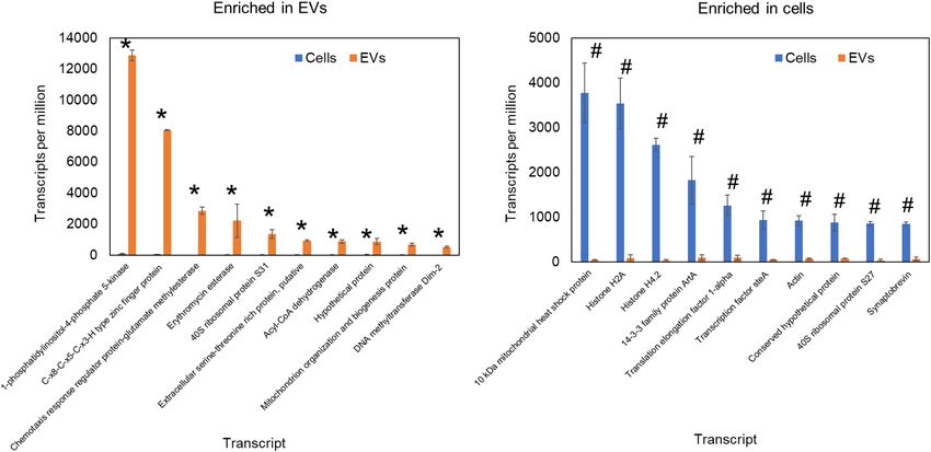
Zamith-Miranda et al.                                                                                      Omics of Fungal Extracellular Vesicles

INTRODUCTION                                                            have shown that heat-killed C. neoformans failed to secrete EVs
                                                                        (Rodrigues et al., 2007). With regard to the cell wall transit,
Cells secrete a variety of molecules to the extracellular               recent studies have shown that this structure is easily penetrated
milieu, from the smallest metabolites to large proteins and             by vesicles (Walker et al., 2018). To further investigate this
glycoconjugates. These secreted molecules range from toxic              issue, we compared lipidomic analysis data of H. capsulatum
catabolites to cell communication molecules, virulence factors,         EVs (Cleare et al., 2020) and whole cells (Burnet et al., 2020).
and enzymes involved in nutrient acquisition. One particularly          We found a significant depletion of the energy storage lipid,
intriguing mechanism of secretion is the release of extracellular       triacylglycerol, and of the mitochondrial lipid, cardiolipin, in
vesicles (EVs). Organisms from all kingdoms have been                   EVs when compared with whole cells (Figure 1). The fact that
described to release EVs (Toyofuku et al., 2019; Woith et al.,          EVs and whole cells have distinct lipid composition suggests
2019; Rayamajhi and Aryal, 2020; Rizzo et al., 2020b). In               they are formed by specific EVs biogenesis process(es), rather
fungi, EVs were first described and partially characterized in          than being a product of cell death, and/or breakdown. Lipidomic
Cryptococcus neoformans (Rodrigues et al., 2007). Since the             analysis of EVs from two Paracoccidioides brasiliensis isolates
original description, different omics approaches have been              of different phylogenetic groups showed differences in sterol
instrumental for the characterization of EVs, from their putative       and fatty acid composition of EVs when compared with whole
mechanisms of biogenesis to their function in the fungal biology.       cells, also suggesting the involvement of specific organelles in
In this article, we review the contribution of different omics          EVs biogenesis (Vallejo et al., 2012a). In addition, the deletion
approaches to study fungal EVs.                                         of the sterol biosynthesis gene Erg6 induced changes in the
                                                                        lipid and protein content of C. neoformans EVs, suggesting a
                                                                        role for sterols in EVs formation (Oliveira et al., 2020). Studies
INITIAL CHARACTERIZATION OF                                             have shown that fungal EVs can originate from intracellular
FUNGAL EXTRACELLULAR VESICLES                                           organelles, such as endosomes (Oliveira et al., 2010; Zarnowski
                                                                        et al., 2018; Zhao et al., 2019; Park et al., 2020), or at
Cryptococcus neoformans EVs comprise a heterogeneous                    the plasma membrane (Rodrigues et al., 2000, 2013; Rizzo
population of lipid bilayer vesicles, including pigment-                et al., 2020a). Morphological studies of C. neoformans showed
containing,        electron-lucid,     electron-dense,      and         structures resembling multivesicular bodies (MBVs) that can fuse
membrane-associated electron-dense vesicles. The first                  to the plasma membrane, resulting in the release of intraluminal
proteomic analysis of cryptococcal EVs led to the identification        MBV vesicles into the fungal periplasm (Takeo et al., 1973).
of 76 proteins involved in a variety of functions, including            Those images suggested that populations of fungal EVs might
virulence, oxidative stress, unfolded-protein response,                 correspond to exosomes, which are mammalian EVs released to
cellular metabolism, protein translation, signal transduction,          the extracellular milieu by the fusion of MVBs to the plasma
cytoskeleton organization, and also proteins found in the               membrane (Raposo and Stoorvogel, 2013). This biogenesis
plasma membrane (Rodrigues et al., 2008). Subsequent                    pathway was supported by recent studies in C. neoformans
studies in Histoplasma capsulatum identified 206 EV-                    (Park et al., 2020) and Candida albicans (Zarnowski et al.,
associated proteins with a similar diversity of functions to            2018), in which deletion of genes affecting MVB formation
C. neoformans EVs, besides proteins involved in cell synthesis          resulted in aberrant vesicles and/or decreased EVs production. In
and remodeling (Albuquerque et al., 2008). Lipidomic analysis of        Saccharomyces cerevisiae, deletion of several regulators of either
H. capsulatum EVs led to the identification of 18 phospholipids,        conventional or unconventional secretion resulted in alterations
including phosphatidylethanolamine, phosphatidylcholine, and            of EVs composition, as measured by proteomic analysis (Oliveira
phosphatidylserine species (Albuquerque et al., 2008). These            et al., 2010). In C. neoformans, the deletion of SEC6, a gene
initial characterization of fungal EVs opened new questions             participating in the post-Golgi secretory pathway, also resulted
about their biogenesis and roles in infection, along with their         in reduced EVs formation (Panepinto et al., 2009). In the
potential use for clinical and biotechnological applications, as        filamentous fungus Neurospora crassa, GFP-localization of SEC-
discussed in the subsequent sections.                                   6, -5, -8, and -15 subunits of the exocyst complex each form a
                                                                        crescent just beyond the cluster of vesicles of the Spitzenkörper
                                                                        at an extending hyphal tip (Riquelme et al., 2014). The exocyst
CELLULAR SITES AND MECHANISMS                                           allows a physical linkage of the vesicle cluster to the apical
OF EXTRACELLULAR VESICLE                                                membrane. The cellular events of these EVs is also a matter of
FORMATION IN FUNGI                                                      debate, including fusion or vesicle budding and secretion (Rizzoli
                                                                        and Jahn, 2007; Miura and Ueda, 2018). These studies show that
The precise cellular sites and mechanisms of fungal EVs                 intracellular regulators of secretory pathways, such as the post-
formation are still not fully defined. At first, there was some         Golgi pathway, participate in fungal EVs formation, similar to
skepticism in the field that EVs may be products of dying cells         what occurs in mammalian EVs.
whereby released lipids self-assembled into vesicles. Skepticism            As observed in protozoan parasites (Marcilla et al., 2014;
about EVs was also fueled by concerns of how such large                 Szempruch et al., 2016) and mammals (Raposo and Stoorvogel,
structures could cross the cell wall, which was viewed as a rigid       2013; Stahl and Raposo, 2019), fungal EVs can also be formed
structure that would preclude vesicular transport. However, we          at the plasma membrane. Immunofluorescence of C. neoformans

Frontiers in Genetics | www.frontiersin.org                         2                                     May 2021 | Volume 12 | Article 648524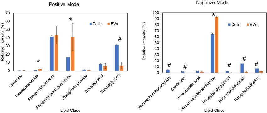
Zamith-Miranda et al.                                                                                                                  Omics of Fungal Extracellular Vesicles

  FIGURE 1 | Comparative lipidomic analysis of H. capsulatum EVs and whole cells. Lipidomics data from previous publications (Burnet et al., 2020; Cleare et al.,
  2020) of yeast cells grown in F12 medium. Each lipid class was normalized by the total ion intensity of the identified lipid species. Lipid classes significantly (p ≤ 0.05
  by Student’s t-test) enriched in extracellular vesicles and whole cells are shown by asterisks and hash signs, respectively. (A) Lipids quantified by mass spectrometry
  in the positive ionization mode. (B) Lipids quantified by mass spectrometry in the negative ionization mode.

surface lipids revealed plasma membrane projections, suggesting                           CELL WALL REMODELING BY
that the plasma membrane could bud and release EVs (Rodrigues                             EXTRACELLULAR VESICLE CARGO
et al., 2000). The participation of the plasma membrane in
fungal EVs formation was also shown using “wall-less” Aspergillus                         The cell wall is responsible for shaping fungal cells and for their
fumigatus cells (Rizzo et al., 2020a). Ultra-resolution microscopy                        resistance to diverse types of stress (Nimrichter et al., 2016).
analyses of these fungal protoplasts demonstrated the occurrence                          Nutrient availability, ambient pH, temperature, osmotic stressors,
of EVs budding from the plasma membrane. Shedding of                                      and other extracellular stimuli can lead to cell wall remodeling,
these plasma membrane-derived EVs was increased during cell                               which includes structural changes in their major components
wall synthesis, suggesting their participation in this process.                           such as chitin, glucan, and glycoproteins (Nimrichter et al., 2016).
Accordingly, protoplast EVs contain cell-wall polysaccharides                             The interplay between rigidity and plasticity of the cell wall is a
and polysaccharide synthases, which are plasma membrane-                                  key factor for fungal adaptation, survival, growth, and virulence
associated enzymes (Rizzo et al., 2020a). These morphological                             (Nimrichter et al., 2016; Beauvais and Latgé, 2018). Although
and compositional studies support the presence of plasma                                  the cell wall synthesis and shaping are classically attributed to
membrane-derived EVs in fungi similar to the mammalian                                    plasma membrane proteins, EVs might also play a role in this
microvesicles (or ectosomes; Raposo and Stoorvogel, 2013;                                 process (Vallejo et al., 2012b; Nimrichter et al., 2016; Ikeda et al.,
Stahl and Raposo, 2019). However, additional mechanisms of                                2018; Zhao et al., 2019; Dawson et al., 2020). In P. brasiliensis,
plasma membrane-derived EVs formation that differ from those                              60% of the non-covalently bound cell wall proteins, detected by
described for mammalian microvesicles have been described in                              proteomic analysis of two distinct isolates, have been described
fungi. In S. cerevisiae, electron tomography studies revealed deep                        in fungal EVs (Longo et al., 2014). The EVs content can vary
invaginations of the plasma membrane organized as two parallel                            deeply depending on the growth conditions and fungal species
membranes extending a few hundred nanometers toward the                                   (Vallejo et al., 2012a,b; Longo et al., 2014; Peres et al., 2015;
cell center. These structures can curve back to the cell surface,                         Alves et al., 2019; Peres da Silva et al., 2019; Cleare et al., 2020).
resulting in fusion with the plasma membrane and EVs formation                            In addition, enrichment of cell wall remodeling enzymes is a
(Rodrigues et al., 2013). Based on these observations, it has been                        conserved feature across fungal EVs (Vallejo et al., 2012b; Longo
proposed that fungal EVs might also originate from cytoplasmic                            et al., 2014; Ikeda et al., 2018; Zhao et al., 2019; Dawson et al.,
content loading into a vesicle derived from the reshaping of                              2020). Cell wall synthases or hydrolases have been found in
the plasma membrane. The mechanisms behind this process are                               EVs from Candida auris, C. albicans, Cryptococcus deneoformans,
currently unknown, but they could represent a new pathway                                 Cryptococcus deuterogattii, C. neoformans, Fusarium oxysporum,
of EVs formation.                                                                         P. brasiliensis, Sporothrix brasiliensis, S. cerevisiae, H. capsulatum,
   Overall, the studies described above show that fungi                                   and Trichoderma reesei (Vallejo et al., 2012b; Wolf et al.,
release exosome-like and microvesicle-like EVs, suggesting a                              2014; Ikeda et al., 2018; Bleackley et al., 2019; de Paula
conserved mechanism of EVs formation in lower and higher                                  et al., 2019; Konečná et al., 2019; Zhao et al., 2019; Cleare
eukaryotes.                                                                               et al., 2020; Dawson et al., 2020; Zamith-Miranda et al., 2020;

Frontiers in Genetics | www.frontiersin.org                                           3                                               May 2021 | Volume 12 | Article 648524
Zamith-Miranda et al.                                                                                                              Omics of Fungal Extracellular Vesicles

Rizzo et al., 2020c). When we compared the proteomic data of                           gain-of-function experiments and showed that addition of
H. capsulatum EVs (Cleare et al., 2020) with whole cells (Burnet                       wild-type EVs to biofilm-deficient strains restored biofilm
et al., 2020), the results showed that EVs were highly enriched                        formation and re-established fluconazole resistance. This work
in cell wall synthases and hydrolases, such as β-glucanase, β-1,3-                     is akin to the previously mentioned work of EV-associated
glucanosyltransferase, chitin synthase, and chitinase (Figure 2).                      Chs3 restoring and rescuing cell wall defects and improving
In S. cerevisiae, supplementation with chitin synthase Chs3-                           tolerance to antifungals (Zhao et al., 2019). Proteomic analyses
enriched EVs rescued the growth of cells treated with the cell                         performed in both studies showed that EVs carry a myriad of
wall-targeting antifungal caspofungin, suggesting that the EVs                         functional enzymes. Interestingly, it is possible that the delivery
cargo per se is sufficient to supply components for cell remodeling                    of a combination of enzymes in EVs allows for higher efficiency
(Zhao et al., 2019). Enzymes that have already been described                          in cell wall remodeling.
in fungal EVs are involved in evasion of the immune system by
modifying cell wall epitopes. The presence of lactate or exposure
to hypoxia induced β-1,3-glucan masking in C. albicans. This                           RNA CONTENT IN FUNGAL
effect was mediated by the secreted exo-β-1,3-glucanase, Xog1,                         EXTRACELLULAR VESICLES
which has been described as an EVs cargo in proteomic studies
(Nimrichter et al., 2016; Konečná et al., 2019; Childers et al.,                      Omic approaches caused a major impact in deciphering
2020; Dawson et al., 2020). However, the direct participation of                       the RNA content carried by EVs, being most of the data
EVs in this process still needs to be confirmed. The ability to                        available characterized using RNA-seq. In fungi, RNA export
actively secrete cell wall synthases and hydrolases through EVs in                     via EVs was originally described in S. cerevisiae, C. albicans,
response to extracellular environmental signals could represent a                      C. neoformans, and P. brasiliensis (Peres et al., 2015). Similar
new mechanism of cell wall remodeling, which could affect the                          to what has been published for mammalian EVs, the fungal
exposure of epitopes during infection, consequently resulting in                       EVs transcripts were composed mainly of small RNA (sRNA)
modulation of the immune response.                                                     sequences of up to 250 nt. The most abundant classes were
                                                                                       non-coding (nc)RNA sequences of the small nucleolar RNA
                                                                                       (snoRNAs), small nuclear RNA (snRNAs), and tRNA types
EXTRACELLULAR VESICLES AND                                                             (Peres et al., 2015). Subsequent transcriptomics analysis of EVs
ANTIFUNGALS                                                                            from H. capsulatum (Alves et al., 2019) and Paracoccidioides
                                                                                       (Peres da Silva et al., 2019) isolates revealed that the small
A seminal work by Andes laboratory showed that EVs were                                ncRNAs were mostly represented by short 25-nt fragments
involved in biofilm formation in C. albicans (Zarnowski                                that aligned to a specific region of a particular mRNA. The
et al., 2018). Defective biofilm formation leads to an                                 presence of anti-sense RNA fragments in EVs might have
increased susceptibility to fluconazole. The authors performed                         a role in gene silencing, maybe similarly to that of micro

  FIGURE 2 | Comparative proteomic analysis of Histoplasma capsulatum extracellular vesicles and whole cells. Proteomics data from previous publications (Burnet
  et al., 2020; Cleare et al., 2020) of yeast cells grown in F12 medium. The relative copy number of proteins in cells or extracellular vesicles was calculated using
  intensity based absolute quantification (iBAQ) by normalizing to the total iBAQ of each sample. Proteins significantly (p ≤ 0.05 by Student’s t-test) enriched in
  extracellular vesicles (A) and whole cells (B) are indicated as asterisk and hash signs, respectively.

Frontiers in Genetics | www.frontiersin.org                                        4                                              May 2021 | Volume 12 | Article 648524
Zamith-Miranda et al. Omics of Fungal Extracellular Vesicles
RNAs (miRNAs) and fungal exonic short interfering RNAs Regarding RNA loading into EVs, in mammals, the autophagy
(Nicolás and Ruiz-Vázquez, 2013; Son et al., 2017). The fungus protein LC3 has been reported as a recruiter of RNA-binding
Malassezia sympodialis, a member of the human skin microbiota, proteins to these compartments (Leidal et al., 2020). In fungi, the
also exports EVs containing 16 to 22 nucleotides long RNAs. composition of RNA in the EVs can be affected by alterations in
However, M. sympodialis lacks an RNAi machinery, suggesting the intracellular vesicle and secretion pathway. The knockout of
that this fungal species might bear an alternative miRNA Golgi reassembly and stacking protein in C. neoformans deeply
production pathway (Rayner et al., 2017). In the dimorphic affected the EVs RNA composition, suggesting a role of the Golgi
fungus Pichia fermentans, the length of EVs RNAs ranges in the EVs RNA loading (Peres et al., 2018). To further evaluate
from 25 to 130 nt. These transcripts are involved in the the existence of a specific mechanism of RNA loading into
transition of this fungus from yeast to pseudohyphal morphology, fungal EVs, we compared the published transcriptomics data of
which occurs in response to specific environmental conditions H. capsulatum EVs with that of the whole cell (Alves et al., 2019).
(Leone et al., 2018). We observed a striking enrichment of specific RNA sequences in
Although most of the fungal EVs transcripts were small EVs, while the most expressed RNAs in the cells were present
ncRNAs, full-length mRNAs have also been found. In general, only in trace amounts in EVs (Figure 3). These results support
they corresponded to genes of metabolic pathways, transcription the hypothesis that RNA sorting to EVs is finely regulated. In
regulation, cell cycle, vesicle-mediated transport, cellular addition, robust RNA-seq data comparing the transcriptomics of
responses to stress, and translation, depending on the species and EVs with that of their corresponding C. albicans cells cultivated
the species isolate studied (Peres et al., 2015; Alves et al., 2019; both under control and mild stress conditions. We observed that
Peres da Silva et al., 2019). An in vitro translation experiment the EVs and the cell transcriptomics was distinct in all growth
has shown that mRNAs carried by P. brasiliensis and P. lutzii conditions and that the RNA content of both EVs and cells was
EVs are functional (Peres da Silva et al., 2019). Based on these modulated under the stress conditions analyzed (Leitão, 2017).
results, it is reasonable to speculate that EVs mRNAs can be
transferred and translated into the host cell, possibly modulating
gene expression that could benefit the pathogen infection and
survival. In Cryptococcus gattii, the EVs derived from a virulent
EXTRACELLULAR VESICLE
strain induced, inside macrophages, survival and proliferation COMPONENTS AS DISEASE
of a less virulent strain, which would normally be cleared by the BIOMARKERS AND CELL BIOLOGY
host cell. This phenotype was decreased by EVs pre-treatment MARKERS
with RNase, supporting a role for EV-associated RNAs in the
transfer of virulence traits (Bielska et al., 2018). However, the While the terms biomarkers and markers are often and
nature of these RNAs and their mechanism of action still needs inappropriately used interchangeably, they have distinct
to be further investigated. definitions. Biomarkers, by definition, are molecular signatures
FIGURE 3 | Comparative transcriptomic analysis of RNA content H. capsulatum extracellular vesicles and whole cells. The expression level is represented as
transcripts per million. Transcripts significantly (p ≤ 0.05 by t-test) enriched in extracellular vesicles (A) and whole cells (B) are shown by asterisk and hash signs,
respectively.
Frontiers in Genetics | www.frontiersin.org 5 May 2021 | Volume 12 | Article 648524Zamith-Miranda et al. Omics of Fungal Extracellular Vesicles
that can be used in the clinic to diagnose or predict the (Rodrigues et al., 2008; Eisenman et al., 2009). So far, however,
appearance or outcome of a disease (Strimbu and Tavel, 2010), only a few studies have investigated the participation of fungal
whereas cell biology markers are molecules that can differentiate EVs during infection in vertebrates. Using a murine model
cell populations, cellular processes or cellular compartments of cryptococcosis, Huang and colleagues demonstrated that
(Zhang et al., 2019). The distinct composition of EVs from the co-injection of C. neoformans with EVs facilitated the
fungi and human cells make them good candidates for clinical yeast transversal of the blood-brain barrier and enhanced the
diagnostic biomarkers and/or disease follow-up, i.e., for early disease development (Huang et al., 2012). In addition, EVs are
assessment of chemotherapeutic outcomes and/or disease associated with a higher fungal burden and an increased lesion
progression. Unfortunately, this subject has been understudied diameter in early stages of sporotrichosis caused by S. brasiliensis
in fungi. In parasites, proteins from the murine malarial parasite (Ikeda et al., 2018). Over the last decade, a number of EVs
Plasmodium yoelii have been detected in proteomics analysis proteomic analysis carried out in diverse pathogenic fungal
of EVs from infected reticulocytes (Martin-Jaular et al., 2011). species described the finding of proteins that are associated with
Similar findings have been recently observed in EVs isolated from fungal virulence, but the association between effect and EVs
plasma of a patient with chronic Chagas disease (Cortes-Serra cargo is still speculative, due to the lack of appropriate molecular
et al., 2020). The use of EVs as biomarkers has been better tools, especially genetically deficient strains for most fungal
explored in cancer biology. For instance, in prostate cancer, species and pharmacological inhibitors. These EV-associated
urinary EVs has been shown to carry RNAs that are signature of proteins include hydrolytic enzymes involved in protein and
the disease outcome and are considered promising biomarker lipid degradation, proteins that protect against host oxidative
candidates (Pang et al., 2020). More recently, the proteomic responses and other types of stress (Table 1).
analysis of EVs and particles from plasma and tissue showed that A combination of virulence factors is loaded in EVs from
they can distinguish between normal and cancer cells with >90% Candida species, including aspartyl proteases (SAPs), adhesion
sensitivity and specificity (Hoshino et al., 2020). molecules, and lipases (Gil-Bona et al., 2015; Vargas et al.,
The discovery of specific markers of fungal EVs would have 2015; Karkowska-Kuleta et al., 2020; Martínez-López et al., 2020;
a major impact in cell biology research toward understanding Zamith-Miranda et al., 2020). In P. brasiliensis, six previously
their biogenesis, traffic, and function. In mammalian cells, good characterized virulence factors were detected in EVs (Vallejo
markers are the tetraspanins CD9, CD63, and CD81 (Andreu et al., 2012b): gp43 (Torres et al., 2013), 14-3-3 (Marcos
and Yáñez-Mó, 2014), which are currently unavailable for fungal et al., 2016), catalase (Tamayo et al., 2017), cytochrome C
EVs. In 2012, Vallejo et al. showed that 63% of the P. brasiliensis peroxidase (Parente-Rocha et al., 2015), superoxide dismutase
EVs proteins had orthologs described in EVs of H. capsulatum, (Tamayo et al., 2016), and PbCDC42 (Almeida et al., 2009).
C. neoformans, and S. cerevisiae (Vallejo et al., 2012b). Dawson In C. neoformans, proteomic studies revealed the presence of
et al. (2020) analyzed proteins that were enriched in EVs from antioxidant enzymes such as catalase, superoxide dismutase,
three different strains of C. albicans when compared to the thioredoxin, thioredoxin reductase, and thiol-specific antioxidant
proteome of whole cells from the same strains. They found protein (Rodrigues et al., 2008). Antioxidant proteins were
47 commonly enriched proteins including Sur7, Evp1, and a found in H. capsulatum EVs, including catalase B, superoxide
variety of cell-wall synthesis and remodeling proteins. It should dismutase, and a thiol-specific antioxidant protein (Albuquerque
be noted, however, that the whole cell fraction that the authors et al., 2008), and A. fumigatus, of which Asp F3 and a putative
analyzed did not include plasma membrane and, therefore, the thioredoxin reductase were found (Souza et al., 2019). Rodrigues
results should be carefully considered (Dawson et al., 2020). and colleagues confirmed the urease and laccase activities in
Due to presence of cell-wall synthases and hydrolases in EVs EVs released by C. neoformans (Rodrigues et al., 2008). Urease
from a variety of species (as discussed above) and since cell improves the survival of C. neoformans inside macrophages by
wall is conserved across the kingdom Fungi, it is reasonable modulating the phagosomal pH (Fu et al., 2018). EVs urease
to speculate that cell-wall synthesis and remodeling proteins seems to be relevant during brain invasion (Huang et al., 2012).
could be a common marker for fungal EVs. The validation of Laccase promotes pathogenesis of cryptococcal infections via
these marker candidates and subsequent development of reagents multiple pathways: (1) synthesizing prostaglandins that may
may open new avenues to study the cell biology of fungal EVs, suppress local inflammatory responses (Erb-Downward et al.,
while biomarkers could be used for translational research as new 2008); (2) inducing extrapulmonary dissemination to the brain
diagnostic and prognostic tools. (Noverr et al., 2004); (3) inhibiting the Th17-type cytokine
response and neutrophils recruitment (Hansakon et al., 2020);
(4) enhancing fungal survival in macrophages by mediating its
EXTRACELLULAR VESICLES AS escape through non-lytic exocytosis (De Oliveira Frazão et al.,
TRANSPORTERS OF VIRULENCE 2020); and (5) catalyzing the synthesis of melanin. However, the
FACTORS role in pathogenesis of these virulence factors present in EVs still
need to be investigated.
Early characterization of EVs from C. neoformans showed Lipidomic, glycomic, and metabolomic studies of fungal EVs
the presence of important previously described virulence have also led to the identification of potential virulence factors.
factors, specifically, glucoronoxylomannan (GXM), melanin, Lipidomic analysis comparing EVs from two P. brasiliensis
monohexosylceramide, laccase, urease, and phosphatase isolates with different degrees of virulence showed distinct
Frontiers in Genetics | www.frontiersin.org 6 May 2021 | Volume 12 | Article 648524Zamith-Miranda et al. Omics of Fungal Extracellular Vesicles
TABLE 1 | Effects and virulence factors in fungal EVs.
Fungus In vivo effect of EVs In vitro effect of EVs Virulence factors carried by EVs
C. neoformans Pathogenesis Stimulates cytokine production and antifungal activity in GXM
macrophages (Oliveira et al., 2010)
(Promotes brain infection) Enhance adhesion and trans endothelial passage through Catalase and superoxide
endothelial cells activating lipid rafts (Huang et al., 2012) dismutase
Protection in G. mellonella and mice models Melanin synthesis (Rodrigues et al., 2008) Urease
of cryptococcosis (Rizzo et al., 2020c)
Melanin/laccase
C. gattii nd* Associated with virulence transference (Bielska et al., 2018) Protein and RNA
C. albicans Yeast EVs: Yeast EVs: Yeast EVs Hyphae
EVs
Protection in G. mellonella and mice models Stimulates macrophages to produce NO and cytokines. **SAPs ***SAPs
of candidiasis (Vargas et al., 2015, 2020) Stimulates dendritic cells to produce cytokine and
up-regulates MHCII and CD86 (Vargas et al., 2015)
Als3 and 4 Als3
Biofilm EVs: PLB PLB5 and
PLC2
Matrix production and biofilm drug resistance (Zarnowski ****Ece1p
et al., 2018)
Hyphae EVs:
Induced TNFα release in THP-1 cells (Martínez-López et al.,
2020)
C. auris Induces adhesion to epithelium and Adhesion to epithelial cells Phosphatase
activation of bone marrow-derived dendritic
cells (Zamith-Miranda et al., 2020)
Peroxisomal catalase
Dendritic cell activation Superoxide dismutase
SAP10
Phospholipases B and D
Thioredoxin Reductase
C. glabrata nd nd Phospholipase B
C. parapsilosis nd nd Lipase (Lip2)
C. tropicalis nd nd SAP
Hwp1-like protein
Lysophospholipase
H. capsulatum nd Inhibits phagocytosis and killing by macrophages and Catalase B, Superoxide
impacts ROS generation (Matos Baltazar et al., 2016; Dismutase and a Thiol-specific
Baltazar et al., 2018) antioxidant protein
P. brasiliensis nd Induces production of proinflammatory mediators and the gp43, 14-3-3, PbCdC42,
M1 polarization of macrophages. catalase, superoxide dismutase
Enhance the fungicidal activity of macrophages (da Silva
et al., 2016)
A. fumigatus Induces the production of TNF-alpha and CCL-2 by Asp F3 and a putative
macrophages thioredoxin reductase
Enhances the antifungal activity of macrophages and
neutrophils (Souza et al., 2019)
A. flavus Protection in G. mellonella model of Induces the production of inflammatory mediators (NO and nd
aspergillosis cytokines) and the M1 polarization of macrophages.
Enhance the fungicidal activity of macrophages (Brauer
et al., 2020)
S. brasiliensis Increase in fungal burden and lesion Enhancement of yeast phagocytosis and fungal burden in 70 KDa-glycoprotein
diameter in a mice model of sporotrichosis dendritic cells.
(Ikeda et al., 2018)
Increase in cytokine production (IL-12p40 and TNF-alpha;
Ikeda et al., 2018)
*nd – not dertermined.
**So far SAP 4 was the only member never reported in yeast C. albicans EVs.
***In hyphae EVs SAP2, 4, 5, 6, 7, 8, 9, and 10 were identified.
****Key proteins were found in hyphae EVs, but the toxin peptide candidalysin was not detected.
Frontiers in Genetics | www.frontiersin.org 7 May 2021 | Volume 12 | Article 648524Zamith-Miranda et al. Omics of Fungal Extracellular Vesicles
phospholipid and sterol contents, which might be associated of their respective yeast cells by macrophages (da Silva
with differential virulence. The more virulent Pb18 isolate et al., 2016; Brauer et al., 2020). The EVs also induce the
had higher ergosterol to brassicasterol ratio than the Pb3 polarization of macrophages toward the proinflammatory M1
strain (Vallejo et al., 2012a), and considering the function of phenotype, which has been associated with high antifungal
ergosterol in triggering macrophage pyroptosis it can represent activity (da Silva et al., 2016; Brauer et al., 2020). Similar
a virulence mechanism (Koselny et al., 2018). EVs from induction of pro-inflammatory cytokines has also been reported
C. albicans and C. auris carry a variety of lysophospholipids for EVs from C. neoformans, C. albicans, and A. fumigatus
(Zamith-Miranda et al., 2020), which might be correlated (Oliveira et al., 2010; Vargas et al., 2015; Souza et al., 2019).
with expression of phospholipases in these organisms. In fact, Conversely, EVs can also impair specific host responses.
lysophosphatidylcholine well-characterized regulators of the host EVs released by M. sympodialis drive the production of
immune response (Soehnlein et al., 2009; Carneiro et al., the cytokine IL-4 by human peripheral blood mononuclear
2013; Gazos-Lopes et al., 2014) and might have a role in cells (Gehrmann et al., 2011). It is believed M. sympodialis
candidiasis virulence. Monohexosylceramides have been found EVs have a function in allergic responses. Proteomic analysis
in EVs released by H. capsulatum, P. brasiliensis, C. neoformans, identified that 10 of 13 previously characterized allergens
C. albicans, and C. auris (Rodrigues et al., 2007; Vallejo produced by the fungus are present in EVs. Two of these
et al., 2012a; Cleare et al., 2020; Zamith-Miranda et al., 2020). proteins were enriched in EVs as compared to fungal cells
Monohexosylceramide has been associated with C. neoformans (Johansson et al., 2018).
ability to grow in neutral and basic pH (Rittershaus et al., Host immune factors, such as antibodies, can induce changes
2006), and to promote C. albicans infection (Rittershaus et al., in the composition of EVs (Matos Baltazar et al., 2016;
2006). N-acetylsphingosine (also known as C2-ceramide), a Baltazar et al., 2018). Incubation of H. capsulatum yeasts
regulator of the T-cell function (Menné et al., 2000; Detre with antibodies against HSP60 (heat-shock protein 60), a
et al., 2006), has been reported in EVs from C. auris (Zamith- protein enriched in cell wall and EVs, significantly changed
Miranda et al., 2020) but its function in virulence still need to the EVs cargo (Matos Baltazar et al., 2016; Baltazar et al.,
be investigated. 2018). This treatment led to an increase in protein content
Peres da Silva et al. (2015) showed that P. brasiliensis and and the virulence factor urease, suggesting a counteraction of
P. lutzii have a polysaccharide (or hydrolysis fragments) with the fungal resistance mechanisms against the host defenses
glycogen structure and a galactofuranosylmannan oligomer (Matos Baltazar et al., 2016). Moreover, the EVs from
as the main glycans in EVs. Small amounts of 1,3- and 1,6- antibody-treated H. capsulatum have an inhibitory effect on
cell wall glucans were also found. Indeed, β-1,3-glucan is phagocytosis by macrophages (Baltazar et al., 2018). EVs
an important cell wall inflammatory pathogen-associated from S. brasiliensis enhanced phagocytosis of the respective
molecular pattern. The study also included glycan and cells and increased the fungal burden in dendritic cells
plant/mammalian lectin microarray profiling of EVs surface, (Ikeda et al., 2018).
revealing the presence of ligands of DC-SIGN receptors, As potential targets for immunotherapies, EVs from
exposed mannose and N-acetylglucosamine residues, and C. neoformans, C. albicans and A. flavus have been shown
N-acetylglucosamine-binding lectin(s) that can potentially to elicit at least a partial protection in the moth Galleria
mediate interaction with the host. P. brasiliensis EVs mellonella or in mice (Vargas et al., 2015; Colombo et al.,
carbohydrate content could indeed be implicated in the 2019; Brauer et al., 2020; Rizzo et al., 2020c). The EVs
transcriptome modulation of murine monocyte-derived cargo seems to be crucial to modulate this response. The
dendritic cells (Peres da Silva et al., 2015). A mechanism of presence of GXM and sterylglucosides in EVs reduces the
fungal resistance to the host defenses by EVs has been shown protective effects of EVs in G. mellonella (Colombo et al.,
in C. neoformans by shutting off the host inflammasome. 2019). In macrophages, C. neoformans EVs containing
Metabolomic analysis identified that the aromatic metabolite GXM trigger a lower antifungal immunological response
DL-Indole-3-lactic acid is secreted inside EVs, which in turn compared to EVs from a strain with reduced GXM production
could impair the inflammasome activation by the host cells (Oliveira et al., 2010). C. albicans EVs activate murine
(Bürgel et al., 2020). macrophages and dendritic cells, inducing a protective
Overall, omics analyses have found a variety of molecules immune response in immunosuppressed mice (Vargas
associated with virulence and regulation of the host immune et al., 2015; Vargas et al., 2020). Proteomic analysis revealed
response. However, how EVs promote virulence with their several candidates that could be involved with this response,
molecules still needs additional investigations. including immunogenic proteins MP65 and Bgl2 (Nisini
et al., 2001; Gil-Bona et al., 2015). Purified MP65 induces
the expression of the antigen presentation protein MHC-II
HOST RESPONSE TO EXTRACELLULAR and co-stimulatory molecules, such as CD86, in dendritic
VESICLES cells (Pietrella et al., 2006). Similarly, the endo-β-1,3-
glucanase Bgl2 has been tested as a vaccine candidate with
The presence of virulence factors and antigens suggests that promising results (Gil-Bona et al., 2015). These studies
fungal EVs could modulate the host response to infection. highlight the potential of fungal EVs as candidates for
A. flavus and P. brasiliensis EVs enhance the phagocytosis vaccine development.
Frontiers in Genetics | www.frontiersin.org 8 May 2021 | Volume 12 | Article 648524Zamith-Miranda et al. Omics of Fungal Extracellular Vesicles
EXTRACELLULAR VESICLES AND characterization of the EVs molecular composition. For instance,
BIOMASS DEGRADATION back in 2008, EVs proteomic analysis in C. neoformans led to
identification of 76 proteins (Rodrigues et al., 2008). Current
Biomass degradation by fungi is of major importance for high-resolution tandem mass spectrometry (HR-MS/MS)-based
agriculture and for biotechnological purposes. In agriculture, approaches (Lesur and Domon, 2015), including nanoflow liquid
fungal infections and degradation of plants can cause major chromatography coupled to HR-MS/MS (nanoLC-HR-MS/MS;
losses, while in biotechnology fungi can be used to convert Sanders and Edwards, 2020), allow to identify and quantify over
biomass into biofuels or other bioproducts of economic value 2,000 proteins in fungal EVs (Zhao et al., 2019; Cleare et al., 2020).
(Almeida et al., 2019; Oh and Jin, 2020). F. oxysporum is In this section, we will cover recent technological advances and
an environmental fungus that attacks cotton crops leading to their current impact and perspectives in analyzing fungal EVs.
significant losses in productivity (Gordon, 2017). F. oxysporum
EVs induce damage on leaves from cotton and Nicotiana RNA-seq
benthamiana, a close relative of tobacco, but its spores or hyphae The RNA yield recovered from EVs is quite variable when we
do not cause the same damage. This effect, however, is not compare samples from different origins and that have been
intrinsic to fungal EVs, since EVs from S. cerevisiae are not isolated using distinct protocols. For fungal cells, there are many
phytotoxic (Bleackley et al., 2019). Similarly, the wheat pathogen media and growth conditions that can affect the number of EVs
Zymoseptoria tritici (Hill and Solomon, 2020) produces EVs, obtained and, consequently, the amount of RNA. Usually, the
which may be a part of its transition from apoplastic, non- EVs RNA yield for C. neoformans, C. albicans, P. brasiliensis,
symptomatic growth to necrotrophy of wheat. While some and H. capsulatum ranges from 1 to 15 ng when the EVs are
carbon-active enzymes associated with EVs were produced under isolated from culture supernatant after two ultracentrifugation
media-grown conditions, additional Zymoseptoria effectors are steps, corresponding to EVs enrichment and washing (Peres et al.,
expected in planta. 2015). Growing fungi in solid media, instead liquid cultures, can
In biotechnology, degradation of cellulose from plants releases improve the EV RNA recovery yield to up to 50 ng, as shown for
carbon for biofuel production (Oh and Jin, 2020). Soluble C. neoformans and C. gattii preparations (Reis et al., 2019).
sugars released by fungal enzymes are built into ethanol, In the recent years, the next-generation sequencing has
lipids, secondary metabolites, or other bioproducts accessible emerged as a robust tool to resolve the diversity of RNA sequences
by fungal fermentation. T. reesei is a fungus that produces in EVs. RNA-seq has enabled major advances in the analysis of
large amounts of cellulolytic enzymes. The proteomic analysis EVs RNAs, allowing the identification of low input amounts of
of EVs produced by this fungus revealed that enzymes with distinct RNA populations (Kim et al., 2017). Such technology
cellulase activity are exported through EVs. The cellulolytic allows the comparison of EVs RNA across samples generated
activity of EVs from T. reesei is induced when the fungus under a variety of experimental designs, such as different growth
is cultivated in the presence of cellulose, suggesting that conditions, stresses, or even interspecies studies (Mateescu et al.,
cargo contained in fungal EVs is altered according to the 2017; Yeri et al., 2018). Given that small RNAs are highly enriched
environment (de Paula et al., 2019). Engineering yeasts for in EVs, most of the library construction protocols focus on the
direct cellulosic degradation into ethanol producing yeasts could fractioning of this RNA population. There are many kits available,
lead to important gains in biofuel production (Oh and Jin, but most of them follow similar procedures, involving multiple
2020). In further support of environmental cues regulating steps for the small RNA purification (Giraldez et al., 2018).
EVs, growth conditions of submerged media vs. solid state Overall, we believe that RNA-seq will have a major impact in
fermentation (SSF) have shown that Aspergilli produce different identifying EVs RNAs that could have a role in the host-pathogen
secreted protein profiles. A. oryzae (Oda et al., 2006) secreted interaction. Especially those sequences regulating expression of
4-6x more protein in SSF. A. brasiliensis ATCC9642 in SSF mRNAs in recipient cells and consequently, affecting the host
produces several differentially expressed proteins which lack immune response or the fungal pathogenicity.
secretion signals, suggesting an alternative route of secretion such
as EVs (Volke-Sepulveda et al., 2016). Dissecting the molecular Recent Advances in Sample Preparation
trafficking of EVs formation would create a novel compartment
for enzyme delivery, or bioproduct collection from a culture
for Mass-Spectrometry-Based
without specialized transport proteins. (multi)Omic Analysis
The eternal challenge of an analytical chemist is to improve
sensitivity, precision, and speed of the instrumentation, enabling
NEW METHODOLOGIES AND the accurate analysis of trace amounts of samples in large scale. In
INSTRUMENTATION this context, the recent developments in single-cell analysis might
have a major impact in analysis of EVs as most of the procedures
The small size and scarce amount of material obtainable in are easily adaptable for analyzing EVs. An important concept is
preparations of EVs represent a major analytical challenge. to keep the volumes and contact surfaces as small as possible
However, advances in omics technologies have immensely during the sample preparation and analysis to reduce losses due
improved the sensitivity, throughput, and robustness to contact absorption. Online sample preparations can eliminate
of the measurements, leading to a more comprehensive losses associated with pipetting and sample transfer. One of such
Frontiers in Genetics | www.frontiersin.org 9 May 2021 | Volume 12 | Article 648524Zamith-Miranda et al. Omics of Fungal Extracellular Vesicles techniques, called SNaPP (simplified nanoproteomics platform ozonolysis into the existing LC-IMS-MS/MS instrumentation for reproducible global proteomics), allows the preparation and to enable characterization of double bonds in lipid standards. analysis of proteomic samples from nanograms of proteins The IMS dimension significantly improves assignment of the (Huang et al., 2016). SNaPP has been successfully used to analyze ozonolysis products to their precursor ions. Nipah virus-like particles (Johnston et al., 2019), which are One important challenge of the metabolomics and lipidomics secreted structures that share many characteristics with EVs, fields is the reliable identification of molecules. Most of the including size and the presence of a lipid membrane. Therefore, identifications to date are validated by comparing MS/MS spectra SNaPP has a great potential to be used for preparing EVs and chromatographic elution profiles to bona fide standards. samples. Another technique to prepare small scale samples, but The inclusion of IMS separations opens a new perspective in that has not yet been applied to study EVs, is the nanoPOTS the identification of small molecules without standards. The (nanodroplet processing in one pot for trace samples; Zhu et al., separation in IMS can be predicted with high precision (
Zamith-Miranda et al.                                                                                        Omics of Fungal Extracellular Vesicles

eliminates the time needed for column regeneration and re-                of EVs functions, omics analyses have played a key role
equilibration, consequently increasing the number of samples              in showing the participation of EVs in cell-wall remodeling
that can be analyzed in a day. The Evosep system coupled to IMS-          and downstream function in antifungal resistance. Fungal EVs
MS allows to analyze up to 60 samples a day with a coverage               have also been shown to carry a variety of virulence factors,
that exceeds 5,000 proteins or more than 1,000 proteins with              which opens new perspectives on how they are delivered to
5-min separation gradients (Meier et al., 2018; Bekker-Jensen             and interact with the host. Here are some major knowledge
et al., 2020). For the analysis of lipids and metabolites, due to         gaps in the field and how omics can contribute to close
the overall lower intrinsic complexity of their structures and MS         such gaps:
fragmentation profile, as compared to peptides, it is possible to
even reduce the time needed for the analysis each sample by an               • Biogenesis and EVs populations. Despite numerous
IMS-MS-based approach, as recently proposed by Zhang et al.                    efforts of the field, the question regarding different EVs
(2016). These authors used an automated SPE platform coupled                   populations, their composition, and biogenesis processes
to IMS-MS to analyze metabolites and xenobiotics in the human                  is still open. Novel EVs purification techniques, such as
urine (Zhang et al., 2016). They employed six SPE columns with a               differential centrifugation and affinity purification, might
broad range of chemical properties that allowed them to capture                allow to separate different EVs populations, which in
distinct sets of molecules. In this configuration, each sample is              combination with genetics can lead to the identification
run 6 times, or 12 times if analyzed in both positive and negative             of biogenesis pathways. Omics analyses can contribute
modes, but each cycle only takes 10 s. Therefore, each sample                  by comprehensively characterizing the composition of
only takes about 2 min to be analyzed, allowing the analysis of                different EVs populations.
hundreds of samples a day.                                                   • Markers and biomarkers. The absence of markers and
                                                                               biomarkers is a major impairment to perform cell biology
Integration of Multi-Omics Data                                                and clinical studies on EVs, respectively. Ideally, would
The combination of multiple omics measurements allows to                       be to perform omics analysis of dozens of fungal species
obtain a much deeper view of the EVs composition. Despite                      to identify EVs markers, whereas for developing clinically
the challenges associated with processing large amounts of                     relevant biomarkers it often requires the analysis of
data, there are excellent tools to handle such tasks, which we                 hundreds to thousands of samples from multiple cohorts.
will not cover in this review. Integrating the results of each                 Therefore, faster and more sensitive techniques will
omics measurement provides an opportunity to understand                        empower such studies.
much deeper details and the processes occurring in EVs and                   • Mechanisms of virulence. Omics analysis will continue
cells. Despite some interpretations might be limited in EVs, the               to detect or identify new virulence factors and their
integration of proteomics with metabolomics and lipidomics, for                mechanisms. A major bottleneck is to study their
instance, might show consistent changes in the levels of enzymes,              mechanisms of action in host cells and animal models. We
substrates, and products. We have applied this approach to                     believe that techniques, such as co-affinity purification,
study whole cells of the multidrug-resistant fungus C. auris,                  followed by nanoLC-HR-MS/MS or an orthogonal
which showed consistent changes in the levels of enzymes with                  analytical approach such as IMS-MS/MS, may have a
their respective lipid and metabolite products in drug-resistant               pivotal role in identifying targets of virulence factors in
strains (Zamith-Miranda et al., 2019). Further integration of                  the host cells, leading to a better understanding of the
such data with RNA-seq results may further provide insights                    pathogenic mechanisms.
into the transcriptional and post-transcriptional regulation of              • Structure-function relationship of fungal EVs molecules.
genes. Nematode parasites, for instance, secrete miRNAs via EVs                Structure-function relationship is another major gap in
that target and modulate the expression of host immune factors                 fungal EVs research. Thus far, most aforementioned studies
(Buck et al., 2014).                                                           that have identified fungal EVs molecules by proteomic,
                                                                               lipidomic, transcriptomic, and other omic approaches are
                                                                               descriptive in nature. In the coming years, investigators
CONCLUDING REMARKS, MAJOR                                                      in the field should make greater strides to study the
KNOWLEDGE GAPS AND                                                             structure-function relationship of some of these fungal
PERSPECTIVES                                                                   molecules, particularly those that have known or potential
                                                                               bioactivity, based on published data on fungi or other
Omic approaches have been contributing to the field of fungal                  pathogen(s). This would require considerable improvement
EVs since the initial characterization of these extracellular                  in (a) gene expression and knockout techniques for fungi;
“organelles.” Proteomic, lipidomic, metabolomic and RNA-                       (b) purification and structural analysis of fungal molecules;
seq technologies enabled a detailed characterization of the                    and (c) chemical and/or enzymatic synthesis of fungus-
molecular composition of the fungal EVs, bringing new insights                 specific molecular targets such as lipids, glycoconjugates,
into their biogenesis, and biological and pathophysiological                   and metabolites.
functions. Multiple omics analyses of mutant strains defective
in different secretory pathway components have shown the                     Technological advances and Science are highly dependent on
participation of the Golgi complex in EVs biogenesis. In terms            each other to progress, which is not different for EVs biology and

Frontiers in Genetics | www.frontiersin.org                          11                                     May 2021 | Volume 12 | Article 648524
Zamith-Miranda et al. Omics of Fungal Extracellular Vesicles
omic analyses. We foresee that advances in omic technologies will Borders Science Program, Brazil. EC and AC were funded
continue having major impact in studying EVs biology. by the Johns Hopkins Malaria Research Institute Pilot Grant
Casadevall_123. RPu was supported by the Brazilian funding
agencies FAPESP, CAPES, and CNPq. RPe and CC were funded
AUTHOR CONTRIBUTIONS by Medical Research Council Centre for Medical Mycology at
University of Exeter (MR/N006364/2). Parts of this work were
All authors contributed to the literature review, writing of the performed in the Environmental Molecular Science Laboratory,
manuscript and revision, and editing of the manuscript. All a United States Department of Energy (DOE) national scientific
authors revised and approved the final version of the manuscript. user facility at PNNL in Richland, WA, United States. EC and AC
are funded by NIAID R01 AI052733. EN was also supported by
R01 AI127465 from NIAID.
FUNDING
DZ-M, JN, and EN were supported by NIH R21 AI124797. ACKNOWLEDGMENTS
EB and EN were supported by a Laboratory Directed Research
and Development project from Pacific Northwest National This review is dedicated to the memory of Luiz Rodolpho
Laboratory (PNNL). AC was supported in part by NIH grants Raja Gabaglia Travassos (1938–2020; Universidade Federal
AI052733, AI15207, and HL059842. We were also very grateful to de Sao Paulo, Escola Paulista de Medicina, Brazil), who
the Biomolecule Analysis and Omics Unit (formerly, Biomolecule made numerous seminal contributions to the fields of fungal
Analysis Core Facility), supported by the NIH/NIMHD grants molecular and cellular biology, and glycobiology. Travassos
2G12MD007592-21 and 5U54MD007592 (to Robert A. Kirken), was one the first investigators who proposed extracellular
for the access to mass spectrometry systems and other analytical vesicles as carriers of fungal cell wall, plasma membrane, and
instruments used in several of the studies described here. IA intracellular bioactive molecules that could eventually modulate
was partially supported by NIH/NIMHD grant 5U54MD007592, mammalian host infection and immune response, and fungal
and a Special Visiting Researcher of the CNPq/Science Without immunoevasion mechanisms.
REFERENCES Bielska, E., Sisquella, M. A., Aldeieg, M., Birch, C., O’Donoghue, E. J., and May,
R. C. (2018). Pathogen-derived extracellular vesicles mediate virulence in the
Albuquerque, P. C., Nakayasu, E. S., Rodrigues, M. L., Frases, S., Casadevall, fatal human pathogen Cryptococcus gattii. Nat. Commun. 9:1556. doi: 10.1038/
A., Zancope-Oliveira, R. M., et al. (2008). Vesicular transport in Histoplasma s41467-018-03991-6
capsulatum: an effective mechanism for trans-cell wall transfer of proteins and Bleackley, M. R., Samuel, M., Garcia-Ceron, D., McKenna, J. A., Lowe, R. G. T.,
lipids in ascomycetes. Cell. Microbiol. 10, 1695–1710. doi: 10.1111/j.1462-5822. Pathan, M., et al. (2019). Extracellular vesicles from the cotton pathogen
2008.01160.x Fusarium oxysporum f. sp. vasinfectum induce a phytotoxic response in plants.
Almeida, A. J., Cunha, C., Carmona, J. A., Sampaio-Marques, B., Carvalho, A., Front. Plant Sci. 10:1610. doi: 10.3389/fpls.2019.01610
Malavazi, I., et al. (2009). Cdc42p controls yeast-cell shape and virulence of Brauer, V. S., Pessoni, A. M., Bitencourt, T. A., de Paula, R. G., de Oliveira Rocha,
Paracoccidioides brasiliensis. Fungal Genet. Biol. 46, 919–926. doi: 10.1016/j.fgb. L., Goldman, G. H., et al. (2020). Extracellular vesicles from Aspergillus flavus
2009.08.004 Induce M1 polarization in vitro. mSphere 5, e00190-20. doi: 10.1128/mSphere.
Almeida, F., Rodrigues, M. L., and Coelho, C. (2019). The still underestimated 00190-20
problem of fungal diseases worldwide. Front. Microbiol. 10:214. doi: 10.3389/ Buck, A. H., Coakley, G., Simbari, F., McSorley, H. J., Quintana, J. F., Le Bihan,
fmicb.2019.00214 T., et al. (2014). Exosomes secreted by nematode parasites transfer small RNAs
Alves, L. R., Peres da Silva, R., Sanchez, D. A., Zamith-Miranda, D., Rodrigues, to mammalian cells and modulate innate immunity. Nat. Commun. 5:5488.
M. L., Goldenberg, S., et al. (2019). Extracellular vesicle-mediated RNA Release doi: 10.1038/ncomms6488
in Histoplasma capsulatum. mSphere 4, e00176-19. doi: 10.1128/mSphere. Bürgel, P. H., Marina, C. L., Saavedra, P. H. V., Albuquerque, P., de Oliveira,
00176-19 S. A. M., and Veloso Janior, P. H. H. (2020). Cryptococcus neoformans
Andreu, Z., and Yáñez-Mó, M. (2014). Tetraspanins in extracellular vesicle secretes small molecules that inhibit IL-1β inflammasome-dependent secretion.
formation and function. Front. Immunol. 5:442. doi: 10.3389/fimmu.2014. Mediators Inflamm. 2020:3412763. doi: 10.1155/2020/3412763
00442 Burnet, M. C., Zamith-Miranda, D., Heyman, H. M., Weitz, K. K., Bredeweg, E. L.,
Baker, E. S., Livesay, E. A., Orton, D. J., Moore, R. J., Danielson, W. F. III., Prior, Nosanchuk, J. D., et al. (2020). Remodeling of the Histoplasma capsulatum
D. C., et al. (2010). An LC-IMS-MS platform providing increased dynamic membrane induced by monoclonal antibodies. Vaccines 8:269. doi: 10.3390/
range for high-throughput proteomic studies. J. Proteome Res. 9, 997–1006. vaccines8020269
doi: 10.1021/pr900888b Carneiro, A. B., Iaciura, B. M., Nohara, L. L., Lopes, C. D., Veas, E. M.,
Baltazar, L. M., Zamith-Miranda, D., Burnet, M. C., Choi, H., Nimrichter, Mariano, V. S., et al. (2013). Lysophosphatidylcholine triggers TLR2- and TLR4-
L., Nakayasu, E. S., et al. (2018). Concentration-dependent protein loading mediated signaling pathways but counteracts LPS-induced NO synthesis in
of extracellular vesicles released by Histoplasma capsulatum after antibody peritoneal macrophages by inhibiting NF-κB translocation and MAPK/ERK
treatment and its modulatory action upon macrophages. Sci. Rep. 8:8065. doi: phosphorylation. PLoS One 8:e76233. doi: 10.1371/journal.pone.0076233
10.1038/s41598-018-25665-5 Childers, D. S., Avelar, G. M., Bain, J. M., Pradhan, A., Larcombe, D. E., Netea,
Beauvais, A., and Latgé, J. P. (2018). Special issue: fungal cell wall. J. Fungi 4:91. M. G., et al. (2020). Epitope shaving promotes fungal immune evasion. mBio
doi: 10.3390/jof4030091 11:e00984-20. doi: 10.1128/mBio.00984-20
Bekker-Jensen, D. B., Martínez-Val, A., Steigerwald, S., Rüther, P., Fort, K. L., Arrey, Cleare, L. G., Zamith, D., Heyman, H. M., Couvillion, S. P., Nimrichter, L.,
T. N., et al. (2020). A compact quadrupole-orbitrap mass spectrometer with Rodrigues, M. L., et al. (2020). Media matters! alterations in the loading and
FAIMS interface improves proteome coverage in short LC gradients. Mol. Cell release of Histoplasma capsulatum extracellular vesicles in response to different
Proteom. 19, 716–729. doi: 10.1074/mcp.TIR119.001906 nutritional milieus. Cell Microbiol. 22:e13217. doi: 10.1111/cmi.13217
Frontiers in Genetics | www.frontiersin.org 12 May 2021 | Volume 12 | Article 648524You can also read