Extracellular vesicles as a promising biomarker resource in liquid biopsy for cancer - Extracellular Vesicles and Circulating ...
←
→
Page content transcription
If your browser does not render page correctly, please read the page content below
Tamura et al. Extracell Vesicles Circ Nucleic Acids 2021;2:148-74
DOI: 10.20517/evcna.2021.06
                                                                                          Extracellular Vesicles and
                                                                                          Circulating Nucleic Acids

Review                                                                                                                        Open Access

Extracellular vesicles as a promising biomarker
resource in liquid biopsy for cancer
Takaaki Tamura1,2, Yusuke Yoshioka1, Shinichi Sakamoto2, Tomohiko Ichikawa2, Takahiro Ochiya1
1
    Department of Molecular and Cellular Medicine, Tokyo Medical University, Tokyo 160-0023, Japan.
2
    Department of Urology, Graduate School of Medicine, Chiba University, Chiba 260-8670, Japan.

Correspondence to: Yusuke Yoshioka, Department of Molecular and Cellular Medicine, Tokyo Medical University, 6-7-1
Nishishinjuku, Shinjuku-ku, Tokyo 160-0023, Japan. E-mail: yyoshiok@tokyo-med.ac.jp

How to cite this article: Tamura T, Yoshioka Y, Sakamoto S, Ichikawa T, Ochiya T. Extracellular vesicles as a promising
biomarker resource in liquid biopsy for cancer. Extracell Vesicles Circ Nucleic Acids 2021;2:148-74.
https://dx.doi.org/10.20517/evcna.2021.06

Received: 19 Feb 2021 First Decision: 8 Mar 2021 Revised: 19 Mar 2021 Accepted: 25 Mar 2021 Available online: 13 May 2021

Academic Editor: Chulhee Choi Copy Editor: Yue-Yue Zhang Production Editor: Yue-Yue Zhang

Abstract
Liquid biopsy is a minimally invasive biopsy method that uses molecules in body fluids as biomarkers, and it has
attracted attention as a new cancer therapy tool. Liquid biopsy has considerable clinical application potential, such
as in early diagnosis, pathological condition monitoring, and tailored treatment development based on cancer
biology and the predicted treatment response of individual patients. Extracellular vesicles (EVs) are lipid
membranous vesicles released from almost all cell types, and they represent a novel liquid biopsy resource. EVs
carry complex molecular cargoes, such as proteins, RNAs [e.g., mRNA and noncoding RNAs (microRNA, transfer
RNA, circular RNA and long noncoding RNA)], and DNA fragments; these cargoes are delivered to recipient cells
and serve as a cell-to-cell communication system. The molecular contents of EVs largely reflect the cell of origin
and thus show cell-type specificity. In particular, cancer-derived EVs contain cancer-specific molecules expressed
in parental cancer cells. Therefore, analysis of cancer-derived EVs might indicate the presence and nature of
cancer. High-speed analytical technologies, such as mass spectrometry and high-throughput sequencing, have
generated large data sets for EV cargoes that can be used to identify many candidate EV-associated biomarkers.
Here, we will discuss the challenges and prospects of EV-based liquid biopsy compared to other biological
resources (e.g., circulating tumor cells and cell-free DNA) and summarize the novel studies that have identified the
remarkable potential of EVs as a cancer biomarker.

                   © The Author(s) 2021. Open Access This article is licensed under a Creative Commons Attribution 4.0
                   International License (https://creativecommons.org/licenses/by/4.0/), which permits unrestricted use, sharing,
                   adaptation, distribution and reproduction in any medium or format, for any purpose, even commercially, as
long as you give appropriate credit to the original author(s) and the source, provide a link to the Creative Commons license, and
indicate if changes were made.

                                                                                                                               www.evcna.com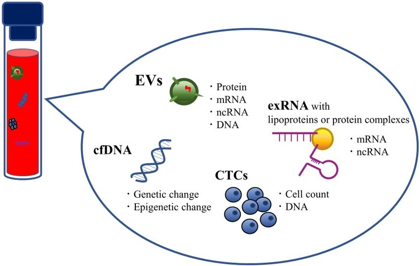
Page 2         Tamura et al. Extracell Vesicles Circ Nucleic Acids 2021;2:148-74   https://dx.doi.org/10.20517/evcna.2021.06

Keywords: Extracellular vesicles, liquid biopsy, cancer biomarker, microRNA

INTRODUCTION
Our body fluids, such as blood, urine, cerebrospinal fluid, saliva, pleural effusion, ascites fluid, breast milk
and seminal plasma, contain various biological molecules[1]. Liquid biopsy is a minimally invasive method
that uses these molecules as biomarkers, and it is emerging as a new tool in the strategy against cancer[2].
The terms "precision medicine"[3] and "personalized medicine"[4] have recently become popularized in the
field of cancer research. Diagnosis and adequate therapy for individual cases of particular cancer types
commonly rely on genetic mutation and gene expression analyses or pathological imaging observations of
cancer lesions. However, such cancer genomic medicine approaches require collection of cancer tissue by
biopsy, which imposes a heavy burden on the patient. In particular, it is difficult to obtain tumor tissue from
organs located in the deeper parts of the body. Therefore, the development of minimally invasive methods,
such as liquid biopsy, is desired. Liquid biopsy initially emerged only for the purpose of genetic diagnosis;
however, it has considerable clinical application potential, such as in early diagnosis, monitoring of
pathological conditions, and tailored treatment development according to cancer biology and the predicted
treatment response[5]. Importantly, because of its minimally invasive nature, liquid biopsy can be scheduled
more often to give more accurate snapshots of the disease at successive time points, which is useful for
measuring temporal tumor burden levels and early evidence of recurrence or therapy resistance[6]
[Figure 1]. Furthermore, liquid biopsy may reflect the genetic profile of more cancer subclones in a patient
than tissue biopsies, which are obtained from only one cancer region[7].

Extracellular vesicles (EVs) have attracted increasing attention as a novel analyte in liquid biopsy[8]. EVs are
lipid membranous vesicles that are released from almost all types of cells, including normal cells as well as
abnormal cells, such as cancer cells. EVs are reported to be correlated with various biological phenomena
and play important roles in cell-to-cell communication via horizontal transfer of cellular cargoes, such as
proteins, RNAs [including mRNA and noncoding RNAs (e.g., microRNA; miRNA, transfer RNA; tRNA
circular RNA; circRNA and long noncoding RNA; lncRNA)], DNA fragments, and lipids[9,10]. Importantly,
the composition of EV cargoes secreted from individual cell types differs greatly depending on the cellular
origin, and the characteristic features of EVs derived from various cancer cells have been revealed. Thus,
cancer-derived EVs can be analyzed to determine the presence and nature of cancer. Furthermore, EVs have
been reported to be found in almost all body fluids[11]. For these reasons, EVs are recognized as a promising
liquid biopsy resource for cancer. In this review, we will discuss the feasibility and practicality of EV-based
liquid biopsy in clinical settings. In the first half, we will argue the advantages and challenges of EV-based
liquid biopsy for clinical application. In the second half, we will summarize recent notable studies
investigating cancer-specific EV-related molecules as cancer biomarkers, with a focus on their biological or
clinical significance.

CANDIDATE ANALYTES IN LIQUID BIOPSY OTHER THAN EVS
Liquid biopsy can target various cancer-associated analytes in multiple body fluids, and several candidate
analytes in liquid biopsy for cancer have been identified. These analytes include circulating tumor cells
(CTCs) and circulating nucleic acids, such as cell-free DNA (cfDNA) and some extracellular RNA (exRNA)
fragments [Figure 2]. Currently, CTCs and cfDNA are the most widely studied target materials for liquid
biopsy[12,13].

CTCs are cancer cells shed by primary or metastatic cancer lesions into the circulation and are considered a
crucial determinant of hematogenous metastasis and recurrence. CTCs contain valuable information about

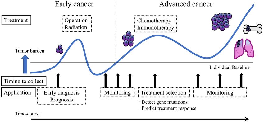
Tamura et al. Extracell Vesicles Circ Nucleic Acids 2021;2:148-74       https://dx.doi.org/10.20517/evcna.2021.06              Page 3

Figure 1. Clinical utility of liquid biopsy in cancer. Liquid biopsy presents a minimally invasive nature and thus has the potential to
impact clinical practice at multiple stages of cancer management. This technique can contribute to early diagnosis, pathological
condition monitoring, and tailored treatment development according to the cancer biology of individual patients. After cancer
treatment, liquid biopsy can support follow-up care by providing early evidence of recurrence or therapy resistance.

Figure 2. Candidate analytes of liquid biopsy. Body fluids contain several promising biomarkers for cancer. Each candidate analyte can
provide considerable information about the cancer biology of individual patients.

the spreading tumor, and early detection of CTCs and treatment of metastatic spread can contribute to
improving disease outcomes. Numerous studies in the past decade have shown that CTCs have potential as
biomarkers to predict cancer metastasis progression and prognosis[14,15]. A high number of CTCs has been
reported to be correlated with clinical outcome[16-19]. However, CTCs are estimated to account for at most
one cell among a hundred million circulating cells; thus, there are generally only a small number of CTCs in
a few milliliters of blood sample. Furthermore, CTCs must be analyzed as soon as possible after collection
because the number of viable cells decreases rapidly. Therefore, analysis of CTCs requires relatively large
volumes of fresh blood and advanced technology with extremely high analytical sensitivity and
specificity[20-22]. To date, numerous technologies are available that are useful for enrichment and detection of
CTCs[23]. Among them, the CellSearch® system has received FDA approval for prognostic clinical evaluation
of several cancer types[24]. However, due to the limited number of CTCs in the blood and the current level of
Page 4         Tamura et al. Extracell Vesicles Circ Nucleic Acids 2021;2:148-74   https://dx.doi.org/10.20517/evcna.2021.06

detection technologies, CTCs have not yet been entirely accepted in the clinic. Research groups have
recently moved towards analyzing CTC contents (e.g., miRNA[25], mRNA[26], and protein[27,28]) for detection
of biomarkers.

cfDNA is a short fragment of nucleic acids found in body fluids, and a component of cfDNA derived from
tumor cells is called circulating tumor DNA (ctDNA). The majority of ctDNA originates from apoptotic
and necrotic tumor cells. Over the past decade, a large number of studies have reported that measuring
ctDNA levels in cancer patients may help in cancer diagnosis and prognosis prediction[29-31]. Interestingly,
ctDNA is reported to be horizontally transferred from cancer cells to normal cells via uptake of apoptotic
bodies, which is one of the EV subclasses (described below), leading to cancer progression[32,33].
Furthermore, ctDNA is also reported to harbor genetic and epigenetic changes present in the original
tumor, and analytical techniques to detect such changes have already been established. By examining these
changes, researchers have shown that ctDNA also helps predict treatment response and recurrence[34-38]. The
major challenge in ctDNA research is that tumor-specific mutations may only represent 0.01% of the total
cfDNA[39,40], thereby increasing the difficulty of detecting rare variants. Moreover, issues limiting cfDNA
testing include its relatively short half-life[41,42]. Therefore, sample processing times are critical, and sample
preservation requires special precautions, which are also significant barriers to practical use of cfDNA
testing in the clinic.

CTC and ctDNA applications are confronted with several challenges, as described above. In short, the
significantly low amounts and fragility of CTCs and ctDNA, which show remarkable variations in amount
among individuals, increase the difficulty of detection. Moreover, CTC and ctDNA applications may be
confined to evaluation of advanced cancer because CTCs are released from cancer tissue that has grown to
the point that it causes metastases and ctDNA is primarily released from apoptotic or necrotic cancer
tissue[43]. Thus, these entities may not be suitable for early diagnosis and may not reflect the current or near-
future state of the disease.

exRNA refers to RNA that is present outside of cells within EVs or associated with platelets, lipoproteins
and protein complexes. Similar to cfDNA, almost all non-EV-associated exRNAs are released into
circulation by passive secretion. In addition to CTCs and cfDNA, non-EV-associated exRNA has also been
well investigated as a candidate analyte in liquid biopsy for cancer. In particular, non-EV-associated
miRNAs are recognized as a significant RNA subtype for biomarker discovery because miRNA profiles are
associated with cancer-specific conditions. The critical issue is miRNA extraction from biofluid samples; the
amounts of miRNA are highly variable among different experiments. Because of the small size of miRNAs
and their attachment to other molecules, reproducible extraction remains an inherent issue[44].

WHAT ARE “EVS”?
We will now describe EVs, which are the main focus of this review. “EV” is a collective term that refers to all
types of lipid membranous vesicles naturally released from all cell types. From the 1970s to the 1980s,
membrane-enclosed vesicle structures were reported in various types of solid tissues, body fluids, and
culture supernatants. Depending on their size and origin, these vesicles are called various names, such as
prostasomes, exosomes, microvesicles, microparticles, shedding vesicles, ectosomes, apoptotic bodies and
oncosomes. Each vesicle type contains slightly different molecular groups due to differences in vesicle
biogenesis and secretion pathways. Nevertheless, their designations have been vaguely defined. Therefore,
the International Society for Extracellular Vesicles (ISEV), founded in Sweden in 2012, recommends the use
of “extracellular vesicle” as a generic term for these vesicles[45,46]. The nomenclature of these vesicles is
detailed in the Minimal Information for Studies of Extracellular Vesicles (MISEV), which is the guideline
Tamura et al. Extracell Vesicles Circ Nucleic Acids 2021;2:148-74   https://dx.doi.org/10.20517/evcna.2021.06   Page 5

advocated by the ISEV[47,48].

EVs are classically classified into three main categories: exosomes (approximately 100 nm), microvesicles
(approximately 1 µm), and apoptotic bodies (greater than 1 µm)[49]. These three classes of EVs differ in size
as well as morphology, content, generation mode, and release mechanism. Exosomes are formed by the
inward budding of early endosomes to form multivesicular bodies (MVBs). These MVBs fuse with the
limiting plasma membrane to release exosomes into the extracellular space. Microvesicles originate by direct
shedding or budding from the plasma membrane. Apoptotic bodies are released from cells undergoing
programmed cell death[50]. Recently, Théry et al[47,51]. developed a more reasonable classification system for
EVs and defined vesicles < 100 nm as small EVs, < 200 nm as medium EVs, and > 200 nm as large EVs.
Moreover, they demonstrated that each EV subtype showed different characteristic protein components,
suggesting that each EV category is generated and secreted through a specific molecular mechanism[47,51].

EVS AS AN ANALYTE IN LIQUID BIOPSY
In 1983, Pan and Johnstone[52] discovered that cells secrete 100 nm-sized vesicles with a lipid membranous
structure and that red blood cells release small vesicles loaded with the transferrin receptor, which is
necessary to synthesize hemoglobin during maturation. They regarded EVs as “garbage bags” that are used
to expel unused molecules from the cell and reported one of the remarkable EV functions; more
importantly, this study demonstrated the presence of proteins in EVs[52,53].

In the 1990s, by analyzing immune cells, Raposo et al.[54] found that EVs affect recipient cells. EVs derived
from B lymphocytes induced antigen-specific MHC class II-restricted T cell responses, indicating that EVs
have functions related to intercellular communication[54].

In 2007, Valadi et al.[55] demonstrated that EVs from the human mast cell line HMC-1 and the mouse mast
cell line MC/9 contain approximately 1300 mRNAs and 121 miRNAs and thus contribute to the exchange
of genetic information between cells. Since that time, miRNAs have been the most widely studied class of
short noncoding RNAs (ncRNAs) in EV cargoes[55].

To date, EVs have been reported to contain various proteins, RNAs [including mRNAs and noncoding
RNAs (miRNAs, tRNAs, circRNAs and lncRNAs)], and DNA fragments[9]. The past few years have seen
extraordinary developments in areas such as mass spectrometry (MS), high-throughput sequencing (HTS)
(e.g., DNA sequencing, chromatin immunoprecipitation sequencing, methylation sequencing, and RNA
sequencing), and big-data analysis[56,57]. These advanced technologies allow us to select analytes contained in
EVs and evaluate them deeply. Significantly, the composition of EV molecular contents reflects the
intracellular status of their cellular origin. Moreover, these molecules are stably preserved due to their lipid-
layered structure; hence, EVs from body fluids can be analyzed after being stored for a relatively long period
of time[58]. A growing body of evidence has suggested that cancer-derived EV cargoes bare a strong
resemblance to the intracellular status of their parental cells[59]. Analysis of EV cargoes can help reveal the
existence, molecular profile and behavior of cancer. Thus, EV-based liquid biopsy contributes to early
cancer diagnosis, monitoring of cancer pathological conditions, and treatment selection according to cancer
biology and the predicted treatment response. In addition, EVs are easily accessible in various kinds of body
fluids and appropriate for sequential collection. For these reasons, EVs have emerged as a promising
biomarker resource and as another kind of liquid biopsy for cancer [Figure 2].
Page 6         Tamura et al. Extracell Vesicles Circ Nucleic Acids 2021;2:148-74   https://dx.doi.org/10.20517/evcna.2021.06

CLASSICAL EV ISOLATION METHODS
Although EVs are a remarkable candidate analyte to detect cancer biomarkers, few EV-associated
biomarkers have been implemented in clinical settings, which is partially due to the lack of adequate
isolation methods[60]. In principle, EVs must be isolated from various biofluids for analysis. Indeed, in
almost all cases, candidate EV-associated biomarkers were detected via isolation processes because target
EV-associated molecules should be distinguished from EV-free molecules in the biofluid; however, a unified
isolation method is not currently available, which is one of the biggest challenges in EV research.

The most popular technique for EV isolation is ultracentrifugation (UC), which separates EVs based on
their size and buoyant density. The UC procedure does not require additional chemicals or pretreatment of
the body fluid samples. Moreover, the combination of UC with other procedures, such as ultrafiltration,
sucrose cushion, and density gradient centrifugation, can increase the purity of the EV fraction[61-63].
However, UC-based procedures generally require dedicated equipment and an extraordinary amount of
time[60]. Moreover, repeated centrifugation can lead to reduced yield due to lost and aggregated EVs[64]. On
the other hand, various polymer-based isolation kits, such as ExoQuick™, Total Exosome Isolation™ and
miRCURY™, which are commercially available, can save time and labor costs, although issues with high
contamination rates and high running costs have been reported[63,65]. An immunoaffinity capture approach
using magnetic beads and monoclonal antibodies targeting EV surface antigens is also commonly used to
isolate EVs[66,67]. Many reports have demonstrated that this approach achieves a selective and high-purity
output; however, there are difficulties in terms of low capacity, low yield and high reagent cost[60]. Size-
exclusion chromatography (SEC) is a technique for separating biological molecules based on molecular size
in which target molecules are isolated by filtration through a resin-packed column. SEC has been reported
to yield highly purified EVs and achieve excellent reproducibility in a relatively short time[60,64]; however, the
existence of EVs in multiple fractions results in a low EV concentration in the obtained samples.
Consequently, subsequent EV analysis often requires an additional concentration step[68]. Recently, many
researchers have studied combined methods integrating multiple isolation techniques together or
conducting them sequentially[69]. Among others, the combination of UC and SEC has demonstrated higher
purity of EVs and better performance in subsequent analyses than the single-step methods[70-72]; however, in
daily clinical settings, combined methods might be avoided due to several factors, such as time
consumption, cost, repeatability, and ease of use. As high-quality EV samples are essential for subsequent
analyses, there is an urgent need for a simple, cost-effective, and reliable technique for both basic research
and clinical practice.

EV-ASSOCIATED PROTEINS
Common EV protein markers
EVs contain abundant proteins that reflect their origin and alterations of their parental cells. Based on the
endosome-based biogenesis pathway, EV-specific protein markers include membrane trafficking-associated
proteins (e.g., Rab family proteins, annexins), MVB-associated proteins (e.g., Alix, Tsg101 and ESCRT
complex), tetraspanins (e.g., CD9, CD63 and CD81) and heat shock proteins (Hsp70 and Hsp90)[73-75]. As
these proteins are common to EVs derived from almost all cell types, they can serve as common positive EV
markers that can confirm the presence of EVs, and isolated EVs can be assessed via downstream proteomic
analyses, such as western blotting (WB), enzyme-linked immunosorbent assay (ELISA), and flow cytometry
(FCM), which use common EV proteins as hallmarks. With regard to checking the quality of isolated EV
samples, MISEV recommends the use of apolipoproteins A1/2 and B and albumin as negative markers of
blood-derived EVs because they are often co-isolated with these molecules[47,76].
Tamura et al. Extracell Vesicles Circ Nucleic Acids 2021;2:148-74   https://dx.doi.org/10.20517/evcna.2021.06   Page 7

EV protein-based platform for cancer diagnosis
Targeting membrane proteins on the surface of EVs is an effective strategy because target cancer-derived
proteins can be directly detected without the use of a large sample volume or time-consuming isolation
processes for EVs. Recently, great efforts have been devoted to establishing clinically useful detection
platforms, including platforms for direct detection of cancer-specific EVs without any isolation or
purification procedures. These platforms mainly consist of specific antibody-based technologies that detect
EV surface proteins, such as ELISA.

Jørgensen et al.[77] established the EV Array, which can detect EVs in unpurified materials in a high-
throughput manner. The EV Array is composed of different capture antibodies located on a microarray
slide, which capture EVs according to their surface proteins, and the target EVs are detected with a cocktail
of biotinylated antibodies against the tetraspanins CD9, CD63, and CD81. The authors validated the
performance of the EV Array by comparing plasma from nonsmall cell lung cancer (NSCLC) patients and
normal healthy subjects[78]. Shao et al.[79] reported that a microfluidic chip platform could distinguish
patients with glioblastoma multiforme (GBM) from normal healthy subjects. This microfluidic chip labeled
with magnetic nanosensors quantifies an EV-specific protein marker (CD63) and glioblastoma-specific
proteins, such as epidermal growth factor receptor (EGFR) and EGFR variant III, on the surface of EVs via
micronuclear magnetic resonance (μNMR). In this study, the authors also indicated that GBM EVs reflect
gene amplification or mutation and predict the therapy response. Surface plasmon resonance (SPR)-based
nanosensors have recently attracted much attention due to their ability to detect a small number of
molecules[80]. Im et al.[81] developed an SPR-based exosome sensor called nanoplasmonic exosomes
(nPLEXs). Each nanohole array of nPLEX is functionalized with antibodies that recognize EV surface
proteins. nPLEX was able to differentiate ascites samples from ovarian cancer patients from healthy controls
with an accuracy of 97% and identified ovarian cancer cell-derived EVs based on their expression of CD24
and EpCAM. Yoshioka et al.[82] also established a highly rapid and sensitive analytical technique called
“ExoScreen”. This assay consists of two kinds of antibodies against proteins on the surface of EVs that are
detectable by photosensitizing beads. A very small sample volume (at least 5 μL) was required to detect EVs
in serum from healthy controls without a complicated isolation process. Moreover, the assay could be
completed within 2 h. In this study, they identified CD147 as a specific EV-surface protein derived from
colorectal cancer cells and revealed that a larger number of CD9/CD147 double-positive EVs could be
detected in serum from colorectal cancer patients than in serum from healthy control subjects using this
assay. Furthermore, Zhao et al.[83] developed a simple microfluidic platform named the “ExoSearch” chip
that allows quantitative isolation of EVs using immunomagnetic beads. An “ExoSearch” chip could detect
ovarian cancer by measuring three EV cancer protein markers, CA-125, EpCAM and CD24. The
development of immune-capturing systems in microchips also provides highly sensitive and reliable
detection of cancer markers without requiring a large sample volume or time-consuming EV isolation
processes.
Other EV protein biomarkers for cancer diagnosis
EV surface proteins and EV lumen proteins are regarded as candidate cancer biomarkers. Numerous studies
have identified cancer-associated EV protein markers using isolation processes, followed by WB, ELISA,
and FCM. Promising EV protein markers identified in clinical studies with patient body fluid samples are
summarized in Table 1.

Clinically validated traditional molecules have also been identified in EVs, such as prostate-specific antigen
(PSA), and they represent novel diagnostic biomarkers. Mitchell et al.[84] demonstrated that the expression
of PSA and PSMA in urinary EVs can act as treatment response markers in prostate cancer. Additionally,
Logozzi et al.[85] showed increased PSA expression on EVs in vitro and in the plasma of prostate cancer
Page 8              Tamura et al. Extracell Vesicles Circ Nucleic Acids 2021;2:148-74            https://dx.doi.org/10.20517/evcna.2021.06

Table 1. A list of EV proteins as potential biomarkers for cancer

Cancer           Biological    Isolation       Detection
                                                                    Markers                       Potential application     Ref.
types            source        method          method
Urinary cancer
Prostate         Plasma        UC              ELISA                PSA                           Diagnosis/Prognosis       [85]
cancer
                 Urine         UC + SUC        ELISA/WB             PSA, PSMA                     Diagnosis/Monitoring      [84]
                 Plasma/Serum UC/ExoQuick      ELISA/WB             Survivin                      Diagnosis/Monitoring      [87]
                 Urine         UC              IP/WB                δ-catenin                     Diagnosis                 [156]
                 Serum         UC              WB                   MDR-1/P-gp, MDR-3,            Predict chemoresistance   [95,157]
                                                                    PABP4                         (Docetaxel)
                 Plasma        -               FCM                  PSMA                          Monitoring/Predict        [158]
                                                                                                  chemoresistance
Bladder          Urine         UC              MS/WB                EH-domain-containing          Diagnosis                 [159]
cancer                                                              protein 4, EPS8L1, EPS8L2,
                                                                    GTPase NRas, Mucin 4,
                                                                    retinoic acid-induced
                                                                    protein3, resistin, alpha
                                                                    subunit of GsGTP binding
                                                                    protein
                 Urine         UC              ELISA                TACSTD2                       Diagnosis                 [160]
                 Urine         UC              MS                   α-1-anti-trypsin, H2B1K       Diagnosis                 [161]
                 Urine         UC              WB                   HEXB, S100A4, SND1,           Diagnosis                 [162]
                                                                    TALDO1, and EHD4
                 Urine         UC              WB                   Periostin                     Diagnosis                 [163]
                 Urine         UC + SUC        WB                   EDIL3                         Diagnosis                 [164]
                 Urine         UC + SUC        FCM                  CD36, CD44, 5T4, basigin,     Diagnosis                 [165]
                                                                    CD73, MUC1, α6-integrin
Renal cancer Urine             UC              MS/WB                MMP9, DKK4, EMMPRIN,          Diagnosis                 [166]
                                                                    CP, PODXL, CAIX, CD10,
                                                                    AQP1, dipeptidase 1,
                                                                    syntenin 1
Female cancer
Breast cancer Serum            ExoQuick        ELISA                Survivin, Survivin2B          Diagnosis/Prognosis       [88]
                 Serum/Plasma ExoQuick         ELISA/WB             CD82                          Diagnosis                 [167]
                 Ascites       UC + SUC        WB                   CD24, EpCAM                   Diagnosis                 [168]
                 Plasma        UC              FCM                  TRPC5                         Prognosis/Predict         [169]
                                                                                                  chemoresistance
                 Serum         UC              FCM                  UCH-L1                        Predict chemoresistance   [92]
                                                                                                  (Anthracycline/taxan)
                 Serum         UC              FCM/WB               HER2                          Predict chemoresistance   [96]
                                                                                                  (Trastuzumab)
                 Plasma        UC              ELISA/FCM            TGFβ1                         Predict chemoresistance   [97]
                                                                                                  (Trastuzumab)
Ovarian          Plasma        -               Exosearch chip       CD24, EpCAM, CA-125           Diagnosis                 [83]
cancer
                 Plasma        UC              WB                   TGFβ1, MAGE3/6                Diagnosis                 [170]
                 Plasma        UC + SUC        WB                   Claudin-4                     Diagnosis                 [171]
                 Ascites       UC + SUC        WB                   E-cadherin                    Diagnosis/Prognosis       [172]
                 Ascites       UC              WB                   MMP2, MMP9, uPA               Diagnosis                 [173]
                 Ascites       UC + SUC        WB                   CD24, L1CAM, ADAM10,          Diagnosis/Prognosis       [174]
                                                                    EMMPRIN
Digestive
cancer
Pancreatic       Serum         UC              FCM                  GPC1                          Diagnosis/Prognosis       [89,175]
cancer
                 Serum         UC + SUC        ELISA                CKAP4                         Diagnosis                 [176]
                 Serum         UC              ELISA                MIF                           Diagnosis/Prognosis       [177]
Tamura et al. Extracell Vesicles Circ Nucleic Acids 2021;2:148-74 https://dx.doi.org/10.20517/evcna.2021.06 Page 9
Plasma - ELISA EpCAM Prognosis [91]
Colorectal Serum Exoquick ELISA CEA Diagnosis [178]
cancer
Serum - ExoScreen CD147, CD9 Diagnosis [82]
Ascites UC WB claudin-3 Diagnosis [179]
Gastric Serum UC FCM/WB HER-2/neu, CCR6, Diagnosis [180]
cancer EMMPRIN, MAGE-1, c-MET
Others
Lung cancer Serum UC ELISA EGFR Diagnosis [181]
(NSCLC) Serum UC ELISA/WB AHSG, ECM1 (with serum Diagnosis [182]
CEA)
Serum - EV array 30 Proteins Diagnosis [78]
Plasma - EV array CD171, NY-ESO-1, PLAP, Diagnosis [183]
Flotilin1
Urine UC WB LRG1 Diagnosis [184]
Melanoma Plasma UC ELISA (Exo Caveolin-1, CD63 Diagnosis/Prognosis [185]
Test)/FCM/WB
Plasma UC WB TYRP2, VLA-4, HSP70, Prognosis [186]
HSP90
Plasma UC/Total ELISA/FCM /WB PDL-1 Predict immunotherapy [91]
Exosome resistance
isolation Kit (Pembrolizumab)
Glioblastoma Serum - µNMR system EGFR, EGFRv , CD63 Prognosis [79]
NSCLC: Non-small cell lung cancer; LSCC: laryngeal squamous cell carcinoma; UC: ultracentrifugation; SUC: sucrose cushion; ELISA: enzyme-
linked immuno-sorbent assay; FCM: flow cytometry; WB: western blotting; IP: immunoprecipitation; MS: mass spectrometry; miRNA: microRNA;
tRNA: transfer RNA; circRNA: circular RNA; lncRNA: long non-coding RNA.
patients. The authors emphasized the failure of current PSA testing in discriminating between benign
prostatic hypertrophy and prostate cancer in terms of both overdiagnosis and overtreatment, which leads to
patient suffering and public and private healthcare expenditures. Moreover, they claimed that EV PSA
might resolve the problem associated with differences in PSA cutoff levels based on age, race and individual
physiological condition. Similar to PSA, some traditional molecules in EVs have been discussed to have
higher relevance to cancer than their total amount in body fluids, such as CEA for colon cancer[86].
Additionally, some EV-associated markers have been reported as diagnostic markers for multiple cancer
types. Khan et al.[87] showed that Survivin, an inhibitor of apoptosis member, could be detected in plasma-
derived EVs from both prostate cancer patients and healthy subjects; however, the relative amount of EV
Survivin was remarkably higher in the plasma of prostate cancer patients. Their subsequent study showed
that the EV Survivin and its alternative splice variants were also elevated in breast cancer patient plasma[88],
which suggests that EV Survivin might be an important diagnostic marker common to several cancer types.
EV proteins can be used to monitor cancer progression and drug resistance
Biomarkers in EVs of several cancer types may be applied for cancer stratification because they change in
response to anticancer therapy. Thus, EV proteins may be used as novel biomarkers to monitor cancer
progression or to identify patients susceptible to anticancer drugs.
Melo et al.[89] reported that glypican-1 (GPC1) could be a specific marker of cancer-derived EVs and that the
existence of GPC1-positive EVs in serum could differentiate patients with pancreatic ductal
adenocarcinoma (PDAC) from patients with benign pancreas disease or from healthy control subjects with
100% sensitivity and specificity. The levels of GPC1-positive EVs were significantly decreased after surgical
resection; moreover, they were related to overall survival (OS) and significantly higher in patients withPage 10 Tamura et al. Extracell Vesicles Circ Nucleic Acids 2021;2:148-74 https://dx.doi.org/10.20517/evcna.2021.06 distant metastasis than in patients with lymph node metastasis only or no metastases[89]. Although this conclusion is controversial, a replication study was performed to validate this finding, and a subsequent discussion was held in ISEV2017[90]. Similarly, Giampieri et al.[91] reported that a higher level of EpCAM- positive EVs before chemotherapy was correlated with shorter progression-free survival (PFS) and OS. In contrast, in this study, an increase in EpCAM-positive EV levels during treatment was correlated with better PFS in PDAC patients[91]. These studies demonstrated that EV-associated proteins could be biomarkers for monitoring tumor burden. The development of chemoresistance is a persistent problem during cancer treatment. Various studies have reported cell-to-cell transfer of multidrug resistance (MDR) efflux pumps as EV cargoes from chemotherapy-resistant cells to chemotherapy-sensitive cancer cells. EVs from doxorubicin-resistant[92] or docetaxel-resistant[93] breast cancer cell lines transferred chemoresistance to recipient cancer cells through P-glycoprotein (P-gp) loaded onto EVs. Moreover, the same phenomenon in paclitaxel-resistant ovarian cancer cells was also caused by the transfer of functional P-gp mediated by EVs[94]. Docetaxel-resistant prostate cancer cells were also reported to proliferate through cell-to-cell transfer of EV P-gp. In this study, serum EVs from prostate cancer patients who were nonresponders to docetaxel therapy protected prostate cancer cells from the cytotoxicity of docetaxel[95]. These studies indicate that EV-P-gp might be a promising marker to predict chemotherapy resistance in several cancer types. Ubiquitin C-terminal hydrolase L1 (UCH-L1) has also been reported as an EV-based predictive biomarker of chemoresistance in breast cancer. UCH-L1 overexpression has been reported to induce upregulation of P-gp levels through the MAPK/ERK signaling pathway, thereby enhancing an MDR phenotype in breast cancer. Ning et al.[92] showed that higher UCH-L1 levels in circulating EVs are correlated with poorer response to adjuvant anthracycline/taxane- based chemotherapy. In addition, they demonstrated that UCH-L1-positive EVs derived from breast cancer cell lines could transfer chemoresistance to recipient cells in vitro, indicating that EVs might be a predictive biomarker of chemoresistance in breast cancer patients. Interestingly, EV-mediated drug resistance has been reported to be relevant for molecular target drugs as well as chemotherapy drugs. Ciravolo et al.[96] reported that 73% of advanced-stage breast cancer patients had HER2-positive EVs in circulation, which hampered the corresponding therapeutic efficacy of trastuzumab monoclonal antibody. Importantly, this study demonstrated that the presence of HER2- positive EVs in the serum of breast cancer patients could be an indicator to predict a patient’s response to trastuzumab therapeutic regimens[96]. Martinez et al.[97] reported that EVs released from HER2 drug-resistant cells contain larger amounts of the immunosuppressive cytokine TGF-β1. Importantly, a recent neoadjuvant clinical trial by the same group that included trastuzumab and lapatinib further demonstrated that HER2- overexpressing breast cancer patients who were nonresponders to HER2 drug therapy had significantly higher amounts of TGF-β1 in circulating EVs than patients who did not respond to HER2 drug therapy. These results suggest that EV-TGF-β1 can be a biomarker for predictive response to this therapeutic regimen against breast cancer[97]. Recently, with the successful development of immune checkpoint inhibitors (ICIs), cancer immunotherapy has attracted worldwide attention as a new cancer treatment. EV proteins may contribute to identifying patients susceptible to ICIs. Tumor cells avoid immune recognition by upregulating the surface expression of programmed death-ligand 1 (PD-L1), which interacts with the programmed death-1 (PD-1) receptor on T cells to elicit the immune checkpoint response. Indeed, immunotherapy with anti-PD-1 antibodies has shown remarkable therapeutic effects against different tumor types[98]. However, for some patients, the therapeutic response has been reported to be rather poor[99,100]. To address this problem, Chen et al.[101] showed that specific EVs reduce the effectiveness of immunotherapy approaches in certain patients with
Tamura et al. Extracell Vesicles Circ Nucleic Acids 2021;2:148-74   https://dx.doi.org/10.20517/evcna.2021.06   Page 11

melanoma. Remarkably, using human melanoma xenografts in nude mice, they showed that metastatic
melanoma cell lines release EVs loaded with PD-L1 on their surface and that interferon-γ increases the
expression of PD-L1 on these vesicles, which suppresses the function of CD8+ T cells, facilitating tumor
growth. Importantly, these authors showed that the level of PD-L1-positive EVs differentiates responders
from non-responders to anti-PD-1 therapy. More importantly, this study provides evidence supporting the
application of PD-L1-positive EVs as a predictive biomarker for anti-PD-1 therapy in melanoma
patients[101].

EV-ASSOCIATED NUCLEIC ACIDS
As described above, high-speed analytical technologies, such as HTS, are powerful tools for identifying
candidate nucleic acid biomarkers. For detection and validation of candidate EV nucleic acid biomarkers,
reverse transcription-quantitative polymerase chain reaction (RT-qPCR) after an isolation process is the
most common detection method. Recently, digital polymerase chain reaction (dPCR) has emerged as a
novel detection method for EV nucleic acids. dPCR is a qPCR technique that provides a sensitive and
reproducible method of measuring the amount of DNA or RNA present in a sample[102]. The tremendous
progress in these technologies during the last few decades has allowed us to analyze EV-associated nucleic
acids derived from diverse body fluids.
EV RNA biomarkers
Pioneering studies of nucleic acids from isolated EVs have identified various miRNAs and mRNAs as the
major components of EVs[55,103]. Subsequently, several important papers reporting on the function of EV-
miRNAs were published and showed that the transferred EV-miRNAs can be active in recipient cells and
modify the cellular phenotype[104-106]. Over the last decade, studies have revealed that EVs contain other
noncoding RNAs, such as tRNAs[107], circRNAs[108] and lncRNAs[109,110]. To date, EV RNAs (as EV cargo)
have received the most attention in terms of cancer diagnosis and prognosis because they are easy to
quantify using conventional methods, such as qPCR, and stable against RNase-dependent degradation in
the circulatory system[111]. Table 2 summarizes the candidate EV RNA markers reported as diagnostic and
prognostic tools for cancer.
EV RNAs for cancer diagnosis
Since the discovery of EV-miRNAs by Valadi et al.[55] in 2007, numerous studies have been performed to
identify diagnostic EV miRNAs for cancer. Taylor et al.[112] showed that eight miRNAs (miR-21, miR-141,
miR-200a, miR-200c, miR-200b, miR-203, miR-205 and miR-214) that were previously characterized as
diagnostic markers for ovarian cancer were also upregulated in circulating EVs derived from ovarian cancer
patients. Rabinowits et al.[113] conducted miRNA-profiling analyses of EV-based liquid biopsy samples and
tumor biopsy samples from lung cancer patients and healthy controls. Their study showed a similarity in
miRNA patterns between EV-based biopsy samples and tumor biopsy samples from lung cancer patients
and demonstrated significant differences between these miRNA patterns and those in EVs from the healthy
control group. This result indicates the potential of EV miRNAs as a liquid biopsy source for lung cancer.
On the other hand, EV miR-1246 was significantly increased in ESCC patient serum samples but was not
elevated in tumor biopsy samples, indicating that the level of EV miRNAs may not necessarily reflect the
amounts in parental cells[114].

Regarding EVs in other body fluids, Nilsson et al.[115] reported that PCA3 and TMPRSS2:ERG mRNA, which
are previously established prostate cancer biomarkers, were also present in urinary EVs from prostate
cancer patients. Additionally, Foj et al.[116] demonstrated that the levels of miR-21-5p, miR-375, and let-7c-
5p were remarkably increased in urinary EVs derived from prostate cancer patients compared with those
Page 12                                                                        Tamura et al. Extracell Vesicles Circ Nucleic Acids 2021;2:148-74       https://dx.doi.org/10.20517/evcna.2021.06

Table 2. A list of EV RNAs as potential biomarkers for cancer

                                     Isolation           Detection
Cancer types Biological source                                            RNA types     Markers                                                   Potential application            Ref.
                                     methods             methods
Uninary cancer
Prostate         Urine               UC + SUC            nestedPCR        mRNA          PSA, PCA-3, TMPRSS:ERG                                    Diagnosis/Monitoring             [115]
cancer
                                                                ®
                 Urine               -                   ExoDx Prostate   mRNA/lncRNA PCA3, ERG, SPDEF                                            Diagnosis                        [118]
                                                         (IntelliScore)
                 Urine               UF/UC               RT-qPCR          miRNA         let-7c, miR-21, miR-107, miR-145, miR-196a-5p, miR-204,   Diagnosis                        [116,187-191]
                                                                                        miR-375, miR-501-3p, miR-574-3p, miR-2909
                 Serum/Plasma        UF/SEC/UC           RT-qPCR          miRNA         let-7i, miR-16, miR-21-5p, miR-24, miR-26a, miR-26b, miR- Diagnosis                        [187,192-196]
                                                                                        30c-5p, miR-34b, miR-92b, miR-93, miR-103, miR-106a,
                                                                                        miR-107, miR-130b, miR-141, miR-181a-2, miR-195, miR-197,
                                                                                        miR-200c-3p, miR-210-3p, miR-223, miR-298, miR-301a,
                                                                                        miR-326, miR-328, miR-331-3p, miR-346, miR-375, miR-
                                                                                        432, miR-574-3p, miR-625, miR-1290, miR-2110
                 Plasma              UF/SEC/UC           RT-qPCR          miRNA         miR-17, miR-20a, miR-23a, miR-130b, miR-198, miR-200b,    Prognosis                        [119,187]
                                                                                        miR-375, miR-379, miR-513a-5p, miR-572, miR-577, miR-
                                                                                        582-3p, miR-609, miR-619, miR-624, miR-1236, miR-1290
                 Serum               UC                  RT-qPCR          miRNA         miR-1246                                                  Prognosis                        [197]
                 Urine               -                   Urine Exosome     lncRNA       p21                                                       Diagnosis                        [122]
                                                         RNA Isolation Kit
                                                         / RT-qPCR
                 Plasma              Total Exosome       RT-qPCR          lncRNA        SAP30L- AS1, SChLAP1                                      Diagnosis                        [198]
                                     Isolation Kit
                 Plasma / Urine      -                   exoRNeasy        mRNA          AR-V7                                                     Predict hormone therapy          [132,133]
                                                         kit/ddPCR                                                                                resistance (Abiraterone and/or
                                                                                                                                                  Enzalutamide)
Bladdar cancer Urine                 UC                  ddPCR            miRNA         miR-21, miR-93, miR-200c, miR-940                         Diagnosis                        [199]
                 Urine               -                   ddPCR            miRNA         miR-21-5p, miR-4454, miR-720, miR-200c-3p, miR-29b-3p, Diagnosis                           [200]
                                                                                        miR-200b-3p
                 Urine               UC                  RT-qPCR          miRNA         miR-375, miR-146a                                         Diagnosis                        [201]
                 Urine               unknown             RT-qPCR          miRNA         miR-146b-5p, miR-155-5p                                   Diagnosis                        [202]
                 Urine               UC                  RT-qPCR          lncRNA        HOTAIR, HOX- AS-2, MALAT1, SOX2, OCT4                     Diagnosis/Prognosis              [203]
                 Serum               ExoQuick            RT-qPCR          lncRNA        PCAT1, UBC1, SNHG16                                       Diagnosis/Prognosis              [204]
                 Serum               UC                  RT-qPCR          lncRNA        UCA1                                                      Diagnosis                        [205]
                 Urine / Serum       UC                  RT-qPCR          circRNA       PRMT5                                                     Diagnosis/Prognosis              [126]
Renal cancer     Serum               UC + IP             RT-qPCR          miRNA         miR-210, miR-1233                                         Diagnosis                        [206,207]
                 Urine               UC                  RT-qPCR          miRNA         miR-150-5p, miR-126-3p in combination with miR-34b-5p,    Diagnosis                        [208]
Tamura et al. Extracell Vesicles Circ Nucleic Acids 2021;2:148-74 https://dx.doi.org/10.20517/evcna.2021.06 Page 13
miR-449a and miR-486-5p
Serum Total Exosome RT-qPCR miRNA miR-224 Prognosis [209]
Isolation Kit
Plasma ExoQuick RT-qPCR miRNA miR-26a-1-3p, miR- let-7-I, miRN-615-3p Prognosis [210]
Urine UC RT-qPCR lncRNA GSTA1, CEBPA, PCBD1 Diagnosis [211]
Female cancer
Breast cancer Plasma FC/UC/ExoQuick RT-qPCR miRNA miR-21, miR-1246 Diagnosis [212]
Plasma UC RT-qPCR miRNA miR-105 Diagnosis [213]
Serum unknown/Total RT-qPCR miRNA miRNA-21, miRNA-222, miRNA-155 Prognosis/Predict [214,215]
Exosome Isolation chemoresistance (miR-21, miR-
Kit 155: Doxorubicin, Paclitaxel)
Serum UC + IP RT-qPCR miRNA miR-200a, miR-200c, miR-205 Diagnosis [216]
Bood/Milk/Ductal UC RT-qPCR miRNA miR-16, miR-1246, miR-451, miR-205 Diagnosis [217]
fluids
Serum ExoQuick RT-qPCR lncRNA HOTAIR Prognossis/Monitoring [129]
Serum ExoQuick RT-qPCR lncRNA SNHG14 Predict chemoresistance [130]
(Trastuzumab)
Plasma UC RT-qPCR mRNA TrpC5, mdr1, MUC1 and flotillin2 Predict chemoresistance [135]
(Anthracycline/taxan)
Serum Total Exosome RT-qPCR mRNA GSTP1 Predict chemoresistance [136]
Isolation Kit (Anthracycline/taxan)
Ovarian cancer Serum UC + IP RT-qPCR miRNA miR-21, miR-141, miR-200a, miR-200c, miR-200b, miR-203, Diagnosis [112]
miR-205, miR-214
Serum Total Exosome RT-qPCR miRNA miR-1246 Predict chemoresistance [218]
Isolation Kit (Paclitaxel)
Digestive
cancer
Pancreatic Serum/Urine UC RT-qPCR miRNA miR-17-5p, miR-21 Diagnosis [219]
cancer
Plasma ExoQuick RT-qPCR miRNA miR-155 Predict chemoresistance [131]
(Gemcitabine)
Saliva UC RT-qPCR mRNA Apbb1ip, ASPN, Daf2, FoxP1, Bco31781, Gng2 Diagnosis [220]
Liver cancer Plasma Total Exosome RT-qPCR tRNA ValTAC-3, GlyTCC-5, ValAAC-5, GluCTC-5 Diagnosis [107]
(HCC) Isolation Kit
Serum Exoquick RT-qPCR circRNA circUHRF1 Diagnosis/immunotherapy [108]
resistance (anti-PD-1)
Serum Exoquick RT-qPCR miRNA miR-21, miR-10b Prognosis [221]
Gastoric Serum unknown RT-qPCR miRNA HOTTIP Diagnosis/Prognosis [124]
cancerPage 14 Tamura et al. Extracell Vesicles Circ Nucleic Acids 2021;2:148-74 https://dx.doi.org/10.20517/evcna.2021.06
Serum UC RT-qPCR lncRNA lncUEGC Diagnosis [123]
Serum Exoquick RT-qPCR circRNA circSHKBP1 Diagnosis [222]
Esophageal Serum Exoquick RT-qPCR miRNA miR-21 Diagnosis [120]
cancer (ESCC)
Colorectal Serum UC RT-qPCR miRNA let-7a, miR-1229, miR-1246, miR-150, miR-21, miR-223, miR- Diagnosis [127]
cancer 23a
Serum ExoQuick RT-qPCR miRNA miR-4772-3p Prognosis [223]
Rectal cancer Plasma miRCURY Exosome RT-qPCR miRNA miR-30d-5p, miR-181a-5p and miR-486-5p Diagnosis [224]
Isolation Kit
Others
Lung cancer Plasma ExoQuick RT-qPCR miRNA miR-151a-5p, miR-30a-3p, miR-200b-5p, miR-629, miR- Diagnosis [225]
100, and miR-154-3p
Lung cancer Plasma IP RT-qPCR miRNA let-7f, miR-20b, miR-30e-3p, miR-223, miR-301 Diagnosis [226]
(NSCLC)
Lung cancer Plasma ExoQuick RT-qPCR miRNA miR-205, miR-19a, miR-19b, miR-30b, miR-20a Monitoring [227]
(LSCC)
Lung cancer Serum UC RT-qPCR miRNA miRNA-222-3p Prognosis/Predict [228]
(NSCLC) chemoresistance (Gemcitabine)
Lung cancer Serum ExoQuick RT-qPCR miRNA miRNA-146a-5p Predict chemoresistance [229]
(NSCLC) (Cisplatin)
Lung cancer Plasma - exoRNeasy mRNA PD-L1 Predict immunotherapy resistance [134]
(NSCLC) kit/ddPCR (anti-PD-1)
Melanoma Plasma - exoRNeasy mRNA PD-L1 Predict immunotherapy resistance [134]
kit/ddPCR (anti-PD-1)
Glioblastoma Serum/Cereblospinal UC RT-qPCR miRNA miR-21 Diagnosis/Prognosis [230]
fluid
Cereblospinal fluid UC RT-qPCR miRNA miR-21 Diagnosis [231]
Serum ExoQuick RT-qPCR miRNA RNU6-1, miR-320, miR-574-3p Diagnosis [232]
Serum - iMER mRNA MGMT, APNG Predict chimoresistance [137]
(Temozolomide)
HCC: Hepatocellular carcinoma; ESCC: esophageal squamous cell cancer; NSCLC: non-small cell lung cancer; LSCC: laryngeal squamous cell carcinoma; UC: ultracentrifugation; SUC: sucrose cushion; UF:
ultrafiltration; IP: immunoprecipitation; RT-qPCR: reverse transcription-quantitative PCR; ddPCR: droplet digital PCR; miRNA: microRNA; tRNA: transfer RNA; circRNA: circular RNA; lncRNA: long non-coding RNA.
from healthy subjects. Analyses of urinary EVs and associated RNAs are difficult to perform because of the low abundance of these molecules and the fact that
the urine volume itself varies greatly over time. However, recently, a new technology has been reported in which zinc oxide nanowires are used to catch
urinary EVs to increase the yield[117]. Moreover, ExoDx® Prostate (IntelliScore) is a simple, non-DRE, urine-based test for prostate cancer that is commercially
available in the United States. This test has been clinically validated for risk stratification of clinically significant prostate cancer (Gleason score ≥ 7) from low-Tamura et al. Extracell Vesicles Circ Nucleic Acids 2021;2:148-74 https://dx.doi.org/10.20517/evcna.2021.06 Page 15 grade prostate cancer (Gleason score 6) and benign prostate disease, thus avoiding unnecessary prostate biopsy. A patient-specific individual risk score is evaluated based on an original algorithm that combines the expression level of three RNAs (PCA3 noncoding RNA, ERG mRNA, and SPDEF mRNA), which are correlated with clinically significant prostate cancer, detected directly in urinary EV RNAs[118]. To date, the potential of many miRNAs as cancer prognostic markers has been reported. Huang et al.[119] showed that increased levels of serum miR-1290 and miR-375 in EVs were correlated with decreased OS in patients with advanced-stage prostate cancer. Tanaka et al.[120] revealed that high levels of circulating EV miR-21 could distinguish esophageal squamous cell cancer (ESCC) patients from patients with benign diseases. The levels of EV miR-21 were also correlated with cancer progression and aggressiveness, indicating that EV miR-21 can serve as a diagnostic biomarker for cancer as well as a therapeutic target. In addition to EV miR-21, EV miR-1246 was also identified as a diagnostic and prognostic marker for ESCC[114]. Regarding prognostic markers for digestive cancers, Matsumura et al.[121]. reported that serum EV miR-19a-3p might be a prognostic biomarker to predict the recurrence of colorectal cancer. Other noncoding RNAs, such as lncRNAs and the less common family of circRNAs in EVs from cancer cells, have also attracted increasing attention. Urinary EV lncRNA p21 was reported to be elevated in prostate cancer patients and able to distinguish prostate cancer patients from those with benign disease[122]. Lin et al.[123] reported that lncRNA upregulated in plasma exosomes from gastric cancer (lncUEGC1) patients could be used to detect early-stage gastric cancer. Serum EV-associated lncRNA HOTTIP was also reported as a diagnostic and prognostic indicator of gastric cancer[124]. Furthermore, Lee et al.[125] demonstrated the prognostic significance of circulating EV ncRNAs [miRNA-21 and lncRNA activated by transforming growth factor beta (lncRNA-ATB)] for human hepatocellular carcinoma. In this study, the OS and PFS rates were significantly lower in patients with higher levels of miR-21 and lncRNA-ATB in EVs[125]. Concerning circRNA, Chen et al.[126] showed that circRNA PRMT5 was highly enriched in both serum and urinary EVs collected from patients with bladder cancer compared with normal individual cohorts. In this study, they also revealed that EV circPRMT5 levels were significantly correlated with cancer metastasis[126]. EV RNAs to monitor cancer progression and drug resistance Similar to EV proteins, EV-RNAs have been reported to have potential as biomarkers for monitoring therapeutic effects or resistance to anticancer therapy. Ogata-Kawata et al.[127] reported that the levels of seven miRNAs (let-7a, miR-1229, miR-1246, miR-150, miR-21, miR-223, miR-23a) in serum EVs were significantly increased in colorectal cancer patients. The serum levels of these miRNAs were significantly decreased after tumor resection, indicating their potential for monitoring tumor burden[127]. Svedman et al.[128] investigated EV-associated miRNA levels in patients with metastatic melanoma before, during, and after MAPK inhibitor therapy. They showed that increased levels of let-7g-5p were correlated with lower tumor burden and could clearly differentiate responders from nonresponders; moreover, an increase in the miR-497-5p level during the treatment course was significantly associated with better PFS rates[128]. Several researchers have investigated EV-associated lncRNAs in patients during chemotherapy. HOTAIR is one of the major lncRNAs that is overexpressed in a variety of cancers and promotes cancer cell proliferation, invasion and migration. Tang et al.[129] reported that EV HOTAIR was more enriched in serum samples from breast cancer patients than in healthy controls. In addition, they showed that a high pretreatment level of EV HOTAIR was correlated with a poor response to neoadjuvant chemotherapy and tamoxifen hormone therapy. In this study, EV HOTAIR was significantly decreased in all patients after surgery compared with before surgery, indicating that serum EV HOTAIR originates from the tumor tissue, and its level is associated with tumor burden and cancer aggressiveness[129].
Page 16       Tamura et al. Extracell Vesicles Circ Nucleic Acids 2021;2:148-74   https://dx.doi.org/10.20517/evcna.2021.06

Regarding EV lncRNAs, Dong et al.[130] have previously shown that isolated EVs from HER2-positive
advanced breast cancer patients who were nonresponders to trastuzumab therapy contained more lncRNA
SNHG14 than EVs from responders. In this study, the authors showed that EV lncRNA-SNHG14 promoted
trastuzumab resistance by activating Bcl-2/apoptosis regulator BAX (Bax) signaling. Similar to this study,
other types of EV RNAs have also been reported to be involved in resistance to anticancer therapy.
Mikamori et al.[131] reported that higher miR-155 expression levels in resected tumor tissue samples from
pancreatic ductal adenocarcinoma patients treated with gemcitabine (GEM) were correlated with a poorer
prognosis. This study demonstrated that the levels of miR-155 in plasma-derived EVs were consistent with
those in pancreatic tissue[131]. The results suggest that circulating EV biomarkers can reflect, to some extent,
the tumor burden and possess potential as real-time monitoring biomarkers for anticancer drug resistance.
Interestingly, they further demonstrated that high expression of miR-155 in pancreatic cancer cell lines
promoted antiapoptotic signaling and EV secretion in vitro. Moreover, they showed that the EVs released
by miR-155-overexpressing PDAC cell lines could transfer chemoresistance-associated molecules (including
miR-155) to other cancer cells and that the recipient cells subsequently acquired chemoresistance to GEM
in vitro. Del Re et al.[132] succeeded in detection of androgen receptor splice variant 7 (AR-V7) in EV RNAs
from castration-resistant prostate cancer (CRPC) patients. The authors showed that EV AR-V7 mRNA
levels were associated with hormonal therapy resistance. OS was significantly shorter among patients with
EVs containing AR-V7 mRNA than among those without AR-V7 mRNA present (3 months vs. 20
months)[132]. Woo et al.[133] reported a practical method for analysis of AR-V7 mRNA in urinary EVs. In this
study, the authors adopted a lab-on-a-disc integrated with six independent nanofiltration units, which
enabled simultaneous processing of six individual samples. EV mRNA was extracted from each 4 mL urine
sample, and AR-V7 and androgen receptor full-length (AR-FL) mRNA levels were quantified by dPCR. The
results showed that higher AR-V7 and lower AR-FL expression was detected in urinary EVs derived from
patients with CRPC than in those from patients with hormone-sensitive prostate cancer. In addition, they
described that AR-V7 transcript levels and the AR-V7/AR-FL ratio in urinary EVs were higher in patients
with advanced prostate cancer[133]. These studies indicate that EV AR-V7 mRNA could be a predictive
biomarker for hormonal therapy resistance in prostate cancer. Some studies have investigated the relevance
between EV RNAs and the response to ICIs. For example, Del Re et al[134]. demonstrated that EV-associated
PD-L1 mRNA levels could be useful for real-time monitoring of the response to anti-PD-1 antibody
therapy. They evaluated PD-L1 mRNA expression levels in plasma EVs from melanoma and NSCLC
patients during nivolumab and pembrolizumab therapy. After 2 months of treatment, the EV PD-L1 mRNA
copy numbers were significantly decreased in responders but remained unchanged in nonresponders[134].
Regarding the EV-based biomarkers associated with breast cancer chemoresistance, Ma et al.[135] performed
profiling analyses of mRNA in circulating EVs from breast cancer patients treated with chemotherapy or
not. The authors demonstrated that four mRNAs (TrpC5, MDR1, MUC1, and flotillin2) were amplified in
EVs from patients with chemotherapy but were not amplified in patients without chemotherapy[135].
Interestingly, considering that TrpC5 was previously shown to regulate P-gp expression in recipient cells,
this study suggested that TrpC5-enriched circulating EVs could promote cancer MDR development in these
patients. More recently, Yang et al.[136] analyzed the levels of glutathione S-transferase P (GSTP1) mRNA in
serum EVs from breast cancer patients treated with anthracycline/taxane-based neoadjuvant chemotherapy.
GSTP1 is an enzyme that has a critical role in cell detoxification. Importantly, they observed that patients
with EVs highly enriched in GSTP1 mRNA showed an inadequate response to chemotherapy[136].

Shao et al.[137] established a microfluidic platform termed immunomagnetic exosome RNA (iMER) analysis,
which integrates immunomagnetic selection targeting EV-surface proteins and real-time qPCR for
collecting EV-associated RNAs into a single microfluidic chip form. Serial measurements of the mRNA
levels of MGMT and APNG, two important enzymes involved in repairing DNA damage induced by
temozolomide for glioblastoma, demonstrated the feasibility of drug resistance monitoring during
You can also read