A multi-layered and dynamic apical extracellular matrix shapes the vulva lumen in Caenorhabditis elegans - eLife
←
→
Page content transcription
If your browser does not render page correctly, please read the page content below
RESEARCH ARTICLE

                                   A multi-layered and dynamic apical
                                   extracellular matrix shapes the vulva
                                   lumen in Caenorhabditis elegans
                                   Jennifer D Cohen1, Alessandro P Sparacio1, Alexandra C Belfi1,
                                   Rachel Forman-Rubinsky1, David H Hall2, Hannah Maul-Newby3, Alison R Frand3,
                                   Meera V Sundaram1*
                                   1
                                    Department of Genetics, University of Pennsylvania Perelman School of Medicine,
                                   Philadelphia, United States; 2Department of Neuroscience, Albert Einstein College
                                   of Medicine, Bronx, United States; 3Department of Biological Chemistry, David
                                   Geffen School of Medicine, University of California, Los Angeles, Los Angeles,
                                   United States

                                   Abstract Biological tubes must develop and maintain their proper diameter to transport
                                   materials efficiently. These tubes are molded and protected in part by apical extracellular matrices
                                   (aECMs) that line their lumens. Despite their importance, aECMs are difficult to image in vivo and
                                   therefore poorly understood. The Caenorhabditis elegans vulva has been a paradigm for
                                   understanding many aspects of organogenesis. Here we describe the vulva luminal matrix, which
                                   contains chondroitin proteoglycans, Zona Pellucida (ZP) domain proteins, and other glycoproteins
                                   and lipid transporters related to those in mammals. Confocal and transmission electron microscopy
                                   revealed, with unprecedented detail, a complex and dynamic aECM. Different matrix factors
                                   assemble on the apical surfaces of each vulva cell type, with clear distinctions seen between Ras-
                                   dependent (1˚) and Notch-dependent (2˚) cell types. Genetic perturbations suggest that chondroitin
                                   and other aECM factors together generate a structured scaffold that both expands and constricts
*For correspondence:
sundaram@pennmedicine.upenn.
                                   lumen shape.
edu

Competing interests: The
authors declare that no            Introduction
competing interests exist.
                                   During tubulogenesis, lumen formation and expansion generally occur in the context of fluid influx
Funding: See page 24               and/or apical extracellular matrix (aECM) secretion (reviewed by Luschnig and Uv, 2014; Navis and
Received: 15 April 2020            Nelson, 2016). Tubular epithelia drive water into the lumen by establishing ionic and osmotic gra-
Accepted: 21 September 2020        dients using various ion pumps and channels; the resulting hydrostatic pressure can stimulate lumen
Published: 25 September 2020       enlargement (Bagnat et al., 2007; Dong et al., 2011; Khan et al., 2013; Kolotuev et al., 2013;
                                   Navis et al., 2013). But ions and water are not the only molecules being secreted into nascent
Reviewing editor: Kang Shen,
                                   lumens; proteoglycans, lipids, mucins, zona pellucida (ZP) domain proteins, and/or other matrix fac-
Howard Hughes Medical
Institute, Stanford University,
                                   tors are also present and can contribute to lumen shaping (Devine et al., 2005; Gill et al., 2016;
United States                      Hwang et al., 2003b; Jaźwińska et al., 2003; Lane et al., 1993; Rosa et al., 2018; Strilić et al.,
                                   2009; Tonning et al., 2005). These aECM factors may act like sponges to bind and organize water
   Copyright Cohen et al. This
                                   molecules and generate outward pushing forces (Lane et al., 1993; Syed et al., 2012), or they may
article is distributed under the
                                   assemble into fibrils or other specialized structures to exert more localized pushing or pulling forces
terms of the Creative Commons
Attribution License, which         on tube membranes (Andrew and Ewald, 2010; Linde-Medina and Marcucio, 2018; Luschnig and
permits unrestricted use and       Uv, 2014; Plaza et al., 2010). aECMs may also bind and present or sequester various signaling mol-
redistribution provided that the   ecules that impact cell identity or behavior (Judge and Dietz, 2005; Perrimon and Bernfield,
original author and source are     2000). aECMs of varying types are present in all tubular epithelia; examples in mammals include the
credited.                          vascular glycocalyx, lung surfactant, and the mucin-rich linings of the gastrointestinal tract and upper

Cohen et al. eLife 2020;9:e57874. DOI: https://doi.org/10.7554/eLife.57874                                                          1 of 33
Research article                                                                                         Developmental Biology

                                   airway (Bernhard, 2016; Johansson et al., 2013; Webster and Tarran, 2018). However, such
                                   aECMs generally appear translucent by light microscopy and are easily destroyed by standard chem-
                                   ical fixation protocols, and thus the organizational structures and lumen-shaping mechanisms of
                                   most luminal matrices remain poorly understood.
                                       Vulva development in the nematode Caenorhabditis elegans has been a paradigm for under-
                                   standing many aspects of cell fate specification and organogenesis (Schindler and Sherwood, 2013;
                                   Schmid and Hajnal, 2015). The vulva tube consists of twenty-two cells of seven different cell types,
                                   organized as seven stacked toroids (vulA, vulB1, vulB2, vulC, vulD, vulE, and vulF) (Sharma-
                                   Kishore et al., 1999; Figure 1). In adult hermaphrodites, the vulva connects to the uterus and serves
                                   as a passageway to allow sperm entry and the release of fertilized eggs. In the 40+ years since vulva
                                   cell lineages were first described (Sulston and Horvitz, 1977), much has been learned about how
                                   different vulva cell fates are specified and how they arrange to form the tube structure. It is known
                                   that the glycosoaminoglycan (GAG) chondroitin promotes initial expansion of the vulva lumen during
                                   morphogenesis (Hwang et al., 2003a), the lumen changes shape and eventually narrows, and then
                                   later, in the adult, collagenous cuticle lines the functional vulva tube (Page, 2007; Sulston and Hor-
                                   vitz, 1977). However, the specific contents, organization, and morphogenetic roles of the luminal
                                   matrix within the developing vulva have remained, for the most part, uncharacterized.
                                       Here, we show that a spatially and temporally dynamic aECM assembles and disassembles within
                                   the vulva lumen during morphogenesis. This transient aECM shares components with the glycocalyx-
                                   like sheath or pre-cuticle matrix that coats other apical surfaces in C. elegans prior to each round of
                                   cuticle secretion (Forman-Rubinsky et al., 2017; Gill et al., 2016; Katz et al., 2018; Kelley et al.,
                                   2015; Labouesse, 2012; Lažetić and Fay, 2017; Mancuso et al., 2012; Priess and Hirsh, 1986;
                                   Vuong-Brender et al., 2017). It contains both fibrillar and granular components, and also extracellu-
                                   lar vesicles, as observed at the ultrastructural level. Different combinations of matrix factors assem-
                                   ble on the apical surfaces of each of the seven different vulva cell types, with particularly clear
                                   distinctions seen between Ras-dependent (1˚) and Notch-dependent (2˚) cell types. Genetic pertur-
                                   bation experiments suggest that chondroitin and other aECM factors together generate a structured
                                   scaffold that has both lumen-expanding and lumen-constraining roles.

                                   Results
                                   Background: vulva tube formation
                                   Specification and generation of the seven vulva cell types occur during the L2 and L3 larval stages,
                                   while toroid formation and other aspects of tube morphogenesis occur during the L4 stage (Fig-
                                   ures 1 and 2). The 22 cells of the vulva are derived from three of six total possible vulva precursor
                                   cells (VPCs), named P3.p-P8.p. Signaling by the epidermal growth factor receptor (EGFR)-Ras-ERK
                                   and Notch pathways specifies one central 1˚ and two flanking 2˚ VPC fates (Figure 1B; Schmid and
                                   Hajnal, 2015; Sternberg and Horvitz, 1989). First, an EGF-like signal from the gonadal anchor cell
                                   (AC) induces P6.p to adopt the 1˚ VPC fate, and that cell then expresses Delta/Serrate/LAG-2 (DSL)-
                                   like ligands to induce its neighbors, P5.p and P7.p, to adopt the 2˚ cell fate. The 1˚ VPC (P6.p)
                                   divides to generate eight descendants: four vulF and four vulE cells. The 2˚ VPCs (P5.p and P7.p)
                                   divide to generate seven descendants each: one vulD, two vulC, one vulB2, one vulB1 and 2 vulA
                                   cells. Primary descendants produce an unknown cue that promotes basement membrane invasion by
                                   the gonadal AC, which is the first step in forming a vulva-uterine connection (Ihara et al., 2011;
                                   Matus et al., 2014; Sherwood and Sternberg, 2003). As they divide, 1˚ descendants detach from
                                   the underlying epidermal cuticle and move dorsally, and also detach from the overlying basement
                                   membrane to allow further AC penetration (Ihara et al., 2011; Matus et al., 2014;
                                   McClatchey et al., 2016). Upon completion of vulva cell divisions, 2˚ descendants migrate inward in
                                   a Rac and Rho-dependent manner, and push the more central 2˚ descendants and the 1˚ descend-
                                   ants further dorsally to generate the vulva invagination seen at early L4 (Farooqui et al., 2012;
                                   Kishore and Sundaram, 2002; Sharma-Kishore et al., 1999; Shemer et al., 2000). As cells of the
                                   same type meet, they eventually fuse to form the seven vulva toroids (Sharma-Kishore et al., 1999).
                                       Ten different stages of L4 vulva morphogenesis (L4.0-L4.9) have been distinguished based on
                                   changing lumen morphology, as observed by differential interference contrast (DIC) microscopy
                                   (Mok et al., 2015). To visualize cell shapes that correspond to each of these stages, we used the

Cohen et al. eLife 2020;9:e57874. DOI: https://doi.org/10.7554/eLife.57874                                                         2 of 33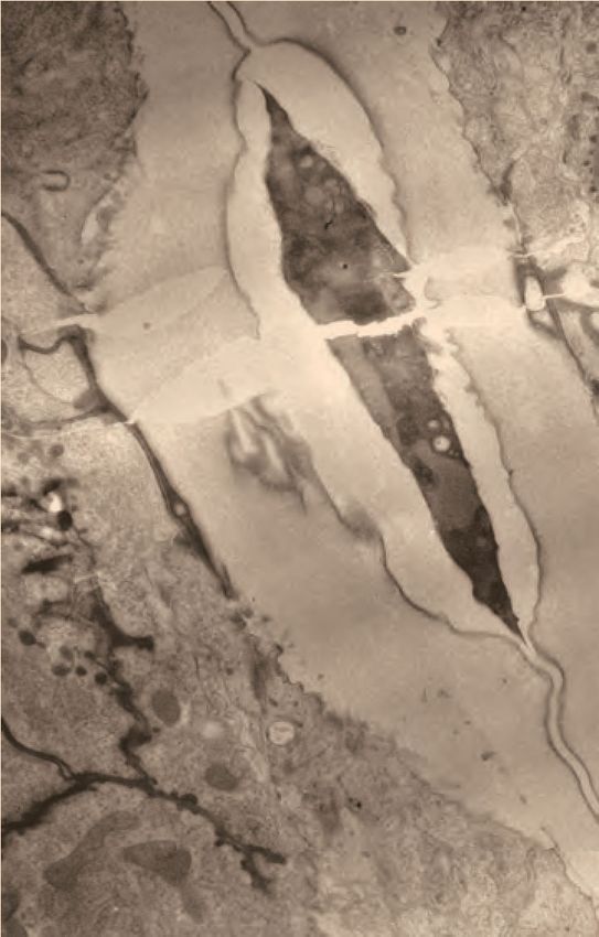
Research article                                                                                                                   Developmental Biology

    A                                                                    D               B   gonad                  AC
                              early L3 larva                         A       P
                                                                                                                             EGF
                                                                         V
                                                                                                             Û         Û           Û
                                                                                                                  DSL        DSL

                                vulva precursor cells
                                       (VPCs)
                                                                                         AA B1 B2 CC D        EE FF FF EE                 D CC B2 B1 AA
                                                                                           Notch-dep.           Ras-dep.                     Notch-dep.
                                                                                           cell types           cell types                   cell types

    C                            mid-L4 larva                                                 D               vulva cells (mid-L4)

                                                                                                   XY view
                                                                                                                                                           D
                                                                                                                                                       A       P
                                                                                                                         lumen                             V
                                               vulva
                                                                                                                                               5 um

                                                                                                                                                           L

                                                                                                  XZ view
    E                    3D renderings of vulva lumen (mid-L4)

                                                                                                                             lumen
                                                                                                                                                       A       P
            D                                                                    D                                                                         R
                    XY view

                                                YZ view

        A       P                                                            L       R
            V                                                                    V
                                                                  5 um
                                                                                              vulA vulB1 vulB2 vulC vulD vulE vulF

    F                                                     adult

                                                          vulva

                                                              eggs

Figure 1. Introduction to vulva development. (A) Cartoon of early L3 larva, indicating the six vulva precursor cells (VPCs) beneath the somatic gonad. (B)
Vulva lineages and cell types. An EGF-like signal from the gonadal anchor cell (AC) induces the primary (1˚) cell fate in the nearest VPC (black), which
then expresses DSL ligands to induce the secondary (2˚) cell fate in the adjacent VPCs (gray). The 1˚ and 2˚ VPCs divide to generate a total of 22
descendants of 7 different cell types. (C) Cartoon of mid L4 larva, showing the vulva lumen. (D) L4.4 stage vulva cells visualized with the membrane
marker MIG-2::GFP (muIs28). The 22 vulval cells are organized into 7 stacked rings (Sharma-Kishore et al., 1999). In the standard lateral or sagittal
view, anterior is to the left and ventral is down. An orthogonal XZ view shows the oblong shape of the lumen. (E) 3D rendering of the L4.4 vulva lumen
generated with Imaris software (BitPlane, Zurich Switzerland), based on imaging of the matrix factor FBN-1 (see Figure 3). The YZ view at right is
comparable to the transverse views of the vulva seen by TEM in Figures 4B and 9C (but note that regions deeper in the Z plane are poorly resolved
here). (F) The adult vulva is a slit-like and cuticle-lined passageway through which eggs are laid.

                                        RhoG marker MIG-2::GFP (Honigberg and Kenyon, 2000) to label all vulva cell membranes
                                        (Figures 1D and 2). At L4.0, the vulva invagination is very narrow, but it enlarges to approximately
                                        10 microns in diameter by the L4.3 stage. Between L4.3 and L4.4, the uterine lumen and the dorsal-
                                        most part of the vulva lumen both expand (Ihara et al., 2011; Matus et al., 2014), and the gonadal
                                        AC fuses with the uterine seam (utse), leaving just a thin part of its membrane as a hymen separating

Cohen et al. eLife 2020;9:e57874. DOI: https://doi.org/10.7554/eLife.57874                                                                                     3 of 33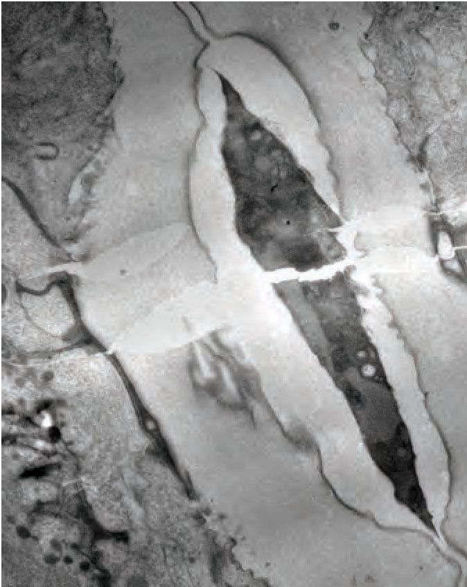
Research article                                                                                                                            Developmental Biology

                                            chondroitin-dependent inflation
                                                                                                                                          “christmas tree stage”
                             L4.0              L4.1                        L4.2                      L4.3             hymen          L4.4                         L4.5      vulE,F
                                                                                                                                                    vulC,D
                                                                                         core                        vulB1,B2                       “shoulder”              “neck”
                                                                                        structure                    “fingers”
   Lumen
   shape

                           L4 cuticle

                                         AC
                       AC
      DIC
      membranes

                             AC
                             AC
                                                                                                                                 *            *                   *         *

                                          “cactus-stage”                                             eversion
                            L4.6                L4.7                       L4.8                       L4.9                        Young Adult
   Lumen

                                                                                      vulD
   shape

                                                           vulB2,C                                               vulD            vulE,F                           vulF   ÛFHOO
                                                                                      vulC                                                           vulC,D
                                                            vulB1,B2                      vulB1,B2               vulC            lumen                                   types
                                                                                                                                                     lips
                                                                                                                   vulB1,B2                                       vulE

                                                                                                                                                 cuticle          vulD

                                                                                                                                                                  vulC
      DIC

                                                                                                                                                                         ÛFHOO
                                                                                                                                                                 vulB2   types

                                                                                                                                                                 vulB1
      membranes

                                                                                                                                                                  vulA

                       *            *      *           *                          *                                                                              *vm1
                                                                       *                              *      *                       *       *                   sex muscles

Figure 2. Cell and lumen shape changes during vulva morphogenesis Sagittal views of the central vulva lumen. Top rows show cartoons of lumen
shape for each L4 sub-stage, as defined by Mok et al., 2015. Middle rows show corresponding DIC images. Bottom rows show confocal slices of vulva
cell membranes marked by MIG-2::GFP (muIs28); cells are colored according to the key at right. Confocal stacks were collected for at least three
animals per stage after L4.3. Luminal core structure is faintly visible beginning at L4.2 (red arrow). At mid-L4 (‘Christmas-tree stage’; Seydoux et al.,
1993), the vulF and vulE cells together define the vulva ‘neck’, the vulD and vulC cells define the vulva ‘shoulder’, the vulB1 and vulB2 cells define the
vulva ‘fingers’, and the vulA cells make the connection between the vulva cells and the surrounding epidermis. *vm1 sex muscles, which attach to the
mature vulva between the vulC and vulD toroids (Sharma-Kishore et al., 1999). Scale bars, five microns.

                                        the two lumens (Sapir et al., 2007; Sharma-Kishore et al., 1999). The vulB1 and vulB2 cells also
                                        develop increasingly concave apical surfaces, creating a ‘Christmas-tree-like’ lumen appearance at
                                        L4.4-L4.5, and a more ‘cactus-like’ appearance by L4.7. In the final morphogenetic stages, collec-
                                        tively termed eversion, further cell shape changes and rearrangements occur to narrow the lumen
                                        and generate the closed lips of the final adult tube structure (Seydoux et al., 1993) (see below). Fol-
                                        lowing eversion, the vulva lumen remains in a closed conformation unless opened by contractions of
                                        the sex muscles, which attach to multiple vulva toroids (Sharma-Kishore et al., 1999). During L4 and
                                        adult stages, each vulva cell type expresses different combinations of known transcription factors,
                                        membrane fusogens, or other molecular markers (Inoue et al., 2004; Inoue et al., 2002;
                                        Inoue et al., 2005; Mok et al., 2015; Shemer et al., 2000; Sternberg and Horvitz, 1989), but the
                                        biological distinctions among the seven cell types are not well understood.
                                           A chondroitin proteoglycan (CPG)-rich luminal matrix is thought to form at the earliest stages of
                                        vulva tube morphogenesis, and to swell with water to exert a uniform pushing force for the lumen

Cohen et al. eLife 2020;9:e57874. DOI: https://doi.org/10.7554/eLife.57874                                                                                                      4 of 33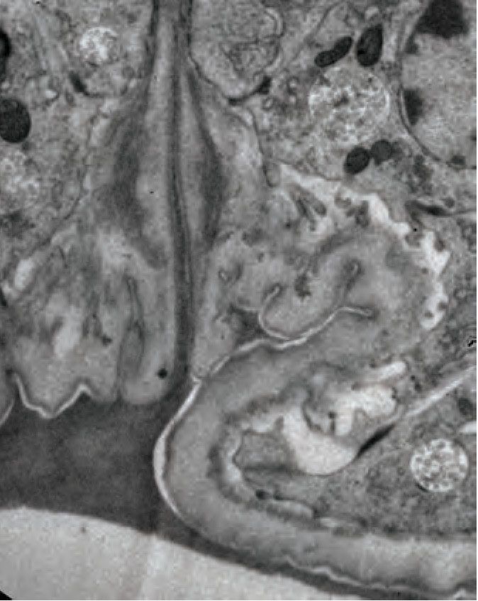
Research article                                                                                                                                                        Developmental Biology

    A              FBN-1                                                                                                         ZP                            signal sequence
                                                                                                                                                               TM domain
                LET-653b                   ZP                             LPR-3                                    LPR                                         potential cleavage site
                                                                                                                                                               chondroitin addition site
                LET-653(ZP)                ZP                             LET-4                                    LRR                                         PAN domain
                                                                                                                                                               EGF-like domain
                                                                                                                                            100 aa
                NOAH-1a                           ZP                      SYM-1                              LRR                                               SfGFP
                                                                                                                                                               mCherry

    B                    L4.4                            L4.8                           C                     L4.7                                   L4.7               L4.8
                                 vulE,F                         vulE,F
                  vulD                                                                                                   vulC
                vulC               vulB2                        vulC,D
                                   vulB1                         vulA,B1,B2
                                   vulA
                                                                                                             F
                                                                                                             D E

                                                                                            merge

                                                                                                                                  merge
                                                                                                         C
    merge

                                                                                                       B2
                                                                                                       B1
                                5 um                                          5 um                     A                 5 um                                                    5 um

                                                                                            membrane

                                                                                                                                  LET-653
    LET-653

                                                                                                                                  NOAH-1
                                                                                            NOAH-1
    FBN-1

   D                      L4.2             L4.3          L4.4            L4.5                          L4.6               L4.7                   L4.8             L4.9
                                                  vulF                                                                                vulC
                                                                                     vulD                                                               vulC
                                                  vulE           vulB2                                                                vulB2
                                                                                     vulC
                                                                 vulB1                                                                vulB1
     FBN-1

                                5 um
      LET-653
      LET-653
        (ZP)

                                                                                                                                                                   ND
      NOAH-1
      LPR-3
      LET-4
      SYM-1

                                                                                                                           ND                    ND                ND

Figure 3. A dynamic aECM fills the vulva lumen during morphogenesis. (A) aECM protein schematics. Genbank accession: FBN-1a (AFN70749.1), LET-
653b (CAH60755.1), NOAH-1a (CCD66686.2), LPR-3 (CAA92030.1), LET-4 (AEZ55699.1), SYM-1 (CAB43345). Full-length FBN-1::mCherry and some LET-
653 fusions were expressed from transgenes; all others were expressed from the endogenous loci tagged by CRISPR-Cas9 genome editing (see
Materials and methods). (B) LET-653::SfGFP (csIs64) and FBN-1::mCherry (aaaIs12) show complementary luminal patterns. Medial confocal slices. Arrow,
Figure 3 continued on next page

Cohen et al. eLife 2020;9:e57874. DOI: https://doi.org/10.7554/eLife.57874                                                                                                                               5 of 33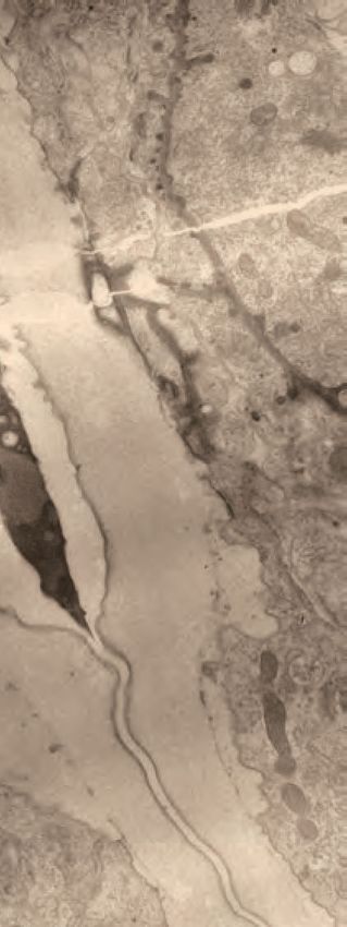
Research article                                                                                                       Developmental Biology

Figure 3 continued
luminal core. Arrowheads, sites of ventro-lateral fibril attachment to vulva cells. Lines, membrane-proximal matrix over 1˚ cells vulE and vulF. Bracket
indicates loss of FBN-1 from the lumen over 1˚ cells during vulva eversion (n = 3/5 L4.7, 4/4 L4.8). (C) NOAH-1::mCherry (mc68) labels matrix spikes that
connect to LET-653-marked luminal fibrils during vulva eversion. Left column shows overlay with cell membrane marker MIG-2::GFP (muIs28) (n = 9 L4.7-
L4.9). Right columns show overlay with LET-653::SfGFP (cs262) (n = 12 L4.7-L4.9). (D) Timeline of vulva morphogenesis showing dynamic matrix patterns.
Each image is a single confocal slice, inverted for clarity. For each fusion, images were collected for at least three animals per stage per strain; most
fusions were imaged in multiple different strains to directly compare the different patterns (as in panels B and C). Solid green underlines indicate stages
with consistent and peak localization; dashed green underlines indicate stages with more variable or weak localization. Fusions shown are FBN-1::
mCherry (aaaIs12), LET-653::SfGFP (cs262), LET-653(ZP)::SfGFP (csIs66), SfGFP::LPR-3 (cs250), NOAH-1::mCherry (mc68), mCherry::LET-4 (cs265), and
SYM-1::GFP (mc85).

                                     expansion (Gupta et al., 2012; Hwang et al., 2003a). Chondroitin antibodies stain the mid-L4 vulva
                                     lumen, though in a disorganized fashion that likely reflects matrix destruction by chemical fixatives
                                     (Bender et al., 2007). Mutants defective in chondroitin biosynthesis have a narrow or ‘squashed’
                                     vulva lumen (Sqv phenotype) (Herman et al., 1999; Hwang et al., 2003a). Genetic screens for Sqv
                                     mutants identified many components of the chondroitin biosynthesis pathway (Herman et al., 1999;
                                     Hwang et al., 2003a; Hwang et al., 2003b), but did not identify any specific chondroitin-modified
                                     proteins, suggesting redundant contributions of multiple CPGs. To date, mass spectrometry has
                                     identified 24 CPG carrier proteins in C. elegans, but it is not yet known which, if any, of these con-
                                     tribute to vulva lumen expansion (Noborn et al., 2018; Olson et al., 2006). One of these known
                                     CPGs is FBN-1, a fibrillin-related ZP protein that is part of the worm’s transient embryonic sheath
                                     matrix that precedes the cuticle (Kelley et al., 2015; Labouesse, 2012; Noborn et al., 2018;
                                     Priess and Hirsh, 1986). Other pre-cuticle aECM proteins also have been observed within the vulva
                                     and other developing tubes (Forman-Rubinsky et al., 2017; Gill et al., 2016), and the adult vulva
                                     becomes cuticle-lined in adults (Page, 2007; Sulston and Horvitz, 1977). These observations sug-
                                     gested to us that a transient sheath-like aECM may exist within the developing vulva lumen.

                                     aECM proteins show dynamic patterns of localization during vulva
                                     lumen morphogenesis
                                     To examine aECM protein expression and localization in the vulva, we examined superfolder (Sf)
                                     GFP or mCherry-based translational fusions generated by transgenic methods or (in most cases) by
                                     CRISPR-Cas9 genome editing of the endogenous loci (Materials and methods). Six different aECM
                                     proteins were assessed (Figure 3A): the ZP proteins FBN-1 (Kelley et al., 2015), LET-653 (Gill et al.,
                                     2016) and NOAH-1 (Vuong-Brender et al., 2017), the lipocalin LPR-3 (Forman-Rubinsky et al.,
                                     2017), and the extracellular leucine-rich repeat only (eLRRon) proteins LET-4 (Mancuso et al., 2012)
                                     and SYM-1 (Davies et al., 1999; Vuong-Brender et al., 2017). We also examined a shortened ver-
                                     sion of LET-653 containing only the ZP domain, because prior studies had demonstrated that this
                                     domain is sufficient for matrix incorporation and excretory duct tube shaping (Gill et al., 2016;
                                     Figure 3A). FBN-1, NOAH-1, and LET-4 each have transmembrane domains (but could be cleaved
                                     extracellularly), while LET-653, LPR-3, and SYM-1 are secreted proteins. These fusions permitted
                                     observations of matrix structure in live L4 animals, without the matrix destruction typically induced
                                     by chemical fixation for immunofluorescence. In each case, these fusion proteins were functional as
                                     assessed by mutant rescue or strain phenotypes (Materials and methods), suggesting that their local-
                                     ization closely approximates the endogenous patterns. Each fusion protein initially appeared at
                                     around the L4.2 stage, and then exhibited a temporally and spatially distinct pattern over the course
                                     of L4 vulva morphogenesis (Figure 3B–D). All six matrix proteins were transient and disappeared in
                                     the adult stage, indicating that they are not components of the mature cuticle, but rather define a
                                     distinct matrix type present only during morphogenesis.
                                        The ZP proteins FBN-1 and LET-653 showed somewhat complementary luminal patterns
                                     (Figure 3B,D). Beginning at L4.2, and as previously reported for LET-653(PAN domain) fragments
                                     (Gill et al., 2016), LET-653 decorated a core structure in the center of the lumen that rises to the
                                     level of the vulD and vulE cells, along with lateral elements that connect this core to the vulA, vulB1
                                     and vulB2 cells and to the surrounding epidermis (Figure 3B,D). The central core structure could
                                     also be detected very weakly by DIC (Figure 2; red arrowhead). This core changed appearance dur-
                                     ing vulva eversion, but remained visible in the most ventral, 2˚-cell-derived region through the L4.9

Cohen et al. eLife 2020;9:e57874. DOI: https://doi.org/10.7554/eLife.57874                                                                         6 of 33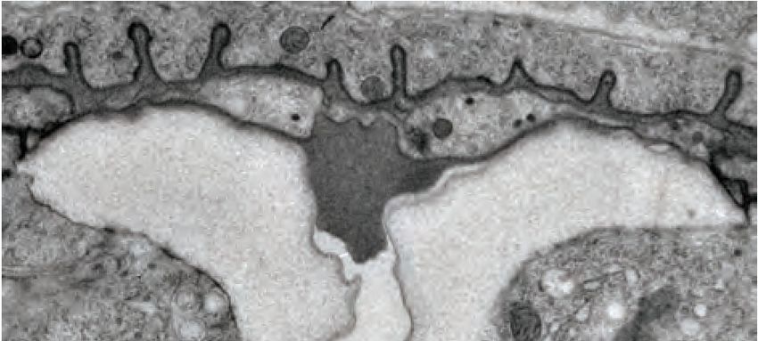
Research article Developmental Biology
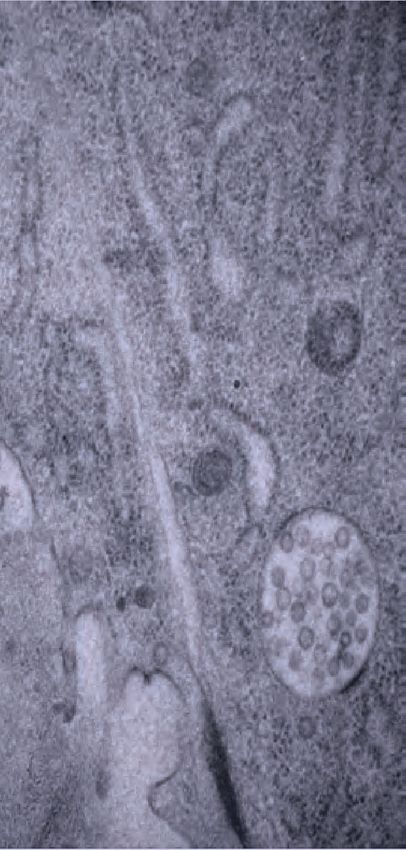
A C granular matrix F lumen pockets I vulD
fibrils
vulva
H K
G
F
I D seam
D
C L R
hyp7
V 500 nm
E
J D core structure G vulF vesicles J striated layer
L4.4, Transverse view (TEM)
AC/utse granular matrix core structure
vulA vulB1 vulB2 vulC vulD vulE vulF
B H* K*
gut F
I
G C E ventro-lateral fibrils H vulE aECM/core border K AC matrix
uterus
muscle
lu
AC/utse
D
E
EVs AC
muscle matrix
granular
J* matrix
broken cuticle
2 microns
Figure 4. Ultrastructural features of the mid-L4 vulva aECM. (A) Transverse serial thin sections of an N2 L4.4-L4.5 stage animal were analyzed by
TEM. The cartoon depicts the vulva lumen in this orientation (see also Figure 1E), and lines indicate the relative locations of different panel images. (B)
Whole vulva view. This thin section captures a portion of the lumen and the cell borders. Vulva cells and AC/utse are pseudo-colored according to the
key shown in A. Lines indicate the relative locations of different panel images, and asterisks indicate that the panel shows a region from a different thin
section of the same animal. The ventral cuticle has broken during specimen processing and oval objects surrounding the specimen are E. coli bacteria.
(C) A rough granular matrix fills the dorsal lumen. (D) Core structure (white arrowheads) rises above vulC and vulD, to the level of vulE. (E) Ventro-lateral
fibrils (white arrowheads) and the ventral edge of the luminal matrix, which has pulled away from the broken cuticle. (F) The interface between vulF (red)
and vulE (yellow) forms a sequestered lumen pocket where matrix accumulates (red and yellow arrowheads). vulD (green) forms another narrow lumen
pocket that is densely populated with fine fibrils (green arrowhead). (G) vulF cells contain large secretory vesicles (black arrows) that are open to the
extracellular space and whose contents resemble the membrane-proximal matrices that line vulF and vulE (red and yellow arrowheads, respectively). (H)
Lateral view of the matrix lining vulE surfaces. (I, J) Examples of the very protrusive surfaces of 2˚-derived cells (vulD and vulB1, respectively) that
interface with fibrils. J also shows the ventral-most border of the aECM, which contains a striated layer (white arrowheads) similar to that seen in
epidermal cuticle (Page, 2007). (K) A fine-grained aECM separates the AC/utse from the rougher granular matrix of the vulva lumen; this AC matrix
contains numerous EVs (orange arrowheads). All scale bars are 500 nm unless otherwise indicated. See Figure 4—figure supplement 1 for uncolored
versions of all images.
The online version of this article includes the following figure supplement(s) for figure 4:
Figure supplement 1. Ultrastructural features of the mid-L4 vulva aECM. Uncolored images from Figure 4 are shown.
stage. Transiently, at the L4.3-L4.5 stages, LET-653 also weakly marked the apical membranes of
most cells. Finally, FBN-1 overlapped with LET-653-marked structures near vulB1 and vulB2 surfaces,
but otherwise was mainly excluded from the core area and instead filled the more dorsal part of the
lumen above the core (Figure 3B,D). During vulva eversion, FBN-1 became excluded from the dor-
sal-most portions of the lumen lined by 1˚-derived cells, such that LET-653 and FBN-1 together
Cohen et al. eLife 2020;9:e57874. DOI: https://doi.org/10.7554/eLife.57874 7 of 33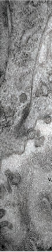
Research article                                                                                            Developmental Biology

                                   appeared to demarcate at least three separate luminal zones roughly corresponding to the regions
                                   outlined by the vulA/B cells, vulC/D cells, and vulE/F cells (Figure 3B).
                                      The isolated LET-653 ZP domain and the other four aECM proteins marked specific apical mem-
                                   brane-proximal regions in a dynamic manner (Figure 3C,D). LET-653(ZP) specifically labelled just the
                                   1˚-derived vulE and vulF cell surfaces at L4.3-L4.5 stages. Previous Fluorescence Recovery After Pho-
                                   tobleaching (FRAP) studies showed that, while it is present, this pool of LET-653(ZP) is relatively
                                   immobile, consistent with matrix incorporation (Gill et al., 2016). NOAH-1 faintly marked all 2˚ vulva
                                   cell surfaces at L4.4-L4.5, but then became increasingly concentrated on vulC and vulD. During vulva
                                   eversion, NOAH-1 prominently marked matrix spikes that protruded from vulC into the lumen, and
                                   these spikes attached to LET-653-marked lateral structures near vulB2 surfaces (Figure 3C,D). These
                                   NOAH-1—LET-653 connections then persisted as the lumen narrowed. LPR-3 briefly marked all vulva
                                   cell apical surfaces at early L4.4, but then became restricted to 2˚ cells and then specifically to vulB1
                                   and vulB2 before largely disappearing by L4.6-L4.7 (Figure 3D). The departure of LPR-3 from vulC
                                   and vulD coincided with the increasingly strong presence of NOAH-1 there. The transmembrane
                                   eLRRon protein LET-4 marked all vulval apical membranes during the late L4.2-L4.7 period, and
                                   thereafter appeared intracellular (Figure 3D). Finally, the secreted eLRRon protein SYM-1 showed
                                   the most limited pattern, labelling vulB1 and vulB2 for just a brief period at L4.4-L4.5 (Figure 3D).
                                   Together, these data reveal that different combinations of aECM factors assemble on the luminal
                                   surface of each vulva cell type. Furthermore, the precise timing of each factor’s appearance and dis-
                                   appearance points to highly regulated mechanisms for matrix assembly and remodeling.

                                   Ultrastructural features of the luminal matrix differ between 1˚ and 2˚-
                                   derived vulva regions
                                   Prior transmission electron microscopy (TEM) studies of the vulva Gill et al., 2016; Herman et al.,
                                   1999 used chemical fixation methods that poorly preserved the luminal matrix and did not capture
                                   the complex luminal structures observed in the live imaging above. To obtain a clearer view of
                                   matrix ultrastructure, we turned to high pressure freezing (HPF) and freeze substitution (Hall et al.,
                                   2012). This method achieved much better matrix preservation and revealed many matrix layers and
                                   fibrils that we could correlate with those observed by light microscopy. Serial thin sections were col-
                                   lected transverse or length-wise to the body axis to obtain a three-dimensional view. Images of mid-
                                   L4 (L4.4-L4.5) and late-L4 (L4-8-L4.9) stage vulvas are shown in Figures 4 and 5 and S1, S2. A striking
                                   feature of both stages is the difference in matrix organization in the dorsal (1˚-derived) vs. ventral (2˚-
                                   derived) portions of the lumen.
                                      At the mid-L4 stage, a rough granular matrix fills the entire vulva lumen, and embedded within it
                                   are a central core structure and numerous ventro-lateral fibrillar elements similar to those seen with
                                   LET-653::SfGFP (Figure 4B–E). Most of the dorsal lumen surrounding the central core contains only
                                   the granular matrix; this corresponds to the region marked by FBN-1::mCherry (Figure 3B) and likely
                                   contains additional CPGs. There is no single luminal channel running through the granular matrix,
                                   which appears organized into multiple wide strips or flaps, each edged with a more electron-dense
                                   border (Figure 4B,C). 1˚-derived vulval cells have relatively smooth apical surfaces lined with thin
                                   matrices that separate them from the granular matrix (Figure 4F–H), while the 2˚-derived cells have
                                   more protrusive apical surfaces lined with numerous fibrils that are embedded within the granular
                                   matrix (Figure 4D–F,I,J).
                                      At the dorsal apex of the vulva, the AC remnant hymen is lined by a layer of finer-grained, elec-
                                   tron-dense matrix that separates it from the vulva granular matrix (Figure 4K). Within this AC matrix
                                   are numerous extracellular vesicles (EVs). The contents and purpose of these EVs are not currently
                                   known, but the AC is a source of multiple signaling molecules (Hill and Sternberg, 1992;
                                   Sherwood and Sternberg, 2003).
                                      The vulF cells contain numerous large (~200 nm) secretory vesicles that resemble those seen in
                                   mammalian goblet cells (Figure 4F,G; Birchenough et al., 2015). These vesicles contain globules
                                   that resemble mucin packets, along with a few membranous intraluminal vesicles (ILVs). The secre-
                                   tory vesicles appear to be dumping their contents within sequestered pockets at the left and right
                                   extremes of the lumen, and these contents then expand spherically upon contacting the outside
                                   environment. The contents within these luminal pockets are continuous with a thin membrane-proxi-
                                   mal matrix layer that likely corresponds to the layer marked by LET-653(ZP)::SfGFP (see Figure 3D
                                   and below). vulE surfaces are decorated by a mesh-like matrix that drapes down along the top

Cohen et al. eLife 2020;9:e57874. DOI: https://doi.org/10.7554/eLife.57874                                                             8 of 33
Research article                                                                                                         Developmental Biology

                                                                                  XWHUXV
    A                                                  B                                                 C
                                           XWHUXV
                                                                           plug
                                                              ÛPDWUL[                                                              lu

                                                                                                         YXO'ILEULOV

                                           vulva
     vulva                                 muscle
     muscle
                                                                             lu

         D
     A       P
         V                               ~1 micron

    D                                                  E                                                 F

                                                                                                                                plug
                                                                                                                                                    XWHUXV
                                                                                                                    ÛPDWUL[
                                                                                                                                lu

                                                                                                               XWHUXV

                           plug                                   MVB
                                                                                                          L     P

                                                                                                           A    R

Figure 5. Ultrastructural features of the late L4 vulva aECM. (A) Longitudinal slice through the vulva of an N2 L4.8-L4.9 stage animal, with orientation
similar to that in confocal images. Vulva and uterine cells are pseudo-colored as in Figure 4B. Pink arrowheads indicate matrix spikes as observed with
NOAH-1::mCherry (see Figure 3C). (B–E) Higher magnification views of the specimen in panel A. (B) Primary vulva cells are covered in a thick
membrane-proximal matrix, and the dorsal-most edge of the lumen is filled with a plug of darkly-staining material. (C) The membrane-proximal matrix
continues over vulD and vulC, but becomes filled with dense fibrils (green arrowheads). (D) Matrix spikes (pink arrowheads) extend from vulC/D into a
cuticle-like matrix below. Various other fibrils (white arrowheads) are present within this ventral matrix. (E) Multi-layered nascent cuticle over vulB2. Note
protrusive surface of vulB2 and multi-vesicular body (MVB) within vulB1. Many MVBs are present in vulva cells at this stage. (F) Longitudinal dorsoventral
slice through uterine cells and the primary vulva matrix and plug of a second N2 L4.8-L4.9 stage animal. Note the numerous EVs (orange arrowheads)
present within the plug. All scale bars, one micron. See Figure 5—figure supplement 1 for uncolored versions of all images.
The online version of this article includes the following figure supplement(s) for figure 5:
Figure supplement 1. Ultrastructural features of the late L4 vulva aECM. Uncolored images from Figure 5 are shown.

                                      border of the core and ventro-lateral fibrils (Figure 4H). This matrix appears as dark membrane-
                                      associated patches when vulE is viewed in cross-section (Figure 4F,G). This matrix may serve as the
                                      barrier that excludes FBN-1 from the ventral fibrillar region (see Figure 3B,D).
                                         vulD and vulC surfaces that sit above the ventro-lateral fibrils (and external to the core) are lined
                                      with thin fibrils that run in a dorsal-ventral orientation, parallel to the cell membranes (Figure 4I).
                                      These fibrils are embedded within the granular luminal matrix rather than forming a separate layer,

Cohen et al. eLife 2020;9:e57874. DOI: https://doi.org/10.7554/eLife.57874                                                                               9 of 33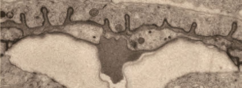
Research article                                                                                            Developmental Biology

                                              LET-653                 LET-653(ZP)                    NOAH-1                        LPR-3
                 A
    wild-type

                           Û
                     Û

                                                         5 um

                 B
    lin-12 (0)

                     Û

                 C
    lin-12 (d)

                      Û

Figure 6. Different vulva cell types produce and assemble different aECMs. (A–C) Panels in left column show cartoons of vulva cell types and lumen
shape at mid-L4. Remaining columns show single confocal slices through the vulva lumen. Fusions used are LET-653(full-length)::SfGFP (cs262), LET-653
(ZP)::SfGFP (csIs66), NOAH-1::mCherry (mc68), SfGFP::LPR-3 (cs250). At least n = 8 L4s were imaged for each strain. (A) In wild-type animals, full-length
LET-653 predominantly labels the core and ventro-lateral fibrils, LET-653(ZP) labels the membrane-proximal matrix over 1˚ cells, NOAH-1 labels
membrane-proximal matrices over 2˚ cells (especially vulC and vulD), and LPR-3 transiently labels membrane-proximal matrices over all cells, but then
becomes concentrated over 2˚ cells (see also Figure 3D). (B) Loss of 1˚ cells in lin-12(0) (null, n137n720) mutants disrupted the luminal core and NOAH-1
localization. Most of the NOAH-1 pattern here is intracellular. (C) Loss of 2˚ cells in lin-12(d) (hypermorphic, n137) mutants disrupted the luminal core
and LET-653(ZP) localization.

                                        and they abut numerous small cellular projections. The fibrils are particularly concentrated in narrow
                                        (~0.5 micron) lumen pockets generated by the complex shape of vulD (Figure 4F). These fibrillar
                                        regions correspond to those surfaces that become strongly marked by NOAH-1::mCherry (see
                                        Figure 3C).
                                           Finally, the ventral-most vulC surfaces, as well as vulB2, vulB1 and vulA, interface with the dense
                                        ventro-lateral fibrils, which run both perpendicular to and parallel with the cell membranes
                                        (Figure 4B,E). The cell surfaces that interface with these fibrils are extremely protrusive (Figure 4J).
                                        At the most ventral edge of the lumen, beneath the core fibrils, matrix layers that resemble those of
                                        the epidermal sheath and nascent cuticle interface with the remaining L4 epidermal cuticle (which
                                        has broken and pulled away somewhat in this specimen) (Figure 4B,E,J).
                                           The late-L4 stage vulva (Figure 5) retains several of the matrix features seen at the earlier stage,
                                        with some notable differences. An electron-dense ‘plug’ resembling the earlier AC matrix is present
                                        at the dorsal apex and contains EVs (Figure 5A,B,F). A similar-appearing matrix is also present at
                                        the ventral opening of the lumen (Figure 5A,D). The 1˚-derived vulE and vulF cells and the 2˚-derived
                                        vulC and vulD cells are now covered with a thick membrane-proximal matrix that somewhat resem-
                                        bles the prior CPG matrix, but with a well-defined, darkly-staining border and a single open channel
                                        that runs through its center (Figure 5A,B,F). The membrane-proximal matrix over vulC and vulD also
                                        contains many dense fibrils that extend down into the matrix below (Figure 5C,D); these likely corre-
                                        spond to the NOAH-1-marked matrix spikes observed by confocal imaging (see Figure 3C). The
                                        remaining vulA, vulB1 and vulB2 cells are covered with a more complex, multi-layered matrix that
                                        resembles the nascent pre-cuticle on nearby epidermal cells (Figure 5D,E). Various fibrillar structures
                                        are embedded within this thick matrix (Figure 5D), possibly corresponding to the core and ventro-
                                        lateral fibrils seen earlier. Thus, just as seen by confocal light microscopy at this stage (Figure 3B),
                                        TEM shows three distinct luminal zones corresponding to the regions outlined by the vulA/B cells,
                                        vulC/D cells, and vulE/F cells.

Cohen et al. eLife 2020;9:e57874. DOI: https://doi.org/10.7554/eLife.57874                                                                      10 of 33
Research article Developmental Biology
L4.1 L4.9
A VAB-10
B L4.5 L4.7
MUP-4 Inverted Merge MUP-4 Inverted Merge
MUP-4 FBN-1
L4.9
MUP-4 Inverted Merge Merge with Dodt ventral view
MUP-4 FBN-1
vulA
vulA vulA
Figure 7. Vulva aECM assembles prior to expression of MUP-4/matrillin. (A) VAB-10::GFP (cas627) marked all apical membranes in the vulva throughout
L4 (including 4/4 L4.1/L4.2 stage animals). (B) MUP-4::GFP (upIs1) marked apical membranes beginning in late L4 (0/3 L4.4/L4.5, 3/3 L4.6/L4.7, 2/2 L4.9).
FBN-1::mCherry (aaaIs12 or aaaEx78) is also shown. At L4.9, MUP-4 expression is particularly strong in the vulA toroid, which surrounds the vulva
opening and connects to the surrounding hyp7 epidermis. The remaining FBN-1 matrix connects to vulA at the left and right sides of the lumen, as
seen in the ventral view.
1˚ and 2˚ vulva cell types produce and assemble different matrices
To better understand the differences between the matrix produced and assembled by 1˚ vs. 2˚ vulva
cell types, we analyzed matrix patterns in lin-12/Notch mutants. LIN-12/Notch promotes 2˚ vs. 1˚
VPC fates, so loss-of-function [lin-12(0)] mutants have only 1˚ vulva cell types, while gain-of-function
[lin-12(d)] mutants have only 2˚ vulva cell types (Greenwald et al., 1983; Sternberg and Horvitz,
1989). lin-12(0) and lin-12(d) mutants both have well-inflated (though mis-shapen) vulva lumens (Fig-
ure 6), indicating that relevant CPGs are made by both sets of vulva cell types. Indeed,
Herman et al., 1999 previously showed that both types of lin-12 mutants require the Sqv chondroi-
tin biosynthesis pathway for lumen inflation.
Close examination of lin-12 mutants suggested, however, that the central core structure was miss-
ing. When LET-653::SfGFP was introduced into lin-12(0) mutants, core structures appeared very mea-
ger or absent (Figure 6B). In lin-12(d) mutants, no core was observed in the central lumen, but some
ventro-lateral elements were still present at the vulB ‘fingers’ (Figure 6C). We conclude that both 1˚
and 2˚ cells are required to generate the core, but that 2˚-derived cells (most likely vulB1 and vulB2)
generate at least some of the ventro-lateral elements independently.
lin-12 mutants also showed changes in the localization of membrane-proximal matrix factors, as
predicted based on the cell fate changes. For example, LET-653(ZP) marked all vulva apical mem-
branes in lin-12(0) mutants, but none in lin-12(d) mutants (Figure 6). Since let-653 is expressed by all
seven vulva cell types (Gill et al., 2016), these data suggest that a 1˚-specific partner is required to
recruit LET-653(ZP) to the membrane-proximal matrix. In contrast, NOAH-1 was mostly absent from
vulva apical membranes in lin-12(0) mutants, but strongly marked the dorsal-most apical membranes
in lin-12(d) mutants (Figure 6). LPR-3 marked some apical membranes in both lin-12(0) and lin-12(d)
Cohen et al. eLife 2020;9:e57874. DOI: https://doi.org/10.7554/eLife.57874 11 of 33Research article Developmental Biology
A let-653;
WT let-653 Is[LET-653(ZP)] fbn-1 let-4 sym-1
L4.4
5 um
L4.7
*
L4-Adult Molt
B C D wild type vulva eversion
% Egl or rupture box 1
0 50 100 1 cells
2 cells
L4.7
L4.8
para-sagittal
WT n = 133
*** ***
let-653 n = 141 box 2
* * *
let-653; Ex[LET-653] n = 109
let-653; Is[LET-653ZP] n = 137
Box 2 height
Egl Rupture L4.9
15 ns
para-sagittal
E wild type L4.7 * **
let-653 L4.7
10
microns
* *
sagittal
* * 5
* *
adult
0
para-sagittal
para-sagittal
53
]
T
ZP
W
t-6
s[
le
;I
* *
* *
53
*
t-6
*
le
F wild type Adult
sagittal para-sagittal Z-projection 3D rendering 3D rendering, YZ
vulC/D, vulE/F
let-653 Adult
sagittal para-sagittal Z-projection 3D rendering 3D rendering, YZ
vulC/D, vulE/F
Figure 8. Individual aECM factors play subtle roles in vulva eversion. (A) DIC images of mutant vulvas at L4.4, L4.7 and L4.9-adult molt. At least 40 L4
animals of each genotype were imaged, including at least five each of the three stages shown. Alleles used: fbn-1(tm290), let-653(cs178) (strains UP3342
and UP3422), let-4(mn105), and sym-1(mn601). Asterisk indicates collapsed lumen morphology in some let-653 mutants (n = 6/22 L4.7, see panel C).
Arrowheads indicate abnormal bulges of the vulA and vulB1/B2 cells in fbn-1 mutants (n = 9/13 L4.9). (B) A small proportion of let-653 mutants had
Figure 8 continued on next page
Cohen et al. eLife 2020;9:e57874. DOI: https://doi.org/10.7554/eLife.57874 12 of 33Research article Developmental Biology Figure 8 continued progeny that hatched in utero (Egl phenotype) or ruptured at the vulva within eight days of reaching adulthood. These phenotypes were rescued by transgenes expressing full-length LET-653 or just the ZP domain. ***p
Research article                                                                                                      Developmental Biology

                              L4.4                 L4.8
         A                                                                     C                   L4.5 (transverse)
                                                                                                                                  D
                                                                    D                let-653                                 L        R
                                                                A       P                                                         V
                                                                    V
                                                                                                vulF
                        let-653                    let-653
                                     n=6                       n=5                             vulE
               FBN-1

                                                                                         vulD

                                     5 um
                                                                                        vulC
                                                               5 um
                                                                                      vulB2
                                                                                              vulB1
          with Dodt

                                                                                                   vulA
           merge

                                                                                                                                      2 microns

                                                                              C’     vulF vesicle
                                                                                     & aggregates
         B                              L4.7
                                                                    D
                                                               A        P
                                                                    V

                         wild-type
                               y                   let-653
               LPR-3

                                                                                                                                      500 nm
                                                               5 um
               NOAH-1

                                                                              D wild-type
               merge
          with Dodt
           merge

                                                                                500 nm

    OVERLAP:             n=0/8                      n=5/6

Figure 9. The ZP protein LET-653 is required for proper organization and remodeling of the vulva aECM. (A) Let-653(cs178) mutants showed relatively
normal patterns of FBN-1::mCherry (aaaIs12) localization. Arrowhead indicates exclusion of FBN-1 from the core region. Bracket indicates exclusion of
FBN-1 from the 1˚ lumen during eversion (n = 5/5). Compare to WT in Figure 3B. (B) Let-653(cs178) mutants showed normal recruitment of SfGFP::LPR-
3 (cs250) and NOAH-1::mCherry (mc68) to 2˚ surfaces, but abnormally delayed clearance of SfGFP::LPR-3 from vulC/D. p=0.0445, Fisher’s exact test. (C)
Figure 9 continued on next page

Cohen et al. eLife 2020;9:e57874. DOI: https://doi.org/10.7554/eLife.57874                                                                        14 of 33
Research article Developmental Biology
Figure 9 continued
Transverse TEM slice of a let-653(cs178) mutant at mid-L4 (~L4.5) stage. Compare to the WT mid-L4 specimen in Figure 4B. Fibrils are present near the
AC/utse (arrow) and the core structure (arrowhead) is not well-defined, unlike in WT (see magnified images in Figure 9—figure supplement 1). Box
indicates region magnified in C’. (C’) An abnormal secretory vesicle in vulF is filled with dark aggregates that match those present in the membrane-
proximal matrix over vulF (red arrowhead) and vulE (yellow arrowheads). A similar matrix continues beneath the AC/utse (see also Figure 9—figure
supplement 1). (D) WT vulF vesicles and matrix for comparison. See also Figure 4G.
The online version of this article includes the following figure supplement(s) for figure 9:
Figure supplement 1. Vulva aECM organization differs between let-653 mutants and WT.
of VAB-10, MUP-4 and MUA-3 more definitively, and to identify the mechanisms that link aECM to
vulva apical membranes.
Transient aECM factors facilitate proper vulva eversion
The complex localization patterns described above suggest important roles for aECM factors in
vulva morphogenesis. Of the six aECM factors described here, only sym-1 mutants are fully viable,
while presumed null mutants of the rest mostly arrest as L1 larvae with excretory tube blockage or
other epithelial tissue-shaping defects (Forman-Rubinsky et al., 2017; Gill et al., 2016;
Mancuso et al., 2012; Pu et al., 2017; Soulavie et al., 2018; Vuong-Brender et al., 2017). To
examine vulva phenotypes in these lethal mutants, we took advantage of rare escapers (for fbn-1) or
used tissue-specific rescue strains (Materials and Methods) to bypass the earlier requirements (for
let-653 and let-4). We were not able to examine noah-1 or lpr-3 mutants in this study because of
their severe epidermal molting defects (Forman-Rubinsky et al., 2017; Vuong-Brender et al.,
2017). All mutants examined had fairly normal vulva lumens at the mid-L4 stage, indicating efficient
CPG-dependent lumen inflation (Gill et al., 2016; Figure 8A). All mutants also assembled a core
structure, as seen by DIC (Figure 8A). However, in some cases, later stage vulvas appeared mis-
shapen or improperly everted (Figure 8A). Specifically, some let-653 mutants had a prematurely col-
lapsed vulva lumen (Figure 8A, asterisk), and most fbn-1 mutants had abnormal bulges of the outer
vulA, vulB1 and vulB2 cells (Figure 8A, arrowheads). We conclude that aECM factors facilitate later
stages of vulva morphogenesis, including vulva eversion.
To better understand these phenotypes, we focused on let-653 mutants. As adults, most let-653
mutants were able to lay eggs; however, a small proportion of mutants ruptured at the vulva and/or
were egg-laying defective (Egl) (Figure 8B). Measurements of the 1˚- and 2˚-derived portions of the
vulva lumen confirmed normal dimensions at the L4.4 stage, but slightly reduced dimensions in a
subset of animals at the onset of eversion at the L4.7 stage (Figure 8C, Figure 8—figure supple-
ment 1). A transgene expressing just the LET-653(ZP) domain reversed the L4.7 defects, and led to
overexpansion of the lumen at L4.4 (Figure 8C, Figure 8—figure supplement 1), consistent with
our prior report (Gill et al., 2016) that this domain has lumen-expanding properties.
Vulva eversion has been described as the vulva ‘turning inside out’ (Seydoux et al., 1993;
Sharma-Kishore et al., 1999), but the specific cellular events involved have never been reported. To
visualize cell positions and shapes during eversion, we used the RhoG marker MIG-2::GFP to label
all vulva cell membranes (Figures 2 and 8D,E), and the daf-6pro::CFP and egl-17pro::YFP marker
combination (Mok et al., 2015) to label the vulE/F and vulC/D cells specifically (Figure 8F). During
wild-type eversion (Figure 8D), all four of these cells elongate in the dorsal-ventral axis to partly
occlude the luminal space. vulC extends a narrow NOAH-1 matrix spike into the core matrix, whose
spokes appear to fold like those of an umbrella as the lumen narrows (Figure 3C). The vm1 sex
muscles also extend ventrally into vulC and vulD (Figures 2 and 8D). Meanwhile, vulA, vulB1 and
vulB2 tilt ventrally, while vulE reaches dorsally to connect to the seam epidermis. By adulthood, vulE
and vulF enclose the bulk of the lumen, vulC and vulD form the vulva lips, and the vulA, vulB1 and
vulB2 cells are excluded from the lumen, and instead form the epidermis surrounding the vulva
opening (Figures 2 and 8D,F).
In let-653 mutants, vulva eversion occurred in a more irregular manner (Figure 8). The anterior
and posterior halves of the vulva showed various asymmetries by the onset of eversion at the L4.7
stage (Figure 8E), and cell shapes and relative cell positions continued to be variably abnormal in
older L4s and adults (Figure 8F). Defects were particularly noticeable in para-sagittal slices and in
Cohen et al. eLife 2020;9:e57874. DOI: https://doi.org/10.7554/eLife.57874 15 of 33Research article Developmental Biology
3D reconstructions of the YZ dimensions; whereas WT adult vulE cells reach far dorsally to connect
to the lateral seam epidermis on the left and right sides of the body (Sharma-Kishore et al., 1999),
some let-653 vulE cells appeared not to reach that far (Figure 8F). Imaging of a cell junction marker
revealed that let-653 morphogenesis defects are not due to failure of cell fusion to form the vulva
toroids (Figure 8—figure supplement 2). Instead, we conclude that LET-653 plays subtle roles to
coordinate the complex cell shape changes that occur during the process of eversion.
LET-653 is required for multiple aspects of vulva aECM organization
To test how LET-653 affects the organization of the vulva matrix, we first assessed the status of other
matrix factors in the let-653 mutant background. let-653 mutants still assembled some type of cen-
tral core structure, as seen by DIC (Figure 8A) and by the exclusion of FBN-1 from this region
(Figure 9A). As in WT, FBN-1 departed from the dorsal-most lumen during eversion (Figure 9A) and
LPR-3 and NOAH-1 still appeared at their proper locations (Figure 9B). However, the normally pre-
cise sequence of LPR-3 clearance was disrupted, such that LPR-3 remained on vulC and vulD apical
surfaces longer than normal, and overlapped significantly with NOAH-1 there (Figure 9B), suggest-
ing improper organization of the vulC/D membrane-proximal aECM.
TEM of a mid-L4 let-653 mutant revealed more dramatic matrix abnormalities (Figure 9C–C’).
Some vulF secretory vesicles contained very disorganized, dark aggregates, and the membrane-
proximal matrices over vulF, vulE, and the AC/utse appeared filled with such aggregates
(Figure 9C’). The luminal matrix also contained many aggregates or short, fibrillar structures rather
than having the uniform granular appearance of WT. Few fibrils were detected near the surfaces of
vulC/D or other 2˚-derived cell types. Instead, many long fibrils accumulated at the dorsal apex of
the vulva, along the AC/utse, where fibrils had not been seen in WT (Figure 9D, Figure 4—figure
supplement 1). The central core region was recognizable but less well-defined than in WT, as it was
interspersed with many aggregates or short fibrils similar to those present in the rest of the luminal
matrix (Figure 9C, Figure 4—figure supplement 1). Thus, although let-653 is not required to build
the luminal core or to establish membrane-proximal matrices per se, it is required for the proper
morphology of these structures and to set up the major dorsal vs. ventral differences in the granular
vs. fibrillar organization of the vulva aECM.
Vulva aECM structures form independently of chondroitin
Thus far, the data indicate that chondroitin plays an early role in vulva lumen inflation, whereas LET-
653 and FBN-1 play later roles in morphogenesis and eversion. However, FBN-1 is a known CPG
(Noborn et al., 2018) and other aECM factors appear to be embedded within the CPG matrix (Fig-
ure 4), suggesting some coordination between the two processes.
To ask if chondroitin affects aECM assembly, we examined aECM reporters in sqv-5 and mig-22
mutants (Figure 10). sqv-5 and mig-22 encode chondroitin sulfate synthases related to human
CHSY1 and CHSY2 (Hwang et al., 2003a; Suzuki et al., 2006), which promote chondroitin biosyn-
thesis and polymerization (Izumikawa et al., 2004). LET-653(ZP) did still assemble on the apical sur-
faces of 1˚ cells in sqv-5 mutants (Figure 10A). However, these null mutants have a very narrow vulva
lumen, which made it difficult to stage L4 animals accurately and assess the localization of full-length
LET-653 or other dynamic aECM factors. Therefore, we turned to mig-22 hypomorphic (rf = reduced
function) mutants, which have a slightly less severe vulva phenotype.
In mig-22(rf) mutants at mid-L4 stage, both the 1˚- and 2˚-derived parts of the lumen were nar-
rower than in wild-type, but lumen length varied between regions (Figures 10B,C and 11A,B). The
vulva ‘neck’ region, defined by vulD, vulE, and vulF (see Figure 2), was longer than in wild-type, and
vulD cells were prematurely elongated along the dorsal-ventral axis (Figure 10B), as previously
described for sqv-3 mutants (Herman et al., 1999). In contrast, the main body of the lumen, defined
by vulA, vulB1, vulB2, and vulC, was shorter. Thus chondroitin has very different effects on the apical
domain size of different cell types.
mig-22(rf) mutants still had a luminal core structure marked by LET-653::SfGFP, though this was
narrower than in wild-type, matching the changed dimensions of the lumen (Figure 10D). mig-22(rf)
mutants also still recruited LET-653(ZP), NOAH-1 and LPR-3 to appropriate apical surfaces
(Figure 10A,E). Thus, chondroitin does not appear essential for assembling the vulva aECM, though
it remains possible that it influences aECM structure in a more subtle way.
Cohen et al. eLife 2020;9:e57874. DOI: https://doi.org/10.7554/eLife.57874 16 of 33Research article Developmental Biology
SfGFP::LET-653(ZP)
A SfGFP::LET-653(ZP) membrane::mCherry B L4.4
10/10
WT
VulD
Aspect ratio
4
WT
5 um ***
3
4/4
10 um
2
sqv-5(lf)
1
mig-22(rf)
10/10 0
2
mig-22(rf)
T
-2
W
ig
m
meas
me
measurements?
asur
as urem
ur emen
ements
en ts?
ts ?
C vulE/F, vulC/D Dodt merge D LET-653::SfGFP Dodt merge
WT
WT
5 um 5 um
mig-22(rf)
mig-22(rf)
L4.5 L4.7
NOAH-1::mCherry NOAH-1::mCherry
SfGFP::LPR-3 Dodt merge SfGFP::LPR-3 Dodt merge
E
WT
WT
5 um 5 um
mig-22(rf)
mig-22(rf)
Figure 10. Vulva aECM structures form independently of chondroitin. (A) Chondroitin mutants showed normal recruitment of LET-653(ZP)::SfGFP
(csIs66) to the membrane-proximal matrix over 1˚ cells. Alleles used were sqv-5(n3611) (n = 4) and mig-22(k141rf) (n = 10). (B) WT vs. mig-22(k141rf)
mutants (L4.4 stage) with membrane marker MIG-2::GFP (muIs28). The vulva neck region was taller and narrower in mutants compared to wild- type,
while the rest of the lumen was shorter and narrower (n = 15, see quantification in Figure 11B). vulD cells (green) showed the most dramatic shape
Figure 10 continued on next page
Cohen et al. eLife 2020;9:e57874. DOI: https://doi.org/10.7554/eLife.57874 17 of 33Research article Developmental Biology Figure 10 continued changes, with an increased aspect ratio (longest axis/shortest axis, p
Research article                                                                                                     Developmental Biology

                                       uniform hydrostatic expansion force. Rather, as shown here, chondroitin proteoglycans act within a
                                       complex luminal scaffold that likely exerts, resists, and distributes multiple different types of vulva
                                       cell- and lumen-shaping forces.

                                       Discussion
                                       The diameter of a tube lumen is ultimately determined by the shape and organization of the cells
                                       that surround that lumen. Two well-known determinants of cell shape are the cytoskeleton and the
                                       ECM. Here, we showed that the luminal aECM within the developing C. elegans vulva tube has a
                                       structural and functional complexity that rivals that of the cytoskeleton. The cytoskeleton consists of
                                       multiple dynamic and interacting components (actin, microtubules, and intermediate filaments) that
                                       are organized into both cytosolic and membrane-anchored fibrils and webs (Fletcher and Mullins,
                                       2010). These cytoskeletal elements can exert both pushing and pulling forces on cell membranes
                                       (Fletcher and Mullins, 2010). Similarly, the vulva luminal matrix contains a variety of both free and
                                       seemingly membrane-attached structural elements. These elements are cell-type specific and highly
                                       dynamic over the course of tube morphogenesis. Removal of individual aECM elements, or sets of
                                       elements, has distinct effects on cell and lumen shape, revealing both lumen- expanding and lumen
                                       constricting roles. Ultimately, these data reveal a complex and dynamic aECM which offers a power-
                                       ful model for investigating aECM assembly, remodeling, and tube-shaping capacity.

                                       Multiple types of matrix shape the vulva lumen
                                       Although it has been clear for over a decade that chondroitin GAGs are required to inflate the vulva
                                       lumen (Herman et al., 1999; Hwang and Horvitz, 2002a; Hwang and Horvitz, 2002b;
                                       Hwang et al., 2003a; Hwang et al., 2003b), other aECM factors involved in shaping had not been
                                       previously described. Here, we showed that the vulva aECM contains multiple discrete elements
                                       (Figure 12). First, a granular matrix containing the CPG FBN-1 (and likely many others) fills the lumi-
                                       nal cavity by mid-L4. Second, distinct types of membrane-proximal aECMs line different vulva cell
                                       types at different stages; these aECM layers contain ZP domain, eLRRon and lipocalin proteins and
                                       appear to be analogous to the embryonic sheath that lines the epidermis (Mancuso et al., 2012;
                                       Priess and Hirsh, 1986; Vuong-Brender et al., 2017). Third, a stalk-like core structure forms within
                                       the central lumen; this core is marked by the ZP protein LET-653 via its PAN domains (Gill et al.,
                                       2016). Finally, the core attaches to different aECM-covered cell surfaces via ventro-lateral fibrils,
                                       also marked by LET-653 PAN domains. Each of these elements may play different roles in lumen
                                       shaping (Figure 12).
                                          The six matrix proteins analyzed here are probably a very small subset of the total components
                                       that make up the various vulva aECM structures, and for the most part their roles in building these
                                       structures remain to be tested. We showed that let-653 is broadly important for proper morphology
                                       of the membrane-proximal matrices over the AC/utse, vulE/F, and vulC/D, and for restricting fibrillar
                                       structures to their proper ventral locations. Neither LET-653 nor any of the other aECM proteins

   A                   B membrane                      C          lumen                   D cuticle-lined
         lumen
        inflation             stabilization                       closure                    mature tube

                                                                   core
                                              sheath                                                     lips
                                                                   fibrils
         CPGs

Figure 12. Model for aECM-dependent shaping of the vulva lumen during morphogenesis. (A) The vulva lumen is initially expanded by chondroitin
proteoglycans (pink arrows). Black; cuticle. Gray; apical membrane. (B) A membrane-proximal aECM appears alongside matrix fibrils and a central core
to halt and/or stabilize vulva expansion. vulE/F aECM; red. vulC/D aECM; green. vulA/B aECM; blue. (C) The lumen narrows in the anterior-posterior
axis. We propose that the central core and fibrils attach to the aECM and underlying membranes and pull ventrally, anteriorly, and posteriorly to shape
the vulva lumen. The aECM changes over time; transient components turn over and the membrane-proximal matrix develops cuticle-like features. (D)
By adulthood, the lumen narrows into a slit and is lined by cuticle.

Cohen et al. eLife 2020;9:e57874. DOI: https://doi.org/10.7554/eLife.57874                                                                     19 of 33
You can also read