A sustained type I IFN-neutrophil-IL-18 axis drives pathology during mucosal viral infection
←
→
Page content transcription
If your browser does not render page correctly, please read the page content below

RESEARCH ARTICLE

                                     A sustained type I IFN-neutrophil-IL-18
                                     axis drives pathology during mucosal viral
                                     infection
                                     Tania Lebratti1†‡, Ying Shiang Lim1†, Adjoa Cofie1, Prabhakar Andhey2,
                                     Xiaoping Jiang1, Jason Scott1, Maria Rita Fabbrizi1§, Ayşe Naz Ozantürk1,
                                     Christine Pham3, Regina Clemens4, Maxim Artyomov2, Mary Dinauer5,
                                     Haina Shin1*
                                     1
                                       Department of Medicine/Division of Infectious Diseases, Washington University
                                     School of Medicine, St Louis, United States; 2Department of Pathology and
                                     Immunology, Washington University School of Medicine, St Louis, United States;
                                     3
                                       Department of Medicine/Division of Rheumatology, Washington University School
                                     of Medicine, St Louis, United States; 4Department of Pediatrics/Division of Critical
                                     Care Medicine, Washington University School of Medicine, St Louis, United States;
                                     5
                                       Department of Pediatrics/Hematology and Oncology, Washington University
                                     School of Medicine, St Louis, United States
*For correspondence:
haina.shin@wustl.edu
†
 These authors contributed           Abstract Neutrophil responses against pathogens must be balanced between protection and
equally to this work                 immunopathology. Factors that determine these outcomes are not well-understood. In a mouse
Present address: ‡Bayer, Crop
                                     model of genital herpes simplex virus-2 (HSV-2) infection, which results in severe genital
Science Division, St Louis, United   inflammation, antibody-mediated neutrophil depletion reduced disease. Comparative single-cell
States; §Department of               RNA-sequencing analysis of vaginal cells against a model of genital HSV-1 infection, which results in
Molecular and Clinical Cancer        mild inflammation, demonstrated sustained expression of interferon-stimulated genes (ISGs) only
Medicine, Northwest Cancer           after HSV-2 infection primarily within the neutrophil population. Both therapeutic blockade of
Research Centre, University of       IFNa/b receptor 1 (IFNAR1) and genetic deletion of IFNAR1 in neutrophils concomitantly
Liverpool, Liverpool, United         decreased HSV-2 genital disease severity and vaginal IL-18 levels. Therapeutic neutralization of IL-
Kingdom                              18 also diminished genital inflammation, indicating an important role for this cytokine in promoting
Competing interests: The             neutrophil-dependent immunopathology. Our study reveals that sustained type I interferon (IFN)
authors declare that no              signaling is a driver of pathogenic neutrophil responses and identifies IL-18 as a novel component
competing interests exist.           of disease during genital HSV-2 infection.
Funding: See page 18
Received: 15 December 2020
Accepted: 21 April 2021
Published: 28 May 2021

Reviewing editor: Joshua T
                                     Introduction
Schiffer, Fred Hutchinson Cancer
                                     Neutrophils are a critical component of the innate immune system. In humans, they are the most
Research Center, United States       abundant leukocytes in circulation and are often among the first wave of immune cells responding to
                                     pathogen invasion. In the context of bacterial or fungal infections, including those that are sexually
   Copyright Lebratti et al. This
                                     transmitted, neutrophils are largely protective and can help eliminate pathogens through a variety of
article is distributed under the
                                     effector functions, including phagocytosis, production of reactive oxygen species (ROS), neutrophil
terms of the Creative Commons
Attribution License, which           extracellular traps (NET) and protease release, and cytokine and chemokine secretion
permits unrestricted use and         (Mayadas et al., 2014; Pham, 2006; Tecchio et al., 2014). In contrast, the role of neutrophils during
redistribution provided that the     viral infection is less clear (Galani and Andreakos, 2015). While neutrophils have been reported to
original author and source are       neutralize several viruses and display protective qualities in vivo (Akk et al., 2016; Jenne et al.,
credited.                            2013; Saitoh et al., 2012; Tate et al., 2009; Tate et al., 2011), they have also been associated with

Lebratti, Lim, et al. eLife 2021;10:e65762. DOI: https://doi.org/10.7554/eLife.65762                                                1 of 30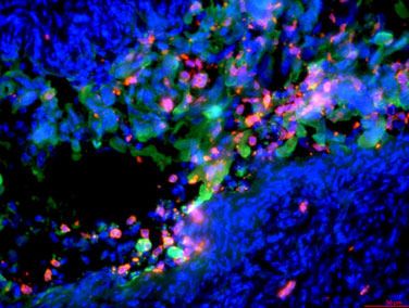
Research article                                                      Immunology and Inflammation Microbiology and Infectious Disease

                                    eLife digest Herpes simplex virus (HSV) is a human pathogen that causes genital herpes, an
                                    incurable disease that results in recurrent sores and inflammation. Infection with HSV induces a
                                    strong antiviral immune response, which results in large numbers of immune cells arriving at these
                                    lesions. But while some of these cells help to control viral replication, others might contribute to the
                                    inflammation that drives the disease.
                                        One of the first immune cells to respond to infection are neutrophils. Although neutrophils are
                                    generally protective, especially against bacteria and fungi, they have also been implicated in tissue
                                    damage and severe inflammation during viral infections. But what determines whether a neutrophil
                                    will help to fight off an infection or increase disease severity is still an open question.
                                        To investigate this, Lebratti, Lim et al. studied mice that had been infected with the genital
                                    herpes virus HSV-2, which is known to cause significant amounts of inflammation in mice. The
                                    experiments revealed that a signaling molecule called type I interferon, which is thought to be
                                    antiviral, causes neutrophils at the site of the infection to produce proteins, such as IL-18, which
                                    trigger an inflammatory reaction. Lebratti, Lim et al. found that type I interferon and IL-18 had
                                    shifting roles during the course of infection. In the early stages, both molecules had a protective
                                    effect, confirming results from previous studies. However, as the infection progressed, sustained
                                    levels of type I interferon signaling in neutrophils led to excess amounts of IL-18.
                                        Lebratti, Lim et al. discovered that blocking interferon signaling or decreasing the levels of IL-18
                                    later during infection unexpectedly reduced the severity of the disease and resulted in less genital
                                    tissue damage. Further experiments also showed that mice infected with another genital herpes
                                    virus called HSV-1 did not experience sustained levels of type I interferon. This may explain why this
                                    virus causes less severe disease in mice.
                                        Understanding how the immune system reacts to viruses could reveal new targets for treatments
                                    of genital herpes. At the moment, there is little information about IL-18 production during genital
                                    herpes in humans. So, the next step is to see whether neutrophils behave in the same way and
                                    whether IL-18 can be detected during human disease. It is possible that the same immune
                                    components could promote disease in other infections too. If so, this work may help uncover new
                                    drug targets for other viral diseases.

                                    tissue damage, loss of viral control, and increased mortality (Bai et al., 2010; Brandes et al., 2013;
                                    Kulkarni et al., 2019; Narasaraju et al., 2011; Vidy et al., 2016).
                                        Type I interferons (IFNs) are potent regulators of neutrophil activity in a multitude of contexts.
                                    Type I IFNs can enhance recruitment of neutrophils to sites of infection, regulate neutrophil function,
                                    and drive immunopathology after infection by different classes of pathogens, including Plasmodium
                                    spp., Candida spp., and Pseudomonas spp. (Majer et al., 2012; Pylaeva et al., 2019; Rocha et al.,
                                    2015). However, type I IFNs can also inhibit neutrophil recruitment to the ganglia by suppressing
                                    chemokine expression after herpes simplex virus (HSV) infection (Stock et al., 2014), suggesting
                                    that the interplay of IFNs and neutrophil activity may be dependent on tissue type and the patho-
                                    gen. The relationship between neutrophil-intrinsic type I IFN signaling and infection outcomes is less
                                    clear. Type I IFNs can promote expression of interferon-stimulated genes (ISGs) and pro-inflamma-
                                    tory cytokines in neutrophils, suggesting a potential role for them in driving immunopathology
                                    (Galani et al., 2017). During Leishmania infection, however, IFNAR signaling appears to suppress
                                    neutrophil-dependent killing of parasites (Xin et al., 2010), which emphasizes the complexity of IFN-
                                    mediated neutrophil responses.
                                        Genital herpes is a chronic, sexually transmitted infection that affects over 400 million people
                                    worldwide (World Health Organization, 2007) and can be caused by two members of the alphaher-
                                    pesvirus family, HSV-2 or the related HSV-1. Genital herpes is characterized by recurrent episodes of
                                    inflammation and ulceration, and the factors that drive disease are unclear. In humans, ulcer forma-
                                    tion is associated with suboptimal viral control and spread during episodes of reactivation
                                    (Roychoudhury et al., 2020; Schiffer and Corey, 2013; Schiffer et al., 2013), while in mouse mod-
                                    els, severity of disease often correlates with susceptibility to infection and the level of viral replica-
                                    tion in the genital mucosa (Gopinath et al., 2018). Neutrophil infiltration into sites of active HSV-2

Lebratti, Lim, et al. eLife 2021;10:e65762. DOI: https://doi.org/10.7554/eLife.65762                                                     2 of 30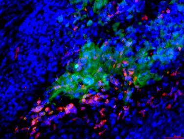
Research article                                                      Immunology and Inflammation Microbiology and Infectious Disease

                                    ulcers has also been reported in humans (Boddingius et al., 1987), but whether these cells are help-
                                    ful or harmful during HSV infection is unknown. While neutrophils have been associated with tissue
                                    damage after multiple routes of HSV-1 infection (Divito and Hendricks, 2008; Khoury-
                                    Hanold et al., 2016; Rao and Suvas, 2019; Thomas et al., 1997), a protective role for neutrophils
                                    after genital HSV-2 infection has also been reported (Milligan, 1999; Milligan et al., 2001), although
                                    the use of non-specific depletion antibodies has muddled the respective contribution of neutrophils
                                    and other innate immune cells such as monocytes, which are known to be antiviral (Iijima et al.,
                                    2011). Furthermore, increased neutrophil recruitment to the HSV-2-infected vaginal epithelial barrier
                                    resulted in greater epithelial cell death, suggesting that neutrophil responses may indeed be patho-
                                    genic (Krzyzowska et al., 2014). However, the factors that distinguish pathogenic vs. non-patho-
                                    genic neutrophil responses during viral infection, including HSV-2 infection, remain ill-defined.
                                        To address this, we evaluated the impact of neutrophils on genital disease severity using two
                                    models of HSV infection that result in low levels (HSV-1) or high levels of inflammation (HSV-2)
                                    (Lee et al., 2020). Between these two states, heightened expression of type I IFN during the resolu-
                                    tion phase of acute infection and sustained expression of ISGs in neutrophils were detected after
                                    HSV-2 infection but not HSV-1. Therapeutic antibody-mediated blockade of IFNa/b receptor
                                    1 (IFNAR1) as well as neutrophil-specific deletion of IFNAR1 reduced both genital inflammation as
                                    well as vaginal IL-18 levels during the resolution phase of acute HSV-2 infection. Accordingly, thera-
                                    peutic neutralization of IL-18 also ameliorated genital disease after HSV-2 infection. Together, our
                                    data demonstrates that sustained type I IFN signaling is a key determinant of pathogenic neutrophil
                                    responses during viral infection, and identifies neutrophil- and type I IFN-dependent IL-18 produc-
                                    tion as a novel driver of inflammation during genital HSV-2 infection.

                                    Results
                                    Neutrophils are a component of severe genital inflammation after
                                    vaginal HSV-2 infection
                                    To determine the role of neutrophils in our model of vaginal HSV-2 infection, wild-type (WT) female
                                    C57BL/6 mice were treated with Depo-Provera (depot medroxyprogestrone, DMPA) to hold mice at
                                    the diestrus phase of the estrus cycle and normalize susceptibility to infection (Kaushic et al., 2003).
                                    Neutrophils were depleted in DMPA-treated mice by intraperitoneal (i.p.) injection of an antibody
                                    against Ly6G, a neutrophil-specific marker, or an isotype control. One day later, mice were inocu-
                                    lated intravaginally with 5000 plaque forming units (PFU) of WT HSV-2 strain 186 syn+ (WT HSV-2).
                                    Neutrophils were effectively reduced up to 6 days post-infection (d.p.i.) in the vagina (Figure 1A)
                                    and the blood (Figure 1—figure supplement 1). In order to focus on genital inflammation, mice
                                    were monitored for 1 week after infection, as progression of disease within the second week of our
                                    infection model is largely indicative of viral dissemination into the central nervous system. In both
                                    cohorts, mild genital inflammation was apparent starting at 4 d.p.i. in a small fraction of mice
                                    (Figure 1B). Over time, progression of disease in the neutrophil-depleted mice was significantly
                                    slower compared to the controls. Remarkably, as late as 7 d.p.i., a proportion of the neutrophil-
                                    depleted group remained uninflamed, in contrast to the isotype control group in which all mice dis-
                                    played signs of inflammation (Figure 1B). To confirm the disparity in disease severity, we examined
                                    the vagina and genital skin by histology. At 6 d.p.i., epithelial denuding and damage was apparent
                                    in the isotype control-treated mice (Figure 1C). In contrast, only a limited amount of epithelial
                                    destruction was observed in neutrophil-depleted mice, with less cellular infiltrates at sites of damage
                                    and in the lumen (Figure 1C). Furthermore, the epithelial layer proximal to areas of damage was
                                    morphologically distinct in isotype control-treated animals compared to neutrophil-depleted ani-
                                    mals, suggesting diverse epithelial responses after infection in the presence or absence of neutro-
                                    phils (Figure 1C). Similarly, destruction of the epidermis and separation of the epidermis from the
                                    dermis were widespread in the genital skin of isotype control-treated mice but not in neutrophil-
                                    depleted mice (Figure 1C). Unexpectedly, differences in genital inflammation and mucosal damage
                                    were largely independent of changes in viral control in the absence of neutrophils, as viral shedding
                                    into the vaginal lumen (Figure 1D) and viral control in the tissue parenchyma (Figure 1E) were simi-
                                    lar between the two groups. Indeed, disease severity was decreased in neutrophil-depleted mice
                                    despite a slight delay in the resolution of viral replication at 5 d.p.i. (Figure 1D).

Lebratti, Lim, et al. eLife 2021;10:e65762. DOI: https://doi.org/10.7554/eLife.65762                                                     3 of 30
Research article Immunology and Inflammation Microbiology and Infectious Disease
A
6 d.p.i. vagina NK1.1- CD11b+ Gr-1+ ** * **
isotype 105
live/dead
FSC-H *** isotype ctrl
SSC-A
NK1.1
PMN/vagina
4 anti-Ly6G
10
29.4
FSC-A 103
PMN
84.9 102
101
live/dead
anti-Ly6G 0 2 4 6
FSC-H
SSC-A
NK1.1 4.6
days post infection
CD11b
FSC-A
Ly6C
PMN D
6.0
8
isotype ctrl
Gr-1 F4/80
log10 PFU/ml
6 ** anti-Ly6G
B 4
**** C isotype anti-Ly6G
inflammation scores
5
isotype ctrl 2
4 anti-Ly6G
** 0
3 1 2 3 4 5 6 7
2 vagina days post infection
1
0
4 5 6 7
E
2
log10 PFU/mg vagina
days post infection
1
skin 0
-1
-2
rl
6G
ct
Ly
e
yp
ti-
an
ot
is
Figure 1. Neutrophil depletion reduces disease severity after HSV-2 vaginal infection. Female C57BL/6J mice were treated with depot
medroxyprogestrone (DMPA) and inoculated intravaginally (ivag) with 5000 plaque forming units (PFU) of herpes simplex virus-2 (HSV-2). One day prior
to HSV-2 inoculation, mice were injected intraperitoneally (i.p.) with 500 mg of rat IgG2a isotype control or anti-Ly6G monoclonal antibody (mAb). (A)
Plots show gating strategy to identify neutrophils in the vagina. Numbers in plots refer to percent of parent population for gated cells (d0, d2: n = 6,
d2: n = 9, d6 isotype: n = 8, d6 anti-Ly6G: n = 10). Depletion was confirmed by flow cytometry in the vagina on the indicated days. (B) Inflammation
scores over the first 7 d.p.i. of mice treated with anti-Ly6G antibody (n = 25) or isotype control (n = 23). Mice showed no signs of disease prior to 4 d.p.
i. (C) Histology of the vagina (top) or genital skin (bottom) at 6 d.p.i. from isotype control (left) or anti-Ly6G antibody-treated mice (right). Red arrows
point to areas of epithelial denuding or damage, black arrows denote the basement membrane. (D) Infectious virus as measured by plaque assay in
vaginal washes collected daily (both groups d1: n = 22, d2: n = 28, d3: n = 15, d4: n = 16, d5: n = 19, d6: n = 6, d7: n = 8). (E) Viral load was measured
in homogenized vaginal tissue collected at 7 d.p.i. from the indicated groups (n = 8). Data in A and E are pooled from two independent experiments,
and data in B and D are pooled from two to four independent experiments. Data in C is representative of two independent experiments. Bars in B
show median with interquartile range. Horizontal bars in A, D, and E show mean. Scale bars show 50 mm. Statistical analysis was performed by two-way
ANOVA on log-transformed data with Bonferroni’s multiple comparisons test (A), repeated measures two-way ANOVA with Geisser-Greenhouse
correction and Bonferroni’s multiple comparisons test (B) repeated measures two-way ANOVA with Bonferroni’s multiple comparison’s test (D) or
Mann-Whitney test (E). *p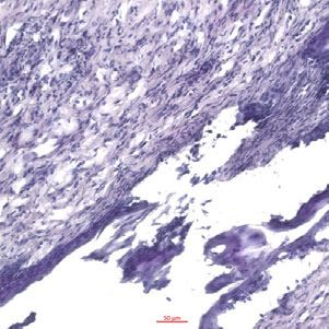
Research article                                                      Immunology and Inflammation Microbiology and Infectious Disease

Figure 1 continued
Figure supplement 2. Neutrophil depletion does not affect magnitude of the immune cell response after HSV-2 infection.
Figure supplement 3. PAD4 is not required for development of genital inflammation during HSV-2 infection.
Figure supplement 4. ROS production and STIM1/STIM2 expression in neutrophils are not required for genital inflammation after HSV-2 infection.

                                       We next evaluated whether the decreased inflammation after neutrophil depletion was due to
                                    changes in the cellular response against HSV-2 infection. We examined the recruitment of Ly6C
                                    + monocytes, NK cells, and CD4 and CD8 T cells (Figure 1—figure supplement 2A), all of which
                                    have been implicated in either the control of HSV or modulation of disease severity (Lee and Ash-
                                    kar, 2012; Shin and Iwasaki, 2013; Truong et al., 2019). To remove intravascular cells and to limit
                                    our analysis to cells within the vagina, tissues were thoroughly perfused prior to collection
                                    (Scott et al., 2018). Unexpectedly, there was no significant difference in the number of
                                    Ly6C + CD11b + cells (Figure 1—figure supplement 2B), NK cells (Figure 1—figure supplement
                                    2C), total CD4 (Figure 1—figure supplement 2D), or CD8 T cells (Figure 1—figure supplement
                                    2E) that were recruited to the vagina over the first 6 days after infection regardless of whether neu-
                                    trophils were present or not. Thus, our data demonstrate that neutrophils do not play a significant
                                    antiviral role in our model of vaginal HSV-2 infection, and rather promote genital inflammation with
                                    minimal impact viral burden and recruitment of other immune cells to the vagina.

                                    Neutrophil extracellular trap formation and oxidative burst are not
                                    major drivers of genital inflammation after HSV-2 infection
                                    We next wanted to determine whether neutrophil-specific effector functions were promoting disease
                                    after HSV-2 infection. NETs have been associated with tissue damage in the context of both infec-
                                    tious (Jenne and Kubes, 2015) and non-infectious disease (Granger et al., 2019). To test whether
                                    NETs play a role in genital disease after HSV-2 infection, we first examined the ability of neutrophils
                                    to form NETs when exposed to HSV-2. In vitro stimulation of neutrophils with HSV-2 resulted in the
                                    enlargement of cell nuclei and the characteristic expulsion of DNA coated in citrullinated histones,
                                    which is a key characteristic of NETs (Figure 1—figure supplement 3A). The formation of NETs
                                    requires input from multiple pathways, including histone citrullination by enzymes such as PAD4,
                                    which leads to chromatin de-condensation and the eventual release of DNA (Li et al., 2010). To gen-
                                    erate animals that were specifically lacking PAD4 in neutrophils, we bred Padi4fl/fl x S100a8-Cre mice
                                    (PAD4 CKO). HSV-2 infection of these mice and their littermate controls demonstrated minimal
                                    impact on genital inflammation (Figure 1—figure supplement 3B) or viral replication (Figure 1—fig-
                                    ure supplement 3C) in the genital mucosa. Thus, our data show that PAD4 expression in neutrophils
                                    and likely NET formation are not the mechanisms by which these cells mediate disease after HSV-2
                                    infection.
                                        We next tested whether ROS production by neutrophils mediated inflammation after HSV-2 infec-
                                    tion. While production of ROS in neutrophils supports antimicrobial activity against a variety of
                                    pathogens (Dinauer, 2019), excessive oxidative stress can be associated with tissue injury
                                    (Mittal et al., 2014). We found that in vitro stimulation of neutrophils with HSV-2 led to an increase
                                    in ROS production compared to unstimulated cells (Figure 1—figure supplement 4A). To deter-
                                    mine whether respiratory burst in neutrophils promoted genital inflammation after HSV-2 infection in
                                    vivo, we infected mice with germline deficiency in Ncf2 (Ncf2 KO), which encodes p67phox, a key
                                    component of the NADPH oxidase complex (Jacob et al., 2017). HSV-2 infection of Ncf2 KO and
                                    heterozygous controls resulted in similar progression of disease (Figure 1—figure supplement 4B)
                                    and did not alter viral titer (Figure 1—figure supplement 4C). To confirm that tested neutrophil
                                    effector functions, including ROS production, had little impact on genital inflammation, we infected
                                    mice in which the calcium-sensing molecules STIM1 and STIM2 were deleted from neutrophils, as
                                    these calcium-sensing molecules cooperatively regulate neutrophil activation and select effector
                                    functions (Clemens et al., 2017). Stim1fl/fl x Stim2fl/fl x S100a8-Cre (STIM1/2 DKO) mice were
                                    infected with HSV-2 and monitored for disease. As expected, there was little difference in genital
                                    inflammation severity between the STIM1/2 DKO and Cre- controls (Figure 1—figure supplement
                                    4D) or viral titers (Figure 1—figure supplement 4E). Together, our data show that ROS production

Lebratti, Lim, et al. eLife 2021;10:e65762. DOI: https://doi.org/10.7554/eLife.65762                                                        5 of 30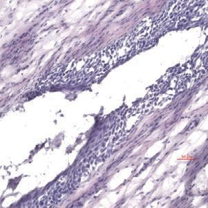
Research article                                                                                                                 Immunology and Inflammation Microbiology and Infectious Disease

                                                          from neutrophils and other cell types play little role in driving genital inflammation after HSV-2
                                                          infection.

                                                          A type I IFN signature distinguishes neutrophil responses after genital
                                                          HSV-1 and HSV-2 infection
                                                          To identify the factors that drove pathogenic neutrophil responses after HSV-2 infection, we turned
                                                          to a complementary model of HSV-1 genital infection that we had previously described (Lee et al.,
                                                          2020). Inoculation with the same dose of HSV-1 and HSV-2 led to profound differences in genital
                                                          inflammation (Figure 2A) despite comparable levels of mucosal viral shedding throughout most
                                                          days after infection (Figure 2—figure supplement 1A; Lee et al., 2020), although resolution of

             A                                                                     B

                                                                                                        CD11b+Ly6G+/vagina
                                      5                           ****                                                       106                                 HSV-1
                 inflammation score

                                                                           HSV-1                                                                       ns
                                                                                                                                             ns                  HSV-2
                                      4                                    HSV-2                                             10   5               ns
                                      3                    ****
                                                                                                                             104
                                      2              *
                                      1    ns                                                                                103

                                      0                                                                                      102
                                           4         5      6         7                                                                d0    d2   d4   d6
                                                days post infection                                                                   days post infection

                                                         mock
            C                                                                      D
                                                                                                            3
                                                                                   inflammation score

                                                                                                                                                            ns       isotype ctrl
                                                                                                                                                  ns                 anti-Ly6G
                                                                                                            2
                                                                                                                                            ns
                                                                                                            1
                                          HSV-2                       HSV-1                                                  ns

                                                                                                            0
                                                                                                                              4             5      6        7
                                                                                                                                      days post infection
    4 d.p.i.

                                      DAPI               HSV      S100A8

Figure 2. Neutrophils are non-pathogenic in a less inflammatory model of vaginal HSV-1 infection. Female C57BL/6J mice were treated with DMPA and
inoculated ivag with 104 PFU HSV-1 McKrae or HSV-2. (A) Inflammation scores were monitored for 7 d.p.i. (HSV-1: n = 14, HSV-2: n = 13). (B)
Neutrophils were counted by flow cytometry in vaginal tissues at the indicated days after HSV-1 or HSV-2 infection (d0: n = 7, day 2: n = 8, day 4: n = 7,
day 6: n = 8). (C) Vaginas were harvested from phosphate buffered saline (PBS)-inoculated (mock), HSV-1- or HSV-2-inoculated mice at 4 d.p.i., and
tissue sections were probed with antibodies against HSV proteins (green) or S100A8 (red). 40 ,6-diamidino-2-phenylindole (DAPI) (blue) was used
to detect cell nuclei. Images are representative of six mice per group. (D) Mice were treated with isotype control or anti-Ly6G mAb as described in
Figure 1 and then inoculated ivag with 104 PFU HSV-1 McKrae. Inflammation scores were monitored for 7 d.p.i. (isotype, anti-Ly6G mAb: n = 10). Data
are pooled from three (A) or two (B–D) independent experiments. Data in C is representative of two independent experiments. Bars show median with
interquartile range (A, D) or mean (B). Scale bars show 50 mm. Statistical significance was measured by repeated measures two-way ANOVA with
Geisser-Greenhouse correction and Bonferroni’s multiple comparisons test (A, D) or two-way ANOVA with Bonferroni’s multiple comparisons test (B).
*p
Research article Immunology and Inflammation Microbiology and Infectious Disease
HSV-2 infection at 6 and 7 d.p.i. was delayed (Figure 2—figure supplement 1A,B). Importantly,
magnitude of the neutrophil response in the vagina was similar between HSV-1- and HSV-2-infected
mice during the course of acute mucosal infection (Figure 2B), and neutrophils could be found infil-
trating sites of both HSV-1- and HSV-2-infected epithelium (Figure 2C). In contrast to HSV-2
A B E
epithelial S100a8, Csf3r
4 d.p.i. 5 d.p.i.
myeloid cells
40 15
cells 1.5 *** *
** *
IFNβ pg/ml
IFNβ pg/ml
1 30
10
0.5 20 ns
5 ns
5 0 10
2 lymphocytes
tSNE 2
tSNE 2
0 -0.5 0 0
PMN
ed
H -1
-2
ed
H -1
-2
SV
SV
SV
SV
ct
ct
fe
fe
H
H
tSNE 1 tSNE 1
in
in
un
un
C mock HSV-1 HSV-2
5 2 5 5
2 2
tSNE 2
0 0 0
tSNE 1
D mock HSV-1 HSV-2 1.2
0.8
0.4
IFN
response 0
tSNE 2
-0.4
tSNE 1
Figure 3. Single cell transcriptome analysis reveals a sustained IFN signature in the neutrophil response against HSV-2. Mice were infected as
described in Figure 2. Vaginas were harvested at 5 d.p.i., and live cells were flow sorted and subjected to high-throughput single-cell
RNA sequencing (scRNA-seq). (A) A t-Distributed Stochastic Neighbor Embedding (tSNE) visualization of 21,633 cells across all mice resolves 17 distinct
clusters in the vaginal tissue. Clusters can be identified as myeloid cells (red border), epithelial cells (blue border), or lymphocytes (purple border).
Neutrophils are encircled in black and contain three distinct clusters (0, 2, and 5). (B) Neutrophils are defined by high expression of S100a8 and Csf3r
(G-SCFR). (C) tSNE plots of vaginal cell clusters from mock-inoculated or HSV-infected mice. Neutrophil populations are circled in black. (D) Distribution
of expression for genes within the Hallmark IFNa Response gene set. (E) Production of IFNb in vaginal washes collected at 4 and 5 d.p.i. as measured
by ELISA (uninfected: n = 8, HSV-1: n = 9, HSV-2: n = 9). scRNA-seq in A-D was performed once. Data in E are pooled from two independent
experiments. Statistical significance was measured by one-way ANOVA with Tukey’s multiple comparisons test. *p
Research article                                                      Immunology and Inflammation Microbiology and Infectious Disease

                                    infection, antibody-mediated depletion of neutrophils with anti-Ly6G antibody prior to inoculation
                                    with HSV-1 did not reduce the development of genital inflammation during the first 7 days after
                                    infection (Figure 2D) and had minimal impact on acute viral control (Figure 2D - Figure 2—figure
                                    supplement 1C). Together, our data suggests that the regulation of the neutrophil response after
                                    HSV-1 or HSV-2 infection was distinct, which may contribute to the differences in disease outcomes
                                    between these infections.
                                        To better understand the differences between pathogenic neutrophil responses after HSV-2 infec-
                                    tion and the non-pathogenic neutrophil responses after HSV-1, we performed single-cell
                                    RNA sequencing (scRNA-seq) on sorted live vaginal cells from a mock-infected mouse or mice
                                    infected with HSV-1 or HSV-2 using the 10x Genomics platform (Zheng et al., 2017). Each sample
                                    was composed of cells from a single animal to better delineate potential subsets within cell popula-
                                    tions, particularly neutrophils. Analysis across 21,633 cells in all samples revealed 17 unique clusters
                                    in the vagina during HSV infection after filtering, including myeloid cells, lymphocytes, and epithelial
                                    cells (Figure 3A). Neutrophils were identified by expression of known cell markers such as S100a8
                                    and Csf3r (Figure 3B). In mock-infected animals, the vaginal neutrophil population was dominated
                                    by cluster 0, and upon infection, at least two additional neutrophil subsets, cluster 2 and cluster 5,
                                    were clearly present (Figure 3C). While HSV-1-infected mice retained all three subpopulations of
                                    neutrophils in the vagina at 5 d.p.i., in HSV-2-infected mice, the presence of cluster 0 was greatly
                                    reduced, and the bulk of the neutrophils was composed of cluster 2 and 5 (Figure 3C). One major
                                    distinguishing characteristic between ‘homeostatic’ cluster 0 and ‘infection’ clusters 2 and 5 was the
                                    extent of ISG expression, in which cluster 0 expressed low levels of genes associated with a type I
                                    IFN response, even in infected animals, while clusters 2 and 5 expressed high levels of these genes
                                    (Figure 3D; Liberzon et al., 2015). Furthermore, the gene expression profile of clusters 2 and 5 was
                                    different between HSV-2 and HSV-1 infection at 5 d.p.i. (Figure 3—figure supplement 1), including
                                    the expression of ISGs (Figure 3C, Figure 3—figure supplement 1). Differential expression of select
                                    ISGs was confirmed by quantitative reverse transcription PCR (qRT-PCR) analysis in the vagina at 5
                                    days after HSV-1 or HSV-2 infection (Figure 3—figure supplement 2). qRT-PCR shows that expres-
                                    sion of CXCL10 (Figure 3—figure supplement 2A,B) and Gbp2 (Figure 3—figure supplement 2C,
                                    D; Glennie et al., 2015) is increased in HSV-2-infected vaginas compared to HSV-1, while IL-15 is
                                    not (Figure 3—figure supplement 2E,F), which supports the accompanying scRNA-seq analysis.
                                    While type I IFN was robustly produced early during acute infection after both HSV-1 and HSV-2
                                    infection (Figure 3—figure supplement 3), IFNb levels were higher in the vaginal lumen after HSV-2
                                    infection compared to HSV-1 at time points corresponding to the onset of genital inflammation
                                    (Figure 3E) despite similar viral burden between the two infection models (Figure 2—figure supple-
                                    ment 1A). IFNb was undetectable in both the vaginal lumen and the parenchyma by 7 days after
                                    both HSV-1 and HSV-2 infection (Figure 3—figure supplement 3B,C). Thus, during viral infection,
                                    distinct neutrophil subsets can be classified by transcriptional profiling, and expression of ISGs sug-
                                    gests that a key difference between a pathogenic and non-pathogenic neutrophil response during
                                    viral infection may be sustained IFN production and signaling.

                                    Sustained cell-intrinsic type I IFN signaling is required for pathogenic
                                    neutrophil responses during HSV-2 infection
                                    We next wanted to test whether type I IFN signaling promoted immunopathology during genital
                                    HSV-2 infection. IFNAR1-deficient mice are highly susceptible to HSV, regardless of the route of
                                    inoculation (Gill et al., 2006; Iversen et al., 2010; Iversen et al., 2016; Reinert et al., 2012;
                                    Royer et al., 2019; Svensson et al., 2007; Wilcox et al., 2016), and rapidly succumb to infection,
                                    mainly due to a loss of viral control. To investigate the temporal effects of type I IFNs in HSV-2 geni-
                                    tal disease, we used an antibody against IFNAR1 to block the receptor at different time points after
                                    infection (Scott et al., 2018). When mice were injected i.p. with anti-IFNAR1 antibody on the day of
                                    HSV-2 inoculation, disease progression was more rapid compared to isotype control-treated animals
                                    (Figure 4—figure supplement 1A), and the mice succumbed to infection at a faster rate (Figure 4—
                                    figure supplement 1B), in a manner similar to IFNAR1-deficient mice (Iversen et al., 2010;
                                    Iversen et al., 2016; Lee et al., 2017; Reinert et al., 2012; Wang et al., 2012). Inflammation and
                                    rapid disease progression were likely due to significantly elevated viral burden in the anti-IFNAR1
                                    antibody-treated mice compared to isotype controls (Figure 4—figure supplement 1C), as HSV is a
                                    highly lytic virus that is capable of independently inducing epithelial tissue damage (Horbul et al.,

Lebratti, Lim, et al. eLife 2021;10:e65762. DOI: https://doi.org/10.7554/eLife.65762                                                     8 of 30
Research article                                                                                  Immunology and Inflammation Microbiology and Infectious Disease

                                    2011). To focus on the effects of persistent IFN signaling in the vagina after HSV-2 infection, we also
                                    treated mice with a single injection of anti-IFNAR1 antibody or an isotype control at 4 d.p.i. In stark
                                    contrast to early anti-IFNAR1 antibody treatment, one treatment with therapeutic IFNAR1 blockade
                                    led to a significant reduction in the severity of inflammation compared to isotype controls
                                    (Figure 4A). Histology of vaginal tissues from isotype-treated controls at 6 d.p.i. showed widespread
                                    epithelial denuding and immune cell infiltrates within the epithelial layer of the vagina (Figure 4B). In
                                    contrast, damage to the vaginal epithelium in anti-IFNAR1 antibody-treated mice appeared to be
                                    localized (Figure 4B), similar to neutrophil-depleted mice (Figure 1C). Similarly, the genital skin of
                                    isotype control-treated mice displayed signs of severe inflammation and destruction of the epider-
                                    mis, while the skin structure of anti-IFNAR1 antibody-treated mice was largely intact (Figure 4B).
                                    Furthermore, IFNAR1 blockade at 4 d.p.i. had little impact on mucosal viral shedding (Figure 4C).

                                        A                                                        ns
                                                                5
                                           inflammation score

                                                                                                             isotype ctrl
                                                                4                                            anti-IFNAR1
                                                                                      ***
                                                                3
                                                                               **                                            B      isotype         anti-IFNAR1
                                                                2
                                                                1     ns
                                                                0
                                                                      4           5       6      7                      vagina
                                                                           days post infection

                                        C
                                                                8             ns
                                                                                              isotype ctrl
                                          log10 PFU/ml

                                                                6                             anti-IFNAR1
                                                                                                                            skin
                                                                4

                                                                2

                                                                0
                                                                      2       4       6
                                                                    days post infection

                                    Figure 4. Inhibition of type I IFN signaling during the resolution phase of infection reduces inflammation after
                                    HSV-2 infection. Mice were infected as described in Figure 2. At 4 d.p.i., mice were injected i.p. with either 1 mg
                                    of anti-IFNAR1 antibody (n = 10–13) or isotype control (n = 7–9) and monitored for disease progression. Mice
                                    showing overt signs of genital inflammation at the time of antibody injection (4 d.p.i.) were excluded from the
                                    study. (A) Inflammation scores of antibody-treated mice over the first 7 d.p.i. (B) Histology of the vagina (top) or
                                    genital skin (bottom) at 6 d.p.i. Red arrows point to areas of epithelial denuding or damage. Black areas denote
                                    the basement membrane. (C) Infectious virus as measured by plaque assay in vaginal washes collected on the
                                    indicated days. Data are pooled from (A, C) or representative of three independent experiments. Bars in A show
                                    median with interquartile range; bars in C show mean. Scale bars show 50 mm. Statistical significance was
                                    measured by repeated measures two-way ANOVA with Geisser-Greenhouse correction and Bonferroni’s multiple
                                    comparisons test (A) or two-way ANOVA with Bonferroni’s multiple comparisons test (C). *p
Research article                                                                                Immunology and Inflammation Microbiology and Infectious Disease

                                          Collectively, these data show that the protective effect of type I IFN on control of genital HSV infec-
                                          tion is limited to the early stages of acute infection, and that sustained IFN signaling in the later
                                          stages of acute HSV-2 genital infection drives inflammation with minimal effect on viral replication.
                                             Single-cell transcriptional profiling data suggested that type I IFN signaling was robust in vaginal
                                          neutrophils after HSV-2 infection (Figure 3D). To determine whether intrinsic IFN signaling in neutro-
                                          phils promoted immunopathology, we deleted IFNAR1 from granulocytes by breeding Ifnar1fl/fl x
                                          S100a8-Cre mice (IFNAR1 CKO). After confirming that IFNAR1 ablation was limited to the neutrophil
                                          population (Figure 5A), IFNAR1 CKO mice and littermate Cre- controls were vaginally infected with
                                          HSV-2. Despite differences in IFNAR1 expression, the number of neutrophils recovered from the
                                          vaginal lumen was similar between the IFNAR1 CKO mice and their Cre- control littermates

                                    neutrophils
        A                                           isotype        B            106
                                                    ctrl                                                                    ctrl
                                                    IFNAR1

                                                                    PMN/wash
                                                                                                                            IFNAR1 CKO
                                                    CKO                         105
              live CD45+
                                                                                104

                                  non-neutrophils                               103
    CD11b

                                                                                                  2       4        6
                                                                                                days post infection
                                                                   C
             Ly6G                                                                           3
                                                                       inflammation score

                                                                                                                              **   ctrl
                                                                                                                                   IFNAR1 CKO
                                                                                                                       **
                                                                                            2
                                  IFNAR1                                                                   *
                                                                                            1
       D                 ctrl          IFNAR1 CKO
                                                                                            0
                                                                                                  4        5           6      7
                                                                                                      days post infection

    vagina
                                                                    E
                                                                                            6
                                                                                                                            ctrl
                                                                           log10 PFU/ml

                                                                                                                            IFNAR1 CKO
                                                                                            4

            skin                                                                            2

                                                                                            0
                                                                                                  2       4        6
                                                                                                days post infection

Figure 5. Type I IFN signaling in neutrophils promotes genital inflammation after HSV-2 infection. (A) IFNAR1 expression on neutrophils and non-
neutrophil hematopoietic cells from the bone marrow of naive Ifnar1fl/fl x S100a8-Cre (IFNAR1 CKO) or Cre- littermate controls. Plot is gated on live
CD45 + cells. CD11b + Ly6G + cells are neutrophils, Ly6G- cells are non-neutrophils. Gray histogram shows isotype staining, black open histogram is
Cre- control, and red open histogram is IFNAR1 CKO. (B) Neutrophils were counted by flow cytometry in vaginal washes collected at the indicated days
from IFNAR1 CKO (n = 4) or Cre- controls (n = 7) that were infected with HSV-2 as described in Figure 1. (C) Inflammation scores for the first 7 d.p.i. of
IFNAR1 CKO (n = 10–13) or Cre- controls (n = 8–11). (D) Histology of the vagina and genital skin at 6 d.p.i. Red arrows point to areas of epithelial
denuding or damage, black arrows denote basement membrane (E) Infectious virus as measured by plaque assay from vaginal washes collected on the
indicated days from IFNAR1 CKO (n = 5–8) or Cre- controls (n = 7–10). Data in C and E are pooled from three independent experiments; data in B are
pooled from two independent experiments; and data in D are representative of two independent experiments. Bars in C show median with
interquartile range, bars in B and E show mean. Scale bars show 50 mm. Statistical significance was measured by mixed-effects analysis with (C) or
without (B, E) Geisser-Greenhouse correction and Bonferroni’s multiple comparisons test. *p
Research article Immunology and Inflammation Microbiology and Infectious Disease
(Figure 5B). Strikingly, although the magnitude of the vaginal neutrophil response was similar, we
found that the severity of genital inflammation presented by the IFNAR1 CKO mice was significantly
reduced compared to the Cre- controls (Figure 5C). As observed after neutrophil depletion, a sub-
set of the IFNAR1 CKO cohort did not develop any signs of inflammation as late as 7 d.p.i.
(Figure 5C). Similar to our observations with therapeutic IFNAR1 blockade, IFNAR1 CKO mice
exhibited less pathology in both the vagina and genital skin compared to Cre- controls (Figure 5D).
Distinct disease outcomes between the Cre- controls and IFNAR1 CKO mice occurred independently
of viral control, as viral loads in the mucosa were similar between the two groups (Figure 5E).
Together, our data demonstrates that tissue inflammation during HSV-2 infection is largely driven by
prolonged type I IFN production, which acts directly upon neutrophils to drive disease.
Sustained type I IFN signaling and neutrophils regulate production of
pathogenic IL-18 in the vagina during HSV-2 infection
Type I IFN stimulation of neutrophils can upregulate ISGs as well as several pro-inflammatory cyto-
kines (Galani et al., 2017). To determine whether type I IFN was driving disease by shaping the cyto-
kine milieu within the vagina, we first measured several pro-inflammatory cytokines in the vagina at 5
d.p.i., in the presence or absence of neutrophils. The production of inflammatory cytokines such as
IL-6 (Figure 6—figure supplement 1A), IL-1b (Figure 6—figure supplement 1B), or TNF (Figure 6—
figure supplement 1C), all of which have been associated with genital inflammation and HSV-2
infection in humans (Gosmann et al., 2017; Masson et al., 2014; Murphy and Mitchell, 2016), was
similar between both neutrophil-depleted and control groups. Production of IFNg (Figure 6—figure
supplement 1D) as well as IL-12p70 (Figure 6—figure supplement 1E), both cytokines associated
with a type I immune response and important for HSV control, was similar between the neutrophil-
depleted and control groups. However, when we measured IL-18, an IL-1 family cytokine that
is primarily known for mediating innate defense (Harandi et al., 2001) and for promoting IFNg pro-
duction from NK cells during genital HSV-2 infection (Lee et al., 2017), we detected a notable differ-
ence between neutrophil-depleted and control mice (Figure 6A), suggesting an unexpected role for
this cytokine in driving disease during HSV-2 infection.
To determine whether type I IFN signaling regulated IL-18 production in the vagina, we assessed
IL-18 levels in the vaginal lumen after therapeutic antibody-mediated IFNAR1 blockade. At 5 d.p.i.,
similarly to neutrophil-depleted mice, we found that IL-18 levels were markedly reduced (Figure 6B).
Importantly, measurement of IL-18 in the vagina of IFNAR1 CKO at 5 d.p.i. also revealed a significant
decrease in cytokine levels compared to littermate controls (Figure 6C).
To determine whether IL-18 was playing a key role in driving immunopathology during genital
HSV-2 infection, we therapeutically administered an IL-18-neutralizing antibody directly at the site of
infection to HSV-2-infected animals starting at 3 d.p.i. in order to promote sufficient antibody con-
centration and activity in the relevant tissue. Remarkably, neutralization of IL-18 led to a considerable
reduction in disease severity (Figure 6D), without any impact on viral control (Figure 6E). To deter-
mine the source of pathogenic IL-18 in the vagina, we probed vaginal tissues for the neutrophil
marker S100A8 and IL-18 at 6 d.p.i. (Figure 6F). Detection of IL-18 and S100A8 around a single
nucleus demonstrated that neutrophils could be a source of IL-18 during vaginal HSV-2 infection
(Figure 6F). However, we also identified IL-18-reactive cells that were negative for S100A8 but in
close proximity to neutrophils (Figure 6F), suggesting the potential for multiple cellular sources of
IL-18. Thus, our data demonstrate that sustained type I IFN signaling in neutrophils leads to the pro-
duction of vaginal IL-18 and reveal IL-18 to be a novel regulator of disease after HSV-2 infection.
Discussion
In this study, we evaluated drivers of a pathogenic neutrophil response using a mouse model for an
important human infection. We found that neutrophils promote genital inflammation without affect-
ing antiviral activity after genital HSV-2 infection, suggesting that the neutrophil response is primarily
immunopathogenic. Depletion of neutrophils led to a significant decrease in disease severity without
altering recruitment of other immune cells or the production of common pro-inflammatory cytokines,
and deficiency in genes controlling neutrophil effector functions such as ROS production and NET
formation had little impact on progression of disease. Comparative analysis of single-cell transcrip-
tional profiles revealed a strong type I IFN signature that was sustained in neutrophils responding to
Lebratti, Lim, et al. eLife 2021;10:e65762. DOI: https://doi.org/10.7554/eLife.65762 11 of 30Research article Immunology and Inflammation Microbiology and Infectious Disease
A B C *
80 80 * 100
***
IL-18 pg/ml
IL-18 pg/ml
IL-18 pg/ml
60 60 75
40 40 50
20 20 25
0 0 0
T
KO
an pe
6G
IF e
1
W
p
AR
y
an oty
Ly
C
ot
N
ti-
1
is
is
AR
ti-
N
IF
D * E 6 ns
5 isotype
inflammation score
isotype
anti-IL-18
log10 PFU/ml
4 anti-IL-18
4
3
*
2
2
1
0 0
4 5 6 7 1 3 5
days post infection days post infection
F IL-18/isotype
DAPI S100A8 Merge
isotype
HSVmock
anti-IL-18
HSV
Figure 6. Sustained type I IFN signaling and neutrophils regulate pathogenic IL-18 levels in the vagina. C57BL/6 mice were treated with anti-Ly6G
(n = 11) or isotype control (n = 12) as described in Figure 1 (A), therapeutically treated with anti-IFNAR1 (n = 12) or isotype control (n = 14) as
described in Figure 5 (B) and infected with HSV-2, or IFNAR1 CKO (n = 7) and Cre- controls (n = 7) were infected with HSV-2 as described in Figure 5
(C). (A-C) Vaginal IL-18 levels were measured by ELISA in washes collected at 5 d.p.i. 100 mg of anti-IL-18 neutralizing antibody (n = 18) or isotype
control (n = 16) was administered ivag at 3, 4, and 5 d.p.i. (D) Inflammation scores of antibody-treated mice over the first 7 d.p.i. (E) Infectious virus as
measured by plaque assay in vaginal washes collected on the indicated days (n = 9–14). (F) Immunofluorescent staining of vaginal tissues collected at 6
d.p.i. or from mock-inoculated mice. Green shows S100A8 (neutrophil), red shows IL-18 or isotype, blue is DAPI. Top row shows HSV-2-infected tissue
probed with isotype control, and bottom two rows show representative images of tissues probed with anti-IL-18 antibody (middle = mock-infected,
bottom = HSV-2-infected). White arrows point to IL-18 + neutrophils. Yellow square demarcates area shown in the inset. Data in A-C, D, and E are
pooled from four independent experiments for each experimental setup. Data in F is representative of two independent experiments. Bars in A-C show
mean and SD, bars in D show median with interquartile range, and bars in E show mean. Scale bars show 50 mm. Statistical significance was measured
by unpaired t-test (A–C), repeated measures two-way ANOVA with (D) Geisser-Greenhouse correction and Bonferroni’s multiple comparisons test, or
mixed-effects analysis with Bonferroni’s multiple comparisons test (D). *pResearch article Immunology and Inflammation Microbiology and Infectious Disease
Figure 6 continued
Figure supplement 1—source data 1. Excel file with individual Bioplex assay values, description of statistical tests and actual p values for Figure 6—
figure supplement 1.
a highly inflammatory genital HSV-2 infection but not a less inflammatory HSV-1 infection, suggest-
ing that host responses to these two related viruses established distinct inflammatory milieus with
divergent effects on responding neutrophils. In contrast to antibody-mediated blockade of IFNAR1
at the time of infection, which led to significantly worse disease outcomes, IFNAR1 blockade just
prior to the resolution phase of acute mucosal infection significantly delayed the progression of gen-
ital inflammation. Importantly, neutrophil-specific deficiency of IFNAR1 markedly reduced the sever-
ity of genital disease after HSV-2 infection, suggesting that persistent IFN signaling drove disease
primarily by acting on neutrophils. Ultimately, this sustained type I IFN signaling in neutrophils pro-
moted the production of pro-inflammatory IL-18, and therapeutic neutralization of this cytokine also
ameliorated disease. Together, our results suggest an axis of type I IFNs, neutrophils, and IL-18 as
the key driver of genital disease in a mouse model of HSV-2 infection, and that sustained type I IFN
signaling is a key factor in distinguishing between pathogenic and non-pathogenic neutrophil
responses during mucosal viral infection.
Type I IFNs are a frontline of defense against viral infection, but models of chronic viral infection,
including lymphocytic choriomeningitis virus (LCMV) (Teijaro et al., 2013; Wilson et al., 2013),
human immunodeficiency virus (HIV) (Meier et al., 2009; Rotger et al., 2010; Sedaghat et al.,
2008; Taleb et al., 2017), and simian immunodeficiency virus (SIV) (Harris et al., 2010;
Jacquelin et al., 2009), reveal the detrimental effect of overexuberant or sustained type I IFN signal-
ing. Notably, prolonged IFN signaling during chronic viral infection can promote immunosuppression
through multiple cellular and molecular mechanisms, and deletion or blockade of IFNAR1 during
chronic LCMV infection can alleviate immunosuppression and enhance long-term viral control
(Cheng et al., 2017; Taleb et al., 2017; Teijaro et al., 2013; Wilson et al., 2013). However, unlike
the LCMV model, early blockade of type I IFN signaling led to more severe disease and a complete
loss of viral control after HSV-2 infection, similar to infections performed on an IFNAR1-deficient
genetic background (Iversen et al., 2010; Iversen et al., 2016; Lee et al., 2017; Leib et al., 1999;
Reinert et al., 2012; Wang et al., 2012), indicating an early antiviral role (Lee et al., 2017;
Luker et al., 2003). Rather, only therapeutic inhibition of sustained IFN signaling diminished disease
without disrupting viral control, thus revealing a heretofore unappreciated, temporal division of the
antiviral and immunopathological effects of type I IFN signaling during HSV-2 infection. The source
of sustained type I IFN production that promotes immunopathology after genital HSV-2 infection is
currently unknown. It is also unclear as to why type I IFN production is sustained during HSV-2, but
not after genital HSV-1 infection. Our data indicates a slight delay in the control of HSV-2 infection
compared to HSV-1 at 6 and 7 d.p.i., but this did not correlate with differences in IFNb levels
between HSV-1 and HSV-2 infection. Differences in viral dissemination to the nervous system
(Lee et al., 2020) or function of viral proteins may account for disparities in type I IFN production
and ultimately, disease severity. HSV encodes numerous proteins that can suppress type I IFN pro-
duction and regulate the signaling pathways (Christensen et al., 2016; Lin and Zheng, 2019;
Melroe et al., 2004), suggesting that production of type I IFN likely occurs from a cell type that is
not directly infected. While plasmacytoid dendritic cells (pDC) are known as robust producers of
type I IFN, they appear to have a limited role during genital HSV-2 infection (Swiecki et al., 2013),
indicating an alternative source of type I IFN, such as conventional DCs (Wilson et al., 2013). In
humans, type I IFN can be detected at active lesion sites during recurrent episodes (Peng et al.,
2009; Roychoudhury et al., 2020), although levels do not correlate with restriction of viral replica-
tion (Roychoudhury et al., 2020). This raises the possibility that type I IFN induction may not be
antiviral and could contribute to ulcer formation, although this hypothesis is yet to be tested. Human
neutrophils from females are also reported to be hyper-responsive to type I IFNs (Gupta et al.,
2020). Although clinical disease recurrence rates between men and women with genital herpes are
similar (Wald et al., 2002), differences in neutrophil sensitivity to type I IFN may have implications
for sex-dependent mechanisms of ulcer development.
Lebratti, Lim, et al. eLife 2021;10:e65762. DOI: https://doi.org/10.7554/eLife.65762 13 of 30Research article Immunology and Inflammation Microbiology and Infectious Disease
Synergistic effects of cytokine signaling have been reported to be important for maximizing cellu-
lar responses to infection through the upregulation of cooperative or independent molecular pro-
grams (Bartee and McFadden, 2013) or through the cross-regulation of receptor signaling
pathways (Ivashkiv and Donlin, 2014). Upon infection, the activity of neutrophils can be modulated
strongly by multiple IFNs, in a variety of tissues. Type I (Ank et al., 2008), type II (Iijima et al., 2008;
Lee et al., 2020), and type III IFNs (Ank et al., 2008; Lee et al., 2020) are all robustly produced dur-
ing HSV-2 infection. While type I and type II IFNs are crucial for the control of HSV-2 replication,
endogenous type III IFNs do not appear to affect either disease severity or viral control, although
exogenous application of type III IFNs can reduce viral burden (Ank et al., 2008; Ank et al., 2006).
Expression of the type III IFN receptor, IFNLR, is limited to very few cell types, including neutrophils
and epithelial cells (Blazek et al., 2015; Mahlakõiv et al., 2015; Sommereyns et al., 2008). As epi-
thelial cells are a major target for HSV-2 replication, dissecting the action of type III IFNs within the
neutrophil and epithelial cell compartments may reveal a more detailed picture of the role type III
IFNs play. The impact of simultaneous type I, II, and III IFN signaling on neutrophil function is cur-
rently unclear, and due to the importance of these molecules in controlling infection, cell-specific
modifications of receptor expression will be required to better understand their impact on neutro-
phil function.
In vitro stimulation of neutrophils with type I IFN leads to the upregulation of many common ISGs
as well as inflammatory genes, including IL-18 (Galani et al., 2017). Importantly, type I IFN may dif-
ferentially regulate expression of IL-18 and IL-1b, another IL-1 family cytokine that depends on cas-
pase-mediated cleavage for activation (Zhu and Kanneganti, 2017). It is unclear whether
neutrophils are directly producing this cytokine in our model of infection and whether IL-18 produc-
tion is dependent on inflammasome activation. As HSV also encodes proteins that can inhibit inflam-
masome activity (Maruzuru et al., 2018), one possibility is that IL-18 is produced by a cell type that
is not productively infected with HSV, such as neutrophils. Alternatively, neutrophil proteases
released in the extracellular space have been reported to cleave and activate proIL-1 cytokines that
are secreted by other cells in a caspase-1-independent manner (Clancy et al., 2018;
Robertson et al., 2006; Sugawara et al., 2001), suggesting a mechanism by which neutrophils may
modulate IL-18 levels without directly secreting the cytokine themselves. Our data show that along
with neutrophils, IL-18 was present in the epithelium in cells that are in close proximity to infiltrating
neutrophils. Although we have not yet confirmed whether this detected IL-18 is bioactive, our data
allude to multiple sources and mechanisms by which pathogenic IL-18 is produced during HSV-2
infection.
During HSV-2 vaginal infection, IL-18 stimulates NK cells to rapidly produce antiviral IFNg
(Lee et al., 2017), and is thought to be important for orchestrating a protective innate immune
response. Accordingly, IL-18-deficient mice are more susceptible to HSV-2 infection (Harandi et al.,
2001) and HSV-1 infection (Fujioka et al., 1999; Reading et al., 2007), presumably due to dysregu-
lation of innate IFNg production and loss of viral control. Our study reveals a novel aspect of IL-18
biology during HSV-2 infection, and that like type I IFN signaling, there may be a temporal compo-
nent to the effects of IL-18 during HSV-2 infection. Currently, the mechanism by which IL-18 pro-
motes disease during genital HSV-2 infection is unknown. In the gut, the role of IL-18 is balanced
between protection and pathology (Jarret et al., 2020; Nowarski et al., 2015). The role of IL-18
during HSV-2 infection appears to be similarly complex, and further study will be required to identify
the compartment on which IL-18 acts and the downstream effects of IL-18 signaling. Additionally,
while our results demonstrate an important role for IL-18, the reduction in disease severity was not
as profound as therapeutic IFNAR blockade in our HSV-2 model of infection. Considering the com-
plex response elicited by type I IFN, our data suggest that other IL-18-independent, IFN-dependent
mechanisms that promote genital inflammation are yet to be elucidated. Nevertheless, therapeutic
neutralization of IL-18 reduced disease without altering viral titers in our model, suggesting that IL-
18 does not have an impact on T-cell-dependent IFNg production (Milligan and Bernstein, 1997;
Nakanishi et al., 2009) or direct antiviral activity. As previous studies have shown that IL-18 is also
dispensable for stimulating IFNg from adaptive memory immune responses (Harandi et al., 2001),
IL-18, along with type I IFN, may present attractive targets for therapeutics aiming to reduce inflam-
mation during genital herpes.
Lebratti, Lim, et al. eLife 2021;10:e65762. DOI: https://doi.org/10.7554/eLife.65762 14 of 30Research article Immunology and Inflammation Microbiology and Infectious Disease
Materials and methods
Mice
Six-week-old female C57BL/6J mice were purchased from Jackson Laboratories and rested for at
least 1 week and infected at a minimum of 7 weeks of age. Ncf2 KO mice and controls were pro-
vided by M.C. Dinauer (Washington University, St Louis) and generated as previously described
(Jacob et al., 2017). Stim1fl/fl x Stim2fl/fl x S100a8-Cre mice were provided by G.A. Clemens (Wash-
ington University, St Louis) and were generated as previously described (Clemens et al., 2017).
Ifnar1fl/fl mice (Ifnar1tm1Uka) were a gift from H.W. Virgin (Kamphuis et al., 2006; Nice et al., 2016).
Padi4fl/fl mice (B6(Cg)-Padi4tm1.2Kmow/J) and S100a8-Cre (B6.Cg-Tg(S100a8-cre,-EGFP)1Ilw/J) were
obtained from Jackson Laboratories and bred at Washington University School of Medicine. Cre- lit-
termates generated from breeding pairs were used as controls. All mice were maintained on a 12 hr
light/dark cycle with unlimited access to food and water. This study was carried out in accordance
with the recommendations in the Guide for the Car and Use of Laboratory Animals of the National
Institutes of Health.
Cell lines and primary cells
Vero Cells (African green monkey kidney epithelial cells, ATCC) were cultured in Dulbeco’s Modified
Eagle Medium (Gibco) containing 1% fetal bovine serum (FBS, Corning) and maintained at 37˚C with
5% CO2. Cells were regularly tested for mycoplasma contamination, and all cells used for this study
were mycoplasma-free. Primary neutrophils were isolated from the bone marrow (BM) of naive
female C57BL/6J mice. A Histopaque gradient was used to isolate primary neutrophils for ROS
assays, while a Percoll gradient was used for NET assays. For Histopaque isolation: 3 ml of Histopa-
que 1119 (Sigma-Aldrich) was overlaid with 3 ml of Histopaque 1077 (Sigma-Aldrich). A single-cell
suspension of isolated BM cells in 1 ml of PBS was layered over the Histopaque gradient. Cells were
centrifuged for 30 min at room temperature (RT), and neutrophils were collected from the bottom
interface. For Percoll isolation: BM cells were resuspended in HBSS (Gibco) with 20 mM HEPES
(Gibco) and layered over 6 ml of 62% Percoll solution (GE Healthcare). Cells were centrifuged for 30
min at RT, and neutrophils were collected from the bottom of the tube. All tissue culture experi-
ments were performed under BSL2 containment.
Viruses and virus quantification
WT HSV-2 186 syn+ (Spang et al., 1983) and HSV-1 McKrae (Williams et al., 1965) were propa-
gated and titered on Vero cells as previously described (Lee et al., 2020). Briefly, for propagation of
virus stocks, Vero cells were plated in T150 tissue culture flasks, inoculated at 0.01 MOI at 80%
confluence, and incubated at 37˚C. Infected cells were harvested 2–3 days after infection, resus-
pended in equal volumes of virus supernatant and twice-autoclaved milk, and sonicated. Lysed cells
were aliquoted and used as viral stock. To titer, Vero cells were plated in six-well plates and inocu-
lated with 10-fold serial dilutions of stock virus. After inoculation, overlay media with 20 mg/ml
human IgG was added to each well and plates were incubated at 37˚C for 2–3 days. To count, Vero
cells were stained with 0.1% crystal violet. All tissue culture experiments were performed under
BSL2 containment. For titration of virus in the vaginal lumen, 50 ul washes with sterile PBS were col-
lected using a pipette and a sterile calginate swab, and diluted in 950 ul of ABC buffer (0.5 mM
CaCl2, 0.5 mM MgCl2, 1% glucose, and 1% FBS in sterile PBS). For titration of virus from tissue, vagi-
nas were harvested into pre-weighed tubes and flash frozen on dry ice. ABC buffer was added to
weighed tissues, which were bead-homogenized and clarified by centrifugation. 10-fold serial dilu-
tions of vaginal washes or tissue homgenate were titered by plaque assays on Vero cells (Lee et al.,
2020).
Mouse infection studies
All mice were injected subcutaneously in the neck ruff once with 2 mg of DMPA (Depo-Provera,
Pfizer) 5–7 days prior to virus inoculation. For experiments in which neutrophils were depleted, mice
were i.p. injected once with 500 mg of anti-Ly6G (clone 1A8) or rat IgG2a isotype control (anti-
trinitrophenol +KLH) (Leinco Technologies) diluted in sterile PBS (Sigma-Aldrich) 1 day prior to inoc-
ulation. For experiments in which IFNAR blockade was conducted, mice were i.p. injected once with
Lebratti, Lim, et al. eLife 2021;10:e65762. DOI: https://doi.org/10.7554/eLife.65762 15 of 30You can also read