Vascular Dysfunction in Diabetes and Obesity: Focus on TRP Channels - Frontiers
←
→
Page content transcription
If your browser does not render page correctly, please read the page content below
REVIEW
                                                                                                                                                published: 26 February 2021
                                                                                                                                           doi: 10.3389/fphys.2021.645109

                                                Vascular Dysfunction in Diabetes and
                                                Obesity: Focus on TRP Channels
                                                Raiana dos Anjos Moraes 1,2 , R. Clinton Webb 3 and Darízy Flávia Silva 1,2*
                                                1
                                                 Laboratory of Cardiovascular Physiology and Pharmacology, Institute of Health Sciences, Federal University of Bahia,
                                                Salvador, Brazil, 2 Postgraduate Course in Biotechnology in Health and Investigative Medicine, Gonçalo Moniz Institute,
                                                Oswaldo Cruz Foundation (FIOCRUZ), Salvador, Brazil, 3 Department of Cell Biology and Anatomy and Cardiovascular
                                                Translational Research Center, University of South Carolina, Columbia, SC, United States

                                                Transient receptor potential (TRP) superfamily consists of a diverse group of non-
                                                selective cation channels that has a wide tissue distribution and is involved in
                                                many physiological processes including sensory perception, secretion of hormones,
                                                vasoconstriction/vasorelaxation, and cell cycle modulation. In the blood vessels, TRP
                                                channels are present in endothelial cells, vascular smooth muscle cells, perivascular
                                                adipose tissue (PVAT) and perivascular sensory nerves, and these channels have been
                                                implicated in the regulation of vascular tone, vascular cell proliferation, vascular wall
                                                permeability and angiogenesis. Additionally, dysfunction of TRP channels is associated
                                                with cardiometabolic diseases, such as diabetes and obesity. Unfortunately, the
                                                prevalence of diabetes and obesity is rising worldwide, becoming an important public
                                                health problems. These conditions have been associated, highlighting that obesity is a
                           Edited by:           risk factor for type 2 diabetes. As well, both cardiometabolic diseases have been linked
           Luciana Venturini Rossoni,
        University of São Paulo, Brazil
                                                to a common disorder, vascular dysfunction. In this review, we briefly consider general
                        Reviewed by:
                                                aspects of TRP channels, and we focus the attention on TRPC (canonical or classical),
            Rafael Menezes da Costa,            TRPV (vanilloid), TRPM (melastatin), and TRPML (mucolipin), which were shown to
        University of São Paulo, Brazil         be involved in vascular alterations of diabetes and obesity or are potentially linked to
                     Osama F. Harraz,
  University of Vermont, United States          vascular dysfunction. Therefore, elucidation of the functional and molecular mechanisms
                  *Correspondence:              underlying the role of TRP channels in vascular dysfunction in diabetes and obesity
                    Darizy Flávia Silva         is important for the prevention of vascular complications and end-organ damage,
                    darizy@gmail.com
                                                providing a further therapeutic target in the treatment of these metabolic diseases.
                   Specialty section:           Keywords: TRP channels, vascular dysfunction, diabetes, obesity, TRPC, TRPM, TRPML, TRPV
         This article was submitted to
                  Vascular Physiology,
               a section of the journal
               Frontiers in Physiology
                                                INTRODUCTION
       Received: 22 December 2020               Diabetes mellitus and obesity are characterized by systemic biochemical and biological
        Accepted: 09 February 2021
                                                abnormalities, including metabolic disturbances, increased oxidative stress (Pandey et al., 2010;
       Published: 26 February 2021
                                                Fülöp et al., 2014; D’souza et al., 2016), and elevated circulating levels of inflammatory markers
                               Citation:        (Panagiotakos et al., 2005; Taha et al., 2019). Obesity is a condition related to disproportionate body
             Moraes RA, Webb RC and
                                                weight for height with an excessive accumulation of adipose tissue (González-Muniesa et al., 2017).
 Silva DF (2021) Vascular Dysfunction
    in Diabetes and Obesity: Focus on
                                                Moreover, obesity represents the strongest risk factor for type 2 diabetes (Censin et al., 2019), and it
                        TRP Channels.           is a common comorbidity among type 2 diabetics (Fajarini and Sartika, 2019). On the other hand,
             Front. Physiol. 12:645109.         diabetes mellitus can be classified into many subtypes, which can be characterized and identified by
      doi: 10.3389/fphys.2021.645109            the presence of hyperglycemia (World Health Organization, 2019).

Frontiers in Physiology | www.frontiersin.org                                          1                                         February 2021 | Volume 12 | Article 645109
Moraes et al.                                                                                                Vascular TRP Channels and Metabolic Diseases

   Unfortunately, the prevalence of these cardiometabolic                       the prevalence of obesity in the United States was 42.4%, and
disorders has been increasing worldwide (Abarca-Gómez et al.,                   the prevalence of severe obesity was 9.2% among adults (Hales
2017; International Diabetes Federation, 2019). Additionally, it                et al., 2020). The study by Sonmez et al. (2019) demonstrated
is evident that diabetes and obesity are related with enhanced                  a high prevalence of obesity in patients with type 2 diabetes,
cardiovascular risk (Ärnlöv et al., 2010; Einarson et al., 2018).               where only 10% of patients with type 2 diabetes had normal body
Moreover, these cardiometabolic disorders have been linked to a                 mass indexes (BMI), while the remaining patients were either
common condition: vascular dysfunction (Schofield et al., 2002;                 overweight (31%) or obese (59%).
Oltman et al., 2006; Sivitz et al., 2007; Farb et al., 2014). For                   Worldwide, an estimated 41 million people died of non-
instance, diabetic and obese individuals can both be affected by an             communicable diseases (NCDs) in 2016, corresponding to 71% of
impaired functional endothelium (Steinberg et al., 1996; Doupis                 all deaths. Cardiovascular diseases (17.9 million deaths), cancer
et al., 2011) and/or increased vasoconstriction (Hogikyan et al.,               (9.0 million deaths), chronic respiratory diseases (3.8 million
1999; Cardillo et al., 2002; Weil et al., 2011; Schinzari et al.,               deaths), and diabetes (1.6 million deaths) were the four greatest
2015), thus leading to vascular complications. Currently, there                 contributors of NCDs related deaths. The increasing mortality
are a large number of studies that have described the mechanisms                rates in diabetic cases are related with the rising prevalence of
of vascular dysfunction, which involve altered transient receptor               obesity and other factors (World Health Organization, 2020).
potential (TRP) channels expression and/or activity, a common                       Obesity has been linked to increased risk of various chronic
event observed in hypertension (Mathar et al., 2010; Alves-Lopes                diseases, including type 2 diabetes, coronary artery disease,
et al., 2020), atherosclerosis (Wei et al., 2013; Zhao et al., 2016),           stroke, and fatty liver (Censin et al., 2019). Moreover, diabetes
pulmonary hypertension (Yu et al., 2004; Yang et al., 2012) and                 is strongly related with nephropathy, retinopathy, neuropathy
pulmonary edema (Jian et al., 2008; Thorneloe et al., 2012).                    (Nathan et al., 2015; Garofolo et al., 2019), and erectile
Therefore, these channels could provide additional targets for                  dysfunction (Kouidrat et al., 2017; Carrillo-Larco et al., 2018).
treatment of these vascular diseases. Furthermore, TRP channels                 These diseases are associated with increased risk of cardiovascular
are involved in diabetic (Evans et al., 2009; Lu et al., 2014;                  disease, elevated mortality, low quality of life (Silveira et al.,
Monaghan et al., 2015; Zhang et al., 2015) and obesity-related                  2020), and increased financial burden to health care systems.
(Zhang et al., 2007; Lee et al., 2015; Sun et al., 2019; Ottolini et al.,       Therefore, diabetes and obesity are considered important global
2020) diseases. TRP superfamily consists of a diverse group of                  public health concerns (Hex et al., 2012).
non-selective cation channels that is divided into six subfamilies
in mammals, which are classified as: canonical or classical
(TRPC), vanilloid (TRPV), melastatin (TRPM), ankyrin (TRPA),                    VASCULAR COMPLICATIONS OF
mucolipin (TRPML), and polycystin (TRPP) (Montell, 2005;                        DIABETES AND OBESITY
Ramsey et al., 2006). Additionally, this superfamily is distributed
throughout a variety of body tissues, such as blood vessels (Mita               Type 2 diabetes is associated with the onset of microvascular
et al., 2010; Gao et al., 2020), heart (Andrei et al., 2016), brain             complications, such as nephropathy, retinopathy and
(Tóth et al., 2005) and bladder (Yu et al., 2011), among others.                neuropathy, as well as macrovascular complications, including
   In this context, the correlation between TRP channels,                       coronary artery disease and cerebrovascular disease (Litwak et al.,
diabetes and obesity have continued to attract growing attention.               2013; Kosiborod et al., 2018). A study by van Wijngaarden et al.
In this review, we briefly consider general features of TRP                     (2017) demonstrated that the greater and more prolonged
channels and focus on TRPC, TRPV, TRPM, and TRPML,                              exposure to hyperglycemia, enhances the risk of both
which have been shown to be potential involved in the vascular                  microvascular and macrovascular complications in patients
dysfunction of diabetes and obesity.                                            with type 2 diabetes (van Wijngaarden et al., 2017). Comparably,
                                                                                intensive glucose control significantly reduced adverse outcomes
                                                                                due to major macrovascular or microvascular events (Patel
OVERVIEW ON DIABETES AND OBESITY                                                et al., 2008). Moreover, obesity and type 2 diabetes mellitus
                                                                                in adolescents, predispose this group to higher vascular
Globally, an estimated 463 million individuals were affected by                 disease risk (Ryder et al., 2020). As well, overweight and obese
diabetes in 2019. The International Diabetes Federation estimates               individuals had an increased risk for major cardiovascular
that there will be 578 million adults with diabetes by 2030, and                events, such as: myocardial infarction, stroke, and heart failure
700 million by 2045. Unfortunately, the global high prevalence of               (Ärnlöv et al., 2010). Additionally, there are several factors that
diabetes continues to increase, with no indications of stabilizing              contribute to the vascular dysfunction associated with diabetes.
(International Diabetes Federation, 2019).                                      Chronic hyperglycemia has been shown to impair endothelium-
   Similarly, the prevalence of obesity is rising in the world. The             dependent vasodilatation in diabetes (Mäkimattila et al., 1996).
global number of girls with obesity rose from 5 million in 1975 to              Elevated advanced glycation end products (AGEs) has been
50 million, and the number of boys increased from 6 million in                  shown to cause endothelial dysfunction (Xu B. et al., 2003; Ren
1975 to 74 million in 2016. As well, the number of adult women                  et al., 2017). Similarly, increased oxidative stress can reduce nitric
with obesity rose from 69 million in 1975 to 390 million, and the               oxide (NO) bioavailability (Nassar et al., 2002; Cho et al., 2013),
number of men grew from 31 million in 1975 to 281 million in                    while augmented peroxynitrite may inactivate endothelial nitric
2016 (Abarca-Gómez et al., 2017). In addition, from 2017 to 2018,               oxide synthase (eNOS) (Chen et al., 2010; Cassuto et al., 2014).

Frontiers in Physiology | www.frontiersin.org                               2                                    February 2021 | Volume 12 | Article 645109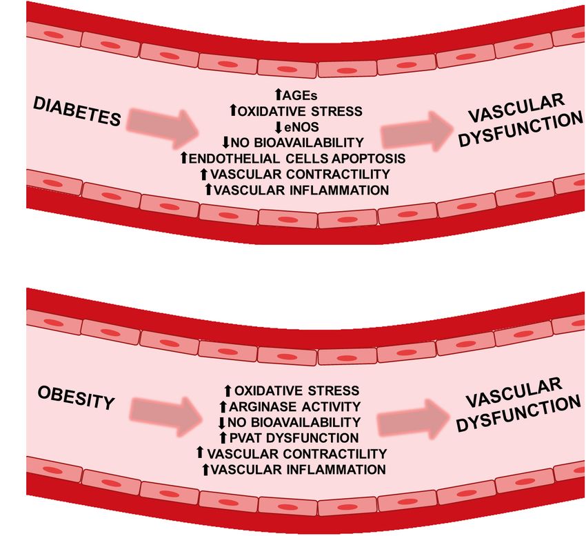
Moraes et al.                                                                                        Vascular TRP Channels and Metabolic Diseases

As well, augmented vascular contractility (Xie et al., 2006;             repletion of intracellular calcium stores (Rosado et al., 2002),
Matsumoto et al., 2014; Lubomirov et al., 2019), increased               vasoconstriction/vasorelaxation (Freichel et al., 2001; Dietrich
vascular inflammation (Zhang et al., 2008; Ku and Bae, 2016),            et al., 2005), secretion of hormones (Togashi et al., 2006; Cheng
and stimulated endothelial cells apoptosis (Sheu et al., 2005,           et al., 2007), cell cycle modulation (Lee et al., 2011; Tajeddine and
Sheu et al., 2008) can cooperate to cause vascular dysfunction           Gailly, 2012), sensory perception (Kichko et al., 2018) and others.
(Figure 1A). There are key processes in obesity which collaborate        This superfamily displays a variety of activation mechanisms,
and lead to impairment of vascular function. These processes             such as ligand binding (Janssens et al., 2016), temperature
include enhanced vascular contractility (Boustany-Kari et al.,           (McKemy et al., 2002), endogenous chemical mediators (Beck
2007; Weil et al., 2011), augmented sympathetic control of               et al., 2006), voltage (Matta and Ahern, 2007), G protein-coupled
vasoconstriction (Haddock and Hill, 2011), elevated oxidative            receptors (Boulay et al., 1997), and tyrosine kinase receptors (Xu
stress (La Favor et al., 2016), increased peroxynitrite (Mason           H. et al., 2003; Vazquez et al., 2004), among other stimuli.
et al., 2011; Gamez-Mendez et al., 2015), perivascular adipose               In blood vessels, TRP channels are present in endothelial
tissue (PVAT) dysfunction (Ma et al., 2010; Bussey et al., 2016),        cells (Ching et al., 2011), vascular smooth muscle cell (VSMC)
increased arginase activity (which can reduce L-arginine and NO          (Johnson et al., 2009), PVAT (Sukumar et al., 2012), perivascular
bioavailability) (Johnson et al., 2015; Bhatta et al., 2017), and        sensory nerves (Zygmunt et al., 1999), and pericytes (Tóth
increased vascular inflammation (Yao et al., 2017; Figure 1B).           et al., 2005), and these channels have been implicated in
Both diabetes and obesity share common mechanisms that                   the regulation of vascular tone (Pórszász et al., 2002; Qian
result in vascular injury. Thus, elucidation of the mechanisms           et al., 2007; Earley et al., 2009), vascular cell proliferation
underlying vascular dysfunction in these cardiometabolic                 (Zhang et al., 2018), vascular wall permeability (Tiruppathi
diseases is essential to provide additional therapeutic targets in       et al., 2002; Paria et al., 2004), and angiogenesis (Hamdollah
the prevention and treatment of these cardiometabolic diseases.          Zadeh et al., 2008; Ge et al., 2009). Additionally, there are a
Interestingly, alterations in TRPs channel expression or/and             large number of studies describing the involvement of TRP
function may contribute to these pathological conditions,                proteins in various pathophysiological conditions. We focus on
making these channels promising therapeutic targets.                     altered expression and/or activity of the TRPC, TRPV, TRPM,
                                                                         and TRPML channels, contributing to vascular dysfunction in
                                                                         obese and diabetic conditions or are potentially associated to
TRP CHANNELS                                                             vascular alterations.

The TRP superfamily was originally discovered in the study
on Drosophila melanogaster, where in response to bright light,           TRP CHANNELS INVOLVED IN
Drosophila mutants behaved as if they were blind, while wild-
                                                                         VASCULAR COMPLICATIONS OF
type flies maintained oriented toward visual cues. Thus, in the
mutated eye, the light-response was transient during sustained           DIABETES AND OBESITY
light (Cosens and Manning, 1969). This mutant was known
as TRP due to the transient response to prolonged intense                The Role of TRPC in the Vasculature
lights, performed by Minke and colleagues (Minke et al., 1975).          Under Diabetic and Obese Conditions
Following these reports, the molecular characterization of the           The TRPC subfamily consists of seven proteins, known as
Drosophila TRP gene was described (Montell and Rubin, 1989).             TRPC1 to TRPC7 (see review of Clapham et al., 2001; Putney,
   In addition, a common feature in the TRP superfamily is its           2005; Dietrich et al., 2010; Mederos y Schnitzler et al., 2018).
tetrameric structure, where each subunit is constituted by six           TRPC channels can form homo- and heterotetramers (Hofmann
transmembrane segments, a pore-forming region between the                et al., 2002; Strübing et al., 2003). Moreover, there is increasing
segments S5–S6 and cytoplasmic amino and carboxyl termini                evidence that TRPC channel members can form receptor-
(For general explanation, see reviews by: Earley and Brayden,            operated channels (ROC) (Soboloff et al., 2005; Peppiatt-
2015; Hof et al., 2019). Mammalian genomes encode 28 distinct            Wildman et al., 2007; Tai et al., 2008; Inoue et al., 2009; Itsuki
TRP protein subunits, and this superfamily is divided into               et al., 2014) and store-operated channels (SOC) (Groschner et al.,
six subfamilies, based on amino acid sequence homology and               1998; Freichel et al., 2001; Xu and Beech, 2001; Xu et al., 2006; Shi
include: TRPC (Wes et al., 1995; Liu et al., 2008), TRPV (Caterina       et al., 2016).
et al., 1997; Smith et al., 2002), TRPM (Tsavaler et al., 2001;              TRPC1, TRPC3, TRPC4, TRPC5, and TRPC6 are expressed
Fujiwara and Minor, 2008), TRPA (Story et al., 2003; Cvetkov             in VSMC (Wang et al., 2004; Evans et al., 2009; Inoue et al.,
et al., 2011), TRPP (Mochizuki et al., 1996; Giamarchi et al.,           2009; Mita et al., 2010) and endothelial cells (Yip et al., 2004;
2010), and TRPML (Sun et al., 2000; Zeevi et al., 2010).                 Gao et al., 2012; Sundivakkam et al., 2012). TRPC channels are
   The TRP superfamily consists of a diverse group of cation             involved in the regulation of vascular tone through different
channels, where most of the channels are non-selective and               signaling pathways. For example, activation of TRPC1 and
permeable to Ca2+ (Gonzalez-Perrett et al., 2001; Feng et al.,           TRPC3 channels in the VSMC can cause depolarization and
2014; Sierra-Valdez et al., 2018). These channels have been shown        vasoconstriction (Reading et al., 2005; Wölfle et al., 2010).
to be involved in many physiological processes, such as responses        Alternatively, TRPC1 channels can be associated with large-
to painful stimuli (Caterina et al., 2000; Davis et al., 2000),          conductance Ca2+ -activated K+ (BKCa ) channels in VSMC,

Frontiers in Physiology | www.frontiersin.org                        3                                   February 2021 | Volume 12 | Article 645109
Moraes et al. Vascular TRP Channels and Metabolic Diseases FIGURE 1 | Vascular dysfunction in diabetes and obesity. Pathophysiological factors leading to vascular dysfunction in (A) diabetic and (B) obese patients. AGEs, advanced glycation end products; eNOS, endothelial nitric oxide synthase; NO, nitric oxide; PVAT, perivascular adipose tissue. indirectly activating cell hyperpolarization (Kwan et al., 2009). Ang-II was due to the alteration of TRPC1/4/5 activity in diabetic As well, TRPC1, TRPC3, and TRPC4 stimulation in endothelial rats (Evans et al., 2009). However, 2-APB and caffeine are non- cells can induce vasodilation through increases in endothelial selective inhibitors and therefore, the general absence of selective Ca2+ , with subsequent generation of NO (Freichel et al., pharmacological tools for TRPC channels is a study limitation. 2001; Huang et al., 2011; Qu et al., 2017) and/or TRPC3 Additionally, 2-APB and caffeine cannot be considered as specific activation can induce endothelium-dependent hyperpolarization reagents to evaluate TRPC1 activity. Therefore, the use of factor (EDHF)-mediated vasodilation (Kochukov et al., 2014). gene knockout or knockdown animals could offer a valuable However, only a few studies have demonstrated the involvement alternative for studying specific functions of TRPC channels in of TRPC channels in the vasculature of diabetic animals the regulation of vascular tone in diabetic conditions. However, and humans and no studies have investigated a role for a limitation of this approach is that when one TRPC channel is TRPC in obesity. downregulated or knocked out it may be compensated by other Evans et al. (2009) showed that angiotensin-II (Ang-II)- TRPCs, as evidenced by Dietrich et al. (2005). Therefore, these induced Ca2+ influx was significantly enhanced in cultured obstacles make difficult to draw correct conclusions about the role aortic VSMC from Goto-Kakizaki (GK) rats, a model of type 2 of TRPC channels on the obesity and diabetes. diabetes, when compared with cells from Wistar-Kyoto (WKY) A study by Chung and colleagues provided the first evidence control rats. TRPC1 and TRPC5 protein expression were similar, that TRPC1, TRPC4 and TRPC6 messenger RNA (mRNA) while TRPC4 protein expression was significantly increased, and and proteins are present in human saphenous vein, and their TRPC6 protein expression was significantly decreased in GK, expression levels are modulated by type II diabetes. The authors compared with WKY values. In GK-VSMC, Ang-II-induced demonstrated that cyclopiazonic acid (CPA)-induced contraction Ca2+ influx was more sensitive to the calcium influx inhibitors of the saphenous vein was greater in diabetic vessels than 2-aminoethoxydiphenyl borate (2-APB) and caffeine, which act the non-diabetic, suggesting that the increased contractility in through the inhibition of the inositol 1,4,5-trisphosphate receptor human diabetes could be partially due to the participation of (IP3 R). Since TRPC1 can be activated by an IP3 R coupling Ca2+ entry through SOC. Additionally, TRPC channels may mechanism, this result suggests a possible increased activation be involved in SOC. Although TRPC4 mRNA expression was of mechanisms contributing to TRPC1 activity. The authors of elevated, protein levels were not significantly different when this study proposed that the elevated calcium influx induced by compared to non-diabetic vessels. TRPC1 and TRPC6 mRNA Frontiers in Physiology | www.frontiersin.org 4 February 2021 | Volume 12 | Article 645109
Moraes et al.                                                                                          Vascular TRP Channels and Metabolic Diseases

levels in diabetic conditions were similar to the control, however,         diabetes and obesity. In pulmonary arteries from streptozotocin
protein expression was decreased in diabetic veins. Even though             (STZ)-treated hyperglycemic lean Zucker (LZ) rats (type I
TPRC protein expression was diminished in the diabetic samples,             diabetic), the TRPM2-L channel isoform was decreased when
the enhanced CPA-induced contraction in diabetic veins might                compared to controls. Contrarily, vascular superoxide levels,
be associated with increased TRPC activity, leading to higher               NADPH oxidase (NOX) activity and lung capillary filtration
capacitative Ca2+ entry (Chung et al., 2009).                               coefficient (Kf) are higher in STZ-treated LZ rats. Interestingly,
   Mita and colleagues demonstrated that TRPC1, TRPC3, and                  inhibition of TRPM2 channel diminished lung Kf in diabetic
TRPC6 mRNAs and proteins were expressed in caudal arteries                  rats but did not affect the Kf in control animals. The
from Wistar rats. However, in addition to the expression of these           authors of this study proposed that in hyperglycemic rats,
TRPC channels, TRPC4 also was expressed at extremely low                    increased oxidative stress activates the TRPM2 channel and
levels in GK rats. In addition, GK rats had a significant increase in       elevates pulmonary endothelial Kf. The decreased TRPM2-
protein expression of TRPC1 and TRPC6 channels or appearance                L expression through chronic hyperglycemia may be due
of TRPC4 channel expression, but not TRPC3, compared with                   to overexposure of superoxide and a subsequent negative
Wistar rats, which is associated with the reduction in cirazoline-          feedback-mediated downregulation. This enhanced the TRPM2
or CPA-induced contractions in GK (Mita et al., 2010).                      activation-mediated increase in Kf that can contribute to
   These authors demonstrated that TRPC channel expression                  the elevated susceptibility to lung complications observed in
levels and function are altered in diabetes (Table 1). However,             individuals with type I diabetes. Taken together, additional
there was a heterogeneity of findings among these studies,                  studies are needed to determine the pulmonary TRPM2 channel
therefore these discrepancies may be explained by a number                  sensitivity in control and diabetic animal models by using
of factors, including: variations in the metabolic profile of               electrophysiological and pharmacological tools (Lu et al., 2014;
the diabetic animals, distinct stages of diabetes, and the type             Figures 2A,B).
of arteries and veins investigated. Nevertheless, the in vivo                   A study developed by Sun et al. (2019) demonstrated that
significance of these findings has not been shown. Additionally,            TRPM2 expression significantly increased in both primary
the role of TRPCs in obesity should also be more completely                 mouse aortic endothelial cells and aortic endothelium from
explored in future studies.                                                 high-fat diet (HFD, 60 kcal% fat)-fed mice. In addition,
                                                                            preincubation of the TRPM2 inhibitor N-(p-amylcinnamoyl)
                                                                            anthranilic acid (20 µM), reduced the impaired insulin-induced
The Role of TRPM2 in the Vasculature                                        relaxation in aortas from HFD-fed mice. Similarly, knockdown
Under Diabetic and Obese Conditions                                         of TRPM2 alleviated endothelial insulin resistance and improved
TRPM2 is activated by H2 O2 (Hara et al., 2002), adenosine 50 -             endothelium-dependent vasodilatation in obese mice. The
diphosphoribose (ADP-ribose) (Heiner et al., 2003; Yu et al.,               authors proposed that free fatty acid-induced H2 O2 activation
2017), nicotinic acid-adenine dinucleotide phosphate (NAADP)                of TRPM2, thereby aggravating endothelial insulin resistance.
(Beck et al., 2006), Ca2+ (McHugh et al., 2003), and temperature            Therefore, downregulation or pharmacological inhibition of
(35–47◦ C) (Togashi et al., 2006; Kashio et al., 2012; Kashio               TRPM2 channels may contribute to treatment of endothelial
and Tominaga, 2017), while adenosine monophosphate (AMP)                    dysfunction associated with the oxidative stress state (Sun
(Beck et al., 2006; Lange et al., 2008) and acidic pH are negative          et al., 2019; Figures 2A,C). Both of these studies indicated that
regulators (Du et al., 2009; Starkus et al., 2010). This channel is         increased oxidative stress, present in diabetes and obesity, are
expressed in VSMC (Yang et al., 2006) and vascular endothelial              modulating the TRPM2 channel (Table 1), leading to elevated
cells (Hecquet et al., 2008), and it is permeable to Ca2+ , Na+             channel activity. In this context, the decreased vascular TRPM2-
(Perraud et al., 2001; Sano et al., 2001; Kraft et al., 2004), and          L expression in the lung from diabetic animals, as shown by Lu
K+ (Sano et al., 2001). Moreover, physiological splice variants of          et al., is due to negative feedback.
TRPM2, including full-length TRPM2 (TRPM2-L) and a short
splice variant (TRPM2-S), have been identified in endothelial
cells (Hecquet and Malik, 2009; Hecquet et al., 2014) and VSMC              The Role of TRPV1 in the Vasculature
(Yang et al., 2006).                                                        Under Diabetic and Obese Conditions
    TRPM2 is involved in endothelial permeability, as                       TRPV1 channels are expressed in endothelial cells (Yang et al.,
demonstrated by H2 O2 -induced Ca2+ influx via TRPM2                        2010), VSMC (Kark et al., 2008), perivascular sensory nerves
channels that results in endothelial hyperpermeability (Hecquet             (Zygmunt et al., 1999; Breyne and Vanheel, 2006), and pericytes
et al., 2008). Moreover, H2 O2 activates TRPM2 to induce                    (Tóth et al., 2005). TRPV1 channels are present in blood
excessive Ca2+ influx, resulting in Ca2+ overload and                       vessels, such as epineural arterioles (Davidson et al., 2006),
consequently, cell death in vascular endothelial cells (Sun                 aorta (Ohanyan et al., 2011; Sun et al., 2013), mesenteric (Sun
et al., 2012). Furthermore, ROS overproduction activates TRPM2              et al., 2013; Zhang et al., 2015), and coronary arteries (Bratz
channels, leading to Ca2+ influx through TRPM2, which induces               et al., 2008). These channels are activated by multiple stimuli,
VSMC migration and proliferation that contributes to neointimal             including heat (∼42–51◦ C) (Tominaga et al., 1998; Cesare
hyperplasia (Ru et al., 2015).                                              et al., 1999), anandamide (Zygmunt et al., 1999), and exogenous
    There are only a few studies that demonstrate changes in                agonists, such as capsaicin and resiniferatoxin (Caterina et al.,
TRPM2 channel expression and/or function associated with                    1997), as well as low pH that acts as a sensitizing agent

Frontiers in Physiology | www.frontiersin.org                           5                                  February 2021 | Volume 12 | Article 645109
Moraes et al. Vascular TRP Channels and Metabolic Diseases
TABLE 1 | TRP channels involved in vascular complications of diabetes and obesity.
TRP channels involved in vascular complications of diabetes and obesity
Channel Diabetic Normal Tissue Drug-induced vascular mRNA in diabetic Protein in References
and/or control effect or other vascular and/or obesity diabetic and/or
obesity model investigations model obesity model
TRPC1 Goto-Kakizaki Wistar- Cultured aortic Angiotensin-II-induced Ca2+ Decrease No change Evans et al.
(GK) (Type 2 Kyoto vascular smooth influx was enhanced in (2009)
diabetes) (WKY) rat muscle cell diabetic rat
TRPC1 Human Type II Human Saphenous vein Cyclopiazonic acid-induced No change Decrease Chung et al.
diabetic non- Ca2+ influx was enhanced in (2009)
diabetic diabetic patient
TRPC1 GK rat Wistar rats Endothelium- Cirazoline- or cyclopiazonic – Increase Mita et al.
denuded acid-induced Ca2+ influx was (2010)
caudal artery decreased in diabetic rat
smooth muscle
strips
TRPC3 GK rat WKY rat Cultured aortic Angiotensin-II-induced Ca2+ Undetectable – Evans et al.
vascular smooth influx was enhanced in (2009)
muscle cell diabetic rat
TRPC3 GK rat Wistar rat Endothelium- Cirazoline- or cyclopiazonic – No change Mita et al.
denuded acid-induced Ca2+ influx was (2010)
caudal artery decreased in diabetic rat
smooth muscle
strips
TRPC4 GK rat WKY rat Cultured aortic Angiotensin-II-induced Ca2+ No change Increase Evans et al.
vascular smooth influx was enhanced in (2009)
muscle cell diabetic rat
TRPC4 Human diabetic Human Saphenous vein Cyclopiazonic acid-induced Increase No change Chung et al.
non- Ca2+ influx was enhanced in (2009)
diabetic diabetic patient
TRPC4 GK rat Wistar rat Endothelium- Cirazoline- or cyclopiazonic TRPC4 mRNA was TRPC4 protein was Mita et al.
denuded acid-induced Ca2+ influx was not detected in not detected in (2010)
caudal artery decreased in diabetic rat Wistar rats, but it Wistar rats, but it
smooth muscle was detectable in was barely
strips GK rats detectable in GK
rats
TRPC5 GK rat WKY rat Cultured aortic Angiotensin-II-induced Ca2+ No change No change Evans et al.
vascular smooth influx was enhanced in (2009)
muscle cell diabetic rat
TRPC6 GK rat WKY rat Cultured aortic Angiotensin-II-induced Ca2+ Decrease Decrease Evans et al.
vascular smooth influx was enhanced in (2009)
muscle cell diabetic rat
TRPC6 Human diabetic Human Saphenous vein Cyclopiazonic acid-induced No change Decrease Chung et al.
non- Ca2+ influx was enhanced in (2009)
diabetic diabetic patient
TRPC6 GK rat Wistar rat Endothelium- Cirazoline- or cyclopiazonic – Increase Mita et al.
denuded acid-induced Ca2+ influx was (2010)
caudal artery decreased in diabetic rat
smooth muscle
strips
TRPM2 Streptozotocin Lean Pulmonary artery Lung capillary filtration – Decrease Lu et al.
(STZ)-treated Zucker rats coefficient (Kf) was enhanced (2014)
lean Zucker in diabetic rat. TRPM2 channel
(LZ) rats (Type I mediated increase in Kf.
diabetes)
TRPM2 High-fat diet Low-fat diet Mouse aortic Preincubation of TRPM2 – Increase Sun et al.
(HFD)-fed mice (LFD)-fed endothelial cells inhibitor N-(p-amylcinnamoyl) (2019)
C57BL/6J for mice and aortas anthranilic acid (20 µM) or
16 weeks. C57BL/6J knockdown of TRPM2
for alleviates obesity-associated
16 weeks. impairment in insulin-evoked
endothelium-dependent
relaxations in obese mice
(Continued)
Frontiers in Physiology | www.frontiersin.org 6 February 2021 | Volume 12 | Article 645109Moraes et al. Vascular TRP Channels and Metabolic Diseases
TABLE 1 | Continued
TRP channels involved in vascular complications of diabetes and obesity
Channel Diabetic Normal Tissue Drug-induced vascular mRNA in diabetic Protein in References
and/or control effect or other vascular and/or obesity diabetic and/or
obesity model investigations model obesity model
TRPV1 Zucker Genetic Branch II and III Capsaicin-induced relaxation – – Pamarthi
diabetic fatty controls mesenteric was similar in diabetic rat. et al. (2002)
(ZDF) rat (Type arteries.
II diabetes) (A portion of
the omental
membrane,
which frequently
contains nerve
trunks, was
maintained)
TRPV1 STZ -induced Sprague- Epineurial Capsaicin-induced constriction – Decrease Davidson
diabetic Dawley arterioles of the (10−6 M) was decreased in et al. (2006)
Sprague- rats sciatic nerve diabetic rat (10–12-week
Dawley duration).
rats
TRPV1 STZ -induced Wistar rats Medial meningeal Capsaicin-induced relaxation – – Dux et al.
diabetic Wistar treated with artery (10−7 M) was abolished in (2007)
rats the solvent (Meningeal blood diabetic rat.
for STZ flow) Capsaicin-induced constriction
(10−5 M) was similar in
diabetic rat.
TRPV1 db/db mice C57BLKS/J Mean arterial Capsaicin-induced increases – Decrease Ohanyan
(Type 2 mice blood pressure in MAP was attenuated in et al. (2011)
diabetes and (MAP) diabetic mouse.
obesity) Aortic tissue
TRPV1 db/db mice C57BLKS/J Coronary Capsaicin-induced increases – – Guarini et al.
mice microvessel in MBF and (2012)
Myocardial blood capsaicin-mediated relaxation
flow (MBF) in coronary microvessels were
attenuated in diabetic mouse.
TRPV1 db/db mice C57BLKS/J Thoracic aortas Dietary capsaicin improves the – Decrease Sun et al.
mice and endothelium-dependent (2013)
mesenteric relaxation in diabetic mouse
arteries compared to db/db mice given
a normal diet.
TRPV1 STZ -induced Sprague- Third branch of Capsaicin-induced relaxation – Decrease Zhang et al.
diabetic Dawley the superior was decreased in diabetic rat. (2015)
Sprague- rats mesenteric artery
Dawley
rats
TRPV1 db/db mice C57BLKS/J Coronary H2 O2 had little potentiating – – DelloStritto
mice arterioles effect on capsaicin-induced et al. (2016)
Coronary blood CBF responses or
flow (CBF) capsaicin-mediated coronary
vasodilation in db/db and
TRPV1 knockout mice.
TRPV1 Human diabetic Human Cutaneous CVC was decreased in – – Marche et al.
(Type 1 non- vascular diabetic patients in (2017)
diabetes) diabetic conductance response to local heating early
(CVC) in the peak.
forearm
TRPV1 High-fat/high- Lean male Coronary arteries Capsaicin-induced relaxation Increase Decrease Bratz et al.
cholesterol Ossabaw was impaired in obese pigs. (2008)
diet- induced miniature
obese male swine for
Ossabaw 24 weeks.
miniature swine
for 24 weeks.
(Continued)
Frontiers in Physiology | www.frontiersin.org 7 February 2021 | Volume 12 | Article 645109Moraes et al. Vascular TRP Channels and Metabolic Diseases
TABLE 1 | Continued
TRP channels involved in vascular complications of diabetes and obesity
Channel Diabetic Normal Tissue Drug-induced vascular mRNA in diabetic Protein in References
and/or obesity control effect or other vascular and/or obesity diabetic and/or
model investigations model obesity model
TRPV1 HFD-fed Normal Small mesenteric Capsaicin (10 µM) – – Haddock and
Sprague-Dawley diet-fed arteries significantly increased the Hill (2011)
rats Sprague- (third-order) amplitude of nerve-mediated
for Dawley rats contraction induced by
20–24 weeks. for 10 Hz stimulation, with a
20–24 weeks. greater effect in control than
obese animals.
TRPV1 HFD-fed mice Normal Aorta Vascular hypertrophy was – – Marshall et al.
C57BL6/ diet-fed mice observed in HFD-fed (2013)
129SVJ for C57BL6/ wild-type but not HFD-fed
12 weeks. 129SVJ for TRPV1 knockout mice.
12 weeks.
TRPV1 Obese Zucker LZ rats Resistance Capsaicin-induced – No change Lobato et al.
(OZ) rats mesenteric relaxation (2013)
arteries was decreased in OZ rats
TRPV1 High-fat, Regular Capsaicin-induced – – Marics et al.
high-sucrose diet-fed Meningeal blood increased meningeal blood (2017)
(HFHS) Sprague- flow flow (100 nM) was greater in
diet-induced Dawley rats obese rat.
obese for 20 weeks. Capsaicin-induced
Sprague-Dawley decreased meningeal blood
rats flow (10 µM) was greater in
for 20 weeks. obese rat.
TRPV4 STZ -induced Sprague- Third or fourth TRPV4-KCa 2.3-mediated – Decrease Ma et al.
diabetic Dawley branches of rat relaxation were impaired in (2013)
Sprague-Dawley rats mesenteric artery diabetic rats
rats
TRPV4 STZ -induced Sprague- Retinal arteriole – Decrease Decrease Monaghan
diabetic Dawley et al. (2015)
Sprague-Dawley rats
rats
TRPV4 db/db mice C57BLKS/J Aortas – Decrease Decrease Gao et al.
and STZ mice (2020)
-induced
diabetic
C57BLKS/J
mice
TRPV4 HFD-fed mice LFD-fed mice Third-order Vasodilatory responses to – – Greenstein
C57BL/6J. C57BL/6J. mesenteric GSK1016970A (TRPV4 et al. (2020)
The diets The diets arteries agonist) in resistance
initiated at age initiated at mesenteric arteries were
5 weeks and age 5 weeks similar between the LFD-
continued at and and HFD-fed mice.
age 6 months. continued at
age
6 months.
TRPV4 HFD-fed mice LFD-fed mice Resistance Vasodilatory response to – – Ottolini et al.
C57BL/6J C57BL/6J mesenteric GSK1016970A was (2020)
for 14 weeks. for 14 weeks. arteries from impaired in HFD mice.
Obese Non-obese mice. Vasodilatory response to
individuals. individuals. Splenius/ GSK1016970A was
temporalis markedly reduced in the
muscle arteries arteries from obese
from human. individuals.
(Tominaga et al., 1998; Cesare et al., 1999). TRPV1 is a non- Activation of TRPV1 by capsaicin promotes the release
selective cation channel, which is permeable to K+ , Na+ , Ca2+ , of neurotransmitters, such as calcitonin gene-related peptide
and Mg2+ (Caterina et al., 1997). (CGRP) (Zygmunt et al., 1999; Wang et al., 2006) from
Frontiers in Physiology | www.frontiersin.org 8 February 2021 | Volume 12 | Article 645109Moraes et al. Vascular TRP Channels and Metabolic Diseases
capsaicin-sensitive nerves, in addition to NO from endothelial diabetic rats. In control and insulin-treated diabetic animals,
cells (Yang et al., 2010; Ching et al., 2011), which can diffuse capsaicin (10−7 M) induced increases in meningeal blood flow,
to adjacent VSMC and cause relaxation. In smooth muscle but in 6-week STZ-induced diabetic rats, capsaicin promoted
cells from skeletal muscle arterioles obtained from the rat and decreases in the blood flow. In contrast, capsaicin at a higher
mice, TRPV1 stimulation causes an increase in intracellular concentration (10−5 M) caused vasoconstriction, which is
Ca2+ concentration, resulting in vasoconstriction (Czikora et al., a non-neurogenic response and was similar in control and
2012). Therefore, the activation of TRPV1 may induce different diabetic animals. The authors demonstrated a reduction in
effects on the vasculature (vasoconstriction, vasodilation, or no the capsaicin-evoked release of CGRP and decrease in the
effect), which can be unique to each vascular bed. For example, density of perivascular and stromal TRPV1-immunoreactive
arteries with sensory neuron innervation and without vascular nerve fibers of the dura mater from diabetic rats, suggesting that
TRPV1 expression are expected to dilate in response to TRPV1 insufficient vasodilator function of meningeal sensory nerves may
activation. However, arteries with elevated smooth muscle contribute to the higher incidence of headaches in diabetics due
TRPV1 expression and without apparent sensory neuronal to perturbation of tissue homeostasis that could induce additional
innervation constrict in response to the same TRPV1 stimulation activation and/or sensitization of meningeal nociceptors (Dux
(Kark et al., 2008; Tóth et al., 2014). Moreover, TRPV1 activation et al., 2007). Further studies are needed to determine if this
by capsaicin induced concentration-dependent biphasic effects, hypothesis can be supported. It is pertinent to highlight the fact
where a low concentration capsaicin evoked dilation, while a that diabetic rats treated with insulin restored the vasodilatory
higher concentration resulted in vasoconstriction of the dural response and the capsaicin-evoked release of CGRP, indicating
vessels (Dux et al., 2003) and skeletal (musculus gracilis) muscle that impairments observed in diabetic animals can be attributed
arterioles (Kark et al., 2008). to the diabetic condition induced by STZ and not to a toxic
Abundant evidence supports the hypothesis that altered action of this drug. Moreover, it is important to note, that the
TRPV1 expression and/or function is associated with vascular current evidences demonstrate that TRPV1 channels expression
dysfunction in diabetes and obesity. The TRPV1 is the most and/or activity in perivascular sensory nerves are reduced under
studied TRP channel in the vasculature, under these metabolic these conditions.
conditions. In humans, a study by Marche et al. (2017) evaluated In an opposite way, in model of obesity, topical administration
cutaneous vascular conductance (CVC) in response to heat by of capsaicin (100 nM) to the dura mater promoted enhanced
using a skin-heating probe, heated to 44 ◦ C to assess heat-induced meningeal blood flow in high-fat high-sucrose (HFHS) diet-
vasodilation. The local heat-induced early peak is mediated fed Sprague–Dawley rats (diets started at 6 weeks of age and
through TRPV1 channels, located on sensory nerves. Therefore, continued for 20 weeks; 45% of total calories as fat) compared
the significantly diminished peak response to local thermal to regular diet-fed rats. However, administration of capsaicin
hyperemia could suggest reduced activity of the TRPV1 channels at 10 µM induced a greater reduction in meningeal blood
at the skin level in type 1 diabetic patients compared to control flow in obese animals compared to controls. In this way,
subjects. This study indicated that the microvascular response dural application of capsaicin resulted in significantly higher
triggered by TRPV1 channels is reduced in type 1 diabetic vasodilator and vasoconstrictor responses in obese animals
patients (Marche et al., 2017). compared to controls. Moreover, this obesity animal model
Zhang et al. (2015) investigated the pharmacological effects was characterized by an increase in CGRP release in response
of capsaicin on mesenteric arteries of STZ-induced diabetic to both concentrations of capsaicin administered, suggesting a
Sprague-Dawley rats. Capsaicin-induced vasodilation was greater TRPV1-mediated CGRP release from meningeal afferent
impaired in the mesenteric arteries of diabetic rats. As well, nerves likely due to a sensitization of the TRPV1 receptor.
TRPV1 expression was reduced in the diabetic preparation when This sensitization may be a consequence of the increase
compared to the control group. The authors indicated that the in proinflammatory cytokines and levels of oxidative stress.
attenuated expression of CGRP and TRPV1 contribute to the Changes in TRPV1-mediated vascular reactions and CGRP
weakened capsaicin-mediated dilation in diabetic mesenteric release, may be related to the enhanced headache susceptibility
arteries (Zhang et al., 2015). In line with previous studies, of obese individuals (Marics et al., 2017). Moreover, Dux et al.
capsaicin-induced relaxation in resistance mesenteric arteries (2007) and Marics et al. (2017) demonstrated divergent results
was markedly decreased in obese Zucker (OZ; genetic model on TRPV1 receptor-mediated neurogenic sensory vasodilation
of obesity) rats compared with LZ rats. However, TRPV1 between diabetic and obese conditions, indicating that different
receptor protein expression was similar between LZ and OZ mechanisms can contribute to modulation of the TRPV1
rats. The authors suggest that the weakened vascular effect channels in each disease.
to anandamide in arteries from this obese model can involve Guarini et al. (2012) showed that capsaicin-mediated increases
reduced activation of C-fiber nerve endings, and this may in myocardial blood flow (MBF), using myocardial contrast
collaborate to the vascular dysfunction observed in OZ rats echocardiography, were reduced in db/db mice, a model of type II
(Lobato et al., 2013). However, one concern about this model is diabetes, and obesity. Similarly, relaxation promoted by capsaicin
due the mutation of the fa gene (cause of obesity in OZ rats) is was attenuated in coronary microvessels from diabetic mice.
not common among humans. Interestingly, myocardial pH was more acidic in diabetic mice
In addition, the study by Dux et al. (2007) evaluated the than control mice and pH-mediated relaxation was attenuated in
TRPV1 receptor-mediated neurogenic sensory vasodilation in coronary microvessels from TRPV1(−/−) and db/db mice. The
Frontiers in Physiology | www.frontiersin.org 9 February 2021 | Volume 12 | Article 645109Moraes et al. Vascular TRP Channels and Metabolic Diseases
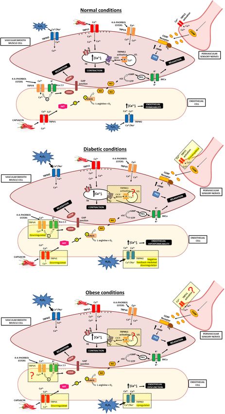
FIGURE 2 | Involvement of TRPs in vascular responses in normal, diabetic and obese conditions. (A) The figure shows the possible mechanisms that can explain
vasodilator influences of TRPV1, TRPV4, TRPM2, and TRPML1 channel present in the vasculature. TRPV1 channel activation causes release of the CGRP from
sensory nerves. CGRP binds to CGRP receptor, inducing augmented levels of cAMP that activates PKA and promotes relaxation of VSMC. TRPV1 activation in
endothelial cells promotes Ca2+ influx and phosphorylation of eNOS and induces NO production. NO active the soluble guanylyl cyclase, that catalyzes the
conversion of GTP to cGMP and active the PKG. The NO/cGMP/PKG activates BKCa that leads to smooth muscle relaxation. Additionally, specific interaction of
(Continued)
Frontiers in Physiology | www.frontiersin.org 10 February 2021 | Volume 12 | Article 645109Moraes et al. Vascular TRP Channels and Metabolic Diseases FIGURE 2 | Continued TRPV4 with KCa 2.3 in endothelial cells promote vasodilation, likely via an EDHF pathway. Moreover, H2 O2 -induced Ca2+ influx via TRPM2 channels in endothelial cells results in endothelial permeability. TRPML1 is closely associated with RyR2. TRPML1 activation provokes Ca2+ signals from a LELs, which can subsequently be augmented by CICR from the SR via RyR2 to induce Ca2+ sparks, leading to BKCa channel activity that result in membrane hyperpolarization, VSMC relaxation. (B) The figure shows the possible alterations in TRPV1, TRPV4, and TRPM2 channel under diabetic conditions. Diabetic conditions promote reduction in the capsaicin-evoked release of CGRP and decrease in the density of perivascular TRPV1. Moreover, a high level of glucose reduces TRPV1 expression and PKA phosphorylation in endothelial cells. Additionality, hyperglycemia is a crucial factor for the diminished TRPV4 expression and impairs the endothelium-dependent vasodilatation. Also, increased oxidative stress activates the TRPM2 channel and results in endothelial hyperpermeability. Besides that, overexposure of superoxide promoted a TRPM2 channel negative feedback-mediated downregulation. Further studies are needed to clarify whether TRPML1 activity and/or expression are altered in the vasculature during diabetes. (C) The figure shows the possible alterations in TRPV1 and TRPM2 channel under obese conditions. Impaired capsaicin-induced vasodilation in arteries is associated with reduced expression of TRPV1 protein and cation influx into endothelial cells under obese conditions. Increased oxidative stress present in obesity are modulating the TRPM2 channel, leading elevated activity of this channel. Further studies are needed to elucidate whether TRPV4 and TRPML1 activity and/or expression are altered in the vasculature during obesity. TRPV1, Transient receptor potential of vanilloid type 1; TRPV4, Transient receptor potential of vanilloid type 4; TRPM2, Transient receptor potential of melastatin type 2; TRPML1, Transient receptor potential of mucolipin type 1; VSMC, vascular smooth muscle cells; NO, nitric oxide; EDHF, endothelium-derived hyperpolarizing factor; eNOS, endothelial nitric oxide synthase; BKCa , large-conductance Ca2+ -activated K+ channel; KCa 2.3, small-conductance Ca2+ -sensitive K+ channels (SKCa ) isoform. PKG, Protein Kinase G; cGMP, cyclic guanosine 30 ,50 -monophosphate; GTP, guanosine 50 -triphosphate; SR, sarcoplasmic reticulum; IP3 R, Inositol 1,4,5-trisphosphate receptor; RyR2, type 2 ryanodine receptors; cAMP, Cyclic adenosine monophosphate; PKA, protein kinase A; CGRP, Calcitonin gene-related peptide; CGRP receptor, Calcitonin gene-related peptide receptor; CICR, calcium-induced calcium release; LELs, late endosomes and lysosomes; H2 O2 , Hydrogen peroxide. authors speculated that TRPV1 channels directly regulate MBF antioxidant effect (Sun et al., 2013). If this conclusion is accurate, and impairment of TRPV1 channels could contribute to vascular then it would indicate a possible target for future research dysfunction that is typically observed in diabetes. As previously on chronic treatment with TRPV1 agonists in the diabetic described, lowering pH is a stimulus for TRPV1 activation. and obesity conditions, evaluating whether these agonists could The study by Guarini et al. (2012) demonstrates a possible attenuate or prevent vascular dysfunction. In addition, these desensitization of TRPV1 in situations of prolonged acidic studies demonstrate new possibilities of capsaicin-rich dietary environment exposure. Further investigation into prolonged recommendations for complementary assistance in the treatment acidic environment on TRPV1 desensitization is necessary. of diabetic patients. A follow-up study by this group reported that acute H2 O2 Similarly, Bratz et al. (2008) demonstrated impaired capsaicin- exposure potentiated capsaicin-mediated coronary blood flow induced vasodilation in coronary arteries from obese Ossabaw (CBF), using the same methodology that was described by swine (diets were provided for 24 weeks; 46% of total kcal Guarini et al. (2012), responses and capsaicin-induced dilation from fat) associated with reduced expression of TRPV1 protein of coronary microvessels in control mice, but H2 O2 had little and cation influx into endothelial cells. On the other hand, potentiating effect on capsaicin-mediated responses in db/db and TRPV1 channel mRNA expression was increased in obese TRPV1 knockout mice. However, after excessive H2 O2 exposure, swine compared with lean controls. The authors concluded CBF and microvessel responses in the control mice resembled that TRPV1 channel signaling is diminished in metabolic those of the attenuated responses seen in TRPV1 knockout syndrome and this disrupted pathway can contribute to the and db/db mice. The author indicated that H2 O2 -induced endothelial dysfunction and the development of coronary artery increases in CBF are promoted, in part, by TRPV1 channels. disease (Bratz et al., 2008). These findings support the notion Moreover, prolonged H2 O2 exposure disrupts TRPV1-dependent that decreased expression of TRPV1 channel and Ca2+ influx coronary vascular signaling, which can cause in-tissue perfusion into endothelial cells promote insufficient vasodilator response, impairments observed in diabetes (DelloStritto et al., 2016). collaborating to the endothelial dysfunction related to diabetic Sun et al. (2013) demonstrated that cultured endothelial cells and obesity conditions. that are exposed to a high level of glucose (30 mmol/L), reduced Together, these studies support a model in which activation of TRPV1 expression and protein kinase A (PKA) phosphorylation TRPV1 channels from endothelial cells and perivascular sensory compared with control cells and that these effects were reversed nerves cause vasodilation. This mechanism may be disrupted by the administration of capsaicin (1 µmol/L). Similarly, in during diabetes and obesity, contributing to vascular dysfunction the aorta and mesenteric arteries from db/db mice, TRPV1 associated with these conditions, resulting in higher incidence of expression and PKA phosphorylation were decreased, but headaches, coronary disease, and tissue perfusion impairment. uncoupling protein 2 (UCP2) level was significantly higher However, Pamarthi et al. (2002) demonstrated that capsaicin- when compared to wild type mice. After dietary administration induced concentration-dependent relaxation of branch II and III of 0.01% capsaicin for 14 weeks, TRPV1 activation induced mesenteric arteries and CGRP nerve density was similar in the PKA phosphorylation and elevated the expression level of Zucker diabetic fatty (ZDF) rat, a model of type II diabetes, and UCP2 in diabetic mice. Moreover, capsaicin ameliorated vascular genetic controls. ZDF rats exhibit obesity, severe hyperglycemia, oxidative stress and increased NO levels in db/db mice. an early hyperinsulinemia and dyslipidemia. Moreover, the The authors concluded that TRPV1 activation by capsaicin obesity is promoted by the fa leptin receptor mutation (Pamarthi might attenuate hyperglycemia-induced endothelial dysfunction et al., 2002), but, as described before, this is not common cause of through a mechanism involving the PKA and UCP2-mediated obesity among humans. Frontiers in Physiology | www.frontiersin.org 11 February 2021 | Volume 12 | Article 645109
Moraes et al.                                                                                          Vascular TRP Channels and Metabolic Diseases

   In contrast, Davidson et al. (2006) reported that capsaicin              intact, suggesting little vascular dysfunction in the mesenteric
induced a concentration-dependent vasoconstriction of                       resistance artery in this obese model. Interestingly, the authors
epineural arterioles of the sciatic nerve from Sprague-Dawley               provided evidence that TRPV1 knockout mice were protected
rats, concluding that vasoconstriction was likely due to the release        from obesity-induced hypertension and vascular hypertrophy
of neuropeptide Y (NPY) contained in nerves that innervate                  (Marshall et al., 2013; Table 1). However, it is important to note
these arterioles. However, vasoconstriction to capsaicin was                that these results differ from studies that have linked decreased
significantly decreased in long-term diabetic rats. This altered            TRPV1 expression or/and function with a worsened phenotype.
response was correlated with the reduced expression of TRPV1                Moreover, there is no significant alteration on the mean arterial
in epineural arterioles in diabetic rats (Davidson et al., 2006).           pressure in TRPV1 knockout mice related to wild-type mice
Moreover, the present evidence shows that TRPV1 channels                    under normal diet. This implies that altered TRPV1 activity can
expression and/or activity in sensory nerves that innervate these           be associated with a compensatory response that counteracts the
arterioles are decreased under diabetic condition. Overall, these           hypertension in this model of obesity. The HFD-wild-type mice
findings are in accordance with findings reported by Haddock                show low-grade inflammation, reducing glucose tolerance and
and Hill (2011). In an animal model of obesity, capsaicin                   raised levels of adipokine that could be involved with modulation
(10 µM) promoted a significant increase in nerve-mediated                   of this channel. Furthermore, it cannot be ruled out that the
vasoconstriction induced by a 10 Hz stimulation in small                    different influences of TRPV1 channels on the vasculature
mesenteric arteries from groups fed a high-fat (diets started at            depend on the tested diabetic or obese animal model. Thus,
6 weeks of age and were provided for 20–24 weeks; containing                additional research is needed to confirm these observations.
43% of total calories as fat) and normal diet, although the effect             Collectively, these findings reveal the downregulated
was greater in control rats (Haddock and Hill, 2011). From                  TRPV1 channel expression is related to the diabetic condition
the results, it is clear that common factors between obesity and            (Figures 2A,B). In obese animal models, these studies
diabetes can modulate TRPV1 channel, leading to the reduced                 demonstrated alterations in TRPV1 channel expression
vasoconstriction. Additional studies to investigate which specific          and/or function, suggesting a role of TRPV1 in obese conditions
mechanisms collaborate to TRP channels modulation in each                   (Figures 2A,C). Nevertheless, the data obtained from these
disease are necessary.                                                      studies are divergent, which can be justified by the use of
   A study by Ohanyan et al. (2011) showed that capsaicin                   different obesity animal models, observed by distinct diet
caused an increase in mean arterial blood pressure (MAP)                    compositions, durations and age of onset of diet intervention,
in mice, but the increase MAP was attenuated in the db/db                   which can result in different metabolic profiles and severity of
mice. In addition, mice were given the ganglion blocker,                    obesity. In addition to the different models, different vascular
hexamethonium, to evaluate the primary actions of capsaicin and             beds were utilized which confound the conclusion’s coalescence.
to eliminate reflex adjustments. Furthermore, this diminished               Overall, these findings demonstrate that mainly TRPV1 channels
capsaicin-induced pressor response was correlated with reduced              in endothelial cells and perivascular sensory nerves are altered
aortic TRPV1 protein expression in db/db mice. Moreover,                    under diabetic and obese conditions.
cultured bovine aortic endothelial cells exposed to capsaicin
augmented endothelin production and endothelin A (ETA )
receptor inhibition reduced the capsaicin-mediated rises in MAP.            The Role of TRPV4 in the Vasculature
Based on these findings, the authors indicated that TRPV1                   Under Diabetic and Obese Conditions
channels are involved in the regulation of vascular reactivity and          TRPV4 is expressed in the aorta (Gao et al., 2020), mesenteric
systemic pressure through production of endothelin, resulting               (Ma et al., 2013), carotid (Hartmannsgruber et al., 2007),
in activation of vascular ETA receptors. Therefore, a decrease in           pulmonary (Martin et al., 2012), cerebral basilar (Han et al.,
vascular TRPV1 channel expression may contribute to vascular                2018), and renal (Soni et al., 2017) arteries, among others, and
dysfunction in diabetes. The authors suggest that this reduced              it can be present in both VSMC (Martin et al., 2012; Soni et al.,
TRPV1 channels could promote sensitization of vasoconstrictor               2017) and the endothelium (Marrelli et al., 2007; Ma et al.,
pathways and reduced functional hyperemia present in diabetic               2013; Han et al., 2018). A broad range of stimuli can lead to
patients (Ohanyan et al., 2011). A limitation of this study was the         TRPV4 activation, including heat (>27◦ C) (Güler et al., 2002;
use of conductance vessels instead of resistance vessels in order           Watanabe et al., 2002b), hypoosmotic conditions (Liedtke et al.,
to evaluate TRPV1 protein expression. Moreover, further studies             2000; Strotmann et al., 2000; Alessandri-Haber et al., 2003), low
should evaluate if substance P and NPY can participate in the               pH and citrate (Suzuki et al., 2003), 5,6- epoxyeicosatrienoic acid
capsaicin-mediated pressor response.                                        (Watanabe et al., 2003), and 4-α-phorbol esters (Watanabe et al.,
   Marshall and colleagues revealed that hypertension and                   2002a,b). TRPV4 is a nonselective cation channel, permeable to
vascular hypertrophy were observed in HFD-fed wild-type                     Ca2+ , Mg2+ and K+ (Voets et al., 2002), and it exhibits moderate
(diets for 12 weeks from 3 weeks of age; 35% fat from                       permeability to Ca2+ (PCa /PNa ∼6) (Strotmann et al., 2000; Voets
lard) but not HFD-fed TRPV1 knockout mice, indicating that                  et al., 2002; Watanabe et al., 2002a).
the onset of vascular remodeling may have an association                        Moreover, there is evidence that TRPV4-mediated stimulation
between TRPV1 and obesity-induced high blood pressure.                      of intermediate-conductance Ca2+ -sensitive K+ channels (IKCa )
Moreover, constrictor and dilator responses to phenylephrine,               and/or small-conductance Ca2+ -sensitive K+ channels (SKCa )
CGRP, and the endothelium-dependent carbachol remained                      channels can promote vasodilation, likely via an EDHF pathway

Frontiers in Physiology | www.frontiersin.org                          12                                  February 2021 | Volume 12 | Article 645109
You can also read