Dopamine Receptor Subtypes, Physiology and Pharmacology: New Ligands and Concepts in Schizophrenia - Frontiers
←
→
Page content transcription
If your browser does not render page correctly, please read the page content below
REVIEW
                                                                                                                                             published: 14 July 2020
                                                                                                                                     doi: 10.3389/fphar.2020.01003

                                           Dopamine Receptor Subtypes,
                                           Physiology and Pharmacology:
                                           New Ligands and Concepts in
                                           Schizophrenia
                                           Jean Claude Martel 1 and Silvia Gatti McArthur 2*
                                           1   Independent Researcher, Amos, QC, Canada, 2 McArthur and Associates, Basel, Switzerland

                                           Dopamine receptors are widely distributed within the brain where they play critical
                                           modulator roles on motor functions, motivation and drive, as well as cognition. The
                                           identification of five genes coding for different dopamine receptor subtypes,
                                           pharmacologically grouped as D1- (D1 and D5) or D2-like (D2S, D2L, D3, and D4) has
                                           allowed the demonstration of differential receptor function in specific neurocircuits. Recent
                                           observation on dopamine receptor signaling point at dopamine—glutamate-NMDA
                                           neurobiology as the most relevant in schizophrenia and for the development of new
                                           therapies. Progress in the chemistry of D1- and D2-like receptor ligands (agonists,
                           Edited by:      antagonists, and partial agonists) has provided more selective compounds possibly
                     Nikolaos Pitsikas,    able to target the dopamine receptors homo and heterodimers and address different
       University of Thessaly, Greece
                                           schizophrenia symptoms. Moreover, an extensive evaluation of the functional effect of
                        Reviewed by:
                                           these agents on dopamine receptor coupling and intracellular signaling highlights
                    Estefanı´a Moreno,
        University of Barcelona, Spain     important differences that could also result in highly differentiated clinical pharmacology.
                    Katerina Antoniou,     The review summarizes the recent advances in the field, addressing the relevance of
        University of Ioannina, Greece
                                           emerging new targets in schizophrenia in particular in relation to the dopamine – glutamate
                    *Correspondence:
                   Silvia Gatti McArthur   NMDA systems interactions.
      silvia.gatti_mcarthur@rmcaa.org
                                           Keywords: schizophrenia, dopamine receptor, NMDA, antipsychotic, psychosis, D1, D2, D3

                   Specialty section:
         This article was submitted to
                  Neuropharmacology,       INTRODUCTION
               a section of the journal
           Frontiers in Pharmacology       The dopaminergic system undergoes a delayed maturation in the brain, suggesting important stabilizing
             Received: 30 April 2020       and integrating functions on neural circuits (Grace, 2016; Ohira, 2020). Schizophrenia (SCZ) is
            Accepted: 22 June 2020         associated with dopamine (DA) neurotransmission alterations during puberty and adult life causing
            Published: 14 July 2020        deficits in motivation, cognition and sensory functions (Simpson and Kellendonk, 2017; Abi-Dargham,
                             Citation:     2018; Grace and Gomes, 2019; Sonnenschein and Grace, 2020). DA release measures in SCZ clinical
Martel JC and Gatti McArthur S (2020)      studies and in preclinical models have clearly documented a fronto-cortical DA hypoactivity and a
       Dopamine Receptor Subtypes,
                                           striatal (mainly dorsal) DA hyperactivity, associated with the occurrence of different SCZ symptoms
 Physiology and Pharmacology: New
               Ligands and Concepts
                                           (Terrillion et al., 2017; McCutcheon et al., 2019; Rao et al., 2019; Li et al., 2020). A summary of the most
                     in Schizophrenia.     recent experimental evidence linking SCZ to DA alterations can be found in Table 1 (McCutcheon et al.,
          Front. Pharmacol. 11:1003.       2020). Recent studies are however questioning the causal role of DA in SCZ in favor of a more “NMDA
      doi: 10.3389/fphar.2020.01003        hypofunction hypothesis” of the disease. The limited SCZ genetic links to dopamine receptors (DR) and

Frontiers in Pharmacology | www.frontiersin.org                                    1                                          July 2020 | Volume 11 | Article 1003
Martel and Gatti McArthur Dopamine Receptors in Schizophrenia
TABLE 1 | Summary of most recent evidence of dopaminergic alterations in schizophrenia.
Method Results References
Functional Impaired PFCx control. Dorsal striatum alterations (McCutcheon et al., 2019)
Imaging
PET studies Increased DA synthesis - release. Reduction during symptoms remission/D2 (Abi-Dargham, 2018; Mitelman et al., 2018; Tseng et al., 2018;
occupancy of antipsychotics./Hypo DA in PFCx. Antipsychotic treatment Avram et al., 2019; D’Ambrosio et al., 2019; Kim et al., 2019; Rao
response./Effect of stress and DA in the reward circuit/DA alterations and white et al., 2019; Sekiguchi et al., 2019; Weidenauer et al., 2020; Girgis
matter reduction./Pre- and postsynaptic alterations./SCZ subtypes./High risk et al., 2020; Brugger et al., 2020; Wulff et al., 2020; Frankle and
SCZ patients. Narendran, 2020)
Post mortem DAT levels./Presynaptic dysregulation. (Tseng et al., 2017; Purves-Tyson et al., 2017)
Genetic/ DA sensitization in SCZ./NMDA DR epigenetic./Cumulative DA genetic and (Oishi et al., 2020; Enge et al., 2020; Faron-Gorecka et al., 2020;
epigenetic response inhibition./DR genetic variants and heterodimerization. Jackson, 2020)
Transcriptional SCZ risk genes control on D2 pathway expression. (Torretta et al., 2020)
Protein level Impact of DA on posttranslational control (Kos et al., 2018)
Developmental Netrin1/DCC on DA neuronal dev./MAM model. (Grace and Gomes, 2019; Sonnenschein and Grace, 2020; Vosberg
et al., 2020)
Biomarker Anti-NMDA antibodies reduces D1 trafficking/Neuromelanin imaging. (Grea et al., 2019; Wengler et al., 2020)
Therapy Review on antipsychotics/Clinical effect of TAAR1 agonist. (Koblan et al., 2020; Willner et al., 2020)
Cognitive DA breakdown and working memory/D2 and cognition, area volumes -IQ./D2- (Bolton and Constantine-Paton, 2018; Veselinovic et al., 2018; Chang
like receptors and executive function. et al., 2020)
Animal models Blonanserine in SCZ-like symptoms rodent models/DA alterations in rodents (Petty et al., 2019; Nakao et al., 2019; Takeuchi et al., 2019)
with NMDA hypofunction/model relevant for prodromal SCZ.
Translational Extensive review from bentch to bed-side. (Abi-Dargham, 2020)
Pharmacology Lumateperone D1 and D2-like antipsychotic profile./Cariprazine new data. (Vyas et al., 2020; Periclou et al., 2020)
Morphology Rodent dorsal striatum synaptosome and Disc1 (Sialana et al., 2018)
the main glutamatergic alterations observed in SCZ imaging elements define the postsynaptic sites with a variety of inputs
studies are among the most compelling reasons for this debate (cholinergic, glutamatergic) in close proximity. DA neurons are
(Coyle et al., 2010; McCutcheon et al., 2020) (see also specialized to receive high volumes of afferent signals and
supplementary material Table 1 for genetic links). This clearly transform this information into a modulatory tone through a
does not question the well documented therapeutic benefit of DR large projection area. It is estimated that one DA neuron provides
antagonists as antipsychotics, but challenges two decades of efforts input to several thousand neurons in the striatum and vice-versa,
to develop new and improved SCZ therapies. This review aims at any given individual striatal neuron is influenced by DA released
providing a summary of the most recent advances in DR control in from more than one hundred DA projections. The DA neuronal
SCZ with focus on DR—glutamate NMDA interactions across the system is often described in terms of DA release (tonic or phasic)
genetic, intracellula,r and synaptic aspects of the disease. (Rampino and several models have tried to explain how multiple functions
et al., 2018). can be effectively impacted by different temporal DA release
patterns (Eshel et al., 2015; Berke, 2018; Lohani et al., 2019;
Mohebi et al., 2019). DA neurons are intrinsic pacemakers, with a
SECTION 1: DOPAMINE RECEPTORS slow (2–4 Hz) rhythmic activity associated with a tonic feed-
forward control on DA receptor activation. The ionic channels/
DA Neurophysiology voltage sensitive mechanisms controlling DA tonic firing activity
DA is a neurotransmitter produced in neuronal terminals by can differ even in within each DA nucleus. DA neurons can also
successive hydroxylation and decarboxylation of tyrosine and fire in rapid bursts in response to relevant (salient) stimuli. This
loaded into synaptic vesicles by the monoamine transporter 2 transient increase in firing rate induces a temporally precise rise in
(VMAT2/SCL18A2). When glutamate is coreleased with DA, DA concentrations that can be synchronized in within local
VGLUT2-mediated glutamate uptake causes vesicular circuits. The lack of canonical synaptic release sites and the low
acidification and increases DA packing (El Mestikawy et al., probability of release for DA containing vesicles allow a scaling of
2011). Released DA is targeted for reuptake by two solute neurotransmitter release as a function firing frequencies
carriers, DAT1/SLC6A3 and DAT/SLCA2, with a prevalence of (Lebowitz and Khoshbouei, 2020). DA neurons in normal
the effect of DAT1. The degradation of DA is under the control of a conditions always contain a “reserve pool” of DA vesicles that
methylation enzyme, COMT (highly expressed in prefrontal are rather insensitive to stimulation and more than half of DA
cortex) and presynaptic monoamine oxidases. The by-product synaptic release sites are functionally silent when stimulated. The
of this oxidation, H2O2 is funneled into the mitochondrial DA system is therefore also sensitive to a local presynaptic
transport chain to support further DA release (Chen and Jonas, modulation from other neurotransmitters (like acetylcholine or
2020). DA release occurs in a rather diffuse manner and endocannabinoids) (Xu et al., 2018). DAT exerts a main
ultrastructural studies show DA neuron axonal arborization and presynaptic master control on DA release as recently
intricate projections covering large areas. DA transmission is demonstrated (Condon et al., 2019; Walters et al., 2020). DA
tightly controlled at presynaptic level, while only varicosity release is in fact directly modulated at the presynaptic terminals by
Frontiers in Pharmacology | www.frontiersin.org 2 July 2020 | Volume 11 | Article 1003Martel and Gatti McArthur Dopamine Receptors in Schizophrenia
a Rho-dependent internalization of DAT. This prolongs DA abundance in complex circuits need to also be taken into
availability after burst stimulation, causing a prolonged account (Hunger et al., 2020). The role of DR in different
postburst increase (>20 min) (Lohani et al., 2018). Differences in neuronal populations in striatum can be an example of this
presynaptic Ca2+ channels and Ca2+ buffering further contribute complexity. D1 and D2 receptors are generally segregated in
to DA release synaptic heterogeneity (Chuhma et al., 2017). Large striatal GABAergic medium spiny neurons (MSNs). D1-MSNs
postexperience DA stimulation phases are important during respond mostly to DA burst signals (Yapo et al., 2017), while
learning procedures and in motivational drive, reward processes optogenetic studies show that the effect of DA burst firing on D2
(Lak et al., 2020; Song and Lee, 2020). Most likely both D1 and D2 is not occluded by the presence of a background DA tone. D2-
receptors subtypes are differentially engaged when in presence of MSNs can therefore respond to a broader range of stimuli
DA burst firing at least in cortical and striatal regions (Hunger (Marcott et al., 2014). Cholinergic interneurons in the same
et al., 2020). Experimental evidence points at presynaptic region also receive an important DA/glutamate corelease input
alterations in DA nerve terminals in the striatal region and in during burst firing. These cholinergic neurons express the
prefrontal cortex in SCZ (Chuhma et al., 2017; McCutcheon et al., receptor D5 (D1-like) responsible for an excitatory response
2020; Weidenauer et al., 2020). Independent groups have reported after a bursts of DA release and D2-like receptors which trigger
alterations in the DAT level or function in SCZ patients (Artiges an hyperpolarization (a pause in the cholinergic signaling
et al., 2017; Tseng et al., 2017; Lucarelli et al., 2019; Sekiguchi et al., sequence) when activated. These events are in temporal
2019), but some of the results are still contradictory (Fusar-Poli sequence with the NMDA activation after glutamate/DA
and Meyer-Lindenberg, 2013). The described SCZ increase in DA corelease creating a specific pattern of activity in these
synthesis/release in the rat dorsal striatum can be reproduced in interneurons (Wieland et al., 2014). In the nucleus accumbens
preclinical models with alterations which resemble SCZ early (nAcc) finally D1 and D2-like receptors work in cooperativity
symptoms (Petty et al., 2019). These general features are (heterodimers) in the same neuronal population and still a local
confirmed in a mouse model of NMDA receptor hypofunction complex coding of response to DA release fluctuations
in GABAergic neurons during development (Nakao et al., 2019), can support motivation and decisional processes (Hamid
in mouse models studying SCZ genetic links to CACNA1C et al., 2016).
(Terrillion et al., 2017) and in Neuregulin 2 KO mice (Yan et al., The original classification of DRs subtypes signaling
2018). Recent data managed to shed further light on the synaptic mechanisms on the basis of cAMP stimulation and/or
proteins involved in DA release, and how these are linked to SCZ inhibition is no longer so useful given the substantial
by genetic studies. For instance both the somato-dendritic and complexity of the heterocomplexes formed by DR. The DR -
axonal release of DA are controlled by RIM protein isoforms in the cAMP cascade is in any case directly linked to mRNA translation
active zone and by the Rab3 counterpart via D2L receptors enhancement via PKA and serine-residues phosphorylation of
(Robinson et al., 2019). Glutamatergic effects on the DA release ribosomal protein S6. So transcriptional - translational control
machinery are most likely indirect and sustained by GABAergic can be considered a specific part of the DRs activation cascade.
interneurons at least in cortical regions (Molinaro et al., 2015). In Only D1 and D2/D3 will be further discussed in this review as
fact, antipsychotic agents do not completely manage DA DR most involved in SCZ related alterations. D5 research did not
synthesis/release alterations, even in presence of efficacy on produce convincing evidence so far of robust SCZ association
psychotic symptoms (Wheeler et al., 2015; Weinstein, 2019). (Hwang et al., 2012) and a link to stress and GABA transmission
is the only new element of relevance for D4 in SCZ psychosis
DR Subtypes (Tan et al., 2019).
DR are integral membrane receptors coupled to G proteins
(Beaulieu and Gainetdinov, 2011; Thal et al., 2018). The D1 Receptors
dopaminergic system signals through “D1-like” D1 and D5 When discussing D1 in the context of SCZ, the most important
receptor subtypes and “D2-like”: D2Short (S), D2Long (L), D3 aspects are certainly related to the prefrontal cortex (PFCx)
and D4 receptor subtypes (Xin et al., 2019). There is some regions and the cognitive deficits observed during the disease
difference in the affinity of DA for D1-like receptors and D2- (Arnsten et al., 2017). D1 activates a postsynaptic Gs/Golf
like receptors, mostly reported on the basis of receptor-ligand protein complex with a final increase in intracellular cAMP
binding studies in recombinant systems (Supplementary levels. PDE1b is the most relevant enzyme for the cAMP
Material: Table 1). D2-like receptors have a 10- to 100-fold degradation upon D1 activation (Yamamoto et al., 2013; Yano
greater affinity for DA than the D1-like family, suggesting that et al., 2018). Two cAMP sensors link D1 activation to the ERK
the balance of D2-like vs. D1-like receptor signaling can change cascade: PKA and NCS-RAP/GEF2. Both proteins are important
depending on extracellular DA concentrations. A general view to trigger neuroplasticity effects (Jiang et al., 2017). Prolonged
supports the specific engagement of D1 receptors in cortical agonist activation of the D1 receptor leads to phosphorylation of
regions when in presence of burst firing (Dreyer et al., 2010; Nair the intracellular domains by G protein coupled receptor (GPCR)
et al., 2014) while DA tonic activity affects only postsynaptic D2- serine and threonine kinases (GRKs) and other kinases like
like receptor signaling (Caravaggio et al., 2020). Differences in GSK3b. They trigger the translocation and coupling of b-
DR affinity may not be however the only relevant factor when arrestins and D1 receptor endocytosis (Wang et al., 2017). The
discussing DR engagement in physiological conditions. The scaffolding function of b-arrestins enables the gathering of
timescale of DR engagement (minutes) and the relative DR various other signaling components (cAMP independent). D1/
Frontiers in Pharmacology | www.frontiersin.org 3 July 2020 | Volume 11 | Article 1003Martel and Gatti McArthur Dopamine Receptors in Schizophrenia
D3 heterocomplexes transactivation can also switch D1 signal 2017). D2S auto-receptors (on dendrites and soma) are known to
toward a cAMP independent cascade (Guitart et al., 2019). D1 inhibits cell firing, activate DA reuptake and inhibit DA
has been the focus of past SCZ research because of its functional synthesis. The work of Purves-Tyson confirms that D2S,
role in the potentiation of postsynaptic NMDA currents via a VMAT2, and DAT mRNAs are significantly decreased in
receptor complex with NR1a/NR2a including PSD95 (Zhang schizophrenia, with no change in DRD3 mRNAs, and DAT
et al., 2009; Desai et al., 2017). D1 activation triggers NR1- protein between groups (Purves-Tyson et al., 2017). Other
CaMKII coupling and enhancement of CaMKII activity; studies have verified that these alterations are sensitive to stress
mGlu5 phosphorylation by MAPK and potentiation of (Sallis et al., 2020) and present in drug-naïve SCZ patients not
the effect of Pin1 - Homer1 (Nai et al., 2010). A previously treated with antipsychotics (Tseng et al., 2018). In the
multicompartment model of this control in striatal medium same presynaptic compartment D2S can inhibit the trace amine
spiny neurons (MSN) involves STEP tyrosine phosphatase receptor TAAR1 with a final potentiating effect on the DA release
(Beutler et al., 2011; Gutierrez-Arenas et al., 2014). The D1- in striatum (Leo et al., 2014; Su et al., 2014). The distribution of
dependent engagement of Fyn kinase leads to an enhancement of TAAR1 is predominantly intracellular thus being uniquely
NMDA NR2b subunit channel activity also of specific relevance positioned to regulate aminergic activity (possibly including
in MSN in striatum (Hu et al., 2010) NMDA – D1 interplay via DAT function) (Asif-Malik et al., 2017). The recent positive
Fyn kinase could be also more broadly relevant across clinical results obtained with the TAAR1 agonist SEP-363856
glutamatergic synapsis in cortical regions given the long term tested as antipsychotic provide a confirmation of the relevance of
effect on the function of ELF2 (David et al., 2020). A more the observed alterations in presynaptic DA release in SCZ (Pei
downstream control on the same path can be made via PKA et al., 2016; Koblan et al., 2020).
activation and by PDE10 inhibitors and similar considerations The D3 receptor is efficiently coupled to Gi/o at pre- and
can be applied to D2 intracellular cascade in MSN (Nishi et al., postsynaptic sites and in cell bodies. Some D3 intracellular
2011; Harada et al., 2020). D1 may be present in heterologous pathways are similar to those observed for D2 (Guitart et al.,
glutamatergic pre-synapsis possibly in heterocomplexes (D3)? in 2019). The D3 receptor can however be sequestered in an
prefrontal cortex and hippocampus with an effect on glutamate inactive state at the membrane level rather than internalized
release (Hikima et al., 2016). (Zhang et al., 2012; Zhang et al., 2016; Zheng et al., 2016). D3 can
work in complex with D1 receptor and thanks to this, D3
D2/D3 Receptors agonists can stimulate cAMP production and even GABA
D2-like receptors (D2/D3) are the main targets of antipsychotics release. This D1/D3 interaction also facilitates non cAMP
(Zhang et al., 2020). The D2 receptor is present in two isoforms related intracellular signaling as demonstrated with biased
D2S and D2L which differ because of a 29 AA insertion in the ligands (Guitart et al., 2019) (see section 3). At postsynaptic
third intracellular loop on D2L (Zuk et al., 2020). Both receptors level in MSN, D3 modulates Ca2+ channels via PLC and PP2B.
can inhibit intracellular cAMP via Gi. The inhibitory effect of D2 At extra-synaptic location (cell bodies) D3 receptors have been
(and D3) on membrane excitability is generally due to the reported to selectively modulate Ca2+ influx through low-voltage
coupling to GIRK channels via Go (Kv 1.1, 1.2, or 1.6 - activated (Ca V 3, T-type) Ca 2+ channels, in a b-arrestin-
possibly Kv3) (Huang et al., 2013; Bonifazi et al., 2019). Both dependent mechanism. In other cases, non-canonical DR
D2S/L receptors can initiate a cAMP-independent pathway by mediated events like the D3 interaction with the ghrelin
promoting the association of a signaling complex containing receptor need to be invoked (in hippocampus) to explain a
AKT1, PP2A, and b-arrestins leading to the activation of both final effect via Galphaq-PLC-IP3-Ca2+ (Kern et al., 2015). The
ERK1/2 and GSK3b signals (Chen et al., 2016). The D2 receptor D3 receptor is able to interact with nicotinic receptors (for
establishes a complex with DISC-1 that facilitates GSK3 instance alpha 4 containing nicotinic receptors) in particular in
mediated signaling and inhibits D2 agonist mediated receptor VTA (Bontempi et al., 2017) and represents a main point of cross
internalization, further enhancing the final D2 mediated effects talk with the cholinergic system (Matera et al., 2019). D3
(Su et al., 2014). Antipsychotics seem to be able to uncouple this turnover is controlled by the EGFR tyrosine kinase signaling
complex (Zheng et al., 2019). The D2S is dominant in the cell cascade (Zhang et al., 2020). EGFR phosphorylates GRK2 which
bodies and projection axons of the dopaminergic cells in then phosphorylates the intracellular domain of the D3 receptor
mesencephalon, while the D2L is a mainly postsynaptic to trigger D3 intracellular receptor degradation (Sun et al., 2018).
receptor strongly expressed by neurons in the striatum and PICK1 instead seems to be able to control surface D3 levels.
nAcc, brain structures targeted by DA terminals. In cell types PICK1 is present in dopaminergic neurons in close proximity
of relevance for SCZ like MSN or cortical pyramidal neurons, with D3 (also D2 and DAT) at cytosolic level and an increase in
D2L is able to trigger PKA activation possibly because of receptor PICK1 lowers the surface density of D3 (Zheng et al., 2016). D3
transactivation (Castellani et al., 2017). DARPP32, RCS, and effects can be increased in presence of NMDA receptor
ARPP16 are the most important PKA targets of the D2 effects hypofunction. Upon NMDA activation CaMKII alpha is
(Walaas et al., 2011). D2L activation can also recruit c-Src to recruited to D3 by rising Ca2+ to increase the CaMKII alpha-
transactivate the PDGF receptor and downstream Ras/Raf/MEK/ mediated phosphorylation of D3, thereby transiently inhibiting
ERK signaling cascade. This pathway represents a main stimulus D3 efficacy (Liu et al., 2009). This CAMKII control on DA/
for dendritic formation in striato-pallidal MSN (Shioda et al., NMDA interplay is potentially very relevant in SCZ and core to
Frontiers in Pharmacology | www.frontiersin.org 4 July 2020 | Volume 11 | Article 1003Martel and Gatti McArthur Dopamine Receptors in Schizophrenia
the therapeutic interventions required to limit D3 overactivation. properties (Hubner et al., 2016). A different type of interaction
See Figure 1 for DR and signal transduction at synaptic level. has been described for D1 and NMDA receptors. In this case the
presence of a membrane cluster in hippocampal neurons has
DR Dimerization and Complexes been convincingly demonstrated during the past decade
As for many GPCRs, all DR subtypes form homo and (Ladepeche et al., 2013). D1 activation is associated with
heterodimers in vivo with effects on native receptors signaling. increased NMDA trafficking to the synaptic surface and vice-
DR dimerization involves transmembrane domains 5 and 6. This versa. The proposed model shows D1 receptors dynamically
interaction can be a transient process, stabilized in presence of retained in clusters in the vicinity of glutamate synapses where
agonists like dopamine or quinpirole (Kasai et al., 2018) and it is they interact with NMDAR. DR activation disrupts this
of potential pathophysiological significance for SCZ. The balance interaction and favors the lateral redistribution of both
of D2 homodimers to monomers has been also associated to receptors. D1Rs moves to extra-synaptic areas, whereas
amphetamine sensitization in animals, a further element related NMDA receptor reaches the glutamatergic postsynaptic
to SCZ (Weidenauer et al., 2020). This is why the generation of density. Most importantly anti-NMDA antibodies from SCZ
bivalent DR ligands has been attempted by several groups (Carli patients disrupt NMDA trafficking and reduce D1 trafficking
et al., 2018). The most common DR heterodimers/tetramers as well. A region contained in the intracellular C-terminus of the
observed in vivo are D1/D2, D1/D3, D1/H3 and D2/A2A D1 receptor is involved in this interaction with the NMDA
(Borroto-Escuela and Fuxe, 2019). They all affect the MAPK receptor (Grea et al., 2019). More complex structures are also
response of these receptor systems, D1/D3 also modify reported in the cortex involving D1, H3 and NMDA receptors
recruitment of b-Arrestin-1 and heterodimer internalization. (Rodriguez-Ruiz et al., 2017).
mGlu5/D2, D2/mu opioid receptor, D2/neurotensin 1 receptor,
and D2/5-HT1a heterodimers have been also described, but not DR Turnover
necessarily in the context of SCZ (Lukasiewicz et al., 2016; Qian Palmitoylation at the C-terminus of the DR protein has been
et al., 2018a; Qian et al., 2018b). They can all be potentially documented for D1, D2, and D3 receptors as reversible switch
relevant for the effects of antipsychotic agents and for the for DR signaling via the cAMP path (Ebersole et al., 2015;
generation of new ligands with unique pharmacological Arango-Lievano et al., 2016). The most important
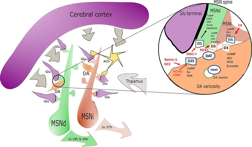
FIGURE 1 | Simplified sketch of the dopamine receptors (DR) connectome in the basal ganglia/striatum with a zoom (right circle) on signal transduction at presynaptic
level in medium spiny neurons (MSN) dendritic boutons. Highlights on the elements associated with SCZ alterations are depicted in red. D1 positive medium spiny neurons
of the direct pathway (MSNd) are in green, inhibitory D2 positive MSN of the indirect pathway (MSNi) are in red. Glutamatergic cortical input - presynaptic terminals are in
magenta. DA “en passant” boutons are indicated in orange and in close proximity of glutamatergic postsynaptic spines. Cholinergic interneurons are in yellow. In the
magnification on the right note the distribution of DR: D2s and D3 are presynaptic in DA terminals; D1/D3 postsynaptic in MSNd and D2L postsynaptic in MSNi. Other
projections are in gray. Abbreviations: ACh, acetylcholine; DA, dopamine; Glu, glutamate; MSNd/i, direct/ indirect path projecting MSN neurons; GPi, internal segment of
globus pallidus; SNr, substantia nigra, reticular part; STh, subthalamic nucleus; other common abbreviation and protein names as cited in text.
Frontiers in Pharmacology | www.frontiersin.org 5 July 2020 | Volume 11 | Article 1003Martel and Gatti McArthur Dopamine Receptors in Schizophrenia
posttranscriptional modification of D2 and D3 receptors is the axon guidance cues, which induce molecular changes in the axonal
N-linked glycosylation that classically affects both correct cell growth cones in response to extracellular levels of DA (via D1 in
surface expression and signaling/internalization (caveolin - complex). The DCC gene keeps being a confirmed SCZ genetic link
chlatrin mediated) (Min et al., 2015). D1 and D2 are localized across several studies (Vosberg et al., 2020) with a particular effect
to different endocytic vesicles after internalization. D1 is recycled on the anatomical connectivity of the nigra/VTA dopaminergic
back to the cell surface in a process controlled by the VPS35 pathways and the final distribution and relative density of DR. In
complex (Wang et al., 2016), while prolonged agonist animal models, SCZ-like symptoms seem to correlate with netrin 1
stimulation causes D2 trafficking into lysosomes and - DCC related alterations in size, complexity and density of DA
subsequent receptor degradation by a Rab5 GTPase controlled spines (medial PFCx layer V pyramidal neurons). Other genetic
pathway (Shioda et al., 2017; Shioda, 2017). A specific SCZ links (for example RGS12) concur on DA synthesis and
presynaptic control on D2S membrane density is exerted by release (Gross et al., 2018; Kos et al., 2018). A common upstream
the L1 close homolog adhesion factor (also a risk gene for SCZ) element affecting the expression of D2, COMT and structural
(Kotarska et al., 2020). Presynaptic D2S receptor density is proteins at presynaptic DA level is the zinc finger element
directly or indirectly affected by ALK and possible ZFN804A (Girgenti et al., 2012), coded by another SCZ risk gene
transactivation mechanisms (He and Lasek, 2020). The overall (Zhou et al., 2020).
complexity of the control of D2 receptor internalization (vs D3)
is possibly justified by the major biological role of D2 surface
density adjustments, required in different circuits depending on SECTION 2: DR ALTERATIONS IN
DA content. A specific example is the D2 vs D3 relative control SCHIZOPHRENIA
by Dysbindin 1 (Leggio et al., 2019). Dysbindin (SCZ risk gene
associated with cognitive symptoms) is mainly expressed in The current understanding of the role of DR in SCZ is in full
hippocampus and dorsolateral (DL) PFCx. It is a component expansion, thanks to developmental brain studies and the
of the multi-subunit complex BLOC-1 where it interacts directly advancements of imaging techniques. DR expression is
with MUTED (also probably associated with SCZ). Both segregated across neuronal populations and associated with
dysbindin and MUTED siRNAs increase cell surface D2 temporal and coupling differences in activation properties. This
receptors and block DA-induced D2 internalization in human distribution is respected in SCZ, while a variety of DR b-arrestin
and rat cells. Dysbindin variants are known to modify the mediated intracellular signaling show clear alterations in SCZ
cognitive response to antipsychotics. This effect is most likely disease models. Some developmental and connectivity aspects of
related to the parallel Dys1/D3 signal reduction that favors a D2 DR distribution are maintained across species and useful for the
component in cortical regions (Leggio et al., 2019). definition of SCZ as a developmental disease across circuits
Other types of control on DR density are exerted at source at (Sonnenschein and Grace, 2020).
the transcriptional level. A recent analysis of proteasome
alterations in SCZ points at spliceosome nuclear protein and Prefrontal Cortex Neurocircuit(s) Affected
calmodulin related pathways. The control on the splice variants by SCZ and DR
of the D2 receptor is exerted by NOVA1 and HNRP (Min et al., Connectivity measures across different SCZ studies are not
2015), and D2 mRNA 3´UTR binding of microRNAs mir-9 and always easy to compare, but some key elements are constant
mir 328 inhibits messenger translation (Shi et al., 2014). across patient groups, detection modalities and data
Development mechanisms are directly impacting on DR interpretation: the involvement of striatal-thalamic and PFCx
expression. In particular DISC-1 can translocate with KLF16 into connections in SCZ (Zhao et al., 2020). Imaging, functional and
the nucleus and recruit SIN3A corepressor to the D1 locus (Suh circadian studies are also in general agreement on the presence of
et al., 2019). The DISC-1 related complex is a main hub that could main alterations in the PFCx of SCZ patients, in particular dorso-
bring more specific information on SCZ developmental aspects in lateral and cingulate regions (Seney et al., 2019). PFCx circuits
terms of consecutive development related alterations in are central to cognitive functions and linked to the different
glutamatergic (NMDA/AMPA) and dopaminergic responses (D1 aspects of cognitive deficits and positive symptoms as observed
+D2+D3) in key SCZ regions like dorso-lateral PFCx and the in SCZ. Dorso-lateral PFCx weaker processing of sensory
striatum (Onishi et al., 2018; Jacobi et al., 2019). The expression information from thalamus is in fact associated with
control can also be exerted more dynamically on the D1 hallucination experiences which are common in > 50% of the
intracellular signal transducers by nuclear receptors like Nr4A1 SCZ patients (Daskalakis et al., 2020). The molecular studies
(Nurr77) (Cirnaru et al., 2019). Another nuclear factor involved in point at parvalbumin positive (PV+) GABAergic interneurons
shaping dopaminergic terminals is Nurr1, highly relevant for the and cortical pyramidal cells networks as both altered in SCZ
D2 receptor network and its circadian cycling (Chung et al., 2014; PFCx and across species in SCZ models (Chung et al., 2018;
Torretta et al., 2020). See Table 1 supplementary material for a Petralia et al., 2020; Wang et al., 2020; Weidenauer et al., 2020).
summary. Until puberty, the DA system maturation is controlled Dopaminergic ascending terminals reaching these neurons are
by the netrin-receptor DCC mediated organization of DA neurons also hypofunctional (Rao et al., 2019). Dopamine release enables
in the meso-cortical limbic system and the projections to PFCx the PFCx to compute and generate spatio-temporally diverse and
(Vosberg et al., 2020). Axon navigation is directed by extracellular specialized outputs, but these are not a linear function of the DA
Frontiers in Pharmacology | www.frontiersin.org 6 July 2020 | Volume 11 | Article 1003Martel and Gatti McArthur Dopamine Receptors in Schizophrenia
release input. Thus, it is quite complex to establish the functional schizophrenia, with possibly few exceptions (amisulpride or 5-
correlates for cortical functions. Rapid, transient changes in DA HT1A partial agonists) (Park et al., 2019; Huang et al., 2020).
transmission in PFCx are observed in response to task events, D3 are expressed by a distinct population of prefrontal
such as cues and rewards whereas prolonged responses are neurons and they also represent the main auto-receptor
relevant to emotional states and motivation (Lohani et al., controlling DA release in prefrontal cortex. D3 expression
2019). DA neurons in the region are mainly coming from the defines an additional class of L5 pyramidal cells that largely
VTA and the terminal density in PFCx is much lower (in terms lack D1 or D2 coexpression. L5 D3-expressing neurons are
of DAT content) when compared to the striatal regions. similar to D1-expressing cells in their synaptic connectivity,
D1 receptors are enriched in pyramidal cells in both layers 5 with projections to contralateral cortex. D3-expressing neurons
(thin-tufted layer) and 6 projecting in turn to contralateral could be distinguished from D1- or D2-expressing neurons by
cortex, striatum, and claustrum. D1 receptors are also present dendritic morphology, intrinsic electro-physiological properties
in interneurons and enriched in a specific population of VIP+ and by the manner in which DA regulates neuronal function. In
calretinin positive interneurons (Anastasiades et al., 2019; Saffari these neurons in fact D3 selectively regulates the dynamics of
et al., 2019). D1 receptors strongly enhance action potential voltage-gated calcium channels localized to the site of action
firing in this subset of cortico-cortical neurons and VIP+ potential initiation in the axon initial segment, with a marked
interneurons and the modulation via D1 receptors can suppression in the generation of high-frequency action potential
influence both excitatory and disinhibitory microcircuits in the bursts. D3 regulates CaV3.2 channels through a non-canonical,
PFCx (Anastasiades et al., 2019). This PV+ interneuron circuits arrestin-dependent pathway. The D3 plays therefore a unique
are a the main point of interaction between mGlu5/NMDA and role in the regulation of pyramidal cell excitability (Clarkson
D1 (D2-like) receptors, both involved in the control of the et al., 2017). The D3 receptor function has received attention
glutamatergic input from pyramidal cells (Nicoletti et al., because it could be a discriminant of the clinical effect of different
2019). D1 is important for the correct migration of the antipsychotics (Girgis et al., 2020) and because of the potential to
dopaminergic terminals which increase throughout adolescence address SCZ negative symptoms. In fact, D3 are associated to a
across species. Developmental studies in netrin-1 receptor DCC cortical circuit important for all the different SCZ symptoms. The
deficient mice demonstrate a role for DA in adolescent brain D3 controlled PFCx projections to hippocampus are interesting
axon growth. DCC controls in fact the extent of this protracted in this sense (Provenzano et al., 2020). The recent paper from
growth by determining where and when DA acts. Pyramidal Meier et al. shows the effect of a preferential D3 partial agonist
neuron morphology studies and cognitive performances show Cariprazine on gamma oscillations in hippocampal slides further
that the lack of DCC causes dopaminergic deficit across PFCx supporting the general assumption that gamma waves could
and morphological changes in pyramidal neurons (Reynolds predict psychosis and in vitro NMDA hypofunction, and that D3
et al., 2018). This process can be influenced by stress. The DA functional reduction can stabilize the alterations of the signal
deficit in PFCx regions following this hypothesis may be then of caused by NMDA hypofunction (Meier et al., 2020). Treatment
developmental origin and caused by morphological alterations response to antipsychotics may be predicted looking at the effect
affecting DA terminals, pyramidal cells and interneurons. on hippocampal- cortical connections and again these changes
D2/3 receptors are also differentially expressed in PFCx and could be in part D3 related (Guma et al., 2019; Blessing et al.,
their activation contribute to specific cognitive processes 2020). The observed hippocampal alterations in some SCZ
(Robinson and Sohal, 2017; Bailey et al., 2020; Papenberg et al., patients (psychotic) also support the presence of hippocampal
2020). D2 are enriched within subcortically projecting L5 immaturity at least in a subgroup of SCZ patients (Alvarez et al.,
pyramidal neurons thick-tufted pyramidal cells, with 2020; Cachia et al., 2020). There is therefore a renewed interest
projections to thalamus and pons, but not contralateral for the hippocampal models in SCZ, because it is possible to
cortex (Yu et al., 2019). These neurons exhibit a prominent study developmental changes which are closer to those observed
hyperpolarization-activated cationic current. In this population, in man and because it is easier to obtain NMDA receptor
pharmacological activation of D2 elicits a profound after hypofunction (Alvarez et al., 2020). In a mouse model of
depolarization that only occurs when NMDA receptors are postnatal NMDA hypofunction (NR1a KO) the effect seems to
coactivated. D2 signal in this case is triggering a Gs- cAMP/ be selectively associated with PV+ interneurons (in cortex and
PKA pathway in a non-canonic manner (Robinson and Sohal, hippocampus among other areas). In this animal model both
2017). D2 are also expressed in PV+ interneurons, a property cortical hypo- and striatal hyperdopaminergic phenotypes can be
acquired during adolescent brain maturation (Urs et al., 2016). observed (Nakao et al., 2019). The reason(s) behind these
The D2 network controls the connection to the hippocampal extensive dopaminergic changes across areas are still not fully
system (Tomasella et al., 2018; Khlghatyan et al., 2019). Species understood, but SCZ genetic data related to ancillary proteins for
related differences in this circuitry could be large, so human data the NMDA receptor function also support this hypothesis. Very
are needed for the correct interpretation of the results (Gonzalez- recent work has also given renewed attention to circuit(s)
Burgos et al., 2019). The cortical D2 mediated effects of the most involving PFCx areas like DL or the orbitofrontal (and
common antipsychotics (antagonists and partial agonists) have cerebellum) in relation to some aspects of negative symptoms
been extensively evaluated. This is mostly because these agents in SCZ (Walton et al., 2018; Brady et al., 2019). It is possibly too
cannot rescue the cognitive impairment associated with early to include a conclusive map of DR expression in within
Frontiers in Pharmacology | www.frontiersin.org 7 July 2020 | Volume 11 | Article 1003Martel and Gatti McArthur Dopamine Receptors in Schizophrenia
these pathways. The DISC-1 developmental mouse model could forebrain differentiation and circuit formation (Brignani and
however help to analyze these circuit(s), considering the main Pasterkamp, 2017), but DA tone also has clear effects on
impairment observed in sociability measures (Sultana and Lee, glutamatergic spine density at adult stage. It is however not
2020). The PV+ interneurons can also be a starting point to clear how SCZ specific NMDA alterations could impact on the
address the network in terms of developmental changes. Recent system. The recent and seminal work of the group of Prof. Groc,
DISC-1 studies report a reduction of spontaneous inhibitory using single molecule-based imaging shows that NMDA
transmission onto L2/3 PV+ interneurons in medial PFCx and a antibodies present in some SCZ patients with psychotic
decreased feed forward inhibition onto L2/3 pyramidal neurons symptoms are specifically changing the surface dynamics and
(Delevich et al., 2020). nanoscale organization of synaptic NMDA and its anchoring
partner the EphrinB2 receptor in synaptic spines in hippocampal
Striatal Circuits Alteration(s) in SCZ and DR neurons, ultimately preventing LTP potentiation (Jezequel et al.,
The main role of the striatum is the integration of cortical and 2017; Jezequel et al., 2018). As expected this causes a small
thalamic glutamatergic projections (Hunnicutt et al., 2016; reduction of the D1 surface expression in the same cellular
McCutcheon et al., 2019). The striatum is at the center of a system (Grea et al., 2019). The associated intracellular DA
DA-sensitive basal ganglia circuit associated with psychosis, SCZ signaling effects however could be more deeply modified
related motor dysfunctions and reward deficits. A summary of all because of this lack of NMDA/D1 interaction. It would be
the direct and indirect evidences of striatal DA alterations in SCZ equally important to study these NMDA-antibody related
was recently published (McCutcheon et al., 2020). All data changes in the context of the striatal circuits in particular on
confirm the presence of presynaptic DA sensitization and MSN D1 mediated signal and during development. The D1
elevated DA synthesis and release capacity (Brugger et al., receptor in dorsal striatum has been also involved in the
2020; Weidenauer et al., 2020). Higher striatal DA synthesis sensorimotor gating alterations observed in SCZ but these
and higher DA release correlated with worsening of psychotic mechanisms needs to be verified in man and with selective
symptoms in SCZ patients and were also supported by agents given the main differences in anatomical connectivity
neuromelanin observation (Weinstein et al., 2017). Excess (Aguilar et al., 2018).
striatal DA in SCZ is not related to changes in DA innervation
(Wengler et al., 2020). There have been extensive efforts to Striatal D2/D3 Receptors and SCZ
describe the neuroanatomy of striatum, and the cellular There are main differences in the DA input across the different
distribution of DR (Soares-Cunha et al., 2016; Clarkson et al., striatal regions. This is particularly true for the D2 receptor
2017). Substantia nigra DA projections mainly reach the dorsal function across dorsal striatum and nAcc. Increased DA D2
striatum (Uchigashima et al., 2016) while ventral tegmental area sensitivity in the nAcc is related to differences in coupling to Go
(VTA) projections from the mesencephalon reach the ventral vs. Gi (Marcott et al., 2018). The striatal D2 related control on
striatum (nAcc). Striatal neurons that receive DA inputs are reward is a key aspect of the effects of antipsychotics. Psychotic
mainly GABAergic medium spiny neurons (MSN). MSN symptoms have been in fact linked to salience changes in the
neurons are the recipients of both DA and glutamatergic (from reward system circuit and blocking D2 controls psychotic
PFCx and thalamus) projections, they represent therefore a core symptoms including a normalization on reward disturbances
neuronal element for both DA and NMDA hypothesis in SCZ. (Han et al., 2020). A direct relationship between D2 receptor
The MSN projecting to the internal segment of globus pallidus/ blockade, normalization of reward processing and symptom
nigra pars reticulata express D1 receptors, while those projecting improvement was recently further supported by a small study
to the external segment of globus pallidus are essentially in antipsychotic-naive first-episode SCZ patients (Wulff et al.,
expressing the D2 receptors. The two types of neurons are 2020). Cognitive flexibility (reversal learning) is another aspect of
finely intermingled across the whole striatum (Ren et al., D1/D2 related deficits that is linked to DA striatal functional
2017). There is also a not so small population of MSN that regional differences (Sala-Bayo et al., 2020). The cellular basis of
express both D1 and D2 receptors. They are usually described as the role of striatal D1 vs. D2 in reward and learning have been
enkephalin receptor positive neurons, they express specifically further clarified by the work of Iino et al., 2020, showing in
the subunit GluA3 of the AMPA receptor and project broadly to rodents the presence of a D2 controlled spine plasticity in MSN,
nuclei containing DA neurons cell bodies, to the nAcc and the that can be reversed with a D2 antagonist (Iino et al., 2020).
ento-peduncular nucleus among others (Perreault et al., 2011). D2 antagonism is still recognized as a main stay of SCZ
The cross talk of interneurons at this level is a main filter on the therapy and the D2 receptor is considered to be directly or
cortical input. Clearly, different DR contribute to the final effect, indirectly responsible for the efficacy of the majority of typical
depending on receptor distribution across different types of and atypical antipsychotics. This is coherent with the general
interneurons (Burke et al., 2018). For example the D1 activity observation of a main role of DA control of cortico-striatal
in MSN is inhibited by the cholinergic tonus (M4 mediated) synchronization of D2-MSN neurons (via D2-GPRIN AKT)
(Nair et al., 2019). In SCZ increased spine density have been (Karadurmus et al., 2019). The tetra complex A2A-D2
observed in dorsal striatum MSN. Converging evidences suggest receptors (plus AC5) is really central to multiple effects of both
a critical role of the dopaminergic system in adapting synaptic adenosine and DR ligands in the striatal region (Ferre et al., 2018;
plasticity of glutamatergic inputs (synaptic spines). Early in Bonifazi et al., 2019). mGlu5 receptor can be also included in a
development, the DA system has fundamental roles in complex interaction with D2-A2A in GABAergic neuronal
Frontiers in Pharmacology | www.frontiersin.org 8 July 2020 | Volume 11 | Article 1003Martel and Gatti McArthur Dopamine Receptors in Schizophrenia
terminals providing a multiple way to increase GABA release mechanisms, as observed with clozapine, the prototypical
(Borroto-Escuela et al., 2016; Sahlholm et al., 2018). It is atypical antipsychotic, to improve treatment compliance
becoming therefore apparent that D2 receptor function is (Aringhieri et al., 2018). Historical perspectives on SCZ drugs
heterogeneous and possibly strictly dependent on the neuronal generally highlight the DA receptor D2 antagonism as main
type expressing the receptor in different cortical and sub-cortical mechanism of action (Madras, 2013), but the pharmacology of
regions. Considering the role of D2 receptor in the control of antipsychotics is much more complex and requires a specific
emotional, cognitive and sensory functions alterations in SCZ it discussion on DR selectivity and serotonin receptor poly-
is therefore important to revisit the molecular aspects of this pharmacology (Butini et al., 2016; Aringhieri et al., 2018;
receptor and possibly even the pharmacology of the different Moritz et al., 2018; Bueschbell et al., 2019). Important
antipsychotics (Quintana and Beaulieu, 2019). For instance the discoveries were made in the DA field during the past decade,
D1/D2 complex (possibly) present in some MSN exhibits the in particular in relation to the pharmacology of DR ligands. DR
remarkable property of a coupling to a Gq- PLC mediated heterodimers have been described in different brain regions and
increase in intracellular calcium release and CAMKII used to explain the complex biological effects associated with
phosphorylation (Perreault et al., 2011). This complex may DR activation (Borroto-Escuela et al., 2018). Exciting data from
represent an interesting new pharmacological target in SCZ. crystallographic studies have supported a wave of drug
The D2S receptor is involved whenever SCZ treatment resistance discovery projects looking for new antipsychotics (Chien
is discussed or phenomena of presynaptic D2 receptor supra- et al., 2010; Wang et al., 2017; Wang et al., 2018). DR
sensitivity induced by antipsychotics (Amato et al., 2019). signaling versatility is further magnified by context dependent
Motivational deficits in SCZ are most likely associated with dissecting signatures or “bias” (Urs et al., 2017) extending the
cortico-striatal circuits involving the VTA, and the ventral potential for optimized pharmacological interventions. It is
striatum (Aberg et al., 2020; Kontaris et al., 2020). Clinical possible for instance to separate b-arrestin mediated signals
observation keep suggesting some involvement of ventral using biased D1 agonists (Urs et al., 2011; Gray et al., 2018).
striatum in the control of motivation, emotions and social Several recent contributions are available on this matter (Vyas
behavior as relevant for negative symptoms in SCZ with et al., 2020). The potential therapeutic applications of biased D2
regular debates on the matter (Fareri et al., 2017; Stepien et al., ligands to new SCZ therapies, has fuelled new interest on D2S
2018; Waltz et al., 2018). Interestingly, D3 receptor expression is vs. D2L or cAMP independent intracellular pathways, looking
enriched in midbrain ventral striatum (including nAcc) (Slifstein for agents with less motor side effects. D2 b-arrestin-biased
et al., 2020) where the receptor is present on pre- and ligands are now available (Park et al., 2016) and they may
postsynaptic locations and can also work in cooperation with provide some pharmacological advantages, at least on the basis
the receptor D1 (in MSN - AKT signal) (Castrellon et al., 2019; of the results in preclinical models (Urs et al., 2017). These
Guitart et al., 2019). The D3 receptor has been linked to control agents are not per se D2 selective since they also interact with
of DA firing in VTA, emotion and reward control in animal the D3 receptor and might require the presence of an
models (Takeuchi et al., 2019), but the lack of selective D3 heteromeric complex with the receptor A2a for the final effect.
ligands has so far hampered specific research on the subject There is therefore a need for a different look at DR ligands
(Correll and Schooler, 2020). Cholinergic interneurons in the pharmacology in vitro. We should possibly reconsider aspects
ventral striatum, particularly those in the insula major of Calleja like receptor internalization or intracellular recycling also for
are highly enriched in D3 receptor, making these cells extremely the main active metabolites or when comparing antagonists and
sensitive to DA from VTA projections. Also in this case a D1/D3 partial agonists (De Vries et al., 2019). See Table 2
complex is probably present. In this region as well as in Supplementary Material for chemical series of DR ligands
cerebellum or other extra-striatal circuits, the D3 receptor has and representative compounds described in section 3.
been linked to thermoregulation and sleep/wakefulness, which
are potentially relevant for the control of some aspects of SCZ DR Ligand Receptor Interactions
(Luo et al., 2018). Calleja islands are also a site related to adult The most interesting finding in the field of DR is certainly the
neurogenesis in ventral striatum across species: these neurons are crystal structure of D2, D3. and D4 receptors and how this was
D3, Erb4 and neuroregulin1 positive. used to identify new series or new mechanisms of ligand
receptor interaction. Homology models are also extremely
helpful for D1 and D5 with some main limitation for specific
SECTION 3. DR LIGANDS AND SCZ domains with reduced identity (Bueschbell et al., 2019). The DA
THERAPIES. THE NEW WAVE OF binding site is contained in a membrane pocket formed by the
LIGANDS WITH POTENTIAL RELEVANCE TM3/5/6/7 with similarities across biogenic amines GPCRs.
FOR THERAPY OR BRAIN IMAGING Molecular docking studies for the D1 receptor were able to
demonstrate the presence of allosteric sites that were further
The discovery that DA effective drugs for treating SCZ is targeted to obtain highly selective positive allosteric modulators
redeemable to the elegant work of Carlsson and Lindqvist in with high potency, weak agonist properties and able to increase
the early 60’s and to the identification, a decade later, of the DA response (cAMP) (Bruns et al., 2018). The mode of
antipsychotics/DA receptor. Atypical antipsychotics developed interaction of biased agonists is different since they fail to
in the 70’s and 80’s, included serotoninergic complementary trigger D1 receptor desensitization in vitro. The current model
Frontiers in Pharmacology | www.frontiersin.org 9 July 2020 | Volume 11 | Article 1003Martel and Gatti McArthur Dopamine Receptors in Schizophrenia supposes a docking in within the DA site, but with differences in allow a more precise approach to specific brain structures and interactions with TM3/5 and extracellular loop 2 (Gray et al., pathways (Cortes et al., 2016; Foster and Conn, 2017). 2018). The rapid advance of the pharmacology of D1 receptors bringing new drugs to the clinic is a clear demonstration of the DR-Ligand Interaction Dynamics and therapeutic impact of research on DR-ligand interactions (Hall Efficacy Studies et al., 2019). For D2/D3 biased ligands the drug design is There are classic aspects of receptor pharmacology like constitutive complicated by the needed poly-pharmacology vs. 5-HT1A or activity or equilibria across receptor conformations which are quite 5-HT2A receptors which contribute to the clinical efficacy and difficult to address with DR, in particular when considering also is intrinsic to some pharmacophore (Ma et al., 2019). The heterocomplexes. It should be however possible to better ligands cocrystallized in the different D2/D3 studies are distinguish antagonists from partial agonists and systematically haloperidol, risperidone, nemonapride and eticlopride, non- discuss on and off rates vs. affinity measures when presenting new selective but potent antagonists (Fan et al., 2020). Thus no DR ligands. Species specific differences are also seldom main difference was expected. In reality the results show acknowledged. This systematic pharmacological work is required differences in D2 inactive conformation that suggest different to make sense of the complex in vivo pharmacology of DR ligands receptor inactive states (Lane et al., 2020). In addition the (in particular D2/D3) also for antipsychotics already on the market. agonist binding pocket in the D2 allows an extension that has The case of D2 and D3 receptors is indeed quite interesting in this been used to study D2 > D3 and D4 selectivity (with agonist sense because of the complexity of the structure/activity database ligands) and to determine the possibility to obtain biased required to select new candidates and validate efficacy in agonists for D2 (Fan et al., 2020). The re-assessment of the comparison to reference antipsychotics. Several groups have D2 interaction profile of different classes of D2 antagonists is generated a variety of synthetic ligands concurring to build also on the way (Zieba et al., 2019). The case of D3 is similar molecular models including dynamic aspects of DR complementing this picture given the variety of new ligands receptor activation over time. In recombinant systems at least, currently available. Subtype-selective compounds have been we witness some amazing activity switches between agonist and sought for more than two decades with difficulties achieving “antagonist” properties across different series that require further sufficient selectivity and central exposure. Clinical PET data dynamic considerations (Tan et al., 2020). Destabilization of D3 have recently provided encouraging results with cariprazine and inactive state(s) and flexibility of the ligands are among the F17464 (Slifstein et al., 2020). More recent D3 over D2 new elements that the most recent model available is proposing ligands have been obtained exploiting the presence of a (Ferraro et al., 2020). Molecular recognition steps, changes in secondary allosteric D3 pocket to generate bitopic ligands hydration of the ligand binding pocket and ligand dependent with long molecular bridges. This strategy has allowed a receptor configuration changes are also important considerations powerful expansion in chemical possibilities even while for D2 and D3 in particular when docking flexible ligands and maintaining the capacity to generate agents with biased establishing comparisons (Pal et al., 2019). Native system activities (Rossi et al., 2017; Bonifazi et al., 2019). The concept pharmacology studies are due to confirm the relevance of the of bitopic ligands is associated with the presence of two observed in vitro differences. It would be indeed interesting to separated regions of the receptor with different vectors obtain a database of consistent functional information for all the relevant for the affinity and the allosteric pocket interaction ligands generated to further advance in the direction of new (usually driving D3/D2 selectivity considerations). Shorter D3 therapeutics. A re-evaluation of known DR ligands in the clinic ligands will necessarily reside instead only in within the on the basis of the latest available molecular model would be useful orthosteric pocket. Some interesting caged ligands for the D2/ to help DR drug developers to build a more integrated view on the D3 orthosteric p ocket could possibly help further efforts, the tools and the information available and needed to pharmacological studies on this subject in native systems move forward. (Gienger et al., 2020). There is a second interesting aspect in the pharmacology of D3 bitopic ligands. It has allowed to show the presence of an alternative mechanism of D3 receptor CONCLUSION internalization independent of b-arrestin and used by group II GPCR (Xu et al., 2019). Considering the excess D2 homodimers This article reviews current knowledge on DR subtypes in SCZ, detected in schizophrenia (Wang et al., 2010), the effects of DA anatomical distribution, and new pharmacological tools that can antagonists on these entities has been specifically explored using help dissect out subtype-specific functions. The aspects of DR bivalent ligands (Pulido et al., 2018; Wouters et al., 2019). A research described hereby are strictly related to SCZ or risk genes molecular model of the homodimer has been also generated for associated with it. What appears is that the current molecular D2 to provide docking information relative to bivalent ligands understanding of Glutamate NMDA - DA interactions in SCZ with different pharmacological properties (for example has improved, but it is still insufficient in particular in brain areas orthosteric and allosteric agents) (Kaczor et al., 2016). Other like the ventral striatum and in relation to negative symptoms. A DR heterodimers were also considered as selective targets for better understanding of the circuit(s) will possibly further reduce this type of ligands (Carli et al., 2018), mainly because the boundaries between cognitive and negative SCZ symptoms differential expression of these dimeric receptor entities may domains (Robison et al., 2020). The DA - NMDA research is Frontiers in Pharmacology | www.frontiersin.org 10 July 2020 | Volume 11 | Article 1003
You can also read


