Distinct neural networks for the volitional control of vocal and manual actions in the monkey homologue of Broca's area
←
→
Page content transcription
If your browser does not render page correctly, please read the page content below
RESEARCH ARTICLE

                                      Distinct neural networks for the volitional
                                      control of vocal and manual actions in the
                                      monkey homologue of Broca’s area
                                      Natalja Gavrilov, Andreas Nieder*

                                      Animal Physiology, Institute of Neurobiology, University of Tübingen, Tübingen,
                                      Germany

                                      Abstract The ventrolateral frontal lobe (Broca’s area) of the human brain is crucial in speech
                                      production. In macaques, neurons in the ventrolateral prefrontal cortex, the suggested monkey
                                      homologue of Broca’s area, signal the volitional initiation of vocalizations. We explored whether
                                      this brain area became specialized for vocal initiation during primate evolution and trained
                                      macaques to alternate between a vocal and manual action in response to arbitrary cues. During
                                      task performance, single neurons recorded from the ventrolateral prefrontal cortex and the
                                      rostroventral premotor cortex of the inferior frontal cortex predominantly signaled the impending
                                      vocal or, to a lesser extent, manual action, but not both. Neuronal activity was specific for volitional
                                      action plans and differed during spontaneous movement preparations. This implies that the
                                      primate inferior frontal cortex controls the initiation of volitional utterances via a dedicated
                                      network of vocal selective neurons that might have been exploited during the evolution of Broca’s
                                      area.

                                      Introduction
*For correspondence:                  The neural basis of cognitive vocal control underlying human speech and its evolutionary emergence
andreas.nieder@uni-tuebingen.de       in the primate lineage remain poorly understood. In humans, a key structure endowing volitional
                                      speech control is Broca’s area in the inferior frontal lobe. Broca’s area classically comprises areas 44
Competing interests: The
                                      and 45 in the ventrolateral prefrontal cortex (vlPFC). These areas are instrumental for producing
authors declare that no
                                      speech and language (Friederici and Chomsky, 2017; Penfield and Roberts, 1959); damage to
competing interests exist.
                                      these areas causes speech production aphasia. Recent evidence suggests that Broca’s area may not
Funding: See page 20                  directly regulate speech articulation but rather affect the cognitive preparation of speech
Received: 04 September 2020           (Dronkers and Baldo, 2010). For instance, cooling of Broca’s area in awake neurosurgical patients
Accepted: 27 January 2021             slows speech without affecting articulation (Long et al., 2016).
Published: 03 February 2021               Several lines of evidence suggest that the neural correlates of human speech could have evolved
                                      from a basic cognitive vocal control system already present in the nonhuman primate frontal lobe. In
Reviewing editor: Ingrid S
Johnsrude, University of Western
                                      macaques, area 44 is located deep in the inferior arcuate sulcus (ASi), whereas area 45 is part of the
Ontario, Canada                       ventral pre-arcuate region (VPA) of the vlPFC. Both areas have been identified as an anatomical
                                      homolog of Broca’s area in the human brain (Petrides, 2014; Petrides and Pandya, 1994;
   Copyright Gavrilov and
                                      Petrides and Pandya, 2002). Anatomically precise electrical stimulation in area 44 of the ASi of
Nieder. This article is distributed
                                      anesthetized monkeys elicits orofacial and laryngeal movements that are in the service of speech
under the terms of the Creative
Commons Attribution License,          production in humans (Petrides et al., 2005). Moreover, in macaques trained to vocalize on com-
which permits unrestricted use        mand, neurons in vlPFC respond specifically in preparation of volitional calls (Gavrilov et al., 2017;
and redistribution provided that      Hage and Nieder, 2013; Hage and Nieder, 2015). Importantly, vlPFC neurons show a strong corre-
the original author and source are    lation between the onset of neuronal activity and the timing of vocal output, suggesting that the
credited.                             vlPFC is forming a decision signal for initiating vocalizations (Gavrilov et al., 2017).

Gavrilov and Nieder. eLife 2021;10:e62797. DOI: https://doi.org/10.7554/eLife.62797                                                    1 of 24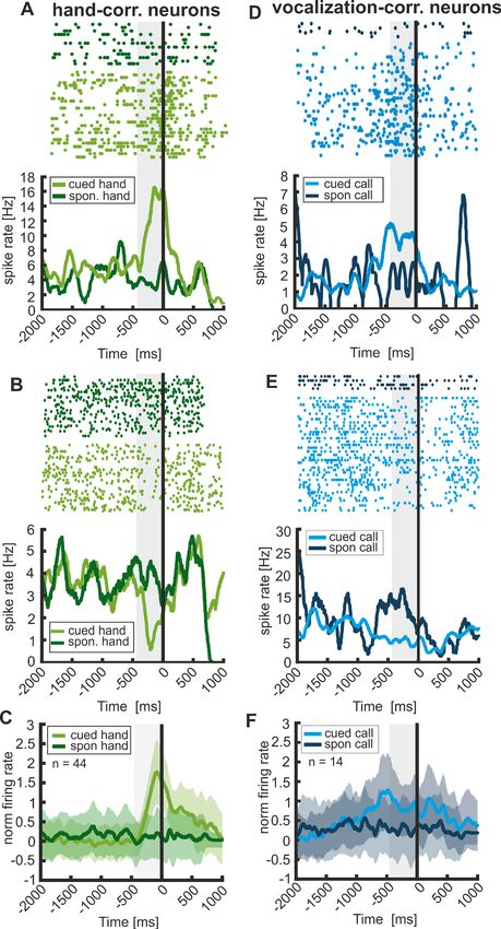
Research article Neuroscience
In order to demonstrate ‘volitional control’, three criteria have to be fulfilled in unison
(Brecht et al., 2019; Nieder and Mooney, 2020). First, responses need to be executed in conse-
quence of an arbitrary instruction stimulus that is neutral in its value or emotional valence. This crite-
rion is important to ensure that motor acts, and vocalizations in particular, are not elicited by
internal (affective/motivational/arousal) status changes in the presence of food or predators
(Fischer and Price, 2017; Jürgens, 1979). Second, responses need to be uttered in a manner that is
temporally contingent to the instruction stimulus. Third, actions need to be reliably withheld in the
absence of an instructive stimulus. In neuropsychological tests, this list of criteria is similarly applied
to differentiate between volitional and affective/spontaneous responses in patients (Cattaneo and
Pavesi, 2014; Hopf et al., 1992). For example, patients with facial paralysis due to damage of
descending pathways from the motor cortex have considerable difficulty smiling or frowning on
command, a condition called ‘voluntary facial paresis’; nevertheless, they smile or frown spontane-
ously in response to their emotional states. Similar dissociations have been observed for vocaliza-
tions: some patients with neurological insults may lose volitional control of their speech, but can still
laugh, scream, or groan when they are happy, frightened, or in pain. To determine the neuronal
basis of different types of motor responses, we adopt a protocol that fulfils these criteria in the cur-
rent study in order to distinguish volitional from spontaneous responses.
Despite advances in our understanding of vocal control mechanisms in primates, it is currently not
known if vocalization-correlated neurons are part of a dedicated vocal network that specifically enco-
des the volitional preparation of vocalizations. After all, the primate PFC is regarded as the central
executive of the brain (Miller and Cohen, 2001) and hosts a variety of cognitive functions necessary
for motor planning and goal-directed action control (Tanji et al., 2007). One hypothesis therefore is
that vlPFC neurons encode the preparation of various volitional actions, irrespective of the effector
organs. In this case, such neurons would not only signal the initiation of vocalizations, but also of
other actions, such as the preparation of hand movements. The alternative hypothesis predicts that
neurons in the vlPFC show functional specialization for the volitional preparation of specific motor
acts; according to this scenario, neurons would be functionally segregated and encode the initiation
of only one type of motor act, for instance, of either vocalizations or hand movements. We tested
these hypotheses and recorded single neuron activity from the vlPFC (the ventral pre-arcuate region,
VPA, and in the fundus of the inferior arcuate sulcus, ASi) and the adjacent rostroventral premotor
cortex (PMrv, part of area 6) of rhesus macaques trained to either vocalize or make a hand move-
ment in response to arbitrary visual cues.
Results
Behavioral performance
We trained two rhesus monkeys in a computer controlled ‘go/nogo’ detection task. The monkeys
either had to vocalize (‘vocal’ trials) or to respond manually by releasing a bar (‘manual’ trials) in
response to the presentation of arbitrary visual stimuli to receive a reward (Figure 1). Two stimuli
per trial types were used: a red cross or a blue square both cued vocalizations; a yellow ring or
green square both prompted hand movements. Vocal trials and hand trials were presented in blocks
within each session. The blocks switched after 25 correctly performed ‘go’ trials. Even block numbers
comprised ‘vocal trials’, whereas odd block numbers consisted of ‘manual’ trials.
We collected data from 20 daily sessions from monkey A and 41 sessions for monkey P. On aver-
age, a session consisted of 4 blocks per condition (monkey A) or five blocks per condition (monkey
P), respectively. In each session, monkey A uttered 84.2 ± 30.9 calls and monkey P elicited
104.5 ± 27.1 calls. First, we analyzed the monkeys’ behavioral performance. To that aim, we used
signal detection theory. To compare the performances for both response types (‘vocal’ and ‘man-
ual’), we calculated ‘hit’ rates, ‘false alarm’ rates, and d’ values for each condition separately. The
production of cued vocalizations was more difficult for the monkeys than the production of cued
hand movements.
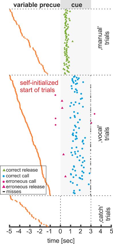
Figure 2 shows a representative session by monkey P comprising of 78 ‘grunt’ vocalizations and
84 ‘manual’ responses. In the ‘vocal’ block, the monkey uttered 73 calls in response to the ‘go’ cue.
However, the monkey also missed to vocalize in 108 ‘go’ trials which resulted in a ‘hit’ rate of 39.3%
(73 of 186 ‘go’ trials). Not a single call was uttered during ‘catch’ trials (trials without a ‘go’-cue) or
Gavrilov and Nieder. eLife 2021;10:e62797. DOI: https://doi.org/10.7554/eLife.62797 2 of 24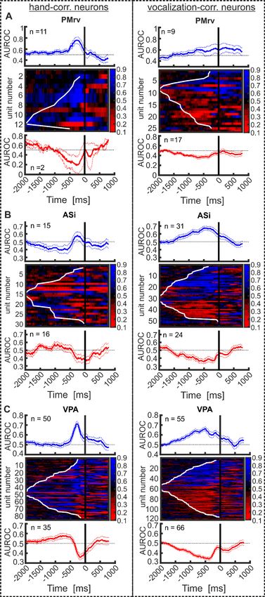
Research article                                                                                                               Neuroscience

                                   Figure 1. Experimental design. Two monkeys were trained in a ‘go/nogo’ detection task. In alternating trial
                                   blocks, the animals had either to vocalize in response to one of two arbitrary visual cues (blue square or red cross)
                                   (A) or to release a bar in response to two other visual stimuli (green square or yellow circle) (B).

                                   during ‘manual’ trials. The remaining five vocalizations were uttered during the wait periods (three
                                   calls; 3.8%) and after cue offset of ‘go’ trials (two calls; 2.6%). During the ‘hand’ block, the monkey
                                   produced a high proportion of hand releases in response to the corresponding ‘go’ stimuli. In con-
                                   trast to ‘vocal’ trials, the animal did not miss to respond in ‘manual’ trials, resulting in a ‘hit’ rate of
                                   100%. In four trials, the monkey erroneously responded with a bar release in ‘vocal’ trials, resulting
                                   in a ‘false alarm’ rate of 2.2% (4/179). For this session, the obtained ‘hit’ and ‘false alarm’ values led
                                   to a mean d’ sensitivity value of 3.1 and 5.4 in ‘vocal’ and ‘manual’ trials, respectively. This shows
                                   that the monkey produced each response type reliably and almost exclusively in response to the cor-
                                   responding visual ‘go’ stimuli.
                                       Throughout the sessions, both monkeys never missed to release the bar in response to the corre-
                                   sponding ‘go’ cue during ‘manual’ trials, which resulted in a ‘hit’ rate (HR) of 100% (Figure 3A,B). At
                                   the same time, the monkeys rarely erroneously released the bar in ‘catch’ and ‘vocal’ trials (‘false
                                   alarm’ (FA) of 12% and 5%, respectively). Because of the high ‘hit’ and low ‘false-alarm’ rates, the d’-
                                   sensitivity values for cued hand movements were well above the threshold of 1.5 during ‘manual’ tri-
                                   als (4.3 ± 0.7 monkey A; 5.3 ± 0.7 in monkey P) (Figure 3C,D).
                                       In ‘vocal’ trials, both monkeys missed to vocalize in response to the ‘go’ cue in about 60% of the
                                   trials (averaged hit rate: 38% monkey A; 55% monkey P). However, both animals almost never called

Gavrilov and Nieder. eLife 2021;10:e62797. DOI: https://doi.org/10.7554/eLife.62797                                                              3 of 24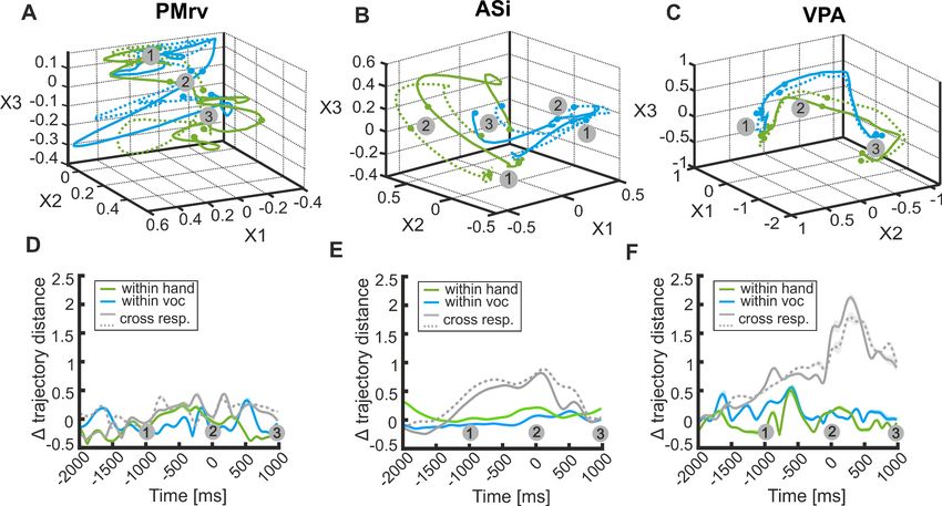
Research article                                                                                                        Neuroscience

                                                                                          in ‘catch’ trials or the ‘manual’ trials. Decent ‘hit’
                                                                                          rates and very low ‘false alarm’ rates in the ‘vocal’
                                                                                          trials cause above threshold discrimination sensi-
                                                                                          tivity (d’) performance on all recording sessions
                                                                                          (average d’-values of 2.4 ± 0.5 in monkey A, and
                                                                                          3.0 ± 0.6 in monkey P).
                                                                                              As a second behavioral parameter, we also
                                                                                          analyzed reaction time (Figure 3E,F). While the
                                                                                          monkeys responded on average within the first
                                                                                          500 ms after the ‘go’ cue onset in the ‘manual’
                                                                                          task (on average 0.51 s and 0.54 s for monkey A
                                                                                          and monkey P, respectively), they uttered cued
                                                                                          vocalizations only over a second later (on average
                                                                                          2.00 s for monkey A and 1.64 s for monkey P).
                                                                                          These relatively long call reaction times are com-
                                                                                          parable to our previous reports with two different
                                                                                          monkeys (Gavrilov et al., 2017; Hage and
                                                                                          Nieder, 2013).

                                                                                          Response type correlated neurons
                                                                                             While the animals performed the task, we
                                                                                             recorded 863 single neurons (286 neurons from
                                                                                             monkey A and 577 neurons from monkey P) in
                                                                                             two regions of the ventrolateral PFC (the ventral
                                                                                             pre-arcuate region, VPA, and inside the inferior
                                                                                             arcuate sulcus, ASi) and the adjacent rostroven-
                                                                                             tral premotor cortex (PMrv, part of area 6rv) (Fig-
                                                                                             ure 4). The main aim was to investigate whether
                                                                                             neurons in these frontal areas would differentiate
                                                                                             between cued initiation of vocalizations and hand
                                                                                             movements.
                                                                                                 We first cleared the data from putative con-
                                                                                             founds with eye-movement-related activity. Since
                                                                                             we measured the eye movements of the monkeys
                                   Figure 2. Example of a single session of monkey P.        that were not required to maintain fixation, neu-
                                   Responses in ‘vocal’, ‘manual’ and ‘catch’ trials are     rons showing eye-movement-related activity
                                   sorted according to the length of the ‘pre-cue’           could be identified (see Materials and methods
                                   signal. Each line represents a single trial; blue circles for details). We found that 37.3% of all recorded
                                   indicate vocal onsets and green triangles indicate        neurons (322/863) exhibited significant eye move-
                                   timing of the bar release. Pink circles and triangles
                                                                                             ment-related activity, and a further 4.2% of the
                                   indicate wrong responses. ‘go’ trials ignored by the
                                                                                             neurons (36/863) showed significant fixation-
                                   monkey (‘misses’) are marked with a horizontal black
                                   bar at trial end.                                         related activity. All these neurons were excluded
                                                                                             from further analyses.
                                                                                                 We then identified neurons that showed activ-
                                                                                             ity which was correlated with the instructed initia-
                                   tion of vocalizations and/or hand movements. To that aim, we used the neurons’ firing rates after
                                   ‘go’ cue presentation in a 450 ms interval immediately before the monkeys’ responses (vocalization
                                   or hand movement). This time window was adjusted to the monkeys’ short average reaction times
                                   during ‘manual’ trials (individual trials with response latencies shorter than 450 ms were discarded)
                                   and thus allowed a comparison of premotor activity for both ‘manual’ and ‘vocal’ trials. Neurons that
                                   significantly modulated their firing rate in this premotor time window prior to hand movements dur-
                                   ing ‘manual’ trials compared to baseline activity were defined as hand-correlated neurons (Wilcoxon
                                   signed rank test; p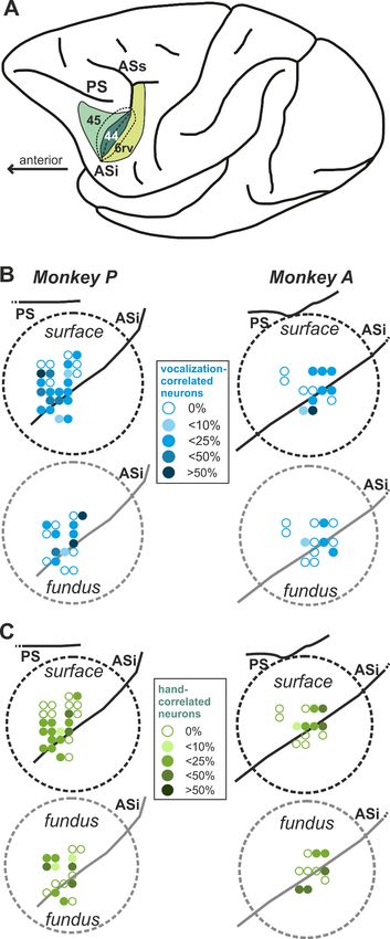
Research article                                                                                                               Neuroscience

                                   Figure 3. Behavioral performance. (A, B) Distribution of ‘hit’ and ‘false alarm’ rates for each response type and
                                   animal separately, averaged over 20 sessions for Monkey A and 41 for monkey P. (C, D) Sensitivity of signal
                                   detection for ‘vocal’ and ‘manual’ trials indicated by the d’ prime value. The dotted line indicates the detection
                                   threshold of 1.5. (E, F) Response probability of ‘vocal’ and ‘manual’ responses in the corresponding trial type.
                                   Normalized and averaged response over sessions are shown. Shaded areas indicate first and third quartiles.

                                   respective example neurons. In this figure, activity in both ‘vocal’ and ‘manual’ trials (only correct tri-
                                   als) is aligned relative to response onset (black vertical line in the histograms). Figure 5A,C,E depicts
                                   three vocalization-correlated neurons, one for each recording area (i.e. PMrv, ASi, and VPA), that
                                   show a significant increase in discharge rate prior to vocal onset. Interestingly, the firing rates of the
                                   same neurons hardly changed in ‘manual’ trials when the monkeys prepared a bar release (note that
                                   some neurons were selective to both vocalization and hand movements, as later discussed in Fig-
                                   ure 6). Overall, we found 20% (26/130), 24% (55/228) and 24% (121/505) vocalization-correlated
                                   neurons in PMrv, ASi, and VPA, respectively. Approximately one-third of vocalization-correlated neu-
                                   rons recorded in PMrv showed increased activity (9/26: 34.6%), whereas the remaining two-thirds of

Gavrilov and Nieder. eLife 2021;10:e62797. DOI: https://doi.org/10.7554/eLife.62797                                                              5 of 24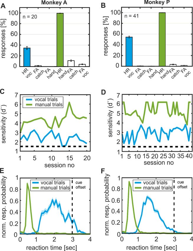
Research article                                                                                                                Neuroscience

                                   Figure 4. Recording sites in the inferior frontal lobe of both monkeys. (A) Lateral view of the left hemisphere
                                   indicating the recording area: parts of the rostroventral premotor cortex (PMrv of area 6), the fundus of the inferior
                                   arcuate sulcus (ASi) encompassing area 44, and the ventral pre-arcuate region containing parts of area 45. The
                                   inferior arcuate sulcus is unfolded, with dotted lines marking the transition from the cortical surface to the gyral
                                   walls. PS, principal sulcus; ASi, inferior arcuate sulcus; ASs, superior arcuate sulcus (B, C) Precise recording sites
                                   inside each recording chamber (dotted circles). Recording sites with a depth >6 mm were defined as ASi sites. For
                                   better overview, recording sites within the fundus of the inferior AS (area 44) are depicted offset on the right side
                                   of each chamber. The proportion of vocalization-correlated neurons (B) in relation to all neurons recorded at a
                                   Figure 4 continued on next page

Gavrilov and Nieder. eLife 2021;10:e62797. DOI: https://doi.org/10.7554/eLife.62797                                                               6 of 24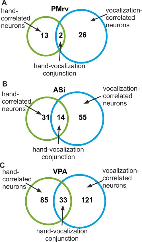
Research article                                                                                                               Neuroscience

                                   Figure 4 continued
                                   specific recording site is coded with different shades of blue. Similarly, the proportion of hand-correlated neurons
                                   (C) in relation to all neurons recorded at a specific recording site is coded with different shades of green color.

                                   vocalization-correlated neurons exhibited decreased activity (17/26: 65.4%) prior to vocal onset. In
                                   the two other areas, the proportion of excited and suppressed neurons was roughly the same. In
                                   ASi, we found 56% excited neurons (31/55) and 44% suppressive neurons (24/55). In VPA, we
                                   detected 45% excited neurons (55/121) and 55% suppressed neurons (66/121).
                                       A comparison of neuronal latencies of vocalization-correlated neurons revealed no significant dif-
                                   ferences between the three areas (median latency for PMrv: 1068 ms before vocal onset; median
                                   latency for ASi: 1099 ms; median latency for VPA: 871 ms; Kruskal-Wallis test, p=0.12).
                                       To summarize activity profiles from excited and suppressed neurons, the normalized activity of all
                                   suppressed neurons was rectified relative to the baseline activity so that negative deflections were
                                   transferred into positive deflections of equal magnitude. The average normalized discharge rates of
                                   all vocalization-correlated neurons in the respective recording areas are shown in Figure 5B,D,F.
                                   They show continuously rising activity toward call onset while hardly any activity change can be seen
                                   for the preparation of hand movements.
                                       We also detected neurons that signaled the preparation of hand movements. Three such neurons
                                   are depicted in Figure 5G,I,K. All three neurons show an increase in firing rates prior to hand
                                   responses while premotor activity remained unchanged in ‘vocal’ trials. In total, we found 10% (13/
                                   130), 13.6% (31/228), and 16.8% (85/505) hand-correlated neurons in PMrv, ASi, and VPA, respec-
                                   tively. The proportion of neurons that increased or decreased its activity prior to the bar release var-
                                   ied widely between the three areas, ranging from 51% of excited neurons in ASi, followed by 59% in
                                   VPA and 85% in PMrv. The averaged and normalized activities of all hand-correlated neurons
                                   (excited and suppressive) for the corresponding recording areas are shown in Figure 5H,J,L and
                                   show a relatively rapid increase in activity shortly before the cued hand movement, but not in prepa-
                                   ration of calls.
                                       Similar to vocalization-correlated neurons, a comparison of neuronal latencies of hand-correlated
                                   neurons revealed no significant difference between the areas (median latency for PMrv: 252 ms prior
                                   to response onset; median latency for ASi: 316 ms prior to response onset; median latency for VPA:
                                   257 ms prior to response onset; Kruskal-Wallis test p>0.66).
                                       The proportion of vocalization-correlated neurons in each area was significantly larger compared
                                   to the proportion of hand-correlated neurons (vocalization-correlated neurons overall (202/863) and
                                   hand-correlated neurons overall (129/863), p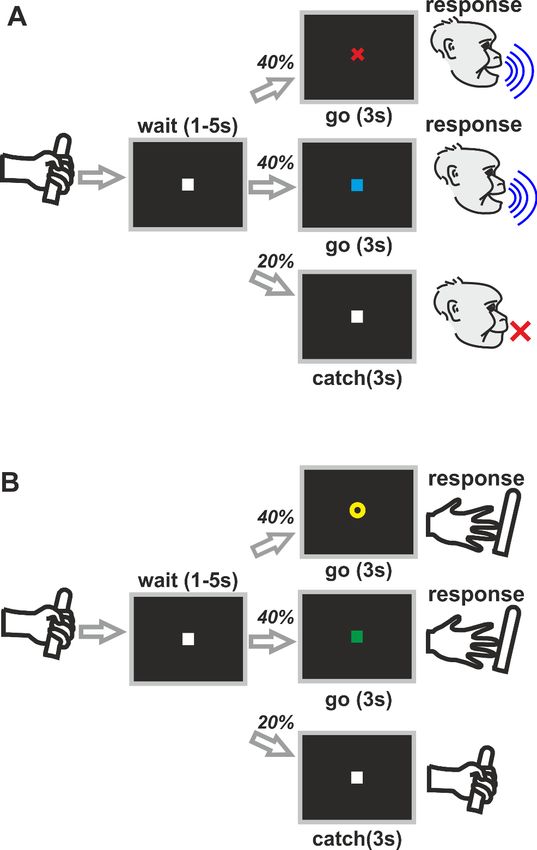
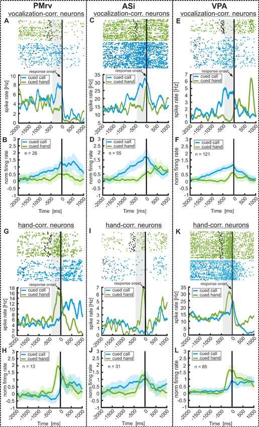
Research article                                                                                                                Neuroscience

Figure 5. Neuronal responses to the preparation of volitional vocalizations and hand movements in PMrv (left), ASi (middle), and VPA (right). Responses
of three example vocalization-correlated neurons and three hand-correlated neurons in ‘vocal’ and ‘manual’ trials as well as averaged and normalized
population responses for the corresponding examples. Responses are recorded in PMrv (left column), ASi (middle column), and VPA (right column). (A,
C,E) Three example vocalization-correlated neurons that show a significant increase of neuronal activity during 450 ms before the instructed vocalization
Figure 5 continued on next page

Gavrilov and Nieder. eLife 2021;10:e62797. DOI: https://doi.org/10.7554/eLife.62797                                                              8 of 24
Research article                                                                                                                   Neuroscience

Figure 5 continued
compared to baseline (450 ms before ‘go’ cue onset). At the same time, the neurons show only minor changes in activity preceding the response in
‘manual’ trials. Upper panel show the raster plot, black dots and asterisks indicate the ‘go’ cue onset in the ‘vocal’ and ‘manual’ trials, respectively. The
lower panel represent the corresponding spike density histogram averaged and smoothed with a Gaussian kernel (150 ms) for illustration. The vertical
black line indicates the response onset. Vocal responses have an average duration of 97 ± 40 ms. (B,D,F) Averaged and normalized activity of all
vocalization correlated neurons recorded in the corresponding area. These-neurons show a significantly different activity prior to cued ‘vocal’ responses
compared to cued ‘manual’ responses (activity tested in 450 ms before response onset; Mann Whitney U test: p
Research article                                                                                                            Neuroscience

                                   Figure 6. Proportions of hand- and vocalization-correlated neurons recorded in PMrv (A), ASi (B), and VPA (C). The
                                   Venn diagrams depict the number of hand-correlated neurons and vocalization-correlated neurons that encode
                                   only one type of the response (‘vocal’ or ‘manual’) or both response types at the same time.

                                   Neuronal population coding
                                   Furthermore, we explored whether and how the entire population of recorded neurons, irrespective
                                   of any response preferences, encoded the two different response types (‘manual’ and/ or ‘vocal’).
                                   Therefore, we performed a multi-dimensional state space analysis (Gaussian-Process Factor Analysis,
                                   GPFA) (Yu et al., 2009) on a population of pseudo-simultaneously recorded neurons for each brain
                                   area separately (PMrv: n = 36; ASi: n = 81; VPA: n = 180; for Details see Materials and methods).
                                   This approach extracts trajectories from the spiking activity of a neuronal population in individual tri-
                                   als. Such trajectories reflect the instantaneous firing rates of the respective neuronal population as
                                   they evolve over time.
                                       Figure 9A,B,C depict each two average population trajectories for ‘vocal’ and ‘manual’ trials in a
                                   space defined by the top three most meaningful dimensions in the respective brain areas. To evalu-
                                   ate the temporal evolution of population activity in each brain area and to different trial types, we
                                   measured Euclidian distances between trial trajectories corresponding to the same trial type (within-
                                   response coding) and different trial types (cross-response coding) (Figure 9D,E,F). For two of the

Gavrilov and Nieder. eLife 2021;10:e62797. DOI: https://doi.org/10.7554/eLife.62797                                                         10 of 24
Research article Neuroscience
Figure 7. Response type correlated activity of vocalization- and hand-correlated neurons preceding cued and
spontaneous responses. Left and right column represent the activity in ‘cued’ and ‘spontaneous’ trials of two
example hand- and vocalization-correlated neurons, respectively (A,B,D,E). Vocalization-correlated neurons show
significantly increased or decreased activity prior to instructed calls but not prior to spontaneously uttered
Figure 7 continued on next page
Gavrilov and Nieder. eLife 2021;10:e62797. DOI: https://doi.org/10.7554/eLife.62797 11 of 24Research article Neuroscience
Figure 7 continued
vocalizations. (C,F) Averaged and normalized population activity of 44 hand-correlated and 14 vocalization-
correlated neurons that were recorded during ‘cued’ and ‘spontaneous’ trials. Hand-correlated neurons show a
significantly different activity prior to cued ‘manual’ responses compared to ‘spontaneous manual’ responses
(activity tested in 450 ms before response onset; Wilcoxon signed rank test: pResearch article Neuroscience
Figure 8. Quality and temporal evolution of response correlated activity for all hand-correlated neurons (left
column) and all vocalization-correlated neurons (right column), recorded in PMrv (A), ASi (B), and VPA (C). Neurons
are sorted based in their activity profile (increased or decreased FR) in the pre-response period compared to
baseline and the latency of response type coding. Top: average AUROC values of all hand- (left) or vocalization-
Figure 8 continued on next page
Gavrilov and Nieder. eLife 2021;10:e62797. DOI: https://doi.org/10.7554/eLife.62797 13 of 24Research article Neuroscience
Figure 8 continued
correlated neurons (right) increasing their FR prior to ‘manual’ response. Middle: AUROC values of each individual
hand- or vocalization-correlated neuron from 2000 ms before to 1000 ms after response onset. The black line at 0
ms indicates the response onset. The white line marks each neuron’s latency of response type discrimination.
Bottom: average AUROC values of all hand- (left) or vocalization-correlated neurons (right) showing decreased
activity prior to response onset. Dashed lines represent the s.e.m.
2018). Thus, the capability to produce volitional vocalizations seems to be evolutionarily delayed in
primates relative to volitional hand movements.
The vlPFC involved in manual and vocal action planning
The primate vlPFC is known to be involved in the planning of different motor acts. On the one hand,
the vlPFC is involved during sensorimotor transformation required for hand actions and gestures
(Simone et al., 2015; Yamagata et al., 2012). Neurons in the vlPFC not only encode object proper-
ties (Rainer and Miller, 2000; Ramirez-Cardenas et al., 2016) and mnemonic information
(Funahashi et al., 1989; Rainer et al., 1999; Eiselt and Nieder, 2013), but also contribute to the
planning and control of hand actions across various contexts, such as during light vs. darkness, and
memory vs. visually-guided actions (Simone et al., 2015). This suggests that the vlPFC integrates
contextual information and generates the goal of the intended action during action planning and
execution. This information may subsequently be distributed to the ventral premotor cortex (PMv)
Figure 9. Temporal evolution of pre-response activity in the whole populations of neurons recorded in PMrv (left), ASi (middle), and VPA (right). (A–C)
Gaussian Process Factor Analysis delineating the state-space of neuronal population activity (PMrv: n = 36 neurons; ASi: n = 81 neurons; VPA: n = 180
neurons) over time for ‘manual’ (green) and ‘vocal’ trials (blue), respectively. Solid and dashed lines of the same color represent each one half of the
trials from the same condition. Each trajectory represents the average of 100 repetitions of GPFA performed on randomly selected 17 trials (VPA) or 15
trials ASi and PMrv for each condition and each neuron. Dots and numbers indicate time point 1000 ms before response onset (1), the response onset
(2) and time point 1000 ms after response onset (3). (D–F) Averaged inter-trajectory Euclidean distance over time, as a measure for within-response
coding (colored lines) and cross-response coding (gray lines). Shaded area around the curves represents the s.e.m.
Gavrilov and Nieder. eLife 2021;10:e62797. DOI: https://doi.org/10.7554/eLife.62797 14 of 24Research article Neuroscience
and inferior parietal lobule (IPL), brain areas involved in visuo-motor transformation of hand actions
(Bonini et al., 2010; Fogassi et al., 2005) to which the vlPFC is connected (Borra et al., 2011;
Gerbella et al., 2010), in order to successfully perform goal-directed hand actions via output to the
primary motor cortex (Battaglia-Mayer and Caminiti, 2019; Borra et al., 2017; Hoshi and Ishida,
2015). The increased dexterity of forelimb, hand, or finger voluntary actions necessary for gestures
also correlates with the expansion of direct cortico-motoneuronal connections along primate evolu-
tion (Lemon, 2008).
Besides hand movements, the vlPFC in nonhuman primates is also implicated in orofacial move-
ments and the planning of vocal output (Eliades and Miller, 2017; Nieder and Mooney, 2020).
Electrical stimulation of area 44 in the ASi, the suggested homologue of the cortex covering the
pars opercularis in Broca’s region in the human brain (Frey et al., 2014), elicits orofacial movements
in anaesthetized macaques (Petrides et al., 2005). The strongly interconnected areas 44 and 45
within the vlPFC contain neurons that selectively discharge when trained macaques prepare an
instructed vocalization (Gavrilov et al., 2017; Hage and Nieder, 2013; Hage and Nieder, 2015).
Moreover, area 44 is strongly connected with ventral premotor cortex (ventral area 6) in which neu-
rons exhibit responses related to mouth movements and vocalizations (Coudé et al., 2011;
Fornia et al., 2018; Shepherd and Freiwald, 2018), but also the rostral part of the inferior parietal
lobule in which neurons also exhibit responses related to mouth movements (Leinonen and Nyman,
1979). The primate lateral frontal cortex is connected with the posterior temporal region and the
adjacent inferior parietal lobule via three major white matter pathways that subserve auditory-vocal
processing: the arcuate fasciculus connects the posterior temporal region (referred to as Wernicke’s
area in humans and involved in the comprehension of language) with the inferior frontal gyrus (IFG)
(incl. Broca’s area in the language dominant hemisphere of humans). In addition, the third branch of
the superior longitudinal fasciculus (SLF III) links rostral inferior parietal lobule with areas 6 and 44,
whereas the second branch of the superior longitudinal fasciculus (SLF II) links area 45 with posterior
inferior parietal lobule (i.e. angular gyrus) (Barbeau et al., 2020; Petrides, 2014; Petrides and Pan-
dya, 1984). This anatomical segregation suggests distinct functional roles of these major pathways
in audio-vocal processing.
As a classical multi-modal association and executive brain area receiving not only visual but also
auditory information, the vlPFC is a well-situated hub for audio-vocal transformation
(Balezeau et al., 2020; Hage and Nieder, 2015; Hwang and Romanski, 2015), top-down cognitive
control (Bichot et al., 2015; Vallentin et al., 2012; Viswanathan and Nieder, 2015), and motor
learning (Petrides, 1982; Petrides, 2005). Further evolutionary innovations for vocal control include
elaborations of the laryngeal motor cortex (LMC; Simonyan, 2014) and projections from the LMC
directly to phonatory and respiratory motoneuron pools as the dominant brain system for human
speech and language (Hage and Nieder, 2016; Nieder and Mooney, 2020).
Segregated vlPFC networks for vocal and manual control
Based on single-neuron recordings in parallel to behavior, we can trace these differences between
these two types of volitional responses back to premotor activity in the inferior frontal lobe. The tar-
geted prefrontal areas ASi and VPA, as well as the premotor area PMrv, do not seem to encode the
general preparation of volitional acts irrespective of the effector organs. Rather, we found clear
specificity of neurons in relation to vocal and manual preparatory activity, particularly in ASi and VPA
of the vlPFC. This indicates segregated networks within the vlPFC that are part of a specialized vocal
and manual control network.
Because the production of volitional vocalizations was cognitively much more demanding for the
monkeys (as indicated by the reaction times and hit rates; see also Hage et al., 2016), the monkeys
were kept motivated in the’ vocal’ trials by providing more reward than in the ‘manual’ trials (in
which smaller and larger rewards were randomly assigned to counterbalance expectation). However,
reward expectancy is unlikely to have caused the observed coding difference: at the behavioral level,
the monkeys responded much faster in ‘manual’ trials in which the overall amount of reward was
smaller (the opposite effect would have been expected if the amount of reward determined the
reaction times). At the neuronal level, premotor activity in ‘vocal’ and ‘manual’ trials reached compa-
rable peak activity prior to response onset. While we cannot exclude that reward expectation might
have had a modulatory influence, differences in reward expectation cannot explain the observed
coding differences.
Gavrilov and Nieder. eLife 2021;10:e62797. DOI: https://doi.org/10.7554/eLife.62797 15 of 24Research article Neuroscience
Our finding of manual responses in the vlPFC concurs with a recent functional magnetic reso-
nance imaging (fMRI) study in macaques. In monkeys that were trained to perform either hand
grasping or ingestive mouth movements in the scanner, comparable moderate blood oxygen level
dependent (BOLD) responses during the respective hand and mouth movements in area 44 were
measured (Sharma et al., 2019). Our single neuron recordings suggest a more vivid involvement of
these regions during volitional and potentially communicative face movements, such as vocalizations
(Hage and Nieder, 2013) and lip smacking (Petrides et al., 2005; Shepherd and Freiwald, 2018).
The specialized network for vocal call initiation might have been exploited and adapted over the
course of primate evolution to serve as essential constituent of the human-specific speech and lan-
guage system.
A specialization of the vlPFC in selecting different types of motor acts was recently also reported
in the human brain based on fMRI. In the study by Loh et al., 2020, subjects were required to select
between competing motor acts: manual, orofacial, nonspeech vocal, and speech vocal actions. It
was found that area 44 reacted effector-dependent and was specifically involved in the selection
between competing orofacial acts, as well as nonspeech vocal and speech vocal acts, but not during
conditional manual responses. In contrast, area 45 was specifically recruited during the selection of
both nonspeech and speech vocal responses, but only during learning (Loh et al., 2020). Our study
in macaques confirms the role of area 44/45 in the cognitive control of vocal acts. However, in con-
trast to the study by Loh et al., 2020, we also found strong neuronal responses prior to hand actions
in the vlPFC, albeit based on a smaller proportion of hand-correlated neurons. This discrepancy may
be due to methodological differences between the two studies. In particular, the spatio-temporal
resolution of the BOLD signal is limited compared to single-cell recordings. Also, the BOLD signal is
less sensitive to detect a relatively small proportion of hand-correlated neuronal activity. Alterna-
tively, the human area 44/45 may exhibit more pronounced neuronal specializations supporting the
speech and language system, and a corresponding reduction of manual activity that is still lacking in
old-world monkeys.
Anatomically intermingled but functionally segregated action
preparation networks in relation to language evolution
Our results in the monkey vlPFC also speak to the proposed evolutionary origins of the human lan-
guage system. The two dominant hypotheses assume that the foundations for language evolution
lay either in the gestural (visual-manual) or the vocal (audio-vocal) domain (Kendon, 2017;
Fitch, 2010). The key question is whether one or the other of these hypotheses shows more cogni-
tive pre-adaptations required for the emergence of language. One of these necessary pre-adapta-
tions of a flexible communicative system is volitional control over motor acts (Ackermann et al.,
2014; Balter, 2010; Ghazanfar et al., 2019) as defined in the introduction. The monkeys’ hand
movements easily suffice these criteria for volition. In addition, the current and other recent behav-
ioral studies showed that monkeys can also produce vocalizations volitionally, albeit only with consid-
erable cognitive effort (Hage et al., 2013; Hage et al., 2016; Pomberger et al., 2019). At a
neuronal level, the current comparison of cued versus spontaneous vocalizations and hand move-
ments showed impressive and highly significant activity differences; neuronal activity in the frontal
lobe remains at baseline level for the monkeys’ spontaneous and non-goal directed vocalizations
and hand movements but is strongly modulated for volitional and goal-directed responses. This cat-
egorical distinction strongly argues that the neuronal coding of cued vocalizations is different from
the coding of spontaneous vocalizations.
Within the monkey vlPFC, we found a co-occurrence of neurons encoding the preparation of dif-
ferent types of volitional motor acts. While one group of neurons represented the planning of
instructed vocalizations, a second and largely separate group of neurons encoded the preparation
of instructed hand movements. Thus, anatomically intermingled but functionally segregated action
preparation networks co-exist in a restricted area of the vlPFC that is the proposed anatomical
homolog of Broca’s area in the human brain. Hence, clear neural links between the planning of vocal
and manual output exist in the nonhuman primate brain prior to the evolution of a human language
production system that can access both systems. This suggest a joint evolution of vocal production
and hand-action systems in close anatomical vicinity (Willems and Hagoort, 2007). The primordial
vocal production network, in particular, might have been expanded during primate evolution and
put to the service of the speech and language system (Anderson, 2010).
Gavrilov and Nieder. eLife 2021;10:e62797. DOI: https://doi.org/10.7554/eLife.62797 16 of 24Research article Neuroscience
Materials and methods
Subjects
Two juvenile male macaque monkeys (Macaca mulatta), each aged 6 years. and weighing 8.5 and
5.5 kg, respectively, were used for the study. The monkeys worked under a controlled water intake
protocol during the experiments and were rewarded with fluid for correct responses. All surgery pro-
cedures were accomplished under aseptic conditions under general anesthesia. All procedures have
been approved by the responsible national authorities (Regierungspräsidium Tübingen, Germany)
under permit ZP 1/15 and comply with German Law and the European Directive 2010/63/EU regulat-
ing use of animals in research.
Behavioral protocol and data acquisition
All recording sessions were carried out in a double-walled, soundproof chamber (IAC Acoustics, Nie-
derkrüchten, Germany). Single-cell recordings were conducted in monkeys trained to perform a
computer-controlled ‘go/nogo’ detection task (Figure 1). In alternating trial blocks, the monkey had
either to vocalize in response to one of two arbitrary visual cues (red cross or blue square for ‘vocal’
trials) or to release its hand from a bar in response to another set of visual cues (yellow ring or green
square for ‘manual’ trials). The monkey started a trial by grabbing a bar (’ready’-response). A visual
‘nogo’-signal (‘pre-cue’) appeared for a randomized time period of 1 to 5 s (white square, diameter:
0.5 deg of visual angle) during which the monkey was not allowed to vocalize or release the bar. In
80% of the trials, the ‘pre-cue’-signal was followed by a colored visual ’go’-signal (red cross or blue
square in the vocalization block; yellow ring or green square in the ‘manual’ block, all stimuli
appeared with equal probability of 40%; diameter: 0.5 deg of visual angle) lasting 3 s. During pre-
sentation of the’ go’-cue, the monkey had to emit a vocalization or release the bar to receive a liquid
reward, respectively. In the vocalization trials, both monkeys produced ‘grunt’ vocalizations. To con-
trol for random responses or timed responses independent from the ‘go’-cue, ‘catch’ trials display-
ing no ‘go’-cue appeared in the remaining 20% of the trials. In ‘catch’ trials, the ‘pre-cue’ remained
unchanged for 3 s and the monkey had to remain silent in ‘vocal’ trials (withhold vocal output), or to
keep holding the bar in ‘manual’ trials depending on the block (’catch’-trials).
Correct responses (‘vocal’ or ‘manual’, respectively) during ‘go’-signals were defined as ‘hits’;
calls or hand releases during ’catch’-trials counted as ’false alarms’ according to the ‘go/nogo’
detection paradigm. To ensure that monkeys were under stimulus control during each session, we
computed d’-sensitivity-values derived from signal detection theory (Nevin, 1969) by subtracting
z-scores (normal deviates) of median ’hit’-rates from z-scores of median ’false alarm’-rates for each
condition separately. Detection threshold for d’-values was set to 1.5 (Gavrilov et al., 2017;
Hage and Nieder, 2013).
Monkeys were head-fixed during the experiment in 57 cm in front of a computer screen and
maintained a constant distance of 5 cm between the monkey’s head and the microphone. Eye move-
ments were monitored via an IR-eye tracking system (ISCAN, Woburn, MA, USA), sampled at 1 kHz
and stored with the Plexon system for subsequent analysis.
Stimulus presentation and behavioral monitoring was automated on PCs running the CORTEX
program (NIH) and recorded by a multi-acquisition system (Plexon Inc, Dallas, TX). Vocalizations and
bar releases were recorded synchronously with the neuronal data. Vocalizations were sampled at 40
kHz via an A/D converter for post-hoc analysis. Both bar releases and vocalizations were detected
automatically. Vocalizations were detected in real-time by a MATLAB program (MathWorks, Natick,
MA) that calculated online several temporal and spectral acoustic parameters. Vocal on- and offset
times were also detected offline by a MATLAB program to ensure precise timing for data analysis.
Reward delivery was automatically initiated 100 ms after call detection, which was typically 500 ms
after the call onset; thus, reward was provided no earlier than 600 ms after the call onset. Reward
for correct hand releases in correct ‘manual’ trials was provided 300 ms after the release of the bar
was detected.
Because of the difference in task difficulty (monkeys find it much harder to vocalize on command
than to release a bar), the monkeys were kept motivated by providing more reward in the’ vocal’ tri-
als than in the ‘manual’ trials. The amount of the reward was kept constant in ‘vocal’ trials, while
smaller and larger rewards were randomly provided from trial to trial in ‘manual’ trials. To ensure
Gavrilov and Nieder. eLife 2021;10:e62797. DOI: https://doi.org/10.7554/eLife.62797 17 of 24Research article Neuroscience
that the monkeys did not simply skip the harder vocalization trials, the block would only switch if the
monkey correctly vocalized in 25 trials (delayed and randomized retry of skipped trials).
Neurophysiological recordings
Extracellular single-cell recordings were performed in the inferior frontal region. The recording well
and craniotomy were centered around the inferior arcuate sulcus (AS). This provided access to the
ventrolateral PFC (ventral pre-arcuate region, VPA, and regions inside the inferior arcuate sulcus,
ASi, at least 6 mm below the cortical surface) and the adjacent rostroventral premotor cortex (PMrv).
The recording sites and the position of the recording chambers were localized using stereotaxic
reconstructions from individual magnetic resonance images (see Figure 4).
Arrays of 4–8 glass-coated tungsten microelectrodes (1 and 2 MW, impedance, Alpha Omega,
Alpharetta GA) were inserted each recording day using a grid with 1 mm spacing. Neurons were ran-
domly selected; no attempts were made to preselect neurons. Signal acquisition, amplification, filter-
ing and offline spike sorting were carried out using the Plexon system. Data analysis was
accomplished using MATLAB (MathWorks). We analyzed all well-isolated neurons with mean dis-
charge rate of more than 1 Hz, recorded during both conditions with at least 7 ‘hit’ trials in each of
the condition.
Neuronal data analysis
Eye movement- and fixation-correlated neurons
Eye movements were monitored via a computer-controlled IR-eye tracking system (ISCAN), sampled
at 1 kHz and stored with the Plexon system for offline analysis. Due to the complex behavioral
design, monkeys were not trained to additionally maintain eye position. However, we observed that
vocalizations were typically accompanied by large eye movements. Therefore, neuronal activity was
analyzed post-hoc to exclude eye movement and eye fixation-related neurons.
To detect saccade-related neuronal activity, we tested whether neuronal activity during the ‘pre-
cue’ period was a function of saccade direction (saccades were defined as eye positions that
changed for more than 4 degrees of visual angle within 4 ms). The directional vectors of the saccadic
eye movement were produced by comparing the eye position 50 ms prior to saccade onset and the
eye position 50 ms after saccade onset. Directional vectors were rearranged into eight groups and
peri-event-time histograms were generated. We performed a non-parametric one-way analysis of
variance (Kruskal-Wallis test) to test for significant differences in firing rate between vector groups
within 200 ms around saccade onset. Neurons that showed saccadic eye-movement-correlated activ-
ity were omitted from analysis on vocalization-correlated and hand-movement-correlated activity.
To detect fixation-related neurons, we tested all neurons that did not show saccade-related neu-
ronal activity (neurons with saccade-related activity were already excluded). ‘Fixation neurons’ are
defined as neurons that increase their firing rates after cue fixation. We, therefore, analyzed the neu-
ronal data during recording periods in which a cue was present (‘pre-cue’, ‘go’-signal) and the ani-
mals fixated this cue stimulus. In these epochs, we analyzed all fixation period of at least 200 ms in
duration. We performed a Wilcoxon sign rank test (pResearch article Neuroscience
trials were determined as vocalization-correlated neurons. Similarly, neurons with significant differ-
ence in FRs in ‘manual’ trials were determined as hand-correlated neurons.
Neuronal latency was estimated based on peristimulus time histograms and was defined by the
first 20 consecutive 50 ms bins that crossed the threshold of three times standard deviation of the
baseline firing rate (averaged activity in 450 ms prior to ‘go’ cue onset). We tested whether the neu-
ronal latency of vocalization- and hand- correlated neurons differed between the areas by a Kruskal-
Wallis test.
We did not analyze neuronal activity after response onset, that is when the monkeys vocalized or
executed a hand movement. This is because an inseparable mixture of response factors emerged
during these responses. For instance, sensory-tactile and motor-execution signals co-occurred dur-
ing the response period in ‘manual’ trials, whereas respiratory and orofacial motor commands as
well as auditory input resulting from the monkeys’ own vocalizations coincided in the response
period in ‘vocal’ trails. Moreover, reward was given immediate after the monkeys’ response, sug-
gesting reward expectation and reward delivery as further confounding factors. For all these rea-
sons, and because our task was only designed to differentiate premotor activity, we refrained from
analyzing neuronal activity during response execution.
Volitional and spontaneous responses
Both monkeys sometimes produced calls (‘grunt’ and ‘coo’ vocalizations) in between trials during
the recording. These calls were uttered without an external cue and therefore defined as spontane-
ous vocalizations. To compare pre-vocal neuronal activity between volitional calls uttered during
cued vocalization trials and spontaneous calls produced in between cued trials, we again analyzed
discharges in a 450 ms window before vocal onset. Included in the analysis were only neurons that
were recorded while the monkeys produced at least three spontaneous vocalizations. We performed
Mann–Whitney U-tests (pResearch article Neuroscience
To test for significance, we used a permutation test. Trials of both conditions were shuffled and
randomly assigned to labels ‘vocal’ and ‘manual’ 1000 times, in order to create a null distribution of
AUROC values around 0.5. A neuron’s actual AUROC values were determined to be significant dif-
ferent from 0.5 if they exceeded the highest or lowest 2.5th percentile of this null distribution
(pResearch article Neuroscience
Author ORCIDs
Andreas Nieder https://orcid.org/0000-0001-6381-0375
Ethics
Animal experimentation: All procedures have been approved by the responsible national authorities
(Regierungspräsidium Tübingen, Germany) under permit ZP 1/15 and comply with German Law and
the European Directive 2010/63/EU regulating use of animals in research.
Decision letter and Author response
Decision letter https://doi.org/10.7554/eLife.62797.sa1
Author response https://doi.org/10.7554/eLife.62797.sa2
Additional files
Supplementary files
. Transparent reporting form
Data availability
Matlab code and data to reproduce Figures 2, 3, 5, 7-9 are publicly available at https://doi.org/10.
17632/3w3whp7r44.1.
The following dataset was generated:
Database and
Author(s) Year Dataset title Dataset URL Identifier
Nieder A 2020 Gavrilov, Nieder_Distinct neural http://dx.doi.org/10. Mendeley Data, 10.
networks for the volitional control 17632/3w3whp7r44.1 17632/3w3whp7r44.1
of vocal and manual actions in the
monkey homologue of Broca’s area
References
Ackermann H, Hage SR, Ziegler W. 2014. Brain mechanisms of acoustic communication in humans and
nonhuman primates: an evolutionary perspective. Behavioral and Brain Sciences 37:529–546. DOI: https://doi.
org/10.1017/S0140525X13003099
Anderson ML. 2010. Neural reuse: a fundamental organizational principle of the brain. Behavioral and Brain
Sciences 33:245–266. DOI: https://doi.org/10.1017/S0140525X10000853
Balezeau F, Wilson B, Gallardo G, Dick F, Hopkins W, Anwander A, Friederici AD, Griffiths TD, Petkov CI. 2020.
Primate auditory prototype in the evolution of the arcuate fasciculus. Nature Neuroscience 23:611–614.
DOI: https://doi.org/10.1038/s41593-020-0623-9, PMID: 32313267
Balter M. 2010. Animal communication helps reveal roots of language. Science 328:969–971. DOI: https://doi.
org/10.1126/science.328.5981.969
Barbeau EB, Descoteaux M, Petrides M. 2020. Dissociating the white matter tracts connecting the temporo-
parietal cortical region with frontal cortex using diffusion tractography. Scientific Reports 10:1–13. DOI: https://
doi.org/10.1038/s41598-020-64124-y, PMID: 32424290
Battaglia-Mayer A, Caminiti R. 2019. Corticocortical systems underlying High-Order motor control. The Journal
of Neuroscience 39:4404–4421. DOI: https://doi.org/10.1523/JNEUROSCI.2094-18.2019, PMID: 30886016
Bichot NP, Heard MT, DeGennaro EM, Desimone R. 2015. A source for Feature-Based attention in the prefrontal
cortex. Neuron 88:832–844. DOI: https://doi.org/10.1016/j.neuron.2015.10.001, PMID: 26526392
Bonini L, Rozzi S, Serventi FU, Simone L, Ferrari PF, Fogassi L. 2010. Ventral premotor and inferior parietal
cortices make distinct contribution to action organization and intention understanding. Cerebral Cortex 20:
1372–1385. DOI: https://doi.org/10.1093/cercor/bhp200, PMID: 19805419
Borra E, Gerbella M, Rozzi S, Luppino G. 2011. Anatomical evidence for the involvement of the macaque
ventrolateral prefrontal area 12r in controlling goal-directed actions. Journal of Neuroscience 31:12351–12363.
DOI: https://doi.org/10.1523/JNEUROSCI.1745-11.2011, PMID: 21865477
Borra E, Gerbella M, Rozzi S, Luppino G. 2017. The macaque lateral grasping network: a neural substrate for
generating purposeful hand actions. Neuroscience & Biobehavioral Reviews 75:65–90. DOI: https://doi.org/10.
1016/j.neubiorev.2017.01.017, PMID: 28108414
Gavrilov and Nieder. eLife 2021;10:e62797. DOI: https://doi.org/10.7554/eLife.62797 21 of 24Research article Neuroscience
Brecht KF, Hage SR, Gavrilov N, Nieder A. 2019. Volitional control of vocalizations in corvid songbirds. PLOS
Biology 17:e3000375. DOI: https://doi.org/10.1371/journal.pbio.3000375, PMID: 31454343
Cattaneo L, Pavesi G. 2014. The facial motor system. Neuroscience & Biobehavioral Reviews 38:135–159.
DOI: https://doi.org/10.1016/j.neubiorev.2013.11.002, PMID: 24239732
Coudé G, Ferrari PF, Rodà F, Maranesi M, Borelli E, Veroni V, Monti F, Rozzi S, Fogassi L. 2011. Neurons
controlling voluntary vocalization in the macaque ventral premotor cortex. PLOS ONE 6:e26822. DOI: https://
doi.org/10.1371/journal.pone.0026822, PMID: 22073201
Cunningham JP, Yu BM. 2014. Dimensionality reduction for large-scale neural recordings. Nature Neuroscience
17:1500–1509. DOI: https://doi.org/10.1038/nn.3776, PMID: 25151264
Dronkers NF, Baldo JV. 2010. Broca’s area. In: Hogan P. C (Ed). The Cambridge Encyclopedia of the Language
Sciences. Cambridge: Cambridge University Press. p. 139–142.
Eiselt AK, Nieder A. 2013. Representation of abstract quantitative rules applied to spatial and numerical
magnitudes in primate prefrontal cortex. Journal of Neuroscience 33:7526–7534. DOI: https://doi.org/10.1523/
JNEUROSCI.5827-12.2013, PMID: 23616557
Eliades SJ, Miller CT. 2017. Marmoset vocal communication: behavior and neurobiology. Developmental
Neurobiology 77:286–299. DOI: https://doi.org/10.1002/dneu.22464, PMID: 27739195
Fischer J, Price T. 2017. Meaning, intention, and inference in primate vocal communication. Neuroscience &
Biobehavioral Reviews 82:22–31. DOI: https://doi.org/10.1016/j.neubiorev.2016.10.014, PMID: 27773691
Fitch WT. 2010. The Evolution of Language. Cambridge University Press.
Fogassi L, Ferrari PF, Gesierich B, Rozzi S, Chersi F, Rizzolatti G. 2005. Parietal lobe: from action organization to
intention understanding. Science 308:662–667. DOI: https://doi.org/10.1126/science.1106138, PMID: 15
860620
Fornia L, Ferpozzi V, Montagna M, Rossi M, Riva M, Pessina F, Martinelli Boneschi F, Borroni P, Lemon RN, Bello
L, Cerri G. 2018. Functional characterization of the left ventrolateral premotor cortex in humans: a direct
electrophysiological approach. Cerebral Cortex 28:167–183. DOI: https://doi.org/10.1093/cercor/bhw365,
PMID: 27920095
Frey S, Mackey S, Petrides M. 2014. Cortico-cortical connections of Areas 44 and 45B in the macaque monkey.
Brain and Language 131:36–55. DOI: https://doi.org/10.1016/j.bandl.2013.05.005, PMID: 24182840
Friederici AD, Chomsky N. 2017. Language in Our Brain: The Origins of a Uniquely Human Capacity.
Massachusetts: MIT Press. DOI: https://doi.org/10.7551/mitpress/9780262036924.001.0001
Funahashi S, Bruce CJ, Goldman-Rakic PS. 1989. Mnemonic coding of visual space in the monkey’s dorsolateral
prefrontal cortex. Journal of Neurophysiology 61:331–349. DOI: https://doi.org/10.1152/jn.1989.61.2.331,
PMID: 2918358
Gavrilov N, Hage SR, Nieder A. 2017. Functional specialization of the primate frontal lobe during cognitive
control of vocalizations. Cell Reports 21:2393–2406. DOI: https://doi.org/10.1016/j.celrep.2017.10.107,
PMID: 29186679
Gerbella M, Belmalih A, Borra E, Rozzi S, Luppino G. 2010. Cortical connections of the macaque caudal
ventrolateral prefrontal Areas 45a and 45B. Cerebral Cortex 20:141–168. DOI: https://doi.org/10.1093/cercor/
bhp087, PMID: 19406905
Ghazanfar AA, Liao DA, Takahashi DY. 2019. Volition and learning in primate vocal behaviour. Animal Behaviour
151:239–247. DOI: https://doi.org/10.1016/j.anbehav.2019.01.021
Hage SR, Gavrilov N, Nieder A. 2013. Cognitive control of distinct vocalizations in rhesus monkeys. Journal of
Cognitive Neuroscience 25:1692–1701. DOI: https://doi.org/10.1162/jocn_a_00428, PMID: 23691983
Hage SR, Gavrilov N, Nieder A. 2016. Developmental changes of cognitive vocal control in monkeys. The Journal
of Experimental Biology 219:1744–1749. DOI: https://doi.org/10.1242/jeb.137653, PMID: 27252457
Hage SR, Nieder A. 2013. Single neurons in monkey prefrontal cortex encode volitional initiation of vocalizations.
Nature Communications 4:2409. DOI: https://doi.org/10.1038/ncomms3409, PMID: 24008252
Hage SR, Nieder A. 2015. Audio-vocal interaction in single neurons of the monkey ventrolateral prefrontal
cortex. Journal of Neuroscience 35:7030–7040. DOI: https://doi.org/10.1523/JNEUROSCI.2371-14.2015,
PMID: 25948255
Hage SR, Nieder A. 2016. Dual neural network model for the evolution of speech and language. Trends in
Neurosciences 39:813–829. DOI: https://doi.org/10.1016/j.tins.2016.10.006, PMID: 27884462
Hopf HC, Müller-Forell W, Hopf NJ. 1992. Localization of emotional and volitional facial paresis. Neurology 42:
1918–1923. DOI: https://doi.org/10.1212/WNL.42.10.1918, PMID: 1407573
Hoshi E, Ishida H. 2015. Elucidating network mechanisms underlying hand actions (Commentary on Simone et al
.) . European Journal of Neuroscience 42:2879–2881. DOI: https://doi.org/10.1111/ejn.13037
Hwang J, Romanski LM. 2015. Prefrontal neuronal responses during audiovisual mnemonic processing. Journal of
Neuroscience 35:960–971. DOI: https://doi.org/10.1523/JNEUROSCI.1328-14.2015, PMID: 25609614
Jürgens U. 1979. Vocalization as an emotional Indicator. A neuroethological study in the squirrel monkey.
Behaviour 69:88–117. DOI: https://doi.org/10.1163/156853979x00412, PMID: 112992
Kendon A. 2017. Reflections on the "gesture-first" hypothesis of language origins. Psychonomic Bulletin &
Review 24:163–170. DOI: https://doi.org/10.3758/s13423-016-1117-3, PMID: 27439503
Koda H, Kunieda T, Nishimura T. 2018. From hand to mouth: monkeys require greater effort in motor
preparation for voluntary control of vocalization than for manual actions. Royal Society Open Science 5:180879.
DOI: https://doi.org/10.1098/rsos.180879, PMID: 30564395
Gavrilov and Nieder. eLife 2021;10:e62797. DOI: https://doi.org/10.7554/eLife.62797 22 of 24You can also read