CD13 is a critical regulator of cell-cell fusion in osteoclastogenesis - Nature
←
→
Page content transcription
If your browser does not render page correctly, please read the page content below
www.nature.com/scientificreports
OPEN CD13 is a critical regulator
of cell–cell fusion
in osteoclastogenesis
Mallika Ghosh1*, Tomislav Kelava2, Ivana Vrhovac Madunic2, Ivo Kalajzic2 &
Linda H. Shapiro1*
The transmembrane aminopeptidase CD13 is highly expressed in cells of the myeloid lineage,
regulates dynamin-dependent receptor endocytosis and recycling and is a necessary component
of actin cytoskeletal organization. Here, we show that CD13-deficient mice present a low bone
density phenotype with increased numbers of osteoclasts per bone surface, but display a normal
distribution of osteoclast progenitor populations in the bone marrow and periphery. In addition, the
bone formation and mineral apposition rates are similar between genotypes, indicating a defect in
osteoclast-specific function in vivo. Lack of CD13 led to exaggerated in vitro osteoclastogenesis as
indicated by significantly enhanced fusion of bone marrow-derived multinucleated osteoclasts in
the presence of M-CSF and RANKL, resulting in abnormally large cells containing remarkably high
numbers of nuclei. Mechanistically, while expression levels of the fusion-regulatory proteins dynamin
and DC-STAMP1 must be downregulated for fusion to proceed, these are aberrantly sustained at
high levels even in CD13-deficient mature multi-nucleated osteoclasts. Further, the stability of
fusion-promoting proteins is maintained in the absence of CD13, implicating CD13 in protein turnover
mechanisms. Together, we conclude that CD13 may regulate cell–cell fusion by controlling the
expression and localization of key fusion regulatory proteins that are critical for osteoclast fusion.
Osteoclastogenesis is a critical process for skeletal growth and development that is tightly regulated by differ-
entiation of myeloid progenitor cells into osteoclasts, which are specialized bone marrow (BM)-derived cells
whose major function is bone resorption1. This process is responsible for bone modeling and remodeling that
ultimately translates into maintenance of bone integrity, skeletal growth and r epair2,3. Deregulation of the bal-
ance between bone destruction and synthesis leads to dramatic outcomes4,5. Loss of osteoclast (OC) generation/
function gives rise to elevated bone formation, resulting in osteopetrosis, characterized by high bone mass and
growth impairment. Alternatively, gain of OC generation/function results in exacerbated bone degradation
that, without equivalent coupling to bone formation, leads to osteoporosis, characterized by low bone mass,
bone weakness and high predisposition to fractures with poor healing progression. OCs form from committed
monocyte progenitors via initial signals provided by two key BM-derived cytokines: macrophage colony stimulat-
ing factor (M-CSF) and receptor activator of NFκB ligand (RANKL), initiating a highly-organized program of
commitment towards terminal differentiation and function6–8. This program requires the precise regulation of
the expression of cell–cell fusion proteins that is critical to the generation of functionally active OCs. Despite our
current knowledge of events leading to the formation of OCs, understanding the regulation of these processes
in homeostatic and pathological conditions is u nclear9–11.
CD13 is a transmembrane metalloprotease widely expressed in all cells of the myeloid lineage, activated
endothelial cells, hematopoietic progenitor and stem cells12–16. Previous studies have implicated CD13 in vascu-
logenesis, tumor cell invasion, inflammatory trafficking and as a receptor for a strain of human coronavirus17–19.
Our novel observations have clearly shown that independent of its peptidase activity, CD13 is a critical regulator
that assembles the molecular machinery enabling diverse cellular processes such as cell–cell adhesion, migration,
membrane organization, dynamin-mediated receptor endocytosis and recycling of cell surface p roteins14,15,20–22.
Taken together, CD13 regulates many of the activities that have been described to be critical for osteoclastogen-
esis and cell–cell fusion. In the current study, we demonstrate that despite a relatively normal distribution of
hematopoietic components in bone marrow and periphery12, bone mass in CD13KO mice is reduced and OC
number per bone surface area is increased while bone formation parameters are normal, indicating that in the
1
Center for Vascular Biology, University of Connecticut Medical School, Farmington, CT 06030, USA. 2Center
for Regenerative Medicine and Skeletal Development, University of Connecticut Dental School, Farmington,
CT 06030, USA. *email: mghosh@uchc.edu; lshapiro@uchc.edu
Scientific Reports | (2021) 11:10736 | https://doi.org/10.1038/s41598-021-90271-x 1
Vol.:(0123456789)
www.nature.com/scientificreports/
absence of CD13, osteoclastogenesis is perturbed without affecting osteoblast function. In addition, the in vitro
cytokine-mediated induction of flow-sorted BM-derived CD13-deficient OC progenitors generated increased
numbers of OCs that were considerably larger in size, contained many more nuclei and resorbed bone much
more efficiently than those from wild type progenitors. Consistent with the primary murine OC data, human
osteoclast-like cells generated from CRISPR- engineered CD13KO U937 cells led to an increase in multinucle-
ated, TRAP+, functional OC compared to the control cells, confirming that CD13 modulates osteoclastogenesis.
Furthermore, we demonstrated that while the expression of certain fusion proteins is typically downregulated in
mature osteoclasts post-fusion23, it is abnormally preserved via post-transcriptional mechanisms in osteoclasts
lacking CD13. We have shown that CD13 is a mediator of homotypic cell interaction and a regulator of molecular
events defining cell membrane organization, fluidity and movement, all processes critical to cell–cell fusion14,15,20.
We hypothesize that CD13 is a negative regulator of cell–cell fusion in osteoclastogenesis and potentially, a
universal modulator of membrane fusion and thus is a novel target for therapeutic intervention in pathological
conditions mediated by defects in cell–cell fusion.
Results
Bone density is reduced in CD13KO mice in vivo. Based on the notion that the highly sustained expres-
sion of CD13 in all cells of the myeloid lineage reflects its important role in myeloid cell biology, we and others
have demonstrated that it contributes to many fundamental cellular processes that impact myeloid cell function
in various tissues. These studies prompted our current focus on the myeloid cells of the bone, the osteoclasts.
We initially examined the effect of a global loss of CD13 on the phenotype of developing bone. Analysis of bone
micro-architecture and function in the cortical and trabecular bone isolated from 8 to 10 week old WT and
CD13KO mice by μ-CT revealed that the femur cortical and trabecular bone density and thickness in CD13KO
mice is reduced compared to WT animals (Fig. 1a,b). Reduced bone volume/total volume (BV/TV, WT vs.
CD13KO; 10.4 vs. 7.3, Fig. 1c) and Trabecular number (Tb.N; Fig. 1d), comparable trabecular thickness (Tb.
Th) (Fig. 1e) with increased trabecular spacing (Tb.Sp; Fig. 1f) was observed in the absence of CD13. Similarly,
bone histomorphometric analysis indicated a trend in reduction in BV/TV (Fig. 1g) with an increased number
of osteoclasts per bone surface (Oc.S/BS, WT vs. C D13KO; 13.8 vs. 18.3, Fig. 1h). In addition, histochemical
+
analysis showed that these osteoclasts were TRAP (in purple) in C D13KO femurs compared to their wildtype
counterparts (Fig. 1i). Bone remodeling is tightly controlled by the coordinated action of bone forming osteo-
blasts and bone degrading osteoclasts whereby an increase in bone resorption with no change in formation can
lead to loss of bone density. To investigate potential underlying causes of CD13-dependent loss of bone density
in CD13KO mice, we evaluated bone formation in vivo in young mice. Importantly, both the mineral apposition
rate (MAR, measurement of the linear rate of new bone deposition), (Fig. 1j) and dynamic bone formation rate
(BFR/BS), (Fig. 1k) were unchanged in CD13 deficient mice. These data demonstrated that the overall bone loss
in CD13-deficient mice is a result of enhanced osteoclast number and activity while bone formation and osteo-
blast function is unaltered.
Defects in bone structure are exaggerated in CD13‑deficient aged mice. Next, we investigated
if the steady state phenotype of lower bone mass observed in CD13 deficient young mice is amplified during
aging. Indeed, μCT and histomorphomtery indicated a significant reduction in BV/TV (WT vs. C D13KO; 15.0
KO
vs. 7.5; Fig. 2a and WT vs. C D13 ; 9.0 vs. 3.4; Fig. 2e), trabecular number (Fig. 2b) and thickness (Fig. 2c), with
a concomitant increase in trabecular spacing (Fig. 2d). Finally, the increase in osteoclast numbers (Oc.S/BS,
WT vs. C D13KO; 12.8 vs. 20.3; Fig. 2f) in CD13-deficient aged mice compared to the wildtype counterpart was
sustained. However, over time, while the MAR (Fig. 2g) was unaffected between genotypes in the aged mice, the
BFR/BS (Fig. 2h) was significantly diminished in older C D13KO animals, indicating that CD13 also impacts oste-
oblast function long-term, which may contribute to the accelerated bone loss in CD13KO aged mice (Fig. 2a,e).
Together, these data strongly suggested a defect in bone structure in the absence of CD13 that is sustained over
time, supporting a contribution of CD13 to homeostatic bone remodeling in vivo.
In vitro osteoclastogenesis is exaggerated in CD13KO cells. The previous data suggested that CD13
may mediate remodeling predominantly at the level of the osteoclasts. Stimulation of flow-sorted monocyte
lineage-committed hematopoietic progenitors with two principal BM cytokines, M-CSF and RANKL, triggers
the expression of molecules involved in cell–cell fusion and functional bone resorption6–8 and the generation of
multinucleated osteoclasts. Flow sorted, BM-derived OCP (CD3− B220− NK1.1− CD11blo/− CD115hi Ly6G+, Sup-
plementary Fig. S1) were differentiated to mature OC in the presence of recombinant M-CSF and RANKL and
stained with TRAP. Analysis of these cultures revealed significant increases in the number of osteoclasts contain-
ing > 3 nuclei per field (2.8-fold) and the average OC size (fourfold) in CD13KO cells grown on bovine cortical
bone slices or plastic (Figs. 3a–c, 4) compared to those generated from WT progenitors. These differences were
evident by d5, suggesting that the lack of CD13 accelerates OC fusion and multinucleation but not OCP prolif-
eration rates, as the cell density (total number of nuclei/dish) was not significantly different between genotypes
(Fig. 3f). In agreement with amplified osteoclastogenesis in vivo in CD13-deficient mice, in vitro multinucle-
ated OC formation (Supplementary Fig. S2a–c) was significantly elevated in OCs generated from CD13KO aged
BM. Similarly, spleen-derived C D13KO cells produced OCs of larger area (threefold), more cells with > 3 nuclei
(2.5-fold) and an increased number of nuclei per cell (threefold) compared to WT (Supplementary Fig. S3). To
assess OC bone resorptive capacity, we utilized a 24-well Osteo assay plate, (Corning), coated with synthetic
bone mimetic that allows measurement of in vitro osteoclast activity. We plated WT and CD13KO flow-sorted
BM-derived OCP on Osteo assay plates in the presence of recombinant M-CSF and RANKL and allowed them to
mature to OC over time. At d10, OCs were removed, individual or multiple resorption pit areas were imaged and
Scientific Reports | (2021) 11:10736 | https://doi.org/10.1038/s41598-021-90271-x 2
Vol:.(1234567890)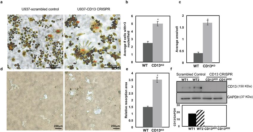
www.nature.com/scientificreports/
Figure 1. Loss of CD13 leads to reduced bone volume and increased number of osteoclasts in 8–10 week old
CD13-deficient mice. µCT reconstruction of cortical (a) and trabecular bone (b). (c–f) Bone morphometry by
µCT analysis; (c) BV/TV(%); Bone volume/Tissue volume, (d) Trabecular number, (e) Trabecular Thickness,
(f) Trabecular Separation. (g–i) Histomorphometric analysis of femurs of WT and CD13KO. Histology of
femurs with (g), BV/TV(%), (h) Oc.S/BS; % OCs per bone surface (i), histochemical detection of T RAP+
osteoclasts (purple, indicated by the arrow). (j) Mineral apposition rate (MAR), (k) Bone formation rate (BFR/
BS) measured by Osteomeasure software (OsteoMetrics, Decatur, USA) (https://www.osteometrics.com). All
samples were scanned, reconstructed, and analyzed in a Scanco µCT40 running Evaluation Program V6.6
RAP+ osteoclasts in
(http://www.scanco.ch/en/systems-solutions/software.html). Histochemical analysis of T
bone sections were imaged with Zeiss fluorescence inverted microscope and analyzed by using Zeiss Zen 2.0 Pro
blue edition software (https://www.zeiss.com/content/dam/Microscopy/Downloads/Pdf/FAQs/zen2-blue-editi
on_installation-guide.pdf). Scale bars-×5; 200 µm, ×10; 100 µm, ×20; 50 µm. Data represents ± SD. (N = 6; WT,
N = 7; CD13KO. **p < 0.01).
the area of resorption quantified by ImageJ. As expected, the increase in OC nuclei/cell positively correlates with
resorption where CD13KO OCs showed increased resorption area (threefold) compared to WT, confirming that
the elevated fusion in CD13KO mice translates into exaggerated functional activity in both young (Fig. 3d,e) and
aged (Supplementary Fig. S2d,e) mice. Importantly, WT cells treated with CD13 blocking antibody (SL13; 1 μg/
ml) prior to fusion in the presence of M-CSF and RANKL led to a significant increase (threefold) in multinucle-
ated OC with greater than 3 nuclei per cell which was analogous to the CD13-deficient OCs, illustrating that
blocking CD13 as well as its absence led to accelerated fusion (Fig. 4a,b) and confirming the CD13 specificity of
osteoclast fusion phenotype.
Osteoclast progenitors with osteoclastogenic potential are similar in WT and CD13KO bone
marrow and periphery. Previously we have shown that the distribution of the hematopoietic population
comprised of early hematopoietic progenitors, myelo-erythroid progenitors, and granulocyte macrophage pro-
Scientific Reports | (2021) 11:10736 | https://doi.org/10.1038/s41598-021-90271-x 3
Vol.:(0123456789)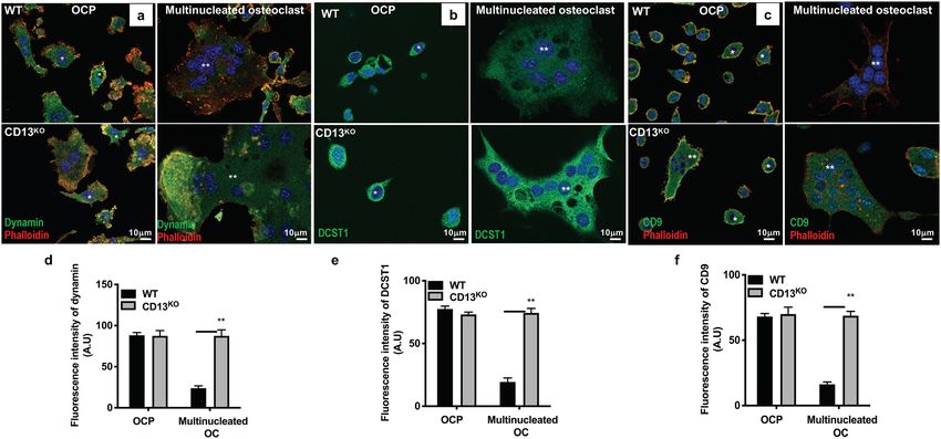
www.nature.com/scientificreports/
Figure 2. CD13 deficiency leads to marked reduction in trabecular bone volume and structure in 18–25 week
old aged mice. (a–d) Bone morphometry by µCT analysis; (a), BV/TV(%); Bone volume/Tissue volume (b),
Trabecular number, (c) Trabecular Thickness (d) Trabecular Separation. (e–h) Histomorphometric analysis
(e), BV/TV(%), (f) Oc.S/BS; % OCs per bone surface, (g) Mineral apposition rate (MAR), (h) Bone formation
rate (BFR/BS) measured by Osteomeasure software (OsteoMetrics, Decatur, USA) (https://www.osteometrics.
com). All samples were scanned, reconstructed, and analyzed in a Scanco µCT40 running Evaluation Program
V6.6 (http://www.scanco.ch/en/systems-solutions/software.html). Data represents ± SD. (N = 6; WT and N = 7;
CD13KO. ***p < 0.001, **p < 0.01, *p < 0.05).
genitors in CD13KO mice were similar to wildtype animals12. Cells that can generate bone-resorbing osteoclasts
reside in both BM and peripheral hematopoietic o rgans24. To determine if differences in osteoclast progenitor
(OCP) frequency are responsible for the loss of bone mass in the absence of CD13, we analyzed the distribu-
tion of primary OCP in both the BM microenvironment and spleen in WT and CD13KO mice. Flow cytometric
analysis (Supplementary Fig. S4) revealed that the OCP profile indicated by CD3−, B220−, NK1.1−, CD11b−/lo,
CD115+, CD117+ in the BM (a, WT vs. CD13KO; 1.7 vs. 1.95) and CD3-, B220-, NK1.1-, CD11b+, Ly6G-, Ly6C+,
CD115+ in spleen (b, WT vs. CD13KO; 0.24 vs. 0.238)24 and common myeloid progenitor population indicated
by lin− c-kit+ Sca-1− CD34+ in the BM (c, WT vs. CD13KO; 0.99 vs. 0.88) is similar between genotypes, indicating
that the absence of CD13 does not change the intrinsic differentiation potential of myeloid cells to osteoclast
progenitors.
Osteoclast‑like cell formation is enhanced in U937 cells expressing CD13 CRISPR. To confirm
that the effect of loss of CD13 in cell fusion extends across species, we utilized human U937 myeloid cells engi-
neered to delete CD13 or express scrambled wildtype control by CRISPR-based technology (Fig. 5f). Differentia-
tion of U937 cells with P MA25–27 for 3 days led to formation of equivalent levels of adherent monocytic cells in
both genotypes (data not shown). Subsequent stimulation with human recombinant M-CSF and RANKL led to
the aggregation of monocyte-like cells followed by differentiation into large multinucleated T RAP+ osteoclast-
like cells over time (8-10d) which was significantly accelerated in cells lacking CD13 compared to scrambled
controls (Fig. 5a–c). TRAP+, multinucleated C D13KO CRISPR cells exhibited increased resorptive activity when
grown in Osteo assay plates for 17 days as indicated by the relative resorption area (Fig. 5d,e), confirming that
the cells generated from U937 are indeed functional osteoclasts and that CD13 is a negative regulator of cell–cell
fusion in both mouse primary cells (BM and periphery) and a human monocytic cell line.
Expression of fusion proteins is dysregulated in CD13KO mature osteoclasts. A number of the
molecular mechanisms mediating osteoclast fusion and multinucleation have been elucidated28. In particular,
the small GTPase dynamin 2 (the major isoform in OC), the “master fusogen” DC-STAMP (DCST1) and the
tetraspanins CD9 and CD81 are common and critical regulators of osteoclast f usion23,29. Our recent studies have
Scientific Reports | (2021) 11:10736 | https://doi.org/10.1038/s41598-021-90271-x 4
Vol:.(1234567890)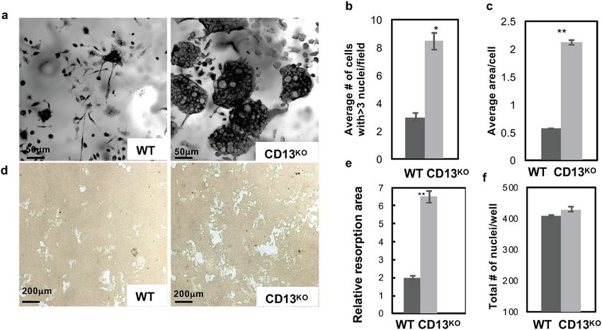
www.nature.com/scientificreports/
Figure 3. TRAP + multinucleated OCs are increased in the absence of CD13 in vitro. (a) Primary murine
BM- derived osteoclast progenitor cells grown on bovine cortical slices in presence of M-CSF and RANKL led
to increased OCs size and number of nuclei/OC in CD13KO cells compared to WT at d5. (b) Average # of cells
with > 3 nuclei/field and (c) Average cell area per OCs in C D13KO are significantly larger than the WT cells.
(d) Area of resorption is significantly higher in CD13KO than WT OCs grown on osteoplates for d10 by phase
contrast imaging. Osteoclasts on cortical slices and cluster of pits formed were imaged using a light microscope
(Olympus Scientific), using Olympus cellSens Dimension V0118 software (Olympus Scientific) (https://www.
olympus-lifescience.com/en/software/cellsens/) and the area of resorption was quantified by Image J (https://
imagej.nih.gov/ij/) (e) and total number of nuclei/well of both genotypes (f) are shown. Scale bars-(a) 50 µm; (d)
200 µm. Data represents ± SEM of three independent experiments. N = 6/genotype, **p < 0.01, *p < 0.05.
Figure 4. Treatment with CD13 blocking Ab leads to increased multinucleated OC formation in WT cells. (a).
TRAP staining of WT, WT + SL13 and C D13KO BM-derived osteoclast progenitor cells grown in presence of
M-CSF and RANKL after 5d on plastic are shown in “red” color. (b). Quantification of Trap+ OC with > 3 nuclei/
cell. TRAP+ osteoclasts were imaged with Zeiss fluorescence inverted microscope and analyzed by using Zeiss
Zen 2.0 Pro blue edition software (https://www.zeiss.com/content/dam/Microscopy/Downloads/Pdf/FAQs/
zen2-blue-edition_installation-guide.pdf). Scale bar; 200 µm. Data represents ± SEM of three independent
experiments. N = 3/genotype, **p < 0.01. Magnification ×5.
Scientific Reports | (2021) 11:10736 | https://doi.org/10.1038/s41598-021-90271-x 5
Vol.:(0123456789)www.nature.com/scientificreports/
Figure 5. Increased Multinucleated OC formation in U937 cells expressing CD13 CRISPR. (a). TRAP staining
of U937 expressing scrambled control or CD13 CRISPR grown in presence of PMA for 3 days followed by
M-CSF and RANKL for 10 d indicated increased size and number of multinucleated OCs in absence of CD13
compared to WT. (b) Multinucleated OC (**) with greater than 3 nuclei per cell and (c), average area of OC.
(d). Phase contrast imaging of area of resorption in U937 cells expressing CD13 CRISPR or scrambled control
grown in presence of PMA followed by M-CSF and RANKL for 17 d on osteoplates. T RAP+ osteoclasts were
imaged with Zeiss fluorescence inverted microscope and analyzed by using Zeiss Zen 2.0 Pro blue edition
software (https://www.zeiss.com/content/dam/Microscopy/Downloads/Pdf/FAQs/zen2-blue-edition_installati
on-guide.pdf). Area of resorption was imaged using a light microscope (Olympus Scientific), using Olympus
cellSens Dimension V0118 software (Olympus Scientific) (https://www.olympus-lifescience.com/en/software/
cellsens/) quantified by Image J (https://imagej.nih.gov/ij/) (e). (f). Immunoblot analysis of CD13 expression
in U937 cells expressing CD13 CRISPR or scrambled control clones. Blots were imaged by ChemiDoc Imaging
system version 3.0.1 (https://www.bio-rad.com/en-us/category/chemidoc-imaging-systems?ID=NINJ0Z15)
(Biorad). A cropped image is shown, see Supplementary Fig. S6 for full-length blots and cropped replicates.
Scale bar-(a) 100 µm; (d) 200 µm. Data represents ± SEM of three independent experiments. N = 3/genotype,
*p < 0.05. Magnification ×10.
shown that CD13 is a potent negative regulator of dynamin-dependent endocytosis of a variety of receptors20–22,
suggesting that CD13 may participate in cell fusion by regulating endocytic processes. Indeed, immunofluores-
cence (Fig. 6) and immunoblot (Fig. 7a,b) analyses of OC lysates derived from flow-sorted WT BM-OCPs dem-
onstrated high levels of dynamin and DC-STAMP expression by 2d-post differentiation, which was subsequently
reduced by 3d when cell fusion and maturation into multinucleated WT osteoclasts is complete, as previously
reported23. However, while dynamin, DC-STAMP and CD9 are highly expressed in C D13KO OCP, rather than
being downregulated, this strong expression is maintained in mature multinucleated OCs (Figs. 6, 7a,b), sug-
gesting that CD13 may impact fusion by regulating the levels of these key fusion and/or endocytic molecules
critical for OC fusion. In addition, flow cytometry of dynamin, DCST1 and CD9 confirmed persistent surface
expression of fusion promoting proteins in multinucleated OC lacking CD13 compared to WT OC (Supplemen-
tary Fig. S5a–c). Furthermore, immunoblot analysis of cell lysates obtained from WT BM-derived progenitor
cells stimulated with M-CSF and RANKL over 3d indicated that CD13 is highly expressed in myeloid progenitor
cells but its expression level is unaltered upon stimulation with M-CSF and RANKL over time (d0-3) (Fig. 7c,d),
consistent with CD13 regulating fusion mechanisms independent of RANKL signaling.
Next, we investigated the overall mechanistic process by which CD13 sustains expression levels of dynamin,
DCST1 and tetraspanins. Quantitative RT-PCR analysis demonstrated that transcript levels for DNM2, DCST1
and tetraspanins CD9 and CD81 were similar in BM progenitor cells stimulated with M-CSF + RANKL over
0-5d, indicating that CD13 does not regulate transcription levels of these proteins (Fig. 8a). To explore if CD13
limits the stability of fusion-regulatory proteins in WT cells, BM-derived OCP were grown in the presence of
M-CSF + RANKL and treated with cycloheximide (100 μg/ml) to inhibit new protein synthesis. While dynamin
and DCST1 protein expression in CD13KO OC remained stable over 8–12 h, loss of dynamin and DCST1 protein
expression occurred by 4–8 h in WT OC, indicating that CD13 controls fusion-regulatory protein turnover and
stability (Fig. 8b,c).
Scientific Reports | (2021) 11:10736 | https://doi.org/10.1038/s41598-021-90271-x 6
Vol:.(1234567890)www.nature.com/scientificreports/
Figure 6. Immunofluorescence analysis of persistent expression of fusion-regulatory proteins in CD13-
deficient multinucleated OC. Expression of the fusion regulatory proteins dynamin (a,d), DCST1 (b,e) and
CD9 (c,f) is maintained in CD13KO multinucleated osteoclasts but not in WT cells (**). High levels of dynamin
co-localize with actin and DCST1 in OCPs (*), imaged using Zeiss LSM 880 confocal fluorescence microscope
and analyzed by Zeiss Zen 2.0 Pro blue edition software (https://www.zeiss.com/content/dam/Microscopy/
Downloads/Pdf/FAQs/zen2-blue-edition_installation-guide.pdf). Scale bar; 10 µm. Data represents average of
three independent experiments. N = 3/genotype. Dynamin, CD9, DCST1; green. Phalloidin; red. Magnification
×63 oil.
Discussion
The fusion of plasma membranes is essential to and indispensable for many physiologic processes such as fer-
tilization through sperm/egg f usion30, muscular development through myoblast f usion31, skeletal development
and maintenance of skeletal integrity through formation of osteoclasts and control of certain viral infections and
spreading through the formation of macrophage giant cells (MGC)32. Thus, this important biological process
directly defines the course of many pathological processes including infertility, skeletal defects (osteoporosis
and osteopetrosis), failure of skeletal repair, failure to maintain prosthetic implants as well as fusion of host and
viral membranes in viral diseases. Clearly, potential common regulators and mechanisms would be attractive
therapeutic targets in these disorders.
Two of the cell types that fuse, osteoclasts and multinucleated giant cells, are derived from a common progeni-
tor, are rendered fusion competent by common molecular mediators and ultimately regulate specialized func-
tions in specific microenvironments. While OC can undergo fusion in both normal or pathological states such
as Paget’s d isease33, macrophages fuse to form MGC primarily under inflammatory conditions such as chronic
granulomatous disease or the foreign body response34. However, the fact that the fusion of OC and MGCs are
governed by common signaling mechanisms again makes these and their component molecules attractive targets
for therapeutic intervention. Interestingly, we have previously shown that in response to ischemic injury, CD13KO
skeletal muscle satellite cells fused more readily than WT cells to form multinucleated myoblasts, suggesting that
CD13 may also participate in fusion of other cell types, thus affecting processes such as skeletal muscle r epair16.
Osteoclastogenesis comprises many steps from the commitment and survival of osteoclast progenitor cells,
their differentiation into mononuclear pre-osteoclasts that fuse to generate multinucleated mature osteoclasts and
finally, activation of osteoclasts for bone resorption. Among the different steps, osteoclast fusion is thought to be
the critical step in this phenomenon. Our data clearly indicate that osteoclast progenitor survival, differentiation
and proliferation is not dependent on CD13 expression, suggesting that CD13 may be specifically involved in
the fusion mechanism to generate multinucleated osteoclasts.
Defective osteoblastic bone forming activity can also contribute to osteolysis. Previously, we have shown that
CD13 expression does not affect mesenchymal stem cell formation or their s urvival16, confirming that elevated
levels of osteoprogenitors in the in vitro osteoblastogenesis analysis is not due to differences in mesenchymal
stem cell formation. We showed that despite an increase in the osteoblast progenitor population in the absence
of CD13, bone formation rate and mineral apposition rate remain unaltered between genotypes, indicating that
impaired skeletal mass is not due to a defect in mature osteoblast function. While dynamic bone formation is
equivalent between genotypes of young mice, C D13KO aged mice have a significantly reduced bone formation
rate compared to their wildtype counterparts, perhaps implicating a CD13 contribution of osteoblast function
in bone remodeling during aging.
Studies have implicated various membrane-associated processes as critical to osteoclastogenesis such as
clustering of membrane tetraspanins35, endocytosis of surface molecules36 as well as the initial increases in
Scientific Reports | (2021) 11:10736 | https://doi.org/10.1038/s41598-021-90271-x 7
Vol.:(0123456789)www.nature.com/scientificreports/
Figure 7. Immunoblot analysis of sustained expression of fusion-regulatory proteins in CD13-deficient OC
over time. Expression of the fusion-promoting proteins dynamin, DC-STAMP (DCST1) and CD9 is aberrantly
sustained in CD13KO but not in WT multinucleated osteoclasts (a,b) in presence of M-CSF and RANKL. A
cropped image is presented, see Supplementary Fig. S7 for full-length blots and Supplementary Fig. S8 for
cropped replicates. (c,d). CD13 expression is unaltered in BM-derived OCs in response to M-CSF and RANKL
stimulation over time. Blots were imaged by ChemiDoc Imaging system version 3.0.1 (https://www.bio-rad.
com/en-us/category/chemidoc-imaging-systems?ID=NINJ0Z15) (Biorad). A cropped image is presented, see
Supplementary Fig. S9 for full-length blots and cropped replicates. Data represents average of two isolates. N = 3/
genotype. **p < 0.01, *p < 0.05.
expression of specific fusogenic molecules, such as DC-STAMP29, OC-STAMP37, dynamin and ATP6V0d2
(ATPase, H + transporting V0 subunit d2) and subsequent decline as osteoclastogenesis proceeds36,38. We see
sustained expression of DC-STAMP, CD9 and dynamin-2 in C D13KO cells while these proteins disappear in paral-
lel WT cultures, suggesting that CD13 regulates fusion by mechanisms leading to decreased fusogen stability and
the absence of CD13 results in sustained fusogen expression and enhanced fusion. Indeed, myeloid cells sorted
for high levels of DC-STAMP expression formed higher numbers of OCs in fusion cultures than DC-STAMP
low populations39. In agreement with these observations, transgenic mice overexpressing DC-STAMP driven
by the actin promoter exhibited an osteoporotic phenotype and osteoclast hyper-multinucleation11, suggesting
that sustained expression leads to bone loss. Clinically, Paget’s disease of the bone (PDB) is a disorder resulting
from osteoclast overactivity of unknown etiology. Osteoclast-like cells isolated from PDB patients carrying a
DC-STAMP genetic variant showed higher DC-STAMP expression levels and greater numbers of nuclei per
cell compared to both healthy controls and PDB patients without the variant40. A distinct gain-of-function DC-
STAMP variant (rs2458413) studied in cells from 158 patients was significantly associated with P DB41. Clearly,
sustained expression of DC-STAMP and other fusogens would promote osteoclastogenesis and likely underlies
the effects of CD13 on bone remodeling.
Scientific Reports | (2021) 11:10736 | https://doi.org/10.1038/s41598-021-90271-x 8
Vol:.(1234567890)www.nature.com/scientificreports/
Figure 8. Fusion-regulatory proteins, dynamin and DCST1 are regulated by a CD13-dependent post
transcriptional mechanism. (a) Quantitative RT-PCR analysis of fusion regulatory transcripts normalized to
GAPDH in flow sorted mouse BM cells stimulated with M-CSF and RANKL over indicated time. Expression
of the genes regulating osteoclast fusion -dynamin 2 (DNM2), DCST1, CD9 and CD81 are highly induced
upon M-CSF and RANKL over time but was equivalent between genotypes. All data was analyzed using
CFX Manager version 3.1 ((https://www.bio-rad.com/en-us/sku/1845000-cfx-manager-software?ID=18450
00) (Biorad). Data represents average of two independent experiments. N = 3/genotype. (b–c) Dynamin and
DCST1 protein stability are enhanced in absence of CD13. Immunoblot analysis of dynamin and DCST1 of
WT and CD13KO BM-derived OC treated with cycloheximide (CHX) for indicated time. Blots were imaged by
ChemiDoc Imaging system version 3.0.1 (https://www.bio-rad.com/en-us/category/chemidoc-imaging-syste
ms?ID=NINJ0Z15) (Biorad). A cropped image is presented, see Supplementary Fig. S10 for full-length blots and
cropped replicates. Data represents average of two isolates. N = 3/genotype. **p < 0.01, *p < 0.05.
We have shown that CD13 regulates dynamin- and clathrin-mediated e ndocytosis21,22, recycling of cell sur-
face proteins20 and Src activation42, each of which have been shown to participate in OC fusion. In the current
study, fusion protein expression in C D13KO OCs is aberrantly sustained by a posttranscriptional mechanism.
Alternatively, organizers of actin-based protrusions are also pivotal in myeloid cell as well as gamete fusion43,
which depends on step-wise reorganization of the actin cytoskeleton, initiated by formation of “podosome-like”
membrane protrusions in myeloid cells44–48. Importantly, overexpression of DC-STAMP generates cells with
numerous cell protrusions and increased fusogenic capacity. Similarly, the abundant filopodia in OC precursors
and OC in active fusion are significantly downregulated as OCs m ature39,47,49. Recently, we have reported that
CD13 is a critical signaling platform that links the plasma membrane to dynamic mediators of actin cytoskel-
etal assembly and rearrangement20. We propose that CD13 may regulate the expression of these endocytic and
fusion regulatory proteins, perhaps by mediating their internalization, endocytic trafficking, recycling and/or
Scientific Reports | (2021) 11:10736 | https://doi.org/10.1038/s41598-021-90271-x 9
Vol.:(0123456789)www.nature.com/scientificreports/
localization. Whether CD13’s regulation of fusogen stability affects formation of membrane protrusions or the
localization of fusion regulators to the site of cell–cell fusion is currently under investigation.
In conclusion, in the present study we demonstrate that CD13 expression controls osteoclastogenesis spe-
cifically at the level of cell–cell fusion. Further investigation into the relationship between CD13 and dynamin,
DCST1 and CD9 and other regulators of osteoclast fusion such as OC-STAMP and the osteoclast receptor αvβ3
integrin28 will clarify mechanisms regulating CD13-mediated cell fusion in osteoclasts as well as in fusion of other
cell lineages including foreign body giant cells and satellite stem cells. Considering the diversity and importance
of pathologies that are influenced by cell–cell fusion, identification of CD13-dependent molecular mechanisms
and signaling that regulate myeloid fusion will provide novel therapeutic approaches in fusion pathologies.
Materials and methods
The authors confirm that all experiments were carried out in accordance with relevant guidelines and regulations.
The ethical approval for all animal care and procedures were carried out in accordance with relevant guidelines
and regulations by the UConn Health Institutional Animal Care and Use Committee.
Animals. Global young (8–10 weeks) or aged (18–25 weeks) wildtype and C
D13KO (C57BL/6J) male and
female mice were generated and housed at the Gene Targeting and Transgenic Facility at University of Con-
necticut School of Medicine12. All procedures were performed in accordance with the guidelines and regulations
approved by the UCONN Health Institutional Animal Care and Use Committee. UConn Health is fully accred-
ited by Association for Assessment and Accreditation of Laboratory Animal Care (AAALAC) International and
Public Health Service (PHS) assurance number is A3471-01 (D16-00295) and USDA Registration Number is
16-R-0025.
Euthanasia by CO2 followed by cervical dislocation was performed and is an accepted method consistent with
AVMA Guidelines for the Euthanasia of Animals to minimize the pain or discomfort in animals.
Reagents. Recombinant mouse and human M-CSF and RANKL were purchased from R&D Systems. Phor-
bol 12-myristate 13-acetate (PMA), Phosphatase, Leukocyte (TRAP) Kit were purchased from Sigma-Aldrich.
Bovine cortical bone slices were a kind gift from Dr. Joseph Lorenzo, University of Connecticut School of
Medicine50. Osteoplates were purchased from Corning.
Antibodies. Antibodies to DC-STAMP1 (DCST1; Biorbyt, orb2242, rabbit polyclonal Ab), Dynamin
(Abcam, ab3457, rabbit polyclonal Ab), CD9 (Abcam, ab223052, rabbit polyclonal Ab), Phalloidin-TRITC
(Sigma-Aldrich, P1951), CD13 (SL13, Millipore, MABC950, rat monoclonal Ab).
Flow cytometry. Antibodies for phenotypic analyses and sorting by flow cytometry used are as follows24;
anti-CD3 (145-2C11), anti-B220 (RA3-6B2), anti-NK1.1 (PK136), anti-CD11b (M1/70), anti-CD115 (AFS98),
anti-Ly6C (Al-21). All antibodies were purchased from Biolegend, BD Biosciences, e-Biosciences. UV Blue Live
dead dye was purchased from Life Technologies. Labeling of cells for flow cytometry and sorting was performed
as described. Briefly, flow cytometry on live cells were performed to obtain OCP cells from BM expressing
CD11blo CD115hi Ly6G+ (CD3/B220/NK1.1)−24 using BD-FACS Aria (BD Biosciences) and data analyzed with
BD FACS DIVA version 9.0 (https://www.bdbiosciences.com/en-us/instruments/research-instruments/resea
rch-software/flow-cytometry-acquisition/facsdiva-software) and FlowJo version 9.9 software (https://www.
flowjo.com/). For flow sorting of OCP, 200 × 106 BM cells isolated from four pooled WT or four pooled C D13KO
mice were run through FACSAriaII to obtain OCP (5–8% of total sorted BM cells) analyzed by FACSDiva.
Surface expression of fusion promoting proteins was performed with goat anti-rabbit dynamin2-Alexa 488
(ProSci; 61-336), rabbit anti-mouse DCST1-Alexa fluor 350 (Bioss Inc; bs-8250R-A350), and rat anti-mouse
CD9-APC (Biolegend, Clone MZ3; 124811) using BD LSRII-A and analyzed by FlowJo version 9.9 software
(https://w
ww.fl owjo.c om/). Goat IgG-Alexa 488 or rabbit IgG-AF350 or rat IgG-APC was used as isotype control.
In vivo analysis of WT and CD13KO mice. Micro‑computed tomography and histomorphometry. Sam-
ples were scanned in a density-calibrated µCT40 (Scanco Medical, Bassersdorf, Switzerland) in PBS at 8 µm3 res-
olution with the following settings: 55 kV, 145 µA, 300 ms integration, 1000 projections/rotation with Gaussian
filtering. Analysis was performed following standard guidelines51. Briefly, femoral trabeculae were auto-con-
toured in a 120-slice region 1 mm proximal to the distal condyles with a lower threshold of 2485 Hounsfield
units (HU), and femoral cortex was auto-contoured in an 80-slice region just distal to the third trochanter with a
lower threshold of 4932HU50,52. All samples were scanned, reconstructed, and analyzed in a Scanco µCT40 run-
ning Evaluation Program V6.6 (http://www.scanco.ch/en/systems-solutions/software.html).
Isolation of hematopoietic progenitor population from bone marrow and spleen. BM cells
were obtained by flushing femur and tibia from WT or CD13KO mice with 10 ml 1 × PBS and 2% heat inactivated
FBS, followed by RBC lysis and filtering through 40 μm cell strainer (BD Biosciences). Total live cells counted
with Countess Automated cell counter (Thermo Fisher Scientific) were stained with antibody cocktail at 4 °C.
Cells from mouse spleen was obtained by gentle crushing the organ between frosted microscopic slides in cold
10 ml 1 × PBS and 2% heat inactivated FBS.
Generation and culture of osteoclast progenitors from bone marrow or spleen. Cells from
mouse BM or spleen were stained with Ab cocktail containing anti-(CD3, B220, NK1.1, CD115, Ly6C) Ab
Scientific Reports | (2021) 11:10736 | https://doi.org/10.1038/s41598-021-90271-x 10
Vol:.(1234567890)www.nature.com/scientificreports/
and subjected to single-cell sorting by BD FACS A ria24. Flow-sorted osteoclast progenitors isolated from BM
[ CD11blo CD115hi Ly6G+ (CD3/B220/NK1.1)−] or spleen [CD11bhi CD115+Ly6Chi (CD3/B220/NK1.1)−] at
a density of 5000–20,000 cells/well (BM) or 20,000–50,000 cells/well (spleen) were seeded in 96-well dish in
α-MEM containing 10%FBS, 1% Penicillin–Streptomycin, 30 ng/ml M-CSF and 30 ng/ml RANKL at 37 degree
C with 5% C O2 for 0-10d. Multi-nucleated osteoclasts were stained with Tartrate-Resistant Acid Phosphatase
(TRAP) staining and assessed by counting cells with more than three nuclei. Average area of osteoclast was
measured by ImageJ software (https://imagej.nih.gov/ij/).
Histomorophometric analysis and TRAP staining52. Dynamic histomorphometry of bone. To evalu-
ate dynamic histomorphometry, mice of 8–10 weeks and 25 weeks of age were injected with calcein 10 mg/
kg (Sigma) seven days and alizarin complexone 30 mg/kg (Sigma) intraperitoneally two days before they were
sacrificed. Seven-micrometer sections of femora were analyzed under a fluorescent microscope (Leica DMR).
Blinded researcher marked bone tissue, single, and double labels in Osteomeasure software (OsteoMetrics, De-
catur, USA) (https://www.osteometrics.com). The software then automatically calculated the mineral apposition
rate (MAR) and bone formation rate (BFR).
Tartrate resistant acid phosphatase (TRAP) staining for osteoclasts. Following 4 days fixation
in 4% PFA, bone samples were placed in 30% sucrose O/N at 4 °C prior cryoembedding (Cryomatrix, Thermo
Fisher Scientific). Sections were cut onto Japanese Cryotape (Cryofilm 2C, Section Lab, Japan) at 7 µm and cross-
linked using Norland Optical Adhesive 61 (Norland Optical) onto glass slides. Bone sections were decalcified
for 30 min at RT and osteoclasts were stained for TRAP using a commercial staining kit (Sigma, St-Louis, MO).
The sections were rinsed with distilled water, counterstained with hematoxylin for 30 s and air-dried. Osteoclasts
were identified as multinucleated cells placed adjacent to the bone surface. Metaphyseal regions of TRAP stained
femora, 1 mm distally from the epiphyseal plate, were analyzed under the microscope (Leica DMR). Blinded
researcher marked osteoclasts and bone surface in Osteomeasure software (Osteo-Metrics) (https://www.osteo
metrics.com). The software then automatically calculated the trabecular volume (BV/TV, %) and osteoclasts per
bone surface (Ocs/BS).
Histochemical analysis of T RAP+ osteoclasts in bone sections were imaged with Zeiss fluorescence inverted
microscope and analyzed by using Zeiss Zen 2.0 Pro blue edition software (https://www.z eiss.c om/c onten
t/d
am/
Microscopy/Downloads/Pdf/FAQs/zen2-blue-edition_installation-guide.pdf).
Flow-sorted osteoclast progenitor cells derived from BM or spleen in α-MEM (GIBCO BRL) containing
10%FBS, 1% Penicillin–Streptomycin, 30 ng/ml M-CSF and 30 ng/ml RANKL grown on plastic or UV-sterilized,
devitalized bovine cortical bone slices (placed in 96-well dishes), at a density of 50,000 cells/well for indicated
time were fixed in 2.5% Glutaraldehyde and TRAP stained according to manufacturer’s instruction (Sigma) and
analyzed by Zeiss Zen 2.0 Pro blue edition software (https://www.zeiss.com/content/dam/Microscopy/Downl
oads/Pdf/FAQs/zen2-blue-edition_installation-guide.pdf).
Bone resorption assay52. Flow-sorted osteoclast progenitors derived from BM were seeded on Osteo
Assay plate (Corning) at a density of 50,000 cells/well in α-MEM containing 10%FBS, 1% Penicillin–Strepto-
mycin, 30 ng/ml M-CSF and 30 ng/ml RANKL for d10. Surface pit formation was measured by removing cells
with 100 μl of 10% bleach solution at RT for 5 min. Wells were washed with deionized water and allowed to
dry. Cluster of pits formed was imaged using a light microscope (Olympus Scientific), using Olympus cellSens
Dimension V0118 software (Olympus Scientific) (https://www.olympus-lifescience.com/en/software/cellsens/)
and the area of resorption was measured by ImageJ software (https://imagej.nih.gov/ij/).
Generation of CD13KO U937 cells by CRISPR‑Cas9 gene editing. Oligos containing the guide RNA
sequence for human CD13: 5′-CAGTGCGATGATTGTGCACA-3′; guide RNA for scrambled control: 5′-CAG
TCGGGCGTCATCATGAT-3′ were cloned into lentiCRISPR v2 (addgene plasmid #52961). Packaging plasmids
psPAX2 and pMD2.G (addgene plasmid #12260 and addgene plasmid #12259) were utilized to generate the len-
tivirus as described20 in presence of 2 ng/µl puromycin to select for lentiCRISPR integration. Multiple C
D13KO
and scrambled control clones confirmed by immunoblot, IF and flow cytometry analysis were employed for OC
generation.
Isolation of TRAP+ osteoclast‑like cells from human monocytic cell line U937. U937 cells
expressing CD13 CRISPR or scrambled c ontrol20 were grown at a density of 100,000 cells/well in 4-well dish
with RPMI 1640 containing 10% heat inactivated fetal bovine serum, 1 mM l-glutamine, 1 mM sodium pyru-
vate, 1% penicillin/ streptomycin and 0.1 µg/ml PMA for 3 days. Non-adherent cells were removed and adherent
cells were stimulated with M-CSF (60 ng/ml) and RANKL (100 ng/ml) in RPMI medium without PMA for an
additional 10 days. Cells were fixed in 2.5% glutaraldehyde and osteoclast-like cells identified by TRAP staining.
Immunofluorescence and microscopy. Flow-sorted osteoclast progenitors were grown on glass cover-
slips that were previously coated with 5 µg/ml fibronectin for indicated time period. Cells were fixed in 4% para-
formaldehyde (Electron Microscopy Sciences) at RT for 30 min, permeabilized with 0.1% Triton-X-100 in PBS
at RT for 5 min. Cells were blocked with blocking buffer containing 5% goat or donkey serum/5% BSA/1 × PBS
at RT for 1 h followed by incubation with primary Ab in blocking buffer at 4 °C for overnight. Cells were washed
and treated with secondary Ab (1:1200) and DAPI (nuclear stain) in blocking buffer at RT for 1 h. Cover-
slips were mounted with ProLong Gold antifade mounting medium (Life Technologies), visualized at excitation
Scientific Reports | (2021) 11:10736 | https://doi.org/10.1038/s41598-021-90271-x 11
Vol.:(0123456789)www.nature.com/scientificreports/
wavelength of 488 nm (Alexa 488), 543 nm (Alexa 594 or TRITC) and 405 nm (DAPI) and imaged by Zeiss LSM
880 confocal fluorescence microscope and analyzed by using Zeiss Zen 2.0 Pro blue edition software (https://
www.zeiss.com/content/dam/Microscopy/Downloads/Pdf/FAQs/zen2-blue-edition_installation-guide.pdf).
Immunoblot analysis. Flow-sorted BM osteoclast progenitors grown in presence of M-CSF and RANKL
for 48 h were LPC (lysophosphatidylcholine)-synchronized by treating with reversible fusion inhibitor LPC
(100 µM) for 12 h followed by washing and growing cells in LPC-free medium for fusion to proceed for 0–72 h.
Cell lysates were harvested in 1% NP40 lysis buffer containing 1× complete Protease Inhibitor cocktail (Roche).
Samples were separated by SDS-PAGE and transferred to nitrocellulose membrane, blocked in 1XTBST con-
taining 5% bovine serum albumin, treated with primary Ab followed by appropriate secondary Ab and imaged
by ChemiDoc Imaging system version 3.0.1 (https://www.bio-rad.com/en-us/category/chemidoc-imaging-syste
ms?ID=NINJ0Z15) (Biorad). β actin or GAPDH were used as loading controls. Gels/blots were cropped and
indicated by dividing lines.
Quantitative RT‑PCR analysis. Total RNA was extracted using TRIZOL reagent (Invitrogen) according
to manufacturer’s instruction. Relative transcript level was normalized to GAPDH level. Primer sequences were
determined using GenBank primer sequences (http://pga.mgh.harvard.edu/primerbank/). Sequence of PCR
primers employed are as follows- Dynamin 2 (Dnm2), 5′-TTCGGGTCTACTCACCACAC-3′ (forward) and
5′-CTCTCGCGGCTGATGAACTG-3′ (reverse); DC-STAMP1 (DCST1), 5′-CGGCGGCCAATCTAAGGTC-3′
(forward) and 5′-CCCACCATGCCCTTGAACA-3′ (reverse); CD9, 5′-ATGCCGGTCAAAGGAGGTAG-3′
(forward) and 5′-GCCATAGTCCAATAGCAAGCA-3′ (reverse); CD81, 5′-CAGATCGCCAAGGATGTGAAG-
3′ (forward) and 5′-GCCACAACAGTTGAGCGTCT-3′ (reverse); GAPDH, 5′-GGATTTGGTCGTATTGGG-3′
(forward), 5′-GGAAGATGGTGATGGGATT-3′ (reverse). All data was analyzed using CFX Manager version 3.1
(https://www.bio-rad.com/en-us/sku/1845000-cfx-manager-software?ID=1845000) (Biorad).
Statistical analysis. Statistical analysis was performed using unpaired, two-tailed Student’s t test using
GraphPad Prism software and results are representative of mean ± SD or ± SEM as indicated. Differences at
p ≤ 0.05 were considered significant.
Disclosures. All animal experiments in this study were reviewed and approved by Animal Care Committee
at University of Connecticut Medical School.
Received: 17 March 2021; Accepted: 7 May 2021
References
1. Teitelbaum, S. L. Bone resorption by osteoclasts. Science 289, 1504–1508. https://doi.org/10.1126/science.289.5484.1504 (2000).
2. Tsukasaki, M. & Takayanagi, H. Osteoimmunology: Evolving concepts in bone-immune interactions in health and disease. Nat.
Rev. Immunol. 19, 626–642. https://doi.org/10.1038/s41577-019-0178-8 (2019).
3. Madel, M. B. et al. Immune function and diversity of osteoclasts in normal and pathological conditions. Front. Immunol. 10, 1408.
https://doi.org/10.3389/fimmu.2019.01408 (2019).
4. Boudin, E., Fijalkowski, I., Hendrickx, G. & Van Hul, W. Genetic control of bone mass. Mol. Cell Endocrinol. 432, 3–13. https://
doi.org/10.1016/j.mce.2015.12.021 (2016).
5. Krakow, D. Skeletal dysplasias. Clin. Perinatol. 42, 301–319. https://doi.org/10.1016/j.clp.2015.03.003 (2015).
6. Arai, F. et al. Commitment and differentiation of osteoclast precursor cells by the sequential expression of c-Fms and receptor
activator of nuclear factor kappaB (RANK) receptors. J. Exp. Med. 190, 1741–1754. https://d oi.o
rg/1 0.1 084/j em.1 90.1 2.1 741 (1999).
7. Fogg, D. K. et al. A clonogenic bone marrow progenitor specific for macrophages and dendritic cells. Science 311, 83–87. https://
doi.org/10.1126/science.1117729 (2006).
8. Jacquin, C., Gran, D. E., Lee, S. K., Lorenzo, J. A. & Aguila, H. L. Identification of multiple osteoclast precursor populations in
murine bone marrow. J. Bone Miner. Res. 21, 67–77. https://doi.org/10.1359/JBMR.051007 (2006).
9. Mensah, K. A., Ritchlin, C. T. & Schwarz, E. M. RANKL induces heterogeneous DC-STAMP(lo) and DC-STAMP(hi) osteoclast
precursors of which the DC-STAMP(lo) precursors are the master fusogens. J. Cell Physiol. 223, 76–83. https://doi.org/10.1002/
jcp.22012 (2010).
10. Miyamoto, H. et al. Osteoclast stimulatory transmembrane protein and dendritic cell-specific transmembrane protein cooperatively
modulate cell–cell fusion to form osteoclasts and foreign body giant cells. J. Bone Miner. Res. 27, 1289–1297. https://doi.org/10.
1002/jbmr.1575 (2012).
11. Yagi, M. et al. DC-STAMP is essential for cell–cell fusion in osteoclasts and foreign body giant cells. J. Exp. Med. 202, 345–351.
https://doi.org/10.1084/jem.20050645 (2005).
12. Winnicka, B. et al. CD13 is dispensable for normal hematopoiesis and myeloid cell functions in the mouse. J. Leukoc. Biol. 88,
347–359. https://doi.org/10.1189/jlb.0210065 (2010).
13. Look, A. T., Ashmun, R. A., Shapiro, L. H. & Peiper, S. C. Human myeloid plasma membrane glycoprotein CD13 (gp150) is identi-
cal to aminopeptidase N. J. Clin. Investig. 83, 1299–1307. https://doi.org/10.1172/JCI114015 (1989).
14. Petrovic, N. et al. CD13/APN regulates endothelial invasion and filopodia formation. Blood 110, 142–150. https://d oi.o rg/1 0.1 182/
blood-2006-02-002931 (2007).
15. Mina-Osorio, P. et al. CD13 is a novel mediator of monocytic/endothelial cell adhesion. J. Leukoc. Biol. 84, 448–459 (2008).
16. Rahman, M. M. et al. CD13 promotes mesenchymal stem cell-mediated regeneration of ischemic muscle. Front. Physiol. 4, 402.
https://doi.org/10.3389/fphys.2013.00402 (2014).
17. Nomura, R. et al. Human coronavirus 229E binds to CD13 in rafts and enters the cell through caveolae. J. Virol. 78, 8701–8708.
https://doi.org/10.1128/JVI.78.16.8701-8708.2004 (2004).
Scientific Reports | (2021) 11:10736 | https://doi.org/10.1038/s41598-021-90271-x 12
Vol:.(1234567890)www.nature.com/scientificreports/
18. Saiki, I. et al. Role of aminopeptidase N (CD13) in tumor-cell invasion and extracellular matrix degradation. Int. J. Cancer 54,
137–143. https://doi.org/10.1002/ijc.2910540122 (1993).
19. Bhagwat, S. V. et al. CD13/APN is activated by angiogenic signals and is essential for capillary tube formation. Blood 97, 652–659
(2001).
20. Ghosh, M. et al. CD13 tethers the IQGAP1-ARF6-EFA6 complex to the plasma membrane to promote ARF6 activation, beta1
integrin recycling, and cell migration. Sci. Signal. https://doi.org/10.1126/scisignal.aav5938 (2019).
21. Ghosh, M., Subramani, J., Rahman, M. M. & Shapiro, L. H. CD13 restricts TLR4 endocytic signal transduction in inflammation.
J. Immunol. 194, 4466–4476. https://doi.org/10.4049/jimmunol.1403133 (2015).
22. Ghosh, M., McAuliffe, B., Subramani, J., Basu, S. & Shapiro, L. H. CD13 regulates dendritic cell cross-presentation and T cell
responses by inhibiting receptor-mediated antigen uptake. J. Immunol. 188, 5489–5499. https://doi.org/10.4049/jimmunol.11034
90 (2012).
23. Shin, N. Y. et al. Dynamin and endocytosis are required for the fusion of osteoclasts and myoblasts. J. Cell Biol. 207, 73–89. https://
doi.org/10.1083/jcb.201401137 (2014).
24. Jacome-Galarza, C. E., Lee, S. K., Lorenzo, J. A. & Aguila, H. L. Identification, characterization, and isolation of a common progeni-
tor for osteoclasts, macrophages, and dendritic cells from murine bone marrow and periphery. J. Bone Miner. Res. 28, 1203–1213.
https://doi.org/10.1002/jbmr.1822 (2013).
25. Kang, K. et al. Inhibition of osteoclast differentiation by overexpression of NDRG2 in monocytes. Biochem. Biophys. Res. Commun.
468, 611–616. https://doi.org/10.1016/j.bbrc.2015.10.167 (2015).
26. Kang, H. S. et al. Receptor activator of nuclear factor-kappaB is induced by a rottlerin-sensitive and p38 MAP kinase-dependent
pathway during monocyte differentiation. Mol. Cells 17, 438–445 (2004).
27. Garcia, D. E., Brown, S., Hille, B. & Mackie, K. Protein kinase C disrupts cannabinoid actions by phosphorylation of the CB1
cannabinoid receptor. J. Neurosci. 18, 2834–2841 (1998).
28. Pereira, M. et al. Common signalling pathways in macrophage and osteoclast multinucleation. J. Cell Sci. https://doi.org/10.1242/
jcs.216267 (2018).
29. Yagi, M., Miyamoto, T., Toyama, Y. & Suda, T. Role of DC-STAMP in cellular fusion of osteoclasts and macrophage giant cells. J.
Bone Miner. Metab. 24, 355–358. https://doi.org/10.1007/s00774-006-0697-9 (2006).
30. Ogle, B. M., Cascalho, M. & Platt, J. L. Biological implications of cell fusion. Nat. Rev. Mol. Cell Biol. 6, 567–575. https://doi.org/
10.1038/nrm1678 (2005).
31. Pajcini, K. V., Pomerantz, J. H., Alkan, O., Doyonnas, R. & Blau, H. M. Myoblasts and macrophages share molecular components
that contribute to cell–cell fusion. J. Cell Biol. 180, 1005–1019. https://doi.org/10.1083/jcb.200707191 (2008).
32. Bracq, L. et al. T cell-macrophage fusion triggers multinucleated giant cell formation for HIV-1 spreading. J. Virol. https://doi.org/
10.1128/JVI.01237-17 (2017).
33. Roodman, G. D. & Windle, J. J. Paget disease of bone. J. Clin. Investig. 115, 200–208. https://doi.org/10.1172/JCI24281 (2005).
34. Seitzer, U., Haas, H. & Gerdes, J. A human in vitro granuloma model for the investigation of multinucleated giant cell and granu-
loma formation. Histol. Histopathol. 16, 645–653. https://doi.org/10.14670/HH-16.645 (2001).
35. Takeda, Y. et al. Tetraspanins CD9 and CD81 function to prevent the fusion of mononuclear phagocytes. J. Cell Biol. 161, 945–956.
https://doi.org/10.1083/jcb.200212031 (2003).
36. Kodama, J. & Kaito, T. Osteoclast multinucleation: Review of current literature. Int. J. Mol. Sci. https://d oi.org/1 0.3 390/i jms21 1656
85 (2020).
37. Khan, U. A., Hashimi, S. M., Bakr, M. M., Forwood, M. R. & Morrison, N. A. Foreign body giant cells and osteoclasts are TRAP
positive, have podosome-belts and both require OC-STAMP for cell fusion. J. Cell Biochem. 114, 1772–1778. https://doi.org/10.
1002/jcb.24518 (2013).
38. Kim, K., Lee, S. H., Ha Kim, J., Choi, Y. & Kim, N. NFATc1 induces osteoclast fusion via up-regulation of Atp6v0d2 and the den-
dritic cell-specific transmembrane protein (DC-STAMP). Mol. Endocrinol. 22, 176–185. https://doi.org/10.1210/me.2007-0237
(2008).
39. Chiu, Y. H. et al. Regulation of human osteoclast development by dendritic cell-specific transmembrane protein (DC-STAMP). J.
Bone Miner. Res. 27, 79–92. https://doi.org/10.1002/jbmr.531 (2012).
40. Laurier, E., Amiable, N., Gagnon, E., Brown, J. P. & Michou, L. Effect of a rare genetic variant of TM7SF4 gene on osteoclasts of
patients with Paget’s disease of bone. BMC Med. Genet. 18, 133. https://doi.org/10.1186/s12881-017-0495-3 (2017).
41. Mullin, B. H. et al. Genetic regulatory mechanisms in human osteoclasts suggest a role for the STMP1 and DCSTAMP genes in
Paget’s disease of bone. Sci. Rep. 9, 1052. https://doi.org/10.1038/s41598-018-37609-0 (2019).
42. Subramani, J. et al. Tyrosine phosphorylation of CD13 regulates inflammatory cell–cell adhesion and monocyte trafficking. J.
Immunol. 191, 3905–3912. https://doi.org/10.4049/jimmunol.1301348 (2013).
43. Runge, K. E. et al. Oocyte CD9 is enriched on the microvillar membrane and required for normal microvillar shape and distribu-
tion. Dev. Biol. 304, 317–325. https://doi.org/10.1016/j.ydbio.2006.12.041 (2007).
44. Faust, J. J. et al. An actin-based protrusion originating from a podosome-enriched region initiates macrophage fusion. Mol. Biol.
Cell 30, 2254–2267. https://doi.org/10.1091/mbc.E19-01-0009 (2019).
45. Oikawa, T. et al. Tks5-dependent formation of circumferential podosomes/invadopodia mediates cell-cell fusion. J. Cell Biol. 197,
553–568. https://doi.org/10.1083/jcb.201111116 (2012).
46. Hartwig, H. et al. Atherosclerotic plaque destabilization in mice: a comparative study. PLoS One 10, e0141019. https://doi.org/10.
1371/journal.pone.0141019 (2015).
47. Wang, Y. et al. FOXO1 mediates RANKL-induced osteoclast formation and activity. J. Immunol. 194, 2878–2887. https://doi.org/
10.4049/jimmunol.1402211 (2015).
48. Soe, K., Hobolt-Pedersen, A. S. & Delaisse, J. M. The elementary fusion modalities of osteoclasts. Bone 73, 181–189. https://doi.
org/10.1016/j.bone.2014.12.010 (2015).
49. Song, R. L. et al. New roles of filopodia and podosomes in the differentiation and fusion process of osteoclasts. Genet. Mol. Res.
13, 4776–4787. https://doi.org/10.4238/2014.July.2.7 (2014).
50. Grcevic, D. et al. The long pentraxin 3 plays a role in bone turnover and repair. Front. Immunol. 9, 417. https://doi.org/10.3389/
fimmu.2018.00417 (2018).
51. Bouxsein, M. L. et al. Guidelines for assessment of bone microstructure in rodents using micro-computed tomography. J. Bone
Miner. Res. 25, 1468–1486. https://doi.org/10.1002/jbmr.141 (2010).
52. Novak, S. et al. Osteoclasts derive predominantly from bone marrow-resident CX3CR1(+) precursor cells in homeostasis, whereas
circulating CX3CR1(+) cells contribute to osteoclast development during fracture repair. J. Immunol. 204, 868–878. https://doi.
org/10.4049/jimmunol.1900665 (2020).
Acknowledgements
This manuscript is dedicated to the memory of Dr. Hector Leonardo Aguila whose love of discovery inspired
this work. We thank Joseph Lorenzo and Judy Kalinowski for the cortical bone slices and for the use of his light
microscope. We also thank Susan Staurovsky and Evan Jellison from CCAM and Flow cytometry core facilities
respectively at UConn Health, for providing technical assistance. We thank Reileigh Fleeher for technical help.
Scientific Reports | (2021) 11:10736 | https://doi.org/10.1038/s41598-021-90271-x 13
Vol.:(0123456789)You can also read