Biomaterials for Cleft Lip and Palate Regeneration - MDPI
←
→
Page content transcription
If your browser does not render page correctly, please read the page content below
International Journal of
            Molecular Sciences

Review
Biomaterials for Cleft Lip and Palate Regeneration
Marcela Martín-del-Campo 1,2 , Raúl Rosales-Ibañez 3 and Luis Rojo 2,4, *
 1    Facultad de Estomatología, Universidad Autónoma de San Luis Potosí, Av. Dr. Salvador Nava No. 2,
      Zona Universitaria, San Luis Potosí (S.L.P.) 78290, Mexico; mar_tin53@hotmail.com
 2    Consejo Superior de Investigaciones Científicas, Instituto de Ciencia y Tecnología de Polímeros,
      Calle Juan de la Cierva, 3, 28006 Madrid, Spain
 3    Laboratorio de Ingeniería Tisular y Medicina Traslacional, Facultad de Estudios Superiores Iztacala,
      Universidad Nacional Autónoma de Mexico, Avenida de los Barrios N 1, Iztacala Tlalnepantla,
      Estado de Mexico 54090, Mexico; rosales_ibanez@unam.mx
 4    Consorcio Centro de Investigación Biomédica en Red CIBER-BBN, Calle Monforte de Lemos S/N,
      28029 Madrid, Spain
 *    Correspondence: rojodelolmo@ictp.csic.es

 Received: 1 March 2019; Accepted: 30 April 2019; Published: 2 May 2019

 Abstract: Craniofacial bone defect anomalies affect both soft and hard tissues and can be caused by
 trauma, bone recessions from tumors and cysts, or even from congenital disorders. On this note,
 cleft/lip palate is the most prevalent congenital craniofacial defect caused by disturbed embryonic
 development of soft and hard tissues around the oral cavity and face area, resulting in most cases,
 of severe limitations with chewing, swallowing, and talking as well as problems of insufficient
 space for teeth, proper breathing, and self-esteem problems as a consequence of facial appearance.
 Spectacular advances in regenerative medicine have arrived, giving new hope to patients that can
 benefit from new tissue engineering therapies based on the supportive action of 3D biomaterials
 together with the synergic action of osteo-inductive molecules and recruited stem cells that can be
 driven to the process of bone regeneration. However, few studies have focused on the application
 of tissue engineering to the regeneration of the cleft/lip and only a few have reported significant
 advances to offer real clinical solutions. This review provides an updated and deep analysis of the
 studies that have reported on the use of advanced biomaterials and cell therapies for the regeneration
 of cleft lip and palate regeneration.

 Keywords: cleft palate; cleft lip; regenerative medicine; bone; craniofacial defects; orofacial disorders;
 musculoskeletal tissue engineering

1. Introduction
      Craniofacial defects generally cause significant negative impacts on the quality of life and
self-esteem of those individuals with musculoskeletal dysfunctionalities. Cleft lip, with or without
cleft palate (CL/P), is the most prevalent congenital craniofacial defect caused by disturbed embryonic
development of soft and hard tissues around the oral cavity and face area [1]. Current treatments for
this orofacial condition generally demand early surgery and face reconstruction procedures that may
be revised during childhood and infancy, causing a great number of patient complaints and economic
burden to health systems that need to be minimized. Due to these reasons, alveolar cleft reconstruction
has been considered one of the most controversial surgical procedures and less invasive therapies
have being demanded since the beginning of the 20th century [2]. Fortunately, tissue engineering is
rapidly providing successful regenerative therapies to several musculoskeletal conditions based on the
synergic triad of using functional biomaterials, in conjunction with the vehiculization and local delivery
of bioactive regenerative molecules and guided or recruited stem cells (Figure 1) that can modulate

Int. J. Mol. Sci. 2019, 20, 2176; doi:10.3390/ijms20092176                           www.mdpi.com/journal/ijms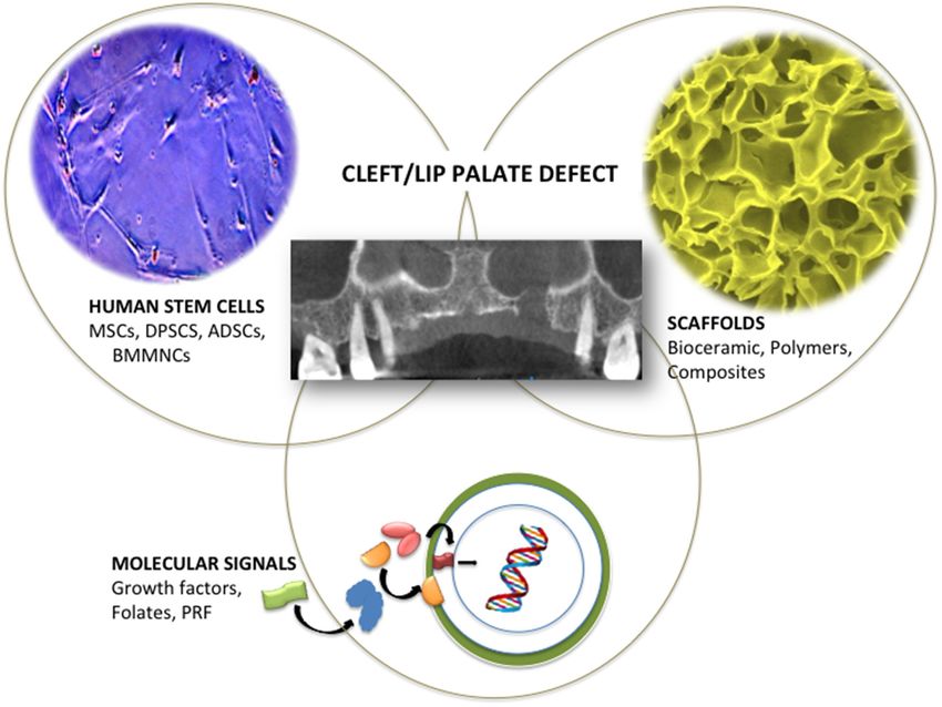
Int. J. Mol. Sci. 2019, 20, x 2 of 13
Int. J. Mol. Sci. 2019, 20, 2176 2 of 13
guided or recruited stem cells (Figure 1) that can modulate the etiopathogenesis of the disease and
its prevalence by promoting the missing self-repairment mechanisms of affected tissues, thus
the etiopathogenesis
improving of the disease
the life conditions of and its prevalence
affected patients. by Thepromoting
functionalthereconstruction
missing self-repairment
of highly
mechanisms of affected tissues, thus improving the life conditions of affected
vascularized bones, such as the craniofacial area, is a key challenge in bone tissue engineering, since patients. The functional
reconstruction of highly vascularized
it depends fundamentally bones, such
on a well-organized as the craniofacial
hierarchical vascular area, is a key
network. Thechallenge
cell survivalin bone
and
tissue engineering,
viability, as well assince the it depends fundamentally
elimination of metabolic wasteon a well-organized
are in charge hierarchical
of the supply vascular
of oxygen network.
and
The cell survival
nutrients carried andoutviability,
by theasblood
well asvessels,
the elimination
in this ofway,
metabolic waste are inof
the restoration charge of the supply of
the neovasculature
oxygen and nutrients
contributes to improve carried
boneout by the blood
functionality [3].vessels,
Scaffold in materials
this way, the restoration
should of the neovasculature
allow vascular regeneration
contributes to improve bone functionality [3]. Scaffold materials
in a fundamental way as well as provide structure, osteonduction and osteoconduction should allow vascular regeneration in
acharacteristics
fundamental way whenasapplied
well as provide structure,
in the field osteonduction
of craniofacial and osteoconduction
regeneration characteristics
[4]. Thus, accordantly with
when applied
different authors, in the
an field
idealof craniofacial
bone regeneration
construction [4]. Thus,
should combine accordantly with
a weightbearing rigid different
scaffoldauthors,
design,
an ideal bone
a porous construction
structure should the
that mimics combine
boneaarchitecture,
weightbearing and rigid scaffold design,
cell-laden materials a porous
that favorstructure
new
that mimics
vascular the bone
formation [5].architecture,
The pore sizeand andcell-laden
shape of amaterials
particularthat favor new
biomaterial playvascular
a key role formation [5].
in vascular
The pore size
ingrowth [6].and shape ofthe
However, a particular
size of biomaterial play a key seems
the interconnections role in vascular
to be more ingrowth [6]. However,
important for the
the size of the interconnections
vascularization of a scaffold when seemscompared
to be morewith important
the poreforsize
the vascularization of a scaffold
[7]. As such, fabrication when
designs,
compared with the
biocompatibility pore size [7]. As
characteristics, such, fabrication
porosity and matrixdesigns,
densitybiocompatibility characteristics,
are of critical consideration [3].porosity
Despite
and
the importance of this knowledge in the study of the craniofacial defect regeneration, thereinhave
matrix density are of critical consideration [3]. Despite the importance of this knowledge the
study
been fewof the craniofacial
studies on CL/P defect
thatregeneration, there have
deepen in assays on thebeenneovascularization
few studies on CL/P ofthat deepen
tissues in assays
through the
on the neovascularization
proposal of new materials.ofThis tissues through
review the proposal
provides an updatedof new and materials. This review
deep analysis of the provides
studies thatan
updated and deep analysis of the studies that have reported on the use of advanced
have reported on the use of advanced biomaterials and cell therapies for the regeneration of cleft lip biomaterials and
cell
andtherapies for the regeneration of cleft lip and palate regeneration.
palate regeneration.
Figure 1. Human stem cells, biomimetic scaffolds, and regenerative molecule signals as fundamental
Figure 1. Human stem cells, biomimetic scaffolds, and regenerative molecule signals as
pieces of the tissue engineering puzzle for cleft/lip palate regeneration.
fundamental pieces of the tissue engineering puzzle for cleft/lip palate regeneration.
1.1. Etiopathogenesis of Orofacial Cleft
1.1. Etiopathogenesis of Orofacial Cleft
Cleft palate (CL/P) malformation occurs as a result of the non-fusion of the primary palate
duringCleft
the palate
fourth (CL/P) malformation
and 12th occurs[2,8].
weeks of gestation as a During
result of the
this non-fusion
period, of theundergoes
the embryo primary palate
rapid
during the fourth and 12th weeks of gestation [2,8]. During this period, the embryo undergoes
changes in shape and growth as the brain expands simultaneously for the formation of the branchial rapid
changes in shape and growth as the brain expands simultaneously for the formation of the
arches responsible for the development of the face and the cranium. Alar structures of the nose are branchial
arches responsible
formed fornasal
by the lateral the development
process while,of during
the facethe
andmandibular
the cranium. Alar structures
processes that takeofplace
the nose are
during
Int. J. Mol. Sci. 2019, 20, x                                                                                         3 of 13
Int. J. Mol. Sci. 2019, 20, 2176                                                                                       3 of 13
formed by the lateral nasal process while, during the mandibular processes that take place during
the eighth week, the shelves ascend above the tongue and then fuse, forming the secondary palate
the eighth week,
completing            the shelves
               the formation         ascend
                                 of the   jaw, above   the tongue
                                                the upper             and then
                                                             lip, alveolus,    andfuse,   forming
                                                                                     primary         the[2].
                                                                                                palate    secondary
                                                                                                             Like anypalate
                                                                                                                        other
completing     the  formation     of the  jaw,  the  upper   lip, alveolus,    and  primary    palate
structural formation in the human body, the entire process is guided by a precise synchronization       [2].  Like any  other
structural
and   balanceformation     in the human
                of cell adhesion,             body, the
                                     proliferation,   andentire    process is regulated
                                                            differentiation,     guided byby   a precise   synchronization
                                                                                                  cell signaling   molecules
and    balance  of cell  adhesion,   proliferation,    and  differentiation,     regulated
from which the family of transforming growth factor beta (TGF-b), fibroblast growth factors   by  cell signaling   molecules
                                                                                                                      (FGFs),
from    which  the  family   of transforming       growth   factor   beta  (TGF-b),    fibroblast
bone morphogenic proteins (BMPs), and sonic hedgehog (SHH) [2,9] stands out. Dysfunctions           growth    factors (FGFs),
                                                                                                                           on
bone morphogenic
these   pathways, mediatedproteinsby(BMPs),     and sonicare
                                      gene regulation,       hedgehog
                                                                responsible(SHH)     [2,9] of
                                                                                for most   stands   out. Dysfunctions
                                                                                              the common                   on
                                                                                                               presentations
these   pathways,    mediated    by  gene   regulation,   are  responsible    for most   of the
of human maxillary alveolar cleft, a bony oronasal communication lined by epithelialized mucosa common      presentations   of
human
and       maxillary
      partially        alveolar
                 erupted          cleft, a bony
                            or unerupted      teethoronasal   communication
                                                     within the    cleft [10].     lined by epithelialized mucosa and
partially  erupted    or  unerupted    teeth   within   the cleft  [10].
       Environmental factors or maternal metabolic imbalances and infections during embryogenesis
       Environmental
ultimately    contributefactors
                            to the or  maternal
                                    etiology        metabolic imbalances
                                                of musculoskeletal               and infections
                                                                        dysfunctionalities         during
                                                                                                being        embryogenesis
                                                                                                        maternal   folic acid
ultimately during
deficiency    contribute
                       the to  the etiology of musculoskeletal
                             periconceptional      period or exposure   dysfunctionalities
                                                                             to alcohol and being       maternal
                                                                                                teratogenic        folic acid
                                                                                                                medications,
deficiency    during    the  periconceptional       period  or   exposure    to  alcohol   and   teratogenic
i.e., retinoids, corticosteroids, and the anticonvulsant phenytoin and valproic acid, which is the main         medications,
i.e., retinoids,
cause             corticosteroids,
        of cleft disorders    [2]. and the anticonvulsant phenytoin and valproic acid, which is the main
cause of cleft disorders [2].
1.2. Prevalence
1.2. Prevalence
      Orofacial cleft conditions have been estimated to have a global annual prevalence of 7.94 cases
      Orofacial cleft conditions have been estimated to have a global annual prevalence of 7.94 cases per
per 10,000 live births with high variances of treated patients across regions and countries (Figure 2)
10,000 live births with high variances of treated patients across regions and countries (Figure 2) [11].
[11]. In some European countries, for example, the prevalence of CL/P has been reported between
In some European countries, for example, the prevalence of CL/P has been reported between 0.53 to
0.53 to 1.59 cases per 1000 live births [12], while the countries that have reported the highest and
1.59 cases per 1000 live births [12], while the countries that have reported the highest and lowest rates
lowest rates were Japan (19.05) and South Africa (3.13), respectively. On the other hand, in the
were Japan (19.05) and South Africa (3.13), respectively. On the other hand, in the American continent,
American continent, the overall case rate is 10.49 per 10,000 live births and this figure is surpassed by
the overall case rate is 10.49 per 10,000 live births and this figure is surpassed by some countries
some countries in South America (i.e., Bolivia with 23.7, Ecuador with 14.96, and Paraguay with
in South America (i.e., Bolivia with 23.7, Ecuador with 14.96, and Paraguay with 13.3). Conversely,
13.3). Conversely, the lowest figures were presented in countries such as Venezuela with 7.92, Peru
the lowest figures were presented in countries such as Venezuela with 7.92, Peru with 8.94, Uruguay
with 8.94, Uruguay with 9.37, and Brazil with 10.12, all for 10,000 live births [13]. Within the USA,
with 9.37, and Brazil with 10.12, all for 10,000 live births [13]. Within the USA, the average prevalence
the average prevalence of cleft lip with or without cleft palate was 7.75 per 10,000 live births,
of cleft lip with or without cleft palate was 7.75 per 10,000 live births, showing differences between
showing differences between ethnicities [14].
ethnicities [14].

       Figure 2.
       Figure     World incidence
              2. World  incidence of
                                  of cleft
                                     cleft lip/palate
                                           lip/palate per
                                                      per surgeon,
                                                           surgeon, anthologist,  and obstetrician
                                                                     anthologist, and obstetrician (SAO)
                                                                                                   (SAO) in
                                                                                                         in each
                                                                                                            each
       country.  Reproduced  from Massenburg     et al. (2018) [11] with permission  from Springer
       country. Reproduced from Massenburg et al. (2018) [11] with permission from Springer ©.      ©.

1.3. Cost at the Health, Social and Economic Level
1.3. Cost at the Health, Social and Economic Level
      CL/P is considered as an anatomical defect of profound aesthetic and functional impact that
      CL/P is considered as an anatomical defect of profound aesthetic and functional impact that
leads to other future alterations, and therefore may negatively impact health-related quality of life,
leads to other future alterations, and therefore may negatively impact health-related quality of life,
and/or speech [12]. Individuals with clefts of the lip, palate, or alveolus often require interdisciplinary
and/or speech [12]. Individuals with clefts of the lip, palate, or alveolus often require
treatment into adulthood and thus they require timely and effective care. In addition, the repercussions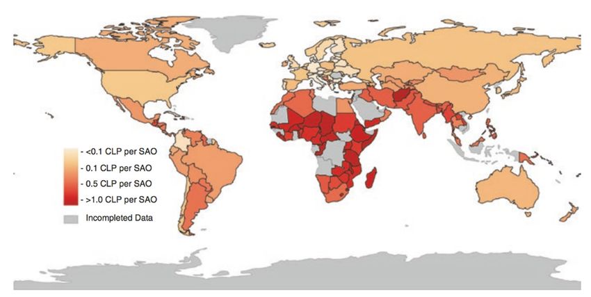
Int. J. Mol. Sci. 2019, 20, x 4 of 13
Int. J. Mol. Sci. 2019, 20, 2176 4 of 13
interdisciplinary treatment into adulthood and thus they require timely and effective care. In
addition, the repercussions of this disease affect the family nucleus and the social environment that
in this
of many cases affect
disease may carry the financial
the family nucleus burden
and the of extensive
social treatment,
environment thatand a variety
in many casesofmay
psychosocial
carry the
challenges
financial [13,15].
burden The economic
of extensive impact
treatment, andofaCL/P therapies
variety on national
of psychosocial health [13,15].
challenges systemsThe is difficult
economic to
estimateofdue
impact to the
CL/P number
therapies onofnational
analyseshealth
and examinations that every
systems is difficult child born
to estimate with
due to athe
CL/P must go
number of
through for
analyses andseveral years. Routine
examinations analysis
that every ofborn
child airway obstruction,
with a CL/P mustin relation to feeding
go through capacity
for several and
years.
nutritional
Routine intake,
analysis ofweight
airwayand growth rates,
obstruction, different
in relation musculoskeletal
to feeding abnormalities,
capacity and nutritional genetic tests to
intake, weight
associate
and growthsyndromes and craniofacial
rates, different examination
musculoskeletal to evaluate
abnormalities, the shape
genetic of associate
tests to the head, syndromes
ears, eyes, nose,
and
jaws and oral
craniofacial cavity need
examination toto be assessed,
evaluate costing
the shape uphead,
of the to $2.4 billion
ears, eyes,per year
nose, according
jaws and oraltocavity
the World
need
Health
to Organization
be assessed, costing[16].
up to $2.4 billion per year according to the World Health Organization [16].
2.
2. Clinical Demands
The
The management
management of of patients
patients with
with CL/P
CL/P pathology
pathology is is complex
complex and and requires
requires aa multidisciplinary
multidisciplinary
approach
approach that
that includes
includes plastic
plastic surgeons,
surgeons, maxillofacial
maxillofacial surgeons
surgeons (cleft
(cleft surgeons),
surgeons), otolaryngologists,
otolaryngologists,
speech/language
speech/languagepathologists,
pathologists,audiologists, dentists,
audiologists, orthodontists,
dentists, psychologists,
orthodontists, geneticists,
psychologists, and social
geneticists, and
workers. Different tissues including bone, dental organs, and soft tissue from the respiratory
social workers. Different tissues including bone, dental organs, and soft tissue from the respiratory system
are largely
system areaffected
largely during
affectedthe CL/P reconstruction
during (Figure 3), (Figure
the CL/P reconstruction therefore3),it therefore
is necessary to necessary
it is standardize
to
the perioperative
standardize managementmanagement
the perioperative of these patients [17].patients [17].
of these
Figure 3. Image
Figure 3. Image of
of aa patient
patient with
with unilateral
unilateral cleft
cleft palate
palate showing
showing thethe different
different tissues
tissues involved
involved (bone,
(bone,
dental
dental organs, respiratory system and soft tissue) that need to be attended during the treatment
organs, respiratory system and soft tissue) that need to be attended during the treatment andand
some
some malformation
malformation around the orofacial
around area responsible
the orofacial for causing respiratory
area responsible for causing and respiratory
speech/language and
problems. Deformation of the arch and dental crowding (A), crossbite dental malposition
speech/language problems. Deformation of the arch and dental crowding (A), crossbite dental (B), and the
deviated nasal septum (C) as revealed by panoramic radiographs showing the maxillary
malposition (B), and the deviated nasal septum (C) as revealed by panoramic radiographs showing defect (circle)
(unpublished
the maxillary data).
defect (circle) (unpublished data).
Regarding the reconstruction of alveolar cleft defects, the most accepted approach consists of the
Regarding the reconstruction of alveolar cleft defects, the most accepted approach consists of
secondary alveolar cleft osteoplasty in the mixed dentition phase [10]. The goal of this surgery is to
the secondary alveolar cleft osteoplasty in the mixed dentition phase [10]. The goal of this surgery is
achieve a normal facial appearance as well as the ability to feed, speak, and hear without affecting
to achieve a normal facial appearance as well as the ability to feed, speak, and hear without affecting
the ultimate facial appearance of the child. To achieve this goal, the most common palatoplasty
the ultimate facial appearance of the child. To achieve this goal, the most common palatoplasty
techniques currently accepted are the von Langenbeck technique, the Bardach 2-flap palatoplasty,
techniques currently accepted are the von Langenbeck technique, the Bardach 2-flap palatoplasty,
the Veau–Wardill–Kilner closure, the 2-stage palatoplasty, and the Furlow palatoplasty [1]. Ultimately,
the Veau–Wardill–Kilner closure, the 2-stage palatoplasty, and the Furlow palatoplasty [1].
there is also variability on the optimal timing to perform palate repair. As transverse facial growth
Ultimately, there is also variability on the optimal timing to perform palate repair. As transverse
is not completed until five years of age, some surgeons have considered retarding cleft palate repair,
facial growth is not completed until five years of age, some surgeons have considered retarding cleft
even to as late as age 8 or 10, to reduce the risk of midface hypoplasia, while others may consider
palate repair, even to as late as age 8 or 10, to reduce the risk of midface hypoplasia, while others
an earlier repair before the age of two, in order to improve speech development and achieve better
may consider an earlier repair before the age of two, in order to improve speech development and
integration in society with less psychosocial impact for the children and families. Taking the middle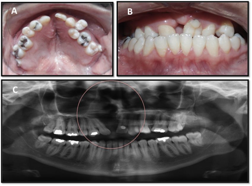
Int. J. Mol. Sci. 2019, 20, 2176                                                                     5 of 13

position, some surgeons have managed cleft palate repair in two stages, with soft palate repair at three
to six months and hard palate repair at 15 to 18 months, while others have advocated a single-stage
repair with both the soft and hard palates being repaired simultaneously. Unfortunately, none of
these surgeries are definitive and may present long-term complications including palatal fistula,
velopharyngeal insufficiency, and midface hypoplasia resulting in facial growth disturbance in multiple
dimensions and cross bite abnormalities such as transverse maxillary hypoplasia that need to be
managed by orthodontic maxillary expansion with fixed appliances and supported by bone grafting in
order to consolidate the dental arch and teeth alignment [1,18].
      Nowadays, the use of autogenous bone is the most widely used type of grafting in bone
regeneration defects [2,19]. However, the availability of autogenous bone is limited and is not free of
tremendous drawbacks, especially in pediatric patients where the availability for harvesting bone may
be limited and thus may not be the ideal graft for alveolar bone reconstruction. In itself, this process is
usually invasive and has the potential for significant morbidities to occur at the donor site, such as
infection, paresthesia, postoperative pain and scarring problems [19,20]. As an alternative, tissue
engineering strategies offer the possibility of using artificial custom made supports for tissues and
cells with the aim for them to be applied in the affected area to promote the regeneration of missing or
damaged tissues.
      The current bioartificial tissues designed for cleft palate reconstruction have been mostly based
on inserted granules isolated with a single tissue layer [10,21]. However, the alveolar cleft defect
typically consists of a two-wall bony defect in which mucoperiosteal flaps are sutured in two layers
to create a new nasal floor and a continuous oral mucosa. As a consequence, the free motion of the
inserted granules negatively affects the dimensional stability and biomechanical properties of the
reconstructed sites, difficulty with the correct closure of these mucoperiosteal flaps, and isolation
from microorganisms that can infect the graft [22]. In order to overcome these limitations, the most
sophisticated approaches to CL/P repair consider the fabrication of biomodels with a 3D shape and
microstructure similar to patients’ bone defects to test the biomechanical properties of bone substitutes
and evaluate the clinical effects with respect to osteogenesis and healing, first in vitro and second in
experimental animals. Several animal models have been utilized for the testing of alveolar cleft grafting
materials including mice, rabbits, cats, dogs, goats, sheep, and monkeys, with rats being the most
referred model among them due to their ease of handling and cost effectiveness. However, these defects
made on rats are significantly smaller in volume than human alveolar defects, thus it is difficult to
extrapolate the results [8,23]. In order to overcome these limitations, according to Pourebrahim et al.,
artificial biomodels created in experimental animals had to fulfill the following criteria: there had to be
a bilateral maxillary alveolar cleft with a 15 mm bony width in each research animal, with demonstrable
oronasal communication, covered by healthy epithelialized mucosa; and there must be functional teeth
on each side [10].
      Some authors have also evaluated in vivo genetically induced CL/P models in rats. It was
described that due to a sevoflurane-induced gene deletion, an incomplete development of the palate
and alveolus was achieved. However, in many cases, the gene defect led to other pathologies and
perinatal lethality, therefore, this methodology has been considered as not suitable to evaluate new
bone grafts [17,24].

Stem Cells Alternative and Growth Factor Assisted Regeneration
      Adult stem cells are considered fundamental for cell therapy because of their unique ability to
self-renew and differentiate into various phenotypes, in addition to being obtained from different
tissues and have been used for craniofacial defect regeneration in tissue engineering. Adipocyte stem
cells (ADSCs) are particularly desirable candidates for musculoskeletal tissue engineering applications
such as cleft lip and palate [10]. In this sense, Pourebrahin et al. proposed the use of adipose tissue
in maxillary alveolar cleft defects, due to their potential for differentiation, the easy accessibility to
this source of cells, and their capability to rapidly expand in vitro. The authors studied the potential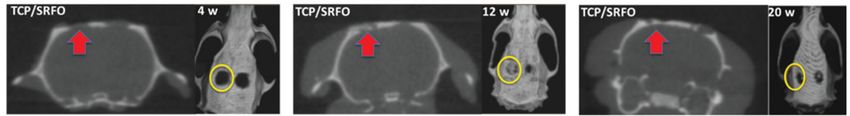
Int. J. Mol. Sci. 2019, 20, 2176 6 of 13
Int. J. Mol. Sci. 2019, 20, x 6 of 13
of ADSCs
studied seeded
the in biphasic
potential boneseeded
of ADSCs substitutes of hydroxyapatite/calcium
in biphasic bone substitutes of triphosphate (HA/TCP) to
hydroxyapatite/calcium
repair maxillofacial bone defects (Figure 4) in a dog model, concluding that they were an
triphosphate (HA/TCP) to repair maxillofacial bone defects (Figure 4) in a dog model, concluding acceptable
alternative
that for the
they were an reconstruction of human
acceptable alternative formaxillofacial bone defects
the reconstruction in the
of human case of limited
maxillofacial autograft
bone defects
availability or morbidity in the donor site [10].
in the case of limited autograft availability or morbidity in the donor site [10].
Figure 4.
4. (Left)
(Left)Scanning
Scanningelectron
electronmicroscope
microscope views
views of the
of the HA/TCP
HA/TCP scaffolds
scaffolds ® seeded
Ceraform
Ceraform ® seeded
with
with Adipocyte
Adipocyte stem(ADSCs)
stem cells cells (ADSCs) used
used for for human
human maxillofacial
maxillofacial reconstruction
reconstruction showingshowing
the abilitythe
of ability
ADSC
of ADSC to
to adhere onadhere on the
the surface surface
of and of and
colonize thecolonize the inner
inner pores of the pores of the
scaffolds. scaffolds.
(Right) (Right)
Alkaline Alkaline
phosphatase
phosphatase analysis of osteogenically
analysis of osteogenically differentiated
differentiated BMSC BMSC
cells after three cells
daysafter three days
of cultivation onof cultivation
bovine hydroxylon
bovine hydroxyl apatite/collagen scaffolds. Reproduced from Pourebrahim et al.
apatite/collagen scaffolds. Reproduced from Pourebrahim et al. (2013) [10] and Korn et al. (2017) [24](2013) [10] and
Kornpermission
with et al. (2017)from
[24] with permission
Elsevier from®Elsevier
and Springer and Springer®, respectively.
, respectively.
Complementary to toADSCs,
ADSCs,another
anothersource
source of of
adult mesenchymal
adult mesenchymal stemstem
cellscells
can be
canisolated from
be isolated
bone marrow
from bone marrow(BMSC)(BMSC)and dentalandpulp (HDPSC).
dental There haveThere
pulp (HDPSC). been multiple
have been examples
multipleof maxillofacial
examples of
bone regeneration
maxillofacial boneusing these sources
regeneration usingofthese
cells.sources
Korn etofal.cells.
demonstrated
Korn et al.that BMSCs could
demonstrated be BMSCs
that used to
promote
could bebone usedformation
to promote in a maxillary defect through
bone formation their osteogenic
in a maxillary defect differentiation
through theirmediated
osteogenicby
BMP-4 (Figure 4) [24], and more recently, Al-Ahmady et al. introduced a novel
differentiation mediated by BMP-4 (Figure 4) [24], and more recently, Al-Ahmady et al. introduced a strategy for alveolar
cleft reconstruction
novel by combining
strategy for alveolar BMSCs seededbyoncombining
cleft reconstruction a collagen BMSCs
sponge seeded
with platelet-rich
on a collagenfibrin (PRF)
sponge
and nano-hydroxyapatite
with platelet-rich fibrin (PRF) [20].and nano-hydroxyapatite [20].
PRF is a platelet concentrate, as a source of growth factors basically used to enhance soft and
hard tissue
tissue healing
healingand andhashasbeen
beenused
usedin plastic andand
in plastic maxillofacial surgery,
maxillofacial in addition
surgery, to many
in addition to tissue
many
engineering
tissue modelsmodels
engineering [25–28].[25–28].
Its advantages include ease
Its advantages of preparation,
include application,
ease of preparation, and absence
application, of
and
chemical alteration. Additionally, previous studies have shown that PRF growth
absence of chemical alteration. Additionally, previous studies have shown that PRF growth factors factors were released
in a time-dependent
were manner, resultingmanner,
released in a time-dependent in prolonged biological
resulting effects [29].
in prolonged In addition,
biological effectsthe fibrin
[29]. In
network of the PRF allows cell migration of endothelial cells essential for angiogenesis,
addition, the fibrin network of the PRF allows cell migration of endothelial cells essential for neurogenesis,
vascularization and subsistence
angiogenesis, neurogenesis, of the graft at
vascularization thesubsistence
and site of regeneration.
of the graft at the site of regeneration.
This is why PRFs have been present as a strong alternative and presumably cost-effective
biomaterial for maxillofacial tissue repair and CL/P CL/P regeneration
regeneration [27].
[27].
3. Biomaterials
3. Biomaterials for
for Soft
Soft and
and Hard
Hard Cleft
Cleft Tissue
Tissue Repair
Repair
Biomaterials play
Biomaterials play aa key
key role
role in
in the
the tissue
tissue engineering
engineering strategy
strategy for
for the
the restoration
restoration of
of missing
missing
tissue and its functionality. In particular, the advances in bone regeneration using biomimetic 3D
tissue and its functionality. In particular, the advances in bone regeneration using biomimetic 3D
scaffolds made of bioceramics, polymers, and composites, using different manufacturing
scaffolds made of bioceramics, polymers, and composites, using different manufacturing methods methods
(i.e.,
(i.e., 3D
3D printing,
printing, cryopolymerization,
cryopolymerization, synthesis,
synthesis, etc.),
etc.), have
have permitted
permitted the
the exploration
exploration of
of new options
new options
for the
for the repair
repair of
of tissues
tissues in
in CL/P
CL/P treatment.
treatment.
3.1. Bioceramics
3.1. Bioceramics
Bioceramics such as hydroxyapatite (HA), α-tricalciumphosphates (αTCP) and β-tricalciumphosphates
Bioceramics such as hydroxyapatite (HA), α-tricalciumphosphates (αTCP) and
(βTCP), demineralized bone matrices, calcium carbonates, calcium sulfates, bioactive glasses, and composite
β-tricalciumphosphates (βTCP), demineralized bone matrices, calcium carbonates, calcium sulfates,
bioactive glasses, and composite materials in combination with bioactive inorganic materials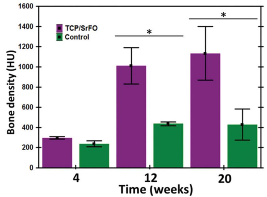
Int. J. Mol. Sci. 2019, 20, 2176 7 of 13
Int. J. Mol. Sci. 2019, 20, x 7 of 13
materials in combination
(bioglasses, etc.) constitutewithan bioactive
importantinorganic
group materials (bioglasses,
of biomaterials usedetc.) constitute an adequate
to manufacture important
group of biomaterials used to manufacture adequate scaffolds in relation
scaffolds in relation to novel treatments for CL/P due to their desired biological properties in terms to novel treatments
for
of CL/P due to their desired
osteoconduction, biological properties
biocompatibility, in terms ofwith
chemical similarity osteoconduction,
natural bone biocompatibility,
and facilitate
chemical similarity with natural bone and facilitate proliferation
proliferation and osteoblast differentiation [30,31]. Janssen et al. described and osteoblast differentiation [30,31].
osteoinductive
Janssen
microstructured βTCP granules, embedded in a glycerol matrix, as an alternative to autologous matrix,
et al. described osteoinductive microstructured βTCP granules, embedded in a glycerol bone
asgrafts
an alternative
for alveolarto autologous
cleft repairbone
becausegrafts
offor alveolar
their abilitycleft repair because
to induce of their ability
bone formation when to induce bone
implanted at
formation when implanted at heterotopic sites in a bilateral alveolar goat cleft model.
heterotopic sites in a bilateral alveolar goat cleft model. These authors hypothesized that the quality These authors
hypothesized
of residual bone that and
the quality
the volumeof residual bone and
of the putty wouldthework
volume of the
at least putty
equal towould work atand,
the autograft leasteven,
equal
tothe
thesurgical
autograft and, even,would
management the surgical management
be superior to the use of would be superior
the regular β-TCP to the use(Figure
granules of the5)regular
[22].
β-TCP
Contrarygranules
to these(Figure 5) [22].
findings, Korn Contrary to these
et al. showed findings,
that when usingKorn et al. showed that when
hydroxyapatite/collagen using
composite
scaffolds, the ossification
hydroxyapatite/collagen of the defect
composite was not
scaffolds, the enhanced,
ossificationprobably due was
of the defect to the micromovements
not enhanced, probably of
theto
due remaining non-resorbable
the micromovements of HA
the particles
remaining after their degradation
non-resorbable of the collagen
HA particles that degradation
after their hampered, asof
in collagen
the the case of autografts,
that hampered, the as
ossification
in the case ofof
theautografts,
defects. Nevertheless, mostofofthe
the ossification thedefects.
investigations using
Nevertheless,
scaffolds
most of thebased on bioceramics
investigations usingare supported
scaffolds basedbyon cellbioceramics
therapy andare growth factorsby
supported and
cellalthough
therapythe and
osteoinduction
growth factors and mechanism
althoughhas the not yet been completely
osteoinduction mechanism revealed,
has not the
yet relationship
been completely between the
revealed,
physical
the and chemical
relationship between features of the osteoinductive
the physical and chemicalbioceramic
features ofandthethe osteogenic differentiation
osteoinductive bioceramic and of
HMSCs and their suitability for craniofacial defect repair including alveolar cleft
the osteogenic differentiation of HMSCs and their suitability for craniofacial defect repair including palate regeneration
has been
alveolar demonstrated
cleft [8,17,19,21,25].
palate regeneration has been demonstrated [8,17,19,21,25].
Figure 5. (Left) Induced bone formation by beta-TCP in the maxillary cleft of goats (A). Material (stars)
Figure 5. (Left) Induced bone formation by beta-TCP in the maxillary cleft of goats (A). Material
is(stars)
reabsorbed by a multinucleated
is reabsorbed osteoclast-like
by a multinucleated cell (arrowhead)
osteoclast-like (B). Elsewhere,
cell (arrowhead) cuboidal osteoblasts
(B). Elsewhere, cuboidal
(black arrow heads) lay down new bone (pink) adjacent to an osteocyte (white
osteoblasts (black arrow heads) lay down new bone (pink) adjacent to an osteocyte (white arrow) inarrow)
its lacuna.
in
Reproduced from Janssen et al. (2017) [22] with permission from SAGE Publications ® . Scale bars:
its lacuna. Reproduced from Janssen et al. (2017) [22] with permission from SAGE Publications ®.
250 (left),250
µmbars:
Scale 25μm (right25A,μm
µm(left), B).(right A, B).
3.2. Polymeric Biomaterials
3.2. Polymeric Biomaterials
Recent advances in macromolecular sciences and tissue engineering methods have made it possible
Recent advances in macromolecular sciences and tissue engineering methods have made it
to efficiently generate several human artificial tissues including the oral mucosa and maxillofacial
possible to efficiently generate several human artificial tissues including the oral mucosa and
bone such as cleft palate [32]. Several synthetic polymer scaffold materials have been used for
maxillofacial bone such as cleft palate [32]. Several synthetic polymer scaffold materials have been
these purposes including poly (ε-caprolactone) (PCL), poly(lactic acid) (PLA), poly(glycerol sebacate)
used for these purposes including poly (ε-caprolactone) (PCL), poly(lactic acid) (PLA), poly(glycerol
(PGS), poly(PGS),
sebacate) (lactide-co-glycolide) (PLGA),(PLGA),
poly (lactide-co-glycolide) or polyhydroxyalkanoates
or polyhydroxyalkanoates (PHA), among
(PHA), amongothers [33].
others
These polymers can be synthesized in large quantities under controlled conditions, thus
[33]. These polymers can be synthesized in large quantities under controlled conditions, thus ensuring ensuring
uniform
uniformandandreproducible
reproducibleproperties
properties while
while reducing
reducing thethe risks
risksofofinfections
infectionsand
andimmunogenicity
immunogenicity [34].
[34].
For example, Flores-Cedillo et al. prepared membrane composites made of multiwall
For example, Flores-Cedillo et al. prepared membrane composites made of multiwall carbon carbon nanotubes
(MWCNTs)
nanotubes with PCL, demonstrating
(MWCNTs) their ability to
with PCL, demonstrating allow
their adhesion
ability andadhesion
to allow proliferation
and of human dental
proliferation of
pulp stem cells (HDPSCs) (Figure 6), and promoting their osteogenic differentiation
human dental pulp stem cells (HDPSCs) (Figure 6), and promoting their osteogenic differentiation toward bone like
phenotypes
toward bone permitting bone regeneration,
like phenotypes permitting boneandregeneration,
thus suitableandforthus
CL/Psuitable
regeneration.
for CL/P regeneration.Int. J. Mol. Sci. 2019, 20, 2176 8 of 13
Int. J. Mol. Sci. 2019, 20, x 8 of 13
Figure 6. Human
Figure 6. Human dental
dental pulp
pulp stem
stem cells
cells seeded
seeded inin multiwall
multiwall carbon
carbon nanotubes
nanotubes with
with PCL
PCL at
at day
day 21
21
with potential
with potential application
application inin CL/P
CL/P regeneration.
regeneration. Osteopontin
Osteopontin labeled antibody was used
used to
to evaluate
evaluate
the expression
expressionofofbone
bonephenotype
phenotype markers,
markers, nuclei
nuclei were
were counter
counter stained
stained with with
DAPIDAPI (unpublished
(unpublished data).
data). Scale10
Scale bars: bars:
µm10 μm 100
(left), (left),
µm 100 μm (right).
(right).
A new generation of advanced 3D polymeric polymeric scaffolds
scaffolds has
has resulted
resulted inin very
very promising
promising results.
results.
Hoshi et al. developed an implant-type tissue-engineered
tissue-engineered cartilage
cartilage using
using aa PLA based scaffold and
evaluated it clinically by inserting it into subcutaneous areas of nasal dorsum in three patients to
correct cleft lip–nose deformity. Subsequently, one year after implantation, the maintenance of the
morphology in the dorsum and apex of the nose of the the patients
patients was
was confirmed
confirmed [35].
[35]. Similar results
were also reported by Puwanun
Puwanun et et al.
al. but using biodegradable electrospun PCL scaffolds with the
ability to
tosupport
supportbone-forming
bone-forming cells andand
cells within cleft palate
within bone defects
cleft palate [36]. Moreover,
bone defects these scaffolds
[36]. Moreover, these
can be developed
scaffolds by incorporating
can be developed hybrid natural
by incorporating hybridderived
naturalbiomaterials such as collagen
derived biomaterials such as or chitosan,
collagen or
that in combination
chitosan, with PCL and
that in combination withPLGA copolymer
PCL and PLGA nanofibers
copolymerserve to offerserve
nanofibers scaffolding
to offeroptions with
scaffolding
superiorwith
options osteogenic
superiorpotential by combining
osteogenic potentialtheby biomimetic
combining and stimulating effects
the biomimetic of natural polymers
and stimulating effects of
and the polymers
natural structuralandandthe
mechanical
structuralstability capabilities
and mechanical of synthetic
stability polymers
capabilities [37–41].polymers
of synthetic On this note,
[37–
an alternative
41]. strategy
On this note, proposedstrategy
an alternative by Zakyproposed
et al. aimed to enhance
by Zaky biocompatibility,
et al. aimed biodegradability,
to enhance biocompatibility,
and material elasticity
biodegradability, andby creatingelasticity
material a biomimetic cellular niche
by creating based on poly
a biomimetic glycerol
cellular nichesebacate
based on(PGS) in
poly
which
glycerolbone marrow
sebacate stromal
(PGS) cells were
in which bonemechanically
marrow stromal stimulated
cells to produce
were their ownstimulated
mechanically extracellular
to
matrix leading
produce to a biochemically
their own mimicking
extracellular matrix environment
leading of bone, while
to a biochemically enabling
mimicking the transmission
environment of
of bone,
mechanical
while forces
enabling with
the the objective
transmission ofofmechanical
treating craniofacial
forces with malformations
the objectiveincluding CL/P
of treating [42].
craniofacial
malformations including CL/P [42].
4. New Manufacturing Techniques for Cleft Palate Reconstruction
4. New Manufacturing
Some Techniques
of the most challenging for Cleftfor
difficulties Palate Reconstruction
craniofacial defect regeneration are derived from the
variety
Someof tissue-specific requirements
of the most challenging and the complexity
difficulties of anatomical
for craniofacial structures in that
defect regeneration are region
derived[43,44].
from
Thus, hierarchical micro-structured and custom-made scaffolds are often
the variety of tissue-specific requirements and the complexity of anatomical structures in required for regenerative
that region
therapies.
[43,44]. Fortunately,
Thus, the current
hierarchical advances in the
micro-structured andfabrication of in situ
custom-made click-chemistry
scaffolds based
are often injectable
required for
formulations, controlled cryopolymerization methods, electrospinning, and 3D direct
regenerative therapies. Fortunately, the current advances in the fabrication of in situ click-chemistry printing of
complex structures with composite biomaterials are able to provide scaffolds
based injectable formulations, controlled cryopolymerization methods, electrospinning, and 3D with adequate nano-,
micro-printing
direct and macro-structure and composition
of complex structures for CL/P
with composite repair. Onare
biomaterials thisable
note,
to Hixon
provideetscaffolds
al. described
with
cryogel scaffolds as tissue-engineered constructs formed at sub-zero temperatures,
adequate nano-, micro- and macro-structure and composition for CL/P repair. On this note, Hixon et with excellent
potential for the
al. described treatment
cryogel of patient-specific
scaffolds bone defects
as tissue-engineered (Figureformed
constructs 7). In addition, thesetemperatures,
at sub-zero authors used
patient-specific 3D-printed molds derived from computed tomography for scaffold fabrication
with excellent potential for the treatment of patient-specific bone defects (Figure 7). In addition, during
the thawing of the cryogels, resulting in a macroporous, sponge-like, and mechanically
these authors used patient-specific 3D-printed molds derived from computed tomography for durable product
for the creation
scaffold of site-specific
fabrication during theimplants
thawing ofin the
the cryogels,
treatmentresulting
of patients
in with CL/P [45]. sponge-like, and
a macroporous,
mechanically durable product for the creation of site-specific implants in the treatment of patients
with CL/P [45].Int.
Int.J.J.Mol.
Mol. Sci. 2019, 20,
Sci. 2019, 20, 2176
x 99of
of13
13
Figure 7. Analysis of a patient custom made patient cryogel. (a) SEM images taken at 1000 and 200X
Figure
(left 7. Analysis
to right). (b) mCTof a3D
patient custom made
reconstruction patient
images cryogel. (a)
representing SEM
both theimages taken
scaffold at 1000
(grey) andinner
and the 200X
(left to right). (b) mCT 3D reconstruction images representing both the scaffold (grey) and
pores with the color bar denoting the size of the pores within the cryogel (left to right). Reproduced the inner
poresHixon
from with the
et al.color bar[45]
(2017) denoting the size of from
with permission the pores
SAGEwithin
®. the cryogel (left to right). Reproduced
from Hixon et al. (2017) [45] with permission from SAGE®.
5. Folic Acid Derivatives as Osteoinductive Molecules for Cleft Palate Regeneration
5. Folic Acid Derivatives as Osteoinductive Molecules for Cleft Palate Regeneration
Maternal folic acid during the periconceptional period is considered to be one of the main causes
Maternal
of clefting folic acid
disorders. during
A recent thepublished
review periconceptional periodVilla
by Fernandez is considered to be one the
et al. [46] highlighted of potential
the main
causes
of of clefting
folic acid as a keydisorders.
bioactiveAcompound
recent review published
to enhance by FernandezofVilla
the effectiveness et al. [46]
biomaterial highlightedand
performance the
potential functions
biological of folic acid as aregeneration
for the key bioactive compound
of tissues to enhance
and organs. the effectiveness
In addition, of biomaterial
new derivatives of folic
performance
acid and biological
bearing bioactive cations functions
such as Sr for
or Znthehave
regeneration
been proven of tissues and organs.
to be promising In addition,
compounds withnewthe
derivatives
ability of folic acid
to accelerate bonebearing
formationbioactive cations such
in craniofacial as Sr
defects orand
[47] Zn have
reduce been proven to be
inflammation promising
[48].
compounds
The therapywithbased
the ability to accelerate
on Sr seems promisingbonedue formation in craniofacial
to its proven defects [47]
action in improving and reduce
preosteoblast
inflammation
replication, [48].
osteoblast differentiation, synthesis of collagen type I, and mineralization of the bone
matrix.TheNonetheless,
therapy based onformulation
any Sr seems promising due to its
should provide anproven action
effective and in improving
consistent waypreosteoblast
to deliver
Sr 2+ ions with
replication, osteoblast
low or thedifferentiation, synthesis pharmacological
absence of secondary of collagen type I, and mineralization
effects. In this regard,ofRojo the et
boneal.
matrix. Nonetheless,
developed a carrier for any
Sr formulation
based on folic should
acid provide an effectivecapacity
with a remarkable and consistent way tobone
of enhancing deliver Sr2+
tissue
ions with and
formation lowsynergic
or the absence
benefits onof cell
secondary
replicationpharmacological effects.
and differentiation In this regard,
processes. Rojo et
In agreement withal.
developed
these authors,a carrier for Sr basedetonal.folic
Martín-del-Campo acid with athat
demonstrated remarkable capacity of
the incorporation ofstrontium
enhancingfolate
bonewithin
tissue
formation
3D and synergic
porous bio-hybrid benefitsprovided
scaffolds on cell replication
an excellent and differentiation
system processes. of
for the regeneration In bone
agreement
tissue with
into
these
the authors, area
craniofacial Martín-del-Campo
(Figure 8) [39]. Theet al.
usedemonstrated that folate
of these strontium the incorporation
derivatives, inofcombination
strontium folate
with
within 3D
HDPSC andporous
biomimeticbio-hybrid scaffolds
scaffolds, provided
is a promising an excellent
alternative system
that can for at
be used theaccessible
regeneration of bone
cost for bone
tissue into the
regeneration, craniofacial
in particular area CL/P
during (Figure 8) [39]. The use of these strontium folate derivatives, in
treatment.
combination with HDPSC and biomimetic scaffolds, is a promising alternative that can be used at
accessible cost for bone regeneration, in particular during CL/P treatment.Int. J. Mol. Sci. 2019, 20, 2176 10 of 13
Int. J. Mol. Sci. 2019, 20, x 10 of 13
Figure 8.
Figure Micro-computed tomography
8. Micro-computed tomography images
images of
of cranial
cranialdefects
defectstreated
treatedwith
withTCP/SrFO
TCP/SrFOscaffolds
scaffoldsatat
4,
12,12,
4, and 20 weeks,
and andand
20 weeks, defect closure
defect on the
closure onside
the of theofimplants
side form the
the implants coronal
form plane (arrows)
the coronal and 3D
plane (arrows)
images (circles) and bone density of the radiographic density (HU) in cranial defects. (*
and 3D images (circles) and bone density of the radiographic density (HU) in cranial defects. (* = = Significant
Significant p < 0.001). Reproduced
differences differences from [39] with
p < 0.001). Reproduced frompermission from the Royal
[39] with permission fromsociety for Chemistry.
the Royal society for
Chemistry.
6. Conclusions and Future Perspectives
The successand
6. Conclusions of synthetic bone grafts is based on their capacity to promote osteoconductivity and
Future Perspectives
osteoinductivity during the formation of new bone growth. In addition, the use of low molecular
The success of synthetic bone grafts is based on their capacity to promote osteoconductivity and
weight compounds such as those derived from folic acid and bioactive cations constitutes a promising
osteoinductivity during the formation of new bone growth. In addition, the use of low molecular
alternative to the use of protein-based growth factors and morphogens, for the preparation of resorbable
weight compounds such as those derived from folic acid and bioactive cations constitutes a
scaffolds in the maxillary defect model to allow osteoconduction and osteoinduction in the defects.
promising alternative to the use of protein-based growth factors and morphogens, for the
In this regard, the use of bioceramics such as calcium phosphate in combination with biomimetic
preparation of resorbable scaffolds in the maxillary defect model to allow osteoconduction and
polymer scaffolds, folic acid derivatives, morphogens, and stem cells are currently considered as the
osteoinduction in the defects. In this regard, the use of bioceramics such as calcium phosphate in
most promising alternative for CL/P regeneration. In addition, emerging bioprinting technologies in
combination with biomimetic polymer scaffolds, folic acid derivatives, morphogens, and stem cells
combination with advanced manufacturing techniques such electrospinning or cryogelation processes
are currently considered as the most promising alternative for CL/P regeneration. In addition,
have permitted the development of new tissue substitutes with a precise control of sizes and shapes
emerging bioprinting technologies in combination with advanced manufacturing techniques such
to recreate the complex physiological, biomechanical, and hierarchical microstructure of biological
electrospinning or cryogelation processes have permitted the development of new tissue substitutes
tissues that are necessary for the regeneration of malformations such as CL/P.
with a precise control of sizes and shapes to recreate the complex physiological, biomechanical, and
hierarchical microstructure
Author Contributions: of have
All authors biological tissues
contributed that
equally aremanuscript.
to the necessary for the regeneration of
malformations such as CL/P.
Funding: This research was supported by the Spanish program MICINN (MAT201573656-JIN) and the Mexican
programs CONACYT (711120) and UNAM-PAPIIT (IA209417).
Author Contributions: All authors have contributed equally to the manuscript.
Acknowledgments: The authors want to acknowledge Christian Navarro Herrera for the images shown in
Figure 1 and
Funding: Ma.research
This Lisseth Flores Cedillo forby
was supported thethe
images shown
Spanish in FigureMICINN
program 6. (MAT201573656-JIN) and the
Mexican programs CONACYT (711120) and UNAM-PAPIIT (IA209417).
Conflicts of Interest: The authors declare no conflict of interest.
Acknowledgments: The authors want to acknowledge Christian Navarro Herrera for the images shown in
References
Figure 1 and Ma. Lisseth Flores Cedillo for the images shown in Figure 6.
1. Moreau,
Conflicts J.L.; Caccamese,
of Interest: J.F.; declare
The authors Coletti, D.P.; Sauk, J.J.;
no conflict Fisher, J.P. Tissue Engineering Solutions for Cleft Palates.
of interest.
J. Oral Maxillofac. Surg. 2007, 65, 2503–2511. [CrossRef]
References
2. Seifeldin, S.A. Is alveolar cleft reconstruction still controversial? (Review of literature). Saudi Dent. J. 2016,
28, 3–11. [CrossRef] [PubMed]
1. Moreau, J.L.; Caccamese, J.F.; Coletti, D.P.; Sauk, J.J.; Fisher, J.P. Tissue Engineering Solutions for Cleft
3. Tian, T.; Zhang, T.; Lin, Y.; Cai, X. Vascularization in Craniofacial Bone Tissue Engineering. J. Dent. Res. 2018,
Palates. J. Oral Maxillofac. Surg. 2007, 65, 2503–2511.
97, 969–976. [CrossRef] [PubMed]
2. Seifeldin, S.A. Is alveolar cleft reconstruction still controversial? (Review of literature). Saudi Dent. J. 2016,
28, 3–11.Int. J. Mol. Sci. 2019, 20, 2176 11 of 13
4. Sun, J.-L.; Jiao, K.; Niu, L.-N.; Jiao, Y.; Song, Q.; Shen, L.-J.; Tay, F.R.; Chen, J.-H. Intrafibrillar silicified collagen
scaffold modulates monocyte to promote cell homing, angiogenesis and bone regeneration. Biomaterials 2017,
113, 203–216. [CrossRef]
5. Mercado-Pagán, Á.E.; Stahl, A.M.; Shanjani, Y.; Yang, Y. Vascularization in bone tissue engineering constructs.
Ann. Biomed. Eng. 2015, 43, 718–729. [CrossRef] [PubMed]
6. Beaumont, M.; DuVal, M.G.; Loai, Y.; Farhat, W.A.; Sándor, G.K.; Cheng, H.-L.M. Monitoring angiogenesis in
soft-tissue engineered constructs for calvarium bone regeneration: An in vivo longitudinal DCE-MRI study.
NMR Biomed. 2010, 23, 48–55. [CrossRef]
7. Bai, F.; Wang, Z.; Lu, J.; Liu, J.; Chen, G.; Lv, R.; Wang, J.; Lin, K.; Zhang, J.; Huang, X. The Correlation
Between the Internal Structure and Vascularization of Controllable Porous Bioceramic Materials In Vivo:
A Quantitative Study. Tissue Eng. Part A 2010, 16, 3791–3803. [CrossRef]
8. Kamal, M.; Andersson, L.; Tolba, R.; Bartella, A.; Gremse, F.; Hölzle, F.; Kessler, P.; Lethaus, B. A rabbit model
for experimental alveolar cleft grafting. J. Transl. Med. 2017, 15, 50. [CrossRef] [PubMed]
9. Marazita, M.L.; Murray, J.C.; Lidral, A.C.; Arcos-Burgos, M.; Cooper, M.E.; Goldstein, T.; Maher, B.S.;
Daack-Hirsch, S.; Schultz, R.; Mansilla, M.A.; et al. Meta-analysis of 13 genome scans reveals multiple cleft
lip/palate genes with novel loci on 9q21 and 2q32-35. Am. J. Hum. Genet. 2004, 75, 161–173. [CrossRef]
10. Pourebrahim, N.; Hashemibeni, B.; Shahnaseri, S.; Torabinia, N.; Mousavi, B.; Adibi, S.; Heidari, F.; Alavi, M.J.
A comparison of tissue-engineered bone from adipose-derived stem cell with autogenous bone repair in
maxillary alveolar cleft model in dogs. Int. J. Oral Maxillofac. Surg. 2013, 42, 562–568. [CrossRef]
11. Massenburg, B.B.; Riesel, J.N.; Hughes, C.D.; Meara, J.G. Global Cleft Lip and Palate Care: A Brief Review.
In Cleft Lip and Palate Treatment; Alonso, N., Raposo-Amaral, C.E., Eds.; Springer International Publishing:
Cham, Switzerland, 2018; pp. 15–23. ISBN 978-3-319-63289-6.
12. Tsangaris, E.; Riff, K.W.Y.W.; Vargas, F.; Aguilera, M.P.; Alarcón, M.M.; Cazalla, A.A.; Thabane, L.; Thoma, A.;
Klassen, A.F. Translation and cultural adaptation of the CLEFT-Q for use in Colombia, Chile, and Spain.
Health Qual. Life Outcomes 2017, 15, 228. [CrossRef]
13. Chavarriaga-Rosero, J.; González-Caicedo, M.X.; Rocha-Buelvas, A.; Posada-López, A.; Agudelo-Suárez, A.A.
Associated Factors with cleft lip and palate in the population attend the “Los Angeles” Children’s Hospital
in Municipality of Pasto (Colombia); 2003–2008. CES Odontol. 2011, 24, 33–41.
14. Tanaka, S.A.; Mahabir, R.C.; Jupiter, D.C.; Menezes, J.M. Updating the epidemiology of cleft lip with or
without cleft palate. Plast. Reconstr. Surg. 2012, 129, 511e–518e. [CrossRef] [PubMed]
15. Zreaqat, M.H.; Hassan, R.; Hanoun, A. Cleft Lip and Palate Management from Birth to Adulthood: An
Overview. In Insights into Various Aspects of Oral Health; Manakil, J.F., Ed.; InTech: London, UK, 2017;
ISBN 978-953-51-3531-9.
16. Hamze, H.; Mengiste, A.; Carter, J. The impact and cost-effectiveness of the Amref Health Africa-Smile Train
Cleft Lip and Palate Surgical Repair Programme in Eastern and Central Africa. Pan Afr. Med. J. 2017, 28.
[CrossRef]
17. Zhang, Z.; Stein, M.; Mercer, N.; Malic, C. Post-operative outcomes after cleft palate repair in syndromic and
non-syndromic children: A systematic review protocol. Syst. Rev. 2017, 6, 52. [CrossRef] [PubMed]
18. De La Pedraja, J.; Erbella, J.; McDonald, W.S.; Thaller, S. Approaches to cleft lip and palate repair. J. Craniofac.
Surg. 2000, 11, 562–571. [CrossRef]
19. Berger, M.; Probst, F.; Schwartz, C.; Cornelsen, M.; Seitz, H.; Ehrenfeld, M.; Otto, S. A concept for
scaffold-based tissue engineering in alveolar cleft osteoplasty. J. Cranio-Maxillofac. Surg. Off. Publ. Eur. Assoc.
Cranio-Maxillofac. Surg. 2015, 43, 830–836. [CrossRef] [PubMed]
20. Al-Ahmady, H.H.; Abd Elazeem, A.F.; Bellah Ahmed, N.E.; Shawkat, W.M.; Elmasry, M.; Abdelrahman, M.A.;
Abderazik, M.A. Combining autologous bone marrow mononuclear cells seeded on collagen sponge with
Nano Hydroxyapatite, and platelet-rich fibrin: Reporting a novel strategy for alveolar cleft bone regeneration.
J. Cranio-Maxillofac. Surg. 2018, 46, 1593–1600. [CrossRef]
21. Martín-Piedra, M.A.; Alaminos, M.; Fernández-Valadés-Gámez, R.; España-López, A.; Liceras-Liceras, E.;
Sánchez-Montesinos, I.; Martínez-Plaza, A.; Sánchez-Quevedo, M.C.; Fernández-Valadés, R.; Garzón, I.
Development of a multilayered palate substitute in rabbits: A histochemical ex vivo and in vivo analysis.
Histochem. Cell Biol. 2017, 147, 377–388. [CrossRef]You can also read