Advisory Document for Wound Bed Preparation in New Zealand
←
→
Page content transcription
If your browser does not render page correctly, please read the page content below
Advisory Document for Wound Bed
Preparation in New Zealand

He ahu umanga ngaio hei whakamahu poka
Advancing Practice and Knowledge in Wound Management

Prepared for New Zealand Wound Care Society by:
Jenny Phillips; NP, RGN, MN, BSc (Hons) – Massey University, Palmerston North.
Christine Gruys; RGON, PGDip HSc -Clinical Nurse Specialist Wound - Taranaki District Health Board
Glenda Dagger; RGON, MHPrac in Nursing, Clinical Nurse Educator, District Nursing Older Adults and
Home Health - Waitemata District Health Board

                                                                                                     1
Published: June 2020                    Review Date: June 2024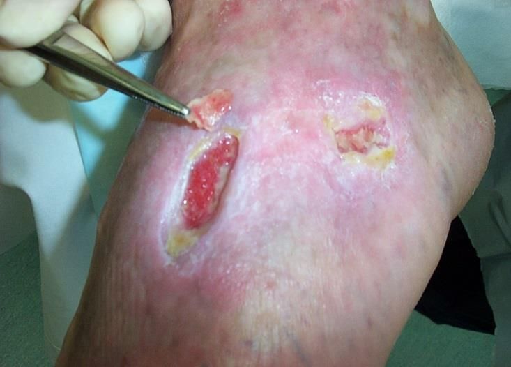
Contents                                                                           Page
       Introduction                                                                       3
       Purpose and Scope                                                                  3
       Factors Affecting Wound Healing                                                    4
       Patient and Wound Assessment                                                       5
       Wound Assessment Models                                                            6
       Pain Assessment and Management                                                     8
       Diagnosis and Treatment Planning                                                   10
       Wound Infection                                                                    11
       Biofilm                                                                            13
       Antiseptics and Antimicrobial Dressings                                            14
       Wound Debridement                                                                  16
       Debridement Considerations                                                         16
       Debridement Description and Methods                                                18
       Future Developments                                                                19
       Summary                                                                            19
       Appendix 1: Debridement Recommendations for Nurses                                 20
       Appendix 2: Wound Product Selection Guide                                          21
       Definitions                                                                        22
       References                                                                         24

Disclaimer
This document has been developed as a guide to support safe wound care practice in Aotearoa/New
Zealand, however, it is not a substitute for education, and wound related competencies, experience,
and the use of clinical judgment. Each health care professional is responsible for their own individual
professional clinical practice and scope of practice as per their regulatory body such as Nursing
Council of New Zealand/ Te Kaunihera Tapuhi o Aoteatoa.

                                                                                                     2
Published: June 2020                     Review Date: June 2024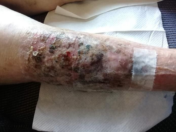
1. Introduction

In accordance with the aims of the New Zealand Wound Care Society (NZWCS) this advisory
document has been designed to guide clinical practice in wound bed preparation.
The NZWCS aims are:
    •    To improve outcomes and quality of life for people with wound and skin integrity problems.
    •    To guide and promote evidence-based practice and wound prevention and management
         education across relevant health care settings.
    •    To be involved at local, national and international level on issues relating to wound
         prevention and management.
Wound Bed Preparation (WBP) is defined as a structured and systematic approach to identify and
remove local barriers to accelerate healing in open wounds. WBP serves to enhance and optimise
healing treatments and advanced therapies.(1)
The NZWCS supports the application of WBP with additional resources such as affiliations to online
learning platforms, wound related guidelines and assessment forms available on the NZWCS website
e.g. Diabetic Foot Ulcer, Pressure Injury, and Venous Leg Ulcer forms.
Key Points:
    •    Wound healability needs to be determined prior to commencement of WBP. A holistic
         assessment of the patient and wound are an essential element of WBP.
    •    Any underlying patient condition, including pain, which will delay healing must, where
         possible, be managed appropriately.
    •    An increased understanding of the importance of biofilms in delaying wound healing has
         resulted in a more structured approach to WBP. Biofilms cannot be seen but may be present
         in 60-100% of chronic wounds. (2) Standard culture methods are not able to capture the
         presence of biofilm or relevant bacteria.
    •    There are several different methods of debridement and it is essential that clinicians choose
         the appropriate method for the patient and the wound they are treating.
    •    Continued disruption of biofilm and ongoing control of bacteria is the next essential step in
         the process. (2)
An interdisciplinary approach is needed to manage complex healable and non-healable wounds to
ensure a holistic and cost-effective approach.

               2. Purpose & Scope

Purpose: The purpose of this document is WBP to enhance and optimise healing in chronic and
complex wounds.
Scope: Health care professionals carrying out WBP and debridement must meet the requirements
of, and practice within, their scope of practice within their professional and employment
organisations. See Appendix 1.

                                                                                                    3
Published: June 2020                     Review Date: June 2024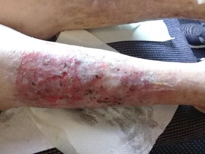
3. Factors Affecting Wound Healing

Complicating factors can have a major impact on the progression of healing. (3) This useful guide to
clinical practice categorises the factors that delay healing into four key groups:
    •    Patient related factors
    •    Wound related factors
    •    Skill and knowledge of the healthcare professional
    •    Resources and treatment related factors. (3)
Patient Related Factors:
Patient related factors need to be identified to determine underlying pathology and associated co-
morbidities. The main factors are best categorised into intrinsic and extrinsic.
Intrinsic local factors - poor venous drainage, lymphoedema, increased skin tension, poor surgical
apposition.
Intrinsic systemic factors - aging, conditions which reduce blood and oxygen flow (such as peripheral
arterial disease), diabetes, obesity, immunological disorders, renal disease, decreased perfusion,
organ failure, sepsis, malignancy.
Extrinsic factors - infection, poor nutrition, tobacco use, radiotherapy, chemotherapy, foreign body,
mechanical pressure, psychosocial, polypharmacy, sensitivity to treatment or product.
Cultural Considerations:
To ensure clinical practice is acceptable, a partnership between the health care professional and the
patient/family (tūroro/whānau) in regard to using conventional and traditional wound healing
modalities is vital(4). Using appropriate cultural models of care such as Te Whare Tapa Wha offers a
cultural framework which may optimise health outcomes for Māori.(5)
Traditional healing modalities such as Rongoā Māori incorporate wellbeing dimensions of cultural
models into wound care and treatment. The healthcare professional is advised to have input from
local Māori healthcare providers for information regarding traditional Māori healing (Rongoā Māori).
Rongoā Māori is informed by ancestral knowledge passed down through the generations and has at
its core the enhancement of Māori wellbeing. Rongoā is formulated into a Māori cultural context in
which understanding of events leading to ill health and its impact is addressed through a range of
culturally bound responses. (4)
Wound Related Factors:
Wound duration, fibroblast senescence, wound size, depth and location on pressure bearing surface
or mobile area, unhealthy wound bed condition, ischaemia, exaggerated inflammatory phase, matrix
degradation and reduced growth factor bioavailability all reduce tissue repair.(6) Infection and the
prolonged presence of bacteria/biofilm in the tissue stimulate chronic inflammation further delaying
healing. (2) The initial response to wound care can guide prediction of healing time and tissue
viability.
Skills and Knowledge of Healthcare Professionals
Healthcare professionals require ongoing wound prevention and treatment education to increase
knowledge and skills so cost-effective and appropriate management is provided. As professionals we

                                                                                                   4
Published: June 2020                     Review Date: June 2024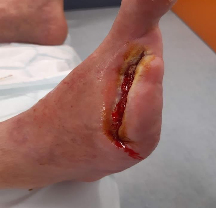
need to critically reflect on our practice to ensure we are providing the best possible care and
outcome for the patient. This involves working with the patient holistically by identifying the
patients’, family/whānau needs and concerns, barriers to healing, and engaging in collaborative
practice.(3)
Resources and Treatment Related Factors
Healthcare professionals require access to a range of wound care products and equipment with
supporting evidence-based procedures. New approaches to wound care continue to be
investigated, such as molecular biomarkers to recognise and target treatment earlier and more
effectively.(7) It is important that new approaches to wound management are accessible,
transferrable and are cost-effective for healthcare professionals to utilise.

               4. Patient and Wound Assessment

Wound assessment is used to describe a wound at a given point in time:

•   Accurate wound assessment and an understanding of the complexities of wound management is
    essential in ensuring that cost-effective and evidence-based interventions are used.
•   Wound assessment will help determine the treatment; practitioners need to ensure they have
    the essential skills required to plan, implement, and evaluate care on an individual basis.
•   Wound assessment should be repeated at any dressing change, or if there is a clinical indication
    for it, to establish indications for healing.
•   Comprehensive assessment of the patient involves inclusion of the whole patient not just the
    hole in the patient.
•   A structured approach incorporating a wound assessment model is necessary and
    recommended.
Wound Tissue Types:

Wound assessment includes the interpretation and classification of tissue in the wound. Tissue
colours (see Figure 1) are often assessed as a percentage within the wound and may include:

•   Necrotic: can be soft and wet, or hard and dry; the colour can be black or dark brown/tan.
•   Slough: moist or wet; typically, yellow or grey/green colouration. Note yellow may indicate
    bone, cartilage, tendon, ligament or fat.
•   Fungating: dry or wet; may bleed easily, and have a mix of colours ranging from black, yellow to
    red, crater or cauliflower appearance.
•   Granulation: moist; pink or red; healthy granulation should be firm and granular which is often
    described like coble-stone or a raspberry; friable tissue may indicate local infection.
•   Hyper or over-granulation: raised moist beefy red tissue; often occurs when the wound is too
    wet, foreign body is present, friction or increased bacteria.
•   Epithelialised tissue: dry, pink or white, final stage of wound healing.

Refer to Appendix 2 for Wound Product Selection Guide and Aim of Treatment, for Chronic Wounds.

                                                                                                  5
Published: June 2020                      Review Date: June 2024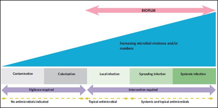
Wound Exudate
Exudate is necessary in wound healing, but when pro-inflammatory cytokines and matrix
metalloproteases (MMPs) are elevated, and growth factors and mitogenic activity are lower, wound
healing is stalled.(8) Assessment of exudate should include type, colour, consistency, amount and
odour. Current dressing regime and determining wound aetiology plus other factors, such as low
serum albumin levels, can also affect exudate levels.(8) Refer to the World Union of Wound Healing
Societies Consensus Document for further information including types of wound exudate. (8)
Wound Edge
To advance epithelialisation, the edge of the wound needs to be moist and at the level with wound
tissue.(9) Unhealthy wound edges include macerated, undermined and rolled: these need to be
addressed to advance wound healing.(9,10) Ensure undermined wounds are not over packed and
leave a gap between the undermined tissue edge and dressing to allow wound advancement.

Figure 1: Tissue Types

 Granulation approx. 60%      Epithelialised approx. 30%          Slough approx. 10%

Wound Assessment Models to Support Wound Bed Preparation:
There are several well-known assessment models which are continually evolving and are now much
more holistic. Listed below are a few examples:

TIME – provides a framework to address factors in the wound bed which delay healing. Components
focus on management of necrotic tissue, infection and exudate.(11)
•   Tissue management
•   Inflammation and infection control

                                                                                                    6
Published: June 2020                     Review Date: June 2024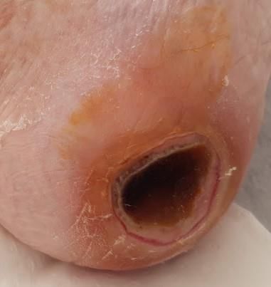
•   Moisture balance
•   Epithelial (edge) advancement

A recent expert review recommended an expansion of TIME which includes a Clinical Decision
Support Tool providing a systematic, structured, evidence-based approach to wound management.
It involves a holistic assessment and involving the multidisciplinary team (MDT) as appropriate,
wound assessment and treatment using TIME, and evaluation.(12)
•   Assess: accurate assessment, measurement and diagnosis of the patient and their wound
•   Bring in the MDT to promote holistic care
•   Control and treat systemic causes
•   Decide appropriate treatment
•   Evaluate treatment and wound management goals

The TRIANGLE of Wound Assessment(9) includes assessment of:
•   The wound bed and types of tissue using TIME
•   Wound edges
•   Peri-wound skin that extends 4cm from the wound bed

HEIDI A structured approach to define health status of patient and tissue type within the wound and
identify associated factors which may impact on the plan of care and the process of healing.(13)
• History: medical, surgical, pharmacological, psychosocial, barriers to healing.
• Examination: the patient as a whole, then the wound.
• Investigations: such as, X-ray, Duplex scan, bloods, wound cultures.
• Diagnosis: follow accepted pathway.
• Intervention (see Appendix 2 Wound Product Selection Guide for Chronic Wounds).

Comprehensive History:
History of presenting wound: site, date of onset, duration (>6 weeks considered chronic wound),
any associated symptoms such as pain, exacerbating factors - include past dressings used and what
has worked what has not.
• History of previous wounds at the same site or fractures affecting the same limb.
• Medical/ Surgical history.
• Medications, over the counter medicines, alternative therapies, rakau rongoā (native flora
    herbal preparations).
• Social history- smoking tobacco/vaping use, alcohol intake, mobility, living status – care and
    supports in place.
• Review of appropriate systems i.e. vascular history - claudication or rest pain for wounds on the
    lower limb.
• Is this an accident which resulted in a wound that treatment could be funded by Accident
    Compensation Corporation (ACC) e.g. skin tear, pressure injury.

Examination:
•   Location of wound(s).

                                                                                                  7
Published: June 2020                    Review Date: June 2024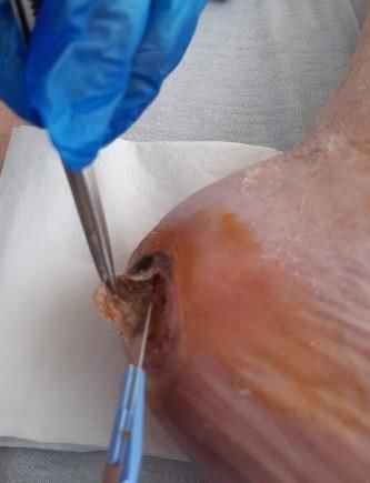
•   Wound measurements including length, width, depth, undermining, sinuses, and tunnels. Note if
    the wound probes to bone there is a risk of developing osteomyelitis and a base-line x-ray is
    recommended, and the treatment plan may include debridement and/or antibiotics.
•   Use TIME to describe the wound bed and surrounding tissue including odour and exudate.
•   For all leg wounds perform basic lower leg assessment including temperature, pulses, and
    capillary refill time. If there is uncertainty whether the blood supply is normal, early referral for
    an advanced lower limb assessment should be considered. These include ankle brachial pressure
    index (ABPI) and non-invasive vascular studies (NIVS).

Document the Assessment:
    •   Utilise your organisational wound care plan or wound assessment tool to guide the
        assessment process.
Investigations: will be determined by the above findings:
    •    Wound swab to identify infection.
    •    Biopsy to rule out neoplasm.
    •    Arterial or venous investigations including ABPI, NIVS or Duplex scan.
    •    X-rays to determine osteomyelitis.
Diagnosis:
Wounds are a result of trauma, venous insufficiency, arterial insufficiency, inflammatory,
neuropathic, pressure, neoplastic or unknown aetiology. To make a diagnosis you need to be able to
consult a clinician who can help with the diagnosis to then be able to facilitate the best treatment
possible.
Interventions:
Discuss proposed plan of care with patient/whānau identifying goals of wound healing. In some
cases, healing may not be possible due to underlying condition such as cancer or peripheral vascular
disease. Management of symptoms e.g. pain, infection, exudate, and odour are the goals of care.
    •    Document your plan of care, review date and timeframe for wound reduction, healing or
         palliation.
    •    Ensure all clinicians are aware of the plan and can implement it.

               5. Pain Assessment

Wound Pain:
Wound pain can be broadly categorised into two main types:
    •    Nociceptive pain results from activity in the neural pathways secondary to actual tissue
         damage; for example, trauma, prolonged pressure causing ischaemia, bruises, burns,
         fractures, and infection. These conditions can all heal, therefore acute nociceptive pain will
         be expected to resolve. (14) Common descriptors include nagging, aching, tender, sharp or
         stabbing pain. (14,15)

                                                                                                       8
Published: June 2020                      Review Date: June 2024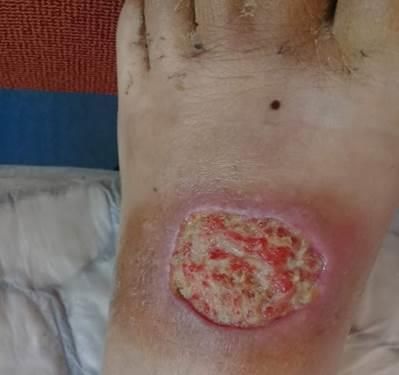
•    Neuropathic pain is caused by nervous system lesions or dysfunction, for example diabetic
         peripheral neuropathy. This pain is more persistent often leading to chronic pain; it is often
         described as shooting, burning, and pins and needles. (14,15)
Note: Allodynia can result from continuous pain; this causes a person to experience pain from a non-
painful stimuli. (15)
Pain can affect sleep, function, and increase anxiety leading to increased cortisol levels that can
affect the immune system and healing, hence pain assessment and management are paramount.(15)
MacLeod(16) defines pain as a combination of sensory, emotional, and cognitive phenomenon and
as a stressor, ongoing pain will interfere with wound healing (see Figure 2).

                                                Spiritual
                                                  Culture
                                              Religion / Belief
                                               Meaning of Life
                                                Helplessness

                Physical                                                       Emotional
                 Diagnosis
                Therapy Side
                                                Total                               Anger
                                                                                 Anxiety, Fear
                  Effects
                 Insomnia
                                                Pain                            Sadness, Loss
                                                                                Disfigurement
              Chronic Fatigue

                                                  Social
                                                Relationships
                                                   Roles
                                              Cultural Attitude

Figure.2 –Adapted from MacLeod. (16)
Pain assessment:
    •    It is essential to be guided by the patient’s description of pain as it applies to them, this will
         assist the healthcare professional to determine whether the pain is acute or chronic,
         neuropathic, and/or nociceptive.
    •    Use a validated pain assessment tool that is appropriate to the population, such as children
         and people with dementia.
    •    Procedural pain is associated with wound cleansing and dressing procedures; and operative
         pain with interventions such as debridement or biopsy. (15)

                                                                                                         9
Published: June 2020                       Review Date: June 2024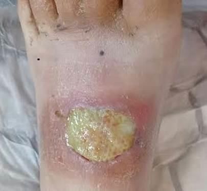
•    When assessing pain note onset, duration, type, location, frequency, persistency, and
         severity. Note precipitating and alleviating factors.
    •    Pain assessment should occur pre, during and post WBP procedures, especially during any
         debridement procedure
    •    An increase or change in pain is a strong indicator of infection.(15)
    •    Painful stimuli below the injury level in patients with spinal cord impairment can lead to an
         increase in spasticity and/or a potentially fatal hypertensive condition called autonomic
         dysreflexia. (17)
    •    Consider what current pharmaceutical and non-pharmaceutical interventions are being
         used, and their effectiveness.
    •    Pain associated with underling conditions such as inflammatory diseases and limb ischaemia
         need to be referred urgently to relevant Clinicians.
Pain Management:
Identify causes of pain and help manage these:(15)
    •    Gain verbal consent prior to any procedure and advise the patient they can stop the
         procedure at any time.
    •    Explore other pain strategies such as allowing the patient to help cleanse/dress their wound,
         massage, and using relaxation techniques such as breathing and music.
    •    Ensure prescribed analgesia is used prior to painful procedures (e.g. oral, inhalation, topical
         and/or intravenous) and assess the effectiveness of this.
    •    Consider factors that can contribute to pain such as infection, maceration, ischaemia, and
         pressure.
    •    Dressing removal and antimicrobial dressings can contribute to pain.
    •    Avoid prolonged wound exposure.

               6. Diagnosis and Treatment Planning:

A comprehensive history, physical examination and diagnostic reasoning will lead to an accurate
diagnosis of the wound and a plan of treatment. A plan of care should include:

    •    Care that is co-designed with patient/family-whānau/carer and relevant interdisciplinary
         team members.
    •    Treatment goals are holistic, realistic, and achievable, some wounds may not heal, seek
         expert advice if there is any doubt.
    •    Patient risk factors for wound healing are identified and incorporated into the plan of care.
    •    Availability of resources, equipment and wound care supplies within your health care
         setting.(3)
    •    For wounds related to/caused by trauma or treatment injury, patients may be eligible for
         Accident Compensation Corporation [ACC] funded wound care related consumables - see
         ACC website.

                                                                                                         10
Published: June 2020                      Review Date: June 2024
7. Wound Infection
Wound infection is a common complication in both acute and chronic wounds. In 2016 the
International Wound Infection Institute (IWII) updated the principles and practice for wound
infection.(18)
Wound infection delays the healing process and, if left untreated, may progress to systemic and life-
threatening sepsis/illness. Early identification and management are important to reduce morbidity
and mortality rates. Addressing local wound infection using a local application of an effective topical
antimicrobial reduces the potential for development of spreading infection and developing antibiotic
resistance. (18)
Stages of Wound Infection:
The approach to treatment will depend on the extent of the infection. The IWII continuum
characterises the relationship between microbes, infection, and recommendations for systemic or
local treatments,(18) see Figure 3.
Figure 3: IWII Wound Infection Continuum. (16)
As microorganisms proliferate in a wound, the host response to the infection may progress from
localised symptoms in and around the wound, to more systemic effects as shown in Table 1.
11
Published: June 2020 Review Date: June 2024Table 1: Signs and symptoms associated with stages of the wound infection continuum.(18) Copyright
permission given by Dr G Sussman, for information retrieved from Wound infection in Clinical Practice via
email on 6th Dec 2019.
Diagnostics to Identify Wound Infection:
The clinician’s observation and understanding of signs and symptoms is important to assist with
identifying developing wound infection. Figure 3 and Table 1 provide guidance on identifying levels
of infection.(18)
Further investigations for clinical microbiology are indicated for:
• Acute wounds with classic signs and symptoms of infection.
• Chronic wounds with signs of spreading or systemic infection.
• Infected wounds that have failed to respond to antimicrobial intervention or are
deteriorating despite appropriate antimicrobial treatment.
• Wounds where the presence of certain species would negate a surgical procedure (e.g. beta
haemolytic streptococci in wound bed prior to skin grafting). (16)
Clinical Microbiological Sampling Techniques include:
• Wound swab using the Levine technique (Table 2).
• Collection of pus in a sterile specimen container.
• Needle aspiration.
• Tissue biopsy: wounds with antibiotic-resistant species. Sample from edge and centre of
wound bed.
Wound Swabs:
Superficial wound swabbing is frequently used, although it has limited use in identifying colonisation
versus infection in chronic wounds, especially if a biofilm is present. However, laboratory
investigation provides clinicians with information about the organisms present in a wound and their
antibiotic sensitivities, which can inform treatment decisions. The Levine technique is considered
the preferred sampling method, see Table 2. (16)
12
Published: June 2020 Review Date: June 2024Table 2 – The Levine Technique adapted from IWII. 16 page 12
Note: Wound Biopsy and Needle aspiration should only be undertaken by staff trained in
local procedures. Advanced diagnostic techniques are not routinely available and must be
ordered by Infection Control Specialist / Microbiologist.
8. Biofilm
A biofilm is described as an aggregate of bacteria tolerant to treatment and the host defence,(2,19)
see Table 3. The subject of biofilm in chronic wounds is complex and a recent position document
has given guidelines for the treatment of biofilm(2); these include:
• WBP and biofilm removal/disruption include use of an appropriate debridement methods to
disrupt biofilm and remove necrotic and devitalised tissue.
• Use of topical surface surfactants which lower surface tension and assist disruption of
potential or actual biofilm formation from the wound surface.
• Use of antimicrobials immediately after debridement to reduce bacterial growth on
vulnerable tissue.
• Repeat the process as necessary, a single debridement is not enough to prevent biofilm
reforming.
• Reassess regularly to see if treatment objectives are being achieved.
13
Published: June 2020 Review Date: June 2024Table 3: The Stages of Biofilm Development Adapted (2)
Stages of Biofilm Formation Stage Description Action to Manage Biofilm
Reversible attachment Micro-organisms attach to surface; Clean the wound – consider using a
this initial attachment is reversible surfactant and/or monofilament fibre
wand or pad
Permanent surface attachment 2- Planktonic microbes attach and form Dressing with surfactant and
4 hours micro-colonies antimicrobial
Protective matrix forms 6-12 Bacteria start to secrete surrounding Continue
hours matrix and micro colonies become
more tolerant to systemic and local
biocides
Increasing tolerance to biocides Without disruption micro colonies Debride regularly and follow up with
2-4 days become fully mature biofilm monofilament wand or pad, surfactant
colonies and further biofilm colonies and antimicrobial
can develop
Reformation 24-72 hours post There is rapid recovery from Repeat whole process at 72 hours
disruption mechanical disruption and a mature,
tolerant biofilm reforms. There are Re-assess wound regularly
24 hours after debridement when
antimicrobials are most effective in
reducing bacteria
9. Antiseptics and Antimicrobial Dressings:
Agents capable of killing (biocidal) or inhibiting (biostatic) micro-organisms are used in conjunction
with debridement to prevent biofilm build up see Table 4. (19)
Treatment goals when using antimicrobial wound dressings are to:
• Prevent the spread of local infection and reduce antibiotic use.
• Disrupt biofilm to advance wound healing.
• Achieve faster resolution of spreading infection in conjunction with oral antibiotics.
How do I know which antimicrobial to choose?
In accordance with your wound assessment, e.g. amount of exudate, tissue type, pain or odour,
dressing availability and the mode and duration of action.(20)
• Use the 2-week rule, use an antimicrobial for 2-weeks then review.
• If there are signs of improvement, and signs and symptoms of infection are resolving,
discontinue the antimicrobial dressing. Often longer use of antimicrobials are required in
chronic wounds.
• If the wound is improving, but the signs and symptoms of infection are still present, continue
for a further 2 weeks with the same antimicrobial or a different one.
• If the wound probes to bone, a baseline x-ray and antibiotics are recommended.
14
Published: June 2020 Review Date: June 2024• If the wound has deteriorated, fully reassess patient holistically, as well as wound factors,
and consider an alternative product or systemic antibiotic treatment.
Table 4: Products Available in New Zealand to Disrupt/Manage Biofilms Adapted from Wounds
UK.(20) For additional product information the Silver Chain Wound Care Manual is recommended.
(21)
Active control Mode of delivery Rationale for use Guidance for use
Iodine Solution, cream, Localised or spreading Dress 2-3x weekly
Povidone iodine ointment, spray, or infection. To prevent wound If not improving at 10-14 days,
Cadexomer iodine impregnated infection or recurrence in re-evaluate and review change
dressings susceptible patients. Rapidly of dressing regime
kills microorganisms and
suppresses biofilm formation.
Medical (UMF) grade honey Liquid, gel sheet, Autolytic debridement Not suitable for highly exuding
impregnated Impede biofilm formation and wounds.
dressing, barrier disrupt established biofilm Ensure direct contact with
cream Reduce odour wound bed
Decrease wound related pain Secondary dressing required
and inflammation *Caution bee venom allergy
Octenidine dihydrochloride Solution and gel Cleanse and decontaminate Solution: leave on 5 min. Apply
(gel used as a wound. Manage wound solution to wound bed, leave
dressing) bioburden/biofilm for 5 minutes.
Autolytic debridement Can be used to soften adhered
Donate moisture to wound dressings
Polyhexamethylenebiguanide Solution, gel, Cleanse/decontaminate Solution: leave on 10-15 min.
(PHMB) impregnated wound Gel: can be used in cavities.
dressings, and Suppress biofilm formation Pads used for conservative
debridement pad Reduce wound odour debridement
Manage wound bioburden
Super oxidised solution of Gel, solution Moist healing environment Apply spray close to wound Do
hypochlorous acid and Physical kill of bacteria not rinse off
sodium hypochlorite Reduces inflammation
Polymeric Hydrophilic Moisturiser Cavity and flat foam dressing
urethane matrix Soothing – reduce pain
contains mild, Includes a silver version
nontoxic wound
cleanser and
surfactant
Silver: Metallic, Nano- Impregnated Manage wound bioburden Some dressings require wetting
crystalline, Ionic dressings Provide antimicrobial barrierto activate Silver, follow
Reduces inflammation manufacturer instructions. Use
for 2 weeks, can be used up to
4 weeks if improving,
otherwise re-assess
Monofilament pads/ Mechanical action Breaks down and removes Should be used moist and may
wands(22) wound debris and skin flakes be used in conjunction with
and keratosis. antimicrobial cleansing
Disrupts and removes biofilm solutions which contain a
surfactant
15
Published: June 2020 Review Date: June 202410. Wound Debridement:
Wound debridement and peri-wound skin care is an essential part of wound bed preparation and
aims to remove non-viable tissue and disrupt established and forming biofilm. (2,10,23,24) Non-
viable tissue and biofilm delay wound healing and provide a focus for infection, prolong the
inflammatory response and obstruct wound re-epithelialisation and contraction. Biofilm can also
form in peri-wound skin debris, hence it is important the wound bed and peri-wound skin, up to 10-
20cm surrounding the wound, is cleansed to remove loose tissue, exudate and skin scales using an
antiseptic or antimicrobial wash or surfactant solution.(10) (see Figure 4) Attention to the wound
edge, such as shaving callus and hyperkeratosis, removing rolled-under and devitalised tissue will
facilitate wound advancement.(10)
Figure 4: Pre and post skin cleansing to remove surface contaminants and skin scales.
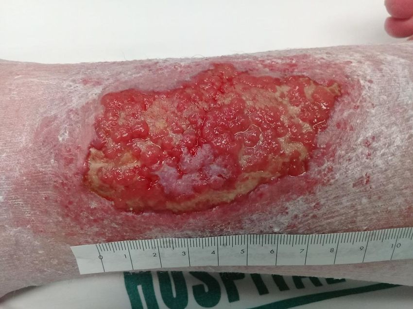
Debridement should be performed more than once to reduce the impact of biofilm reformation,
initially at least every 72 hours if not 48 hours.(2,23) To aid biofilm removal it is recommended
debridement leads to pinpoint bleeding as tolerated by the patient (see Figure 5.(10) Ongoing
wound assessment will determine what level of debridement is required and when it is no longer
needed. It is paramount that products which disrupt biofilm reformation are used (see Table 4).
Debridement Considerations
• Healability of the wound and patient condition.
• Patient safety and informed consent.
• Inform the patient they can stop the procedure at any time.
• Healthcare professional knowledge and skill level.
• Adhere to local debridement / wound care policies and protocols.
• Patient participation (e.g. washing/showering the wound and surrounding skin).
• Environment and resources (e.g. lighting, magnifying devices assist with more accurate
assessment and debridement, safe body positioning).
• Prevent cross-infection (hand hygiene, PPE, sterile equipment, and dressings).
• Manufacturers’ instruction should always be followed along with local policies and procedures.
• In larger complex wounds debridement may be achieved over a period of days or weeks.
• Adequate perfusion to aid wound healing.
16
Published: June 2020 Review Date: June 2024• Avoid debridement that causes any pain in autoimmune disorders such as pyoderma
gangrenosum.
• Pain management.
• Identified devitalised tissue and structures (e.g. tendon, bone).
• Risk of bleeding (anatomical areas, bleeding disorders and anticoagulant therapy).
• Risk of infection in immunosuppressed patients.
• Wounds located on the feet, hands, and face require specialist involvement to ensure safe and
effective treatment.
• Avoid debridement on stable, hard, dry eschar in ischemic wounds on the lower limb/feet(25).
Devitalised tissue should be removed at each dressing change. Forefoot debridement causing pinpoint bleeding.
Figure 5: Debridement
Autolytic debridement: a hydrogel has softened the Conservative debridement: metal forceps were used
non-viable tissue on a chronic burn injury. to remove the slough causing no bleeding or pain. A
dressing was used to further debride the firm slough.
Figure 6: Autolytic and Conservative Debridement (photos taken the same day).
17
Published: June 2020 Review Date: June 2024Methods of Debridement
Debridement methods will depend on the skill level and competency of the registered health
professional. To help guide practice Table 5 lists debridement options, methods and the level of skill
recommended.
Table 5: Debridement Description and Methods
Debridement Description and Methods
Autolytic: Slowest method using wound products to provide moisture donation, a highly selective
process involving macrophage and endogenous proteolytic enzymes which liquefy and separate
necrotic tissue and eschar from healthy tissue. Autolytic debridement may be further facilitated by
scoring eschar.(10,24,26)
Autolytic Methods Skill Level
Dry Necrosis: use moisture denoting products e.g. hydrogels, enzymatic, Low
hydrocolloids, combination dressings including soft silicones, polyurethane films.
Moist to Wet Non-Viable Tissue: use wound products to aid debridement and
Low
obtain moisture balance, e.g.: antimicrobials (see Table 4), enzymatic, alginates,
hydrofibers, cellulose dressings, foam dressings, and composite dressings.
Practice point: Autolytic debridement with occlusive dressings is contraindicated for
infected wounds. Dressings that support autolytic debridement should not be left in
place for longer than 3 days so that the progression of wound debridement and any
onset of infection can be closely monitored.
Mechanical: Physical force is applied to the surface of the wound to disrupt and remove non-
viable tissue and debris. (10,24)
Mechanical Methods Skill Level
Monofilament wands and pads(22), gauze or antiseptic sponges, to break down and Low
remove wound debris, skin scales and keratosis.
Dry, or wet to dry, gauze dressings: this can be painful and is not recommended Low
with newer options available.
Pressure irrigation, pulsed lavage, hydrotherapy, low frequency ultrasound Moderate
Sharp: Includes surgical and conservative debridement. (10,24,27)
Sharp Methods Skill Level
Sharp using scalpel and scissors under topical or general anaesthetic to viable High
bleeding tissue. Usually performed in theatre or specialist clinic.
Conservative using scalpels, curettes, scissors, and forceps causing minimal pain or Moderate to
bleeding. See Figures 5, 6 and 7. High
Biological: Use of sterile larvae (from Lucilla Seratica) which selectively remove Moderate
non-viable tissue through excretions and secretions of proteolytic enzymes and
mouth hooks which liquefy and ingest non-viable tissue and bacteria.(28).
18
Published: June 2020 Review Date: June 2024Note necrosis is naturally separating at Conservative sharp debridement, Post debridement with no bleeding. A
the skin and wound edge. Advise the using a forcep and blade to remove hydrogel was used to soften and deslough
patient the wound may appear deeper or the dead tissue causing no pain. the wound bed.
larger when non-viable tissue is removed.
Figure 7: Conservative Sharp Debridement
RECOMMENDATION: For additional practice information we advise reading: (22)
Murphy C, Atkin L, Swanson T, Tachi M, Tan YK, Vega de Ceniga M, Weir D, Wolcott R. International
consensus document. Defying hard-to-heal wounds with an early antibiofilm intervention strategy: wound
hygiene. J Wound Care 2020; 29(Suppl 3b):S1–28.
11. Future developments
New approaches to wound care and treatments have been and continue to be investigated. These
include gene expression profiling.(29) Wound dressing products derived from animals, advanced
interventions and technologies and therapies are already in use. Access to patient genetic data by
clinicians in future treatments will play a major role in determining wound outcomes not previously
utilised. (28)
12. Summary
At the time of publishing, this advisory document contains the most up to date evidence available
around the area of wound bed preparation; however, new products and methods are constantly
coming into the market and need to be evaluated as to their effectiveness and acceptability to
health professionals and patients. What remains constant is the need for holistic and
comprehensive wound assessment. Wound bed preparation is undertaken by health professionals,
such as Registered Nurses working in New Zealand, who are managing/overseeing patient care. They
require to have the knowledge and skills to provide safe and effective wound care.
19
Published: June 2020 Review Date: June 2024Appendix 1
Recommendations for Wound Debridement undertaken by Nurses in New Zealand. (29)
The purpose of the New Zealand Wound Care Society advisory document is to provide robust,
evidence-based advice on wound bed preparation for clinicians to use in practice. In regards to
conservative sharp, surgical sharp and ultrasonic wound debridement these types of debridement
involve clinical knowledge and skills that need to be acquired, and are attached to relevant rigorous
competency and education programmes alongside mandatory clinical preceptorship. This document
does not credential the nurse to undertake these types of debridements in clinical practice.
Credentialing is the responsibility of the employing organisation of the nurse. The competence
framework for the domains of practice can be found on the Nursing Council of New Zealand / Te
Kaunihera Tapuhi o Aoteatoa website. The purpose of Nursing Council, working under The Health
Practicioners Competence Assurance Act (2003) is to regulate nursing practice and ensure the
competence of the nurse, in order to protect public safety.
There is currently (as at 2020) no wound debridement algorithm for nurses in New Zealand and the
subject has been ad hoc, relying on international publications to guide practice. Conservative sharp
wound debridement is currently undertaken by healthcare professionals, with a current Annual
Practicing Certificate, who have had their competence framework to practice assessed in their
respective working environments, alongside clinical mentoring and supervision.
A suggested pathway for Registered Nurses with a current Annual Practicing Certificate, wanting to
undertake conservative sharp and ultrasonic wound debridement is to explore options to expand
their practice. This could include undertaking postgraduate level 8 study in biological science for
practice, and a specialty practice wound care paper to achieve a Post Graduate Certificate in Health
Science. Additionally, extensive experience in wound care nursing and clinical supervision in
conservative sharp wound debridement by a qualified mentor such as a Nurse Practitioner, Wound
Clinical Nurse Specialist or Vascular/General Surgeon. Attendance at wound debriding courses are
encouraged to expand knowledge (attendance does not mean you are clinically able to perform
debridement). The employing organisation will need to credential the nurse, however there is
currently no framework associated with competency for conservative sharp wound debridement in
New Zealand.
The act of conservative sharp wound debridement remains the responsibility of the individual nurse
under the goverance of their employers own credentialing policy, to ensure practice is safe,
competent, repeatable, and within the confines of the nurses’ advanced knowledge, skill acquisition
and scope of practice.
20
Published: June 2020 Review Date: June 2024Appendix 2: Wound Product Selection Guide for Chronic Wounds (Adapted). (31)
ALERT: Any pressure related wound requires pressure relief. This includes diabetic foot ulcers. Wounds located on the lower leg or
foot should have a vascular assessment (e.g. ABPI/TBPI) before compression is commenced.
Wound Type Aim of treatment: Aim of primary dressing: Secondary dressing:
Healing -Protect and encourage -Low adherent to prevent -Island dressing if using non-
Wound epithelialisation/granulation damage to wound bed and/or adherent primary
hydrocolloid to enhance moisture -Absorbent pad
balance and encourage
epithelialisation
Dry Eschar -Conserve eschar in dry state to -To protect and keep wound dry -Soft pad, gentle cotton or
encourage healing beneath, or and inhibit infection i.e. non- tubular bandaging
if ischaemic, until surgical moisture donating anti-microbial
review. wipe, dry gauze/ combine
Moist Eschar -If signs of good perfusion --Absorb moisture as autolysis -Foam
(edges) evident (pink, intact takes place -Absorbent pad
surrounding skin) debride i.e. hydrofibre®/ hypertonic
slough saline dressing/foam See section on debridement
-Enhance autolytic
debridement by scoring, ensure
moisture balance
Slough-low -Promote autolytic -Donate moisture i.e. hydrogel or -Hydrocolloid
exudate debridement hydrocolloid -Foam
-Moisture balance -Cadexomer iodine, medical -Simple absorbent pad
-De-slough wound grade (UMF) honey, enzymatic
debride
Slough- -Accelerate autolytic -Moisture reduction, prevent -Foam
moderate to debridement/absorb exudate. maceration -Absorbent pad
high exudate -infection prevention i.e. See section on debridement
hydrofibre, foam, hypertonic
saline dressing, cadexomer
iodine, Manuka UMF honey
Clinically -Reduce local bacterial load -Contain/control exudate -Contain exudate
infected- -Accelerate autolytic -Surfactant cleanser & -Simple absorbent pad
mod/high debridement/absorb exudate monofilament wand/pad -Extra absorbent pad eg SAG
exudate Disrupt biofilm -Prevent maceration technology
-Systemic antibacterial -Reduce bacterial burden i.e.
BIOFILM treatment if indicated silver dressings,
hydrofibre®, foam, hypertonic
saline dressing, cadexomer iodine
medical grade (UMF) honey,
Malodorous/ Establish if palliative wound -Absorb odour & exudate -Simple absorbent pads
fungating- -Manage odour//absorb -charcoal dressing changed frequently
moderate/hi exudate -Control infection (bacterial or -Super absorbent pad if
gh exudate -Manage bleeding fungal) silver hydrofibre® and changed less frequently
-Address pain foams, calcium alginates,
absorbent foam pads to reduce Consider referral for, or biopsy
dressing frequency (dispose of to establish aetiology and
dressings promptly to reduce potential surgical intervention
room odour)
Topical anaesthetic preparations
as prescribed
21
Published: June 2020 Review Date: June 2024Definitions
Autolytic - The breaking down of cells or tissues by their own enzymes. Also called self-digestion.
Angiogenesis - Process of vascularisation of tissue involving development of new capillary blood
vessels.
Arterial insufficiency - Reduced arterial blood flow in the artery due to narrowing of the lumen
leading to ischaemia.
Bacterial bioburden – The presence of bacteria that is sufficient to delay or stop wound healing
without causing the classic inflammatory signs and symptoms of infection.
Biofilm – A biofilm is described as an aggregate of bacteria tolerant to treatment and the host
defence.
Cleansing solutions – Wound cleansing solutions include sterile normal saline, sterile water, potable
water, commercial cleansing agents containing surfactant, and topical antiseptics.
Competence - The combination of skills, knowledge, attitudes, values, and abilities that underpin
effective performance of a health professional.
Credential - A qualification, achievement, quality, or aspect of a person's background, especially
when used to indicate their suitability for something.
Conservative sharp wound debridement - The removal of loose avascular tissue without pain or
bleeding.
Debride – Involves the removal of non-viable tissue on a wound bed to promote wound healing.
Delayed healing – Occurs when there is minimal or no change in wound size after 4 weeks of
treatment.
Epithelialisation – Takes place following the formation of granulation tissue in the base of the
wound and occurs as epithelial cells migrate across this new tissue to form a barrier between the
wound and the environment.
Eschar, dry stable – Firm, dry necrotic tissue with an absence of drainage, oedema, erythema or
fluctuance. It is black or brown in colour and is attached to the wound edges and wound base.
Eschar, soft boggy – Soft necrotic tissue, which is black, brown, grey, or tan in colour. It may be
firmly or loosely attached to the wound edges and wound base. Fluctuance and drainage may be
present.
Exudate - Liquid material composed of serum, fibrin, cellular debris and white blood cells that
escapes from the tissues into the wound. Can be serous, haemoserous, sanguineous or purulent.
Fungating – A wound with cancerous or non-cancerous rapidly growing tissue which is generally
cauliflower-like in appearance.
Gangrene – Death or decay of body tissue which may involve bacterial infection. Is usually due to
loss of blood supply to the affected area and can be wet or dry.
Granulation tissue – New connective tissue and tiny blood vessels that form on the wound bed
during the healing process. It appears as firm, red, moist, pebbled healthy tissue.
Healable wound – Wounds are healable when the cause can be treated, there is adequate blood
flow for healing and risk factors that impede healing can be mitigated. Normal wound healing occurs
22
Published: June 2020 Review Date: June 2024in a predictable trajectory. However wound healing trajectories can be heterogeneous and non-
uniform and some wounds present with a prolonged wound healing trajectory.
Hypergranulation tissue – Granulation tissue which is in excess of what is needed for healing.
Presents as beefy red, moist tissue that extends above the level of the skin (proud flesh) and is
caused by excess moisture in the wound, friction on the wound surface, infection, or a foreign body
in the wound. Delays wound healing by preventing or slowing epithelial cell migration across the
wound surface.
Infection – A disease in a part of your body that is caused by bacteria or a virus.
Ischaemia - Deficiency of blood caused by functional constriction, or obstruction of, a blood vessel to
a part.
Maceration - softening and breaking down of skin or wound surface resulting from prolonged
exposure to moisture/fluid.
Maintenance wound – A potentially healable wound that is not healing/slow-to-heal due to patient,
wound and/or health system barriers.
Necrosis - death of tissue.
Neuropathy - functional disturbance and/or pathological changes in the peripheral nervous system.
Non-healable wound – Wound not able to heal due to insufficient blood supply, an inability to treat
the cause of the wound (malignant wounds) or an inability to treat factors impacting wound healing
(immune compromised patient).
Non - touch Aseptic Technique – Technique used to limit the transfer of microorganisms from one
person to another by minimising the microbe count and preventing cross contamination; includes
sterile, no-touch and clean technique. The technique chosen is based on the clinical condition of the
client, aetiology of the wound, location of the wound, invasiveness of the procedure, goal of care
and agency policy.
Oedema- Presence of abnormal amounts of fluid in intercellular tissue spaces.
Peri-wound - The area of skin around a wound.
Potable water – Tap water that is deemed safe to drink by local water authorities.
Sharp wound debridement - A surgical procedure that uses scissors, scalpels and other sharp
instruments to cut away or remove infected/non-viable tissue. It improves the wound's appearance
and promotes enhanced healing.
Slough - Soft, moist necrotic tissue, brown, tan, yellow or green in colour. May be thin or thick and
consistency may be fibrous, stringy, or mucinous. Is firmly or loosely attached to the wound edges
and base.
Wound cleansing - Removal of dirt, loose metabolic waste, or foreign material.
Wound debridement - Removal of adherent, dead or contaminated tissue from a wound inclusive of
necrotic material, eschar, devitalised tissue, serocrusts, infected tissue, hyperkeratosis, slough, pus,
haematomas, foreign bodies, debris, bone fragments or any other type of bio-burden from a wound
with the objective to promote wound healing.
23
Published: June 2020 Review Date: June 2024References:
1. Sibbald RG, Williamson D, Orsted HL et al. Preparing the wound bed-debridement, bacterial
balance, and moisture balance. Ostomy Wound Manag. 2000;46(11):14–37.
2. Wounds International. Management of wound biofilm made easy [Internet]. 2017. p. 1–6.
Available from: https://www.woundsinternational.com/resources/details/management-of-
wound-biofilm-made-easy
3. European Wound Management Association. Position Document: Hard-to-heal wounds: a
holistic approach. European Wound Management Association. 2008. 1–19 p.
4. BPAC. Demystifing Rongoā Māori: traditonal Maori healing. BPAC [Internet]. 2018;(13):32–6.
Available from: https://www.healthnavigator.org.nz/health-a-z/r/rongoā-māori/
5. Mason D. Review of Whaiora: Māori Health Development. 2nd ed. Auckland: Oxford
University Press; 1998. 235 p.
6. Wounds UK. Best practice statement: improving holistic assessment of chronic wounds.
London; 2018.
7. Lindley, L.E, Stojadinovic, M.D, Pastar, I, Tomic-Canic M. Biology and biomarkers for wound
healing. Plast Reconstr Surg. 2016;138(3):18S-28S.
8. World Union of Wound Healing Societies (WUWHS) Consensus Document. Wound exudate:
effective assessment and management. Wound Repair Regen. 2019.
9. Wounds International. Triangle of wound assessment made easy. 2015;(May):1–6.
10. Murphy C, Atkin L, Swanson T, Tachi M, Tan YK, Vega de Ceniga M, Weir D WR. Consensus
Document Defying hard-to-heal wounds with an early antibiofilm intervention strategy:
wound hygiene. Vol. 29, Journal of Wound Care. 2020.
11. Leaper DJ, Schultz G, Carville K, Fletcher J, Swanson T, Drake R. Extending the TIME concept:
What have we learned in the past 10 years? Int Wound J. 2012;9(SUPPL. 2):1–19.
12. Moore Z, Dowsett C, Smith G, Atkin L, Bain M, Lahmann NA, et al. TIME CDST: An updated
tool to address the current challenges in wound care. J Wound Care. 2019;28(3):154–61.
13. Rice J. Wound care: Introduction HEIDI assessment and tissue types [Internet]. 2016 [cited
2018 Aug 24]. Available from: https://www.ausmed.com/articles/wound-care-assessment/
14. Nicholson B. Differential diagnosis: nociceptive and neuropathic pain. Am J Manag Care.
2006;12(SUPPL. 9):256–62.
15. Wounds International. Wound infection and pain management made easy. Vol. 1. 2010.
16. Macleod R. The pain of it all. In 2017. Available from:
https://www.nzwcs.org.nz/index.php?option=com_content&view=article&id=111:new-
zealand-wound-care-society-8th-national-conference-2017&catid=19:conferences
17. Rabchevsky AG, Kitzman PH. Latest Approaches for the Treatment of Spasticity and
Autonomic Dysreflexia in Chronic Spinal Cord Injury. Neurotherapeutics. 2011;8(2):274–82.
18. International Wound Infection Institute. Wound Infection in Clinical Practice. Wounds
International. 2016.
19. Brambilla R, Hurlow J, Landis S, Wolcott R. Innovations in hard-to-heal wounds. 2016.
20. Wounds UK. Best practice statement. The use of topical antimicrobial agents inwound
management. Third. Wounds UK. London; 2015.
21. Carville K. The Wound Care Manual [Internet]. 7th Editio. Carville K, editor. Australia: Silver
24
Published: June 2020 Review Date: June 2024Chain; Available from: https://www.silverchain.org.au/wa/referrers/wound-care-manual/
22. National Institute for Health and Care Excellence. The Debrisoft® monofilament debridement
pad for use in acute or chronic wounds. NICE National Institute for Health and Care
Excellence. 2019.
23. Sibbald RG, Orsted HL, Coutts PM, Keast DH. Best practice recommendations for preparing
the wound bed. Adv Skin Wound Care. 2007;20(7):406–7.
24. Schultz, Gregory; Sibbald, G; Falanga, V; Ayello, E; Dowsett, C; Harding, K; Romanelli, M;
Stacey, M; Teot, L; Vanscheidt W. Wound bed preparation: a systematic approach to chronic
wounds. Br J Community Nurs. 2003;8(6 Suppl).
25. The European Pressure Ulcer Advisory Panel, National Pressure Injury Advisory Panel and
PPPIA. Prevention and Treatment of Pressure Ulcers/Injuries: Clincal Practice Guideline. The
International Guideline. 3rd ed. 2019.
26. Wound Union of Wound Healing Societies. Consensus Document: wound exudate effective
assessment and management. 2019.
27. Canadian Association For Enterostomal Therapy. Evidence-based recommendations for
conservative sharp wound debridement [Internet]. Canada; 2011. Available from:
https://scholar.google.co.nz/scholar?q=Evidence-
Based+Recommendations+for+Conservative+Sharp+Wound+Debridement+(2011)&hl=en&as
_sdt=0&as_vis=1&oi=scholart
28. Denny Y. Larval Therapy. Tissue Issue [Internet]. 2017; Available from:
https://www.nzwcs.org.nz/images/publications/tissue_issue_newsletters/Feb_Mar_2017.pdf
29. Strohal R, Dissemond J, Jordan O’Brien J, Piaggesi A, Rimdeika R, Young T, et al. EWMA
Document: Debridement. J Wound Care. 2013;22:S1–52.
30. Nursing Council of New Zealand. Competencies for Registered Nurses [Internet]. Wellington;
2007. Available from: http://www.nursingcouncil.org.nz/Nurses/Scopes-of-
practice/Registered-nurse
31. Taranaki District HealthBoard. Wound Product Management Guide. 2019.
25
Published: June 2020 Review Date: June 2024You can also read