Viral Infections and Cutaneous Drug-Related Eruptions
←
→
Page content transcription
If your browser does not render page correctly, please read the page content below
REVIEW
                                                                                                                                                published: 10 March 2021
                                                                                                                                          doi: 10.3389/fphar.2020.586407

                                           Viral Infections and Cutaneous
                                           Drug-Related Eruptions
                                           Eleonora Anci 1, Camille Braun 1,2, Annalisa Marinosci 1, Frédérique Rodieux 3, Elise Midun 1,2,
                                           Maria-Jose Torres 4 and Jean-Christoph Caubet 1*
                                           1
                                             Pediatric Allergy Unit, University Hospitals of Geneva and University of Geneva, Geneva, Switzerland, 2Pediatric Allergy Unit,
                                           University Lyon 1 Claude Bernard, Villeurbanne, France, 3Division of Clinical Pharmacology and Toxicology, University Hospitals of
                                           Geneva, Geneva, Switzerland, 4Allergy Unit, Hospital Regional Universitario de Málaga, Universidad de Málaga, Ibima-Bionand-
                                           Aradyal, Málaga, Spain

                                           In the general population, up to 10% of children treated by antibiotics have cutaneous
                                           adverse drug reaction, but allergy is confirmed in less than 20% of patients. Most of the
                                           non-allergic reactions are probably due to virus, such as enterovirus acute infection or
                                           Ebstein-Barr Virus (EBV) acute infection or reactivation. Especially in children, viruses have
                                           the propensity to induce skin lesions (maculopapular rash, urticaria) due to their skin
                                           infiltration or immunologic response. In drug-related skin eruptions, a virus can participate
                                           by activating an immune predisposition. The culprit antibiotic is then the trigger for reacting.
                                           Even in severe drug-induced reactions, such as Drug Reaction with Eosinophilia and
                                           Systemic Symptoms (DRESS) syndrome, viruses take part in immune phenomena,
                                           especially herpes viruses. Understanding the mechanisms of both virus- and drug-
                                           induced skin reaction is important to develop our clinical reflection and give an
                          Edited by:
                                           adaptive care to the patient. Our aim is to review current knowledge on the different
             Alastair George Stewart,
The University of Melbourne, Australia     aspects and potential roles of viruses in the different type of drug hypersensitivity reactions
                      Reviewed by:         (DHR). Although major advances have been made those past year, further studies are
                          Chun Wu,         needed for a better understanding of the link between viruses and DHR, to improve
 Bristol Myers Squibb, United States
               Paulo Ricardo Criado,
                                           management of those patients.
Faculdade de Medicina do ABC, Brazil
                                           Keywords: drug, hypersensitivity, allergy, virus, mechanism
                  *Correspondence:
               Jean-Christoph Caubet
     jean-christoph.caubet@hcuge.ch        INTRODUCTION
                    Specialty section:     Drug allergy is a major public health problem, associated with a high morbidity and mortality, as well
         This article was submitted to     as elevated medical costs (Macy, 1998; MacLaughlin et al., 2000; Solensky, 2013; Solensky, 2014; van
          Translational Pharmacology,      Dijk et al., 2016). The clinical pictures, and the underlying mechanisms are very heterogeneous
               a section of the journal    (Macy, 1998; MacLaughlin et al., 2000; Solensky, 2013; Solensky, 2014; van Dijk et al., 2016). Thus,
            Frontiers in Pharmacology
                                           diagnosis of drug allergies is difficult and a challenge for the treating physician (Macy, 1998;
           Received: 23 July 2020
      Accepted: 06 November 2020
         Published: 10 March 2021
                             Citation:     Abbreviations: ADR, adverse drug reaction; APC, antigen presenting cells; BL, betalactam; COX, cyclooxygenase; CYP,
                                           cytochrome P; DRESS, drug rash with eosinophilia and systemic symptoms; HIV, human immunodeficiency virus; HSV, herpes
       Anci E, Braun C, Marinosci A,
                                           simplex virus; DIHS, drug induced hypersensitivity syndrome; NSAID, nonsteroidal anti-inflammatory drugs; PGE2, pros-
Rodieux Fé, Midun E, Torres M-J and
                                           taglandin E2; SJS, Stevens-Johnson syndrome; SMX, sulfamethoxazole; n-SMX, nitrososulfamethoxazole; TMP, trimethoprim;
Caubet J-C (2021) Viral Infections and     TEN, toxic epidermal necrolysis; EBV, Epstein-Barr virus; EV, enteroviruses; RSV, respiratory sincitial virus; GCS, Gianotti-
  Cutaneous Drug-Related Eruptions.        Crosti syndrome; MI, mononucleosis infectious; NRTI, nucleoside reverse transcriptase inhibitor; HR, homing receptor; CLA,
       Front. Pharmacol. 11:586407.        cutaneous lymphocyte-associated antigen; SAg, superantigen; PRR, pattern recognition receptor; SCAR, severe cutaneous
    doi: 10.3389/fphar.2020.586407         adverse reactions syndrome; DPT, drug provocation test.

Frontiers in Pharmacology | www.frontiersin.org                                     1                                            March 2021 | Volume 11 | Article 586407
Anci et al. Viral Infection and Drug Allergy
MacLaughlin et al., 2000; Solensky, 2013; Solensky, 2014; van Dijk mediators involved: e.g., the mast cells with urticarial/
et al., 2016). A further problem is overdiagnosis. It is common, anaphylaxis are involved in off-target pharmacological activities
particularly during childhood, as the drug allergy may be transient of certain drugs on mast cells receptors (MRGPRX2); the blocking
and allergy tests are difficult, cumbersome, of limited sensitivity of enzymes like cyclooxygenase in nonsteroidal anti-inflammatory
and expensive. One of these confounding factors are virus drugs (NSAID) can lead to exacerbated asthma or urticaria; and
infections, as they constitute the major cause of skin eruptions blocking the degradation of bradykinin by angiotensin converting
in childhood and represent an important differential diagnosis in enzyme (ACE) inhibitors may lead to angioedema.
patients with a suspicion of drug allergy (Goodyear et al., 1991).
Indeed, common clinical manifestations of drug allergy
i.e., maculopapular exanthema and urticaria, are similar to viral- Mechanisms of Viral-Induced Skin
induced rashes. Some viral infections are name-giving for drug- Eruptions
induced exanthemas (rubeola like or measles like exanthemas) and Skin eruptions are among the most common causes of
distinction is difficult during the acute phase. Avoidance of the consultations at primary care physicians, particularly
potential incriminated drug is usually recommended, although paediatricians: it has been found that up to 17% of paediatric
“threating through” can be considered as an option with close emergency consultations are motivated by occurrence of a skin
monitoring of the patient. eruption (Kramkimel et al., 2010; Landolt et al., 2013). The major
In addition, viral infections may be involved by providing a co- causes are infections, most notably viruses. Despite the relatively
factor for immune stimulation. Numerous clinical observations high frequency of this problem, epidemiologic data are scarce
suggest that viral infections promote or aggravate drug-related skin (Folster-Holst and Kreth, 2009a). The estimated prevalence of
rashes (Ponvert et al., 1999; Shiohara and Kano, 2007; Caubet et al., maculopapular virus-linked exanthemas is estimated to be 158.3/
2011). Epstein Barr Virus (EBV) is one of the best known examples 10,000 (CI: 142.3–174.4) (Vega Alonso et al., 2003). Based on
with a higher rate of skin eruptions in EBV-infected patients typical morphological feature, six classical exanthemas have been
treated by betalactams (BL) antibiotics (Chovel-Sella et al., described at the beginning of the 20th century, i.e., measles or
2013). Another example is the apparent role of herpes viruses rubeola, scarlet fever, rubella, Filatow–Dukes disease (fourth
in the pathogenesis of severe drug-related reactions, particularly in disease), erythema infectiosum (fifth disease), and exanthem
the Drug Reaction with Eosinophilia and Systemic Symptoms subitum (sixth disease) (Keighley et al., 2015). Exanthemas not
(DRESS), which is increasingly discussed in the literature included in the previous list are referred to “atypical exanthemas”
(Descamps et al., 2001; Kano et al., 2006; Shiohara et al., 2006). (Drago et al., 2012). The majority of exanthema are caused by
Based on a selection of best quality papers, the aim of this non-polio enteroviruses, respiratory viruses (adenoviruses,
manuscript is to review current knowledge on the different rhinoviruses, parainfluenza viruses, respiratory syncytial virus,
aspects and potential roles of viruses in the different types of influenza viruses), acute EBV, human herpes viruses (HHV) 6
drug hypersensitivity reactions (DHR). and 7, parvovirus B-19 and norovirus (Hogan, 1996; Leiste et al.,
2008). Among enterovirus, the most commonly involved are
Coxsackie virus A16 and EV71, responsible for hand, foot and
PATHOMECHANISMS mouth disease, typically in children (He et al., 2017). Different
clinical aspects have been described based on the morphological
DHR Classification aspects of primary lesions (i.e., erythematous, papular, vesicular,
The traditional classification of Rawlings and Thompson urticarial-like, pustular, or petechial) and the most common types
proposed a sub-classification of adverse drug reactions (ADR) are maculopapular exanthema and maculovesicular exanthema
into type A reactions, which are due to the pharmacological (Schneider et al., 2013).
activity of the drug (80% of all ADR). Type B reactions comprise The mechanisms by which a virus leads to the development
about 15–20% of all ADR: they involve DHR (Rawlins, 1981). of skin eruption have been explored since the 60s (Mims, 1964;
The DHR have been shown to be induced by different and Mims, 1966). They are complex and are still not well defined in
distinct mechanisms. The drug or drug metabolite usually acts as many aspects. The occurrence of a rash induced by a virus may
a hapten, which is able to bind by covalent bonds to a protein and depend on virus ability to grow in dermal and epidermal cells.
thus forms an antigen that is able to induce IgE- or T cell- Indeed, viruses are able to infiltrate skin and infect tissue cells,
mediated allergic reactions (White et al., 2015). Drugs can also via fixation to cellular receptors or intracellular penetration
stimulate the immune system directly, namely by binding by non- (Laksono et al., 2016). Particularly, it has been shown that skin
covalent bonds (pharmacological interaction) to immune manifestations can be induced in part by a direct viral
receptors like HLA or T-cell receptor (TCR); this so-called p-i cytopathic effect (inclusions, ballooning, vacuolation and
mechanism stimulate exclusively T-cells (Pichler et al., 2002). necrosis) which may lead to macroscopical modification such
The third mechanism is summarized as “pseudo-allergy,” term as edema and hemorrhage, generating the skin lesions (Geck
that is controversial, where the drug interferes with inflammatory et al., 1964; Agol, 2012). Theoretically, any circulating virus, free
mechanisms or activates inflammatory cells like mast cells, or cell-associated, which localizes in a skin blood vessel can
eosinophils, neutrophils, etc. without involving the specific infect the vessel wall (or pass through) and grow in extravascular
immune system. Such pseudo-allergic reactions manifest as tissues, giving rise to a skin eruption (Mims, 1966). Skin cell
clinical pictures mimicking allergy, depending on the cells/ lesions induce discharge of pro-inflammatory products,
Frontiers in Pharmacology | www.frontiersin.org 2 March 2021 | Volume 11 | Article 586407Anci et al. Viral Infection and Drug Allergy
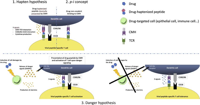
especially damage (or danger) signals, cytokines and non-covalent manner following the p-i model, or an altered
chemokines (Smith, 1972; Folster-Holst and Kreth, 2009b). repertoire of endogenous peptides following drug binding to
Keratinocytes are probably important actors of non-specific MHC (Todd, 2006).
inflammation, through the fixation of the virus and the Another theory that explain this interplay between drug and
secretion of different signals (Strittmatter et al., 2016). In infection is the danger hypothesis which was firstly proposed by
addition to the direct effect of the virus, immunologic Matzinger since the early 1990s (Das et al., 2011). This model
mechanisms induced by the virus can also be involved in the states that the primary driving force of the immune system is to
development of a skin lesion. Indeed, viral-induced cell- protect against danger (Anderson and Matzinger, 2000).
mediated responses might be responsible for damage through Presentation of an antigen in the absence of danger results in
a nonspecific inflammatory reaction (Parham and Janeway, tolerance, while the presence of a danger signal will result in a full-
2009). Recruitment of adaptive immune cells is permitted by blown immune response. Indeed, three different elements are
the interaction between inflamed endothelium receptors and needed to elicit an immune response. Signal 1 represents the
skin-addressing markers on the lymphocyte surface, for interaction between the MHC-restricted antigen and the T-cell
example the CLA (Cutaneous Lymphocyte Antigen) (Schon receptor. Signal 2 is represented by the co-stimulatory
et al., 2003; Clark, 2010). molecule–receptor interactions and a series of pro-
From another point of view, viruses can also lead to inflammatory cytokines such as IL-2, TNF-α, and IFN-γ that
exanthema by a local delayed (type 4) hypersensitivity reaction act indirectly on antigen presenting cells to up-regulate the
within the dermis to various pathogens, such as in Gianotti-Crosti expression of co-stimulatory molecules. Signal 3 represents
syndrome, where exanthema is typically papulo-vesicular, but polarizing cytokines that act directly on T-cells, and lead to
neither viral particles nor antigens have been demonstrated in the either TH1 or TH2 immune responses. The danger signal can
skin lesions (Gianotti, 1979). This syndrome would results from result from chemical, physical or viral stress. This theory was
an immunologic response rather than a primary manifestation of proposed to partially explain the reactions in HIV patients.
an infection (Lowe et al., 1989; Magyarlaki et al., 1991; Hofmann Regarding IgE-mediated hypersensitivity reactions, there is no
et al., 1997; Folster-Holst and Kreth, 2009b). data in the literature indicating a link between viruses and IgE
However, it is unknown why skin rashes are seen in only a mediated drug reactions. However, the implication of viruses in
small proportion of all generalized virus diseases, and the IgE-mediated food allergy is well-known and similarly, a potential
characteristic distribution of skin lesions in different virus role of viruses in these reactions is probable (Muraro et al., 2014).
exanthema remains unclear (Mims, 1966). Genetic and Further studies are needed to explore this important aspect.
individual susceptibility may play an important role to the
development of skin lesions and should be taken into account
to understand the complexity of the problem. Non-immune ROLE OF VIRUS IN BENIGN
mechanisms (i.e., sensitivity to histamine, antigen-antibody NONIMMEDIATE REACTION
complexes clearing by reticuloendothelial system) may be
involved as personal immunological factors necessary to Viral Infection as a Differential Diagnosis
develop an allergic reaction (Levine, 1965). A common situation in clinical practice, and particularly in
pediatric, is the appearance of a benign exanthema or urticaria
(i.e., without any danger signs) in patients treated by antibiotics,
Potential Interaction Between Virus and mainly BL, and NSAID (Bigby, 2001; Thong and Tan, 2011).
Drug It is difficult to distinguish urticaria-like exanthemas from
The interaction between virus immunity and drug hypersensitivity “classical” urticaria, which is characterized by wheal and flare
are multiple and complex (White et al., 2015) (Figure 1). The reactions: in “classical” urticaria, the manifestation is acute after
heterologous immunity models is an enlarged vision that takes into drug intake (min to hours) vs. urticaria-like exanthemas, which
account the specific HLA-restriction and the minimal co- appear after days, often together with macular exanthematic
stimulatory signals observed in drug-related Severe Cutaneous lesions. Classical urticarial lesions lastAnci et al. Viral Infection and Drug Allergy
65.9% of the children with a negative drug provocation test One of the hypothesis regarding the mechanisms for the
(DPT), the most frequent being enteroviruses (Picornavirus) development of skin eruption occurring in patients with
(Caubet et al., 2011). Similarly, Atanaskovic-Markovic et al. infectious mononucleosis and concomitantly treated by
found that 333 children (22%) tested positive for a virus or antibiotics, appears to be a transient virus-mediated immune
Mycoplasma pneumoniae infection among 1,026 children with a alteration (Thompson and Ramos, 2017). In patients with EBV
suspicion of nonimmediate hypersensitivity reactions infection, the CD8+ T cell population is typically expanded,
(Atanaskovic-Markovic et al., 2016). Only two of them were leading to the secretion of INF-γ and interleukine-2 (IL-2).
confirmed to be allergic to the culprit drug (Atanaskovic- This has been shown to inhibit the TH2-response (IL-4, 5, 6,
Markovic et al., 2016). This suggests that in patients 9, 13) (Schissel et al., 2000; Banerjee et al., 2014) and the anti-
developing an exanthema or delayed-appearing urticaria inflammatory IL-10 secretion, while the TH1-response is
while taking concomitantly a drug, viral infection is frequent; activated (Onodi-Nagy et al., 2015). These alterations could set
and that reaction to the drug taken can be detected only rarely. the stage for a loss of antigenic tolerance and the development of a
Possibly the combination of viral infection—facilitating the reversible DHR (Shiohara and Kano, 2007). Thus, the
drug reaction, is transient, and the single drug may be administration of an antibiotic, especially ampicillin, would
tolerated. The virus infections would represent the co- then be the trigger for activation of this anti-IL-10 pro-TH1
stimulatory factor enhancing drug reactions. response, leading to the maculopapular rash (Thompson and
However, in these studies, a virus has not been found in all Ramos, 2017).
patients with a skin eruption during a BL treatment. It can be Conversely, recent studies suggest that a true long lasting
explained by the fact that not all viruses have been tested in antibiotic hypersensitivity might be a lot more prevalent than
those studies. From another point of view, we cannot exclude previously thought, during the acute EBV infection in patients
that the positivity of PCR or serology was due to a previous treated by amoxicillin (Renn et al., 2002; Onodi-Nagy et al.,
infection or an acute infection without any link with the 2015). Some authors found positive lymphocyte transformation
current rash. tests (LLTs) to the incriminated antibiotic (Renn et al., 2002), as
Clinically it is very difficult, and often impossible to well as positive delayed intradermal and patch-tests in those
differentiate a rash of viral origin or secondary to a drug patients (Jappe, 2007; Onodi-Nagy et al., 2015). Authors also
allergy. Although blood tests are not routinely performed in described positive DPT or severe DHR upon re-exposure to the
our current clinical practice for exanthema or urticaria, it has beta-lactam at distance of the initial reaction (Jappe, 2007).
been recently suggested that some tests could be helpful to Thus, it is recommended to assess these reactions with a
distinguish between viral- and drug-induced skin eruptions. As complete allergic workup, and discuss a DPT.
an example, Hari Y et al. have shown that in viral exanthemas, Long lasting HS may be supported by EBV which
IFN-γ is increased in most serum samples from different acute continuously co-activates immune response and prevents
viral diseases, while in drug-induced exanthemas, IL-5 alone or in apoptosis of drug specific T-cell, as it has been found in EBV-
combination with granzyme B and perforin are often found to be induced malignant diseases (Chen, 2011). This anti-apoptotic
increased – together with some eosinophilia (Hari et al., 1999; capacity of EBV could be responsible to the maintenance of
Bellini et al., 2013). Another example is the potential role of lymphocytes, which will then be activated by antibiotic
thymus and activation-regulated chemokine (TARC/CCL17) administration (Chen, 2011; Lindsey et al., 2016).
which plays an important in TH2 immune responses. Thus, a Interestingly, it has been suggested that ampicillin can directly
link between serum TARC levels and HHV-6 reactivation in induce the reactivation of EBV, leading to a skin eruption. Thus,
patient with DRESS has been found and serum TARC levels have Saito-Katsuragi et al. reported the case of a 23-year-old woman
been suggested to be a useful indicator to differentiate DRESS/ with a Still’s disease, who developed a maculopapular rash after
DIHS with HHV-6 reactivation from other drug eruptions an ampicillin treatment. She developed serum IgG antibody
(Ogawa et al., 2014). against EBV-VCA 1 week after. The authors performed two
DPT with intravenous ampicillin, resulting in a recurrence of
the maculopapular rash 24–48 h after the treatment intake. They
The EBV Example as a Co-Factor for monitored the concentration of EBV DNA in blood and found a
Drug-Induced Skin Eruptions significant increase of EBV DNA levels after the injection of
The best illustration for the drug-related exanthemas during a ampicillin and just before the appearance of the skin rash. Further
viral infection is those occurring after antibiotic administration in studies are needed to confirm the hypothesis by which ampicillin
patients with an acute EBV infection. Indeed, it has been shown would be responsible for a reactivation of EBV, which would then
that the incidence of skin rash is higher in EBV patients treated by trigger the skin eruption.
antibiotic (typically ampicillin) compared to EBV patients EBV continues to be one of the most important models to
without associated antibiotic treatment (i.e., 27.8–90% and understand interaction between drugs and concomitant acute or
3–10%, respectively) (Pullen et al., 1967; Copeman and chronic viral infections. Lymphocyte stimulation and direct
Scrivener, 1977; Luzuriaga and Sullivan, 2010). No association stimulation of the virus appears to be the most likely
with age, gender, ethnicity or allergic history appears to be hypotheses. However, further researches are needed for a
correlated with rash development after antibiotic treatment in better understanding of the mechanisms involved in the
EBV patients (Chovel-Sella et al., 2013). dysregulation of the immune system, leading to a reaction.
Frontiers in Pharmacology | www.frontiersin.org 4 March 2021 | Volume 11 | Article 586407Anci et al. Viral Infection and Drug Allergy
ROLE OF VIRUS IN SEVERE (Takahashi et al., 2009). Thus, there are some evidence that
NONIMMEDIATE REACTIONS HHV-6-related mechanisms exist to explain at least partially
the complications of DRESS.
A variety of severe, rare, potentially life-threatening, drug The importance of drug exposure could be integrated with
reactions are described, for which recent evidences suggest an those of viral interplay in a recent model: the heterologous
intimate relationship with reactivation of specific virus: the immunity model. Furthermore, active viral replication is not
DRESS syndrome, the Stevens-Johnson syndrome (SJS) as well required in this abovementioned model, so the evidences of
as the Toxic epidermal necrolysis (TEN) and transitional forms viral reactivation highlighted during SCARs development may
(Tohyama and Hashimoto, 2011). just represent a tangential event. There is still a need of further
studies to highlight differences between patients with or without
DRESS Syndrome viral reactivation. In this context, a retrospective case series of 29
The DRESS syndrome is a drug-induced delayed reaction with an pediatric patient with DRESS, reported that those who were
estimated incidence ranging from one case among 1,000 to 10,000 HHV-6 positive experimented a significantly greater severity
drug exposures (Fiszenson-Albala et al., 2003). It is most and a longer hospitalization compared to HHV-6 negative
frequently associated with administration of aromatic subjects (11.5 days vs. 5 days, p 0.039) (Ahluwalia et al.,
anticonvulsants, antidepressants, sulfonamides and sulfones, 2015). Even in adults, patients with HHV-6 reactivation
anti-inflammatory drugs, antibiotics, angiotensin-converting showed longer course and more severe organ involvement
enzyme inhibitors and beta-blockers (Kardaun et al., 2013). It than others, suggesting a possibly prognostic significance of
has been suggested that viruses play an important role in the HHV-6 (Tohyama et al., 2007; Asano et al., 2009).
physiopathology of DRESS (Redwood and et al., 2018). Further researches should also emphasize on reactivation of
Hypotheses are based on the evidence of virus replication other latent viruses too. Apparently, viral activation follows an
(primo-infection or reactivation) during the development of identifiable chronological pathway and seems to implicate several
disease (Descamps et al., 2001; Ichiche et al., 2003; Picard viruses in the present order: firstly EBV and/or HHV-6, followed
et al., 2010). Human herpes virus 6 (HHV-6) was the first by HHV-7 and soon after CMV (Cho et al., 2017). The
chronic persistent virus incriminated in the pathology of simultaneous appearance of multiple concomitant viral
DRESS (Descamps et al., 1997), being now considered, for reactivations would be explained by the ability of herpes virus
some, as a specific and sensitive diagnostic criteria (Shiohara to reactivate others virus. The role of the EBV in the development
et al., 2007; Watanabe, 2018). of multi-organ involvement of DRESS is discussed particularly
However, the role of HHV replication remains controversial as because infectious mononucleosis-like symptoms are observed
a study did not find a significant correlation between HHV DNA during the early phase of DRESS (Tohyama and Hashimoto,
load and DRESS diagnosis (Ushigome et al., 2012). Several studies 2011). Furthermore, Mardivirin et al. investigate the possibility of
reported that HHV replication does not occur early in the clinical a drug-induced flare-up of DRESS due to antibiotic prescription.
course of DRESS and generally, viremia is observed greater than Amoxicillin seemed to be an aggravating factor, probably due to
2 weeks following symptoms onset (White et al., 2015). These the same pathomechanism of amoxicillin-induced rash in EBV
data suggest that viral reactivation itself is not involved in the infected patients (Mardivirin et al., 2010).
onset of DRESS, but rather than some viruses, in particular of the Finally, hypothesis for DRESS syndrome pathophysiology
herpes group, may be involved in the prolonged clinical course of include interaction between different factors: 1) genetic
DRESS (Ishida et al., 2014). susceptibility factors, such as HLA type or cytochrome p450
The expansion of CD4+ T cells and CD8+ T cells during HHV- polymorphism (Cho et al., 2017); 2) viral infection (primo-
6 reactivation seems to be an important feature in many patients infection or replication) inducing a particular pre-activated
with DRESS’s multiple organ failure (Pritchett et al., 2012). In immune state; and 3) drug as a final trigger for the immune
addition, it has been found that patients with HHV-6 reactivation reaction. Virus reactivation could also be the trigger for relapse of
have significant higher serum levels of TNF-α, compared to DRESS syndrome (Tan and Chan, 2016), as seen in chronic
patients without HHV-6 reactivation. In vitro and in vivo diseases. Besides, it is interesting to note that similarities are
studies showed that TNF-α and other cytokines participate in highlighted between DRESS and autoimmune disease
reactivation of CMV through the induction of CMV immediate mechanisms (Michels and Ostrov, 2015).
early gene expression, leading to the initiation of the viral
replication. CMV IE gene has a high level of homology with SJS and TEN
HHV-6 U95 gene and it is possible that TNF-α interacts Similar observations have been made in SJS and TEN. These
identically with it (Watanabe, 2018). The serum thymus and syndromes are most commonly caused by DHR rather than
activation-regulated chemokine (TARC) levels are also found to viruses (such as EBV, CMV, HHV-6, HSV, Varicella zoster
be higher in DRESS patients with HHV-6 replication than those virus, hepatitis A virus and HIV) (Stutman, 1987;
without. TARC may be able to directly activate HHV-6 through a Werblowsky-Constantini et al., 1989; Lam et al., 2004; Bay
TARC receptor, or induce a relative immunosuppression through et al., 2005; Pereira et al., 2007; Cruz et al., 2010; Wetter and
the activation of regulatory T cells (Tregs) (Watanabe, 2018). This Camilleri, 2010; Khalaf et al., 2011; Kunimi et al., 2011; Kim et al.,
is in accordance with some observations of dysfunction of Tregs 2012; Sotelo-Cruz, 2012; Ferrandiz-Pulido and Garcia-Patos,
and plasmacytoid dendritic cells in the DRESS syndrome 2013; Irungu et al., 2017). In about 30% of cases of SJS and
Frontiers in Pharmacology | www.frontiersin.org 5 March 2021 | Volume 11 | Article 586407Anci et al. Viral Infection and Drug Allergy
FIGURE 1 | Potential immune mechanisms involved in the interactions between viruses and drug.
TEN, no causative drug is identified, and in 15%, drug 1993; Rzany et al., 1993; Temesgen and Beri, 2004). The
responsibility is deemed unlikely (Duong et al., 2017). Since pathogenesis and the reason for the greater propensity for
now, over 200 drugs have been associated with SJS/TEN, most HIV-infected patients to develop DHR to a great variety of
commonly sulfonamides and BL antibiotics (Roujeau et al., 1995; drugs that can be particularly severe, remain unknown. It may
Forman et al., 2002; Sheridan et al., 2002). be related to their greater exposition to medication compared to
To date it is still not clear if the virus is a potential co-factor or general population and/or to a higher incidence of co-infection
trigger. Expression of viral DNA fragments in the keratinocyte with EBV and CMV (Cytomegalovirus) (Smith et al., 1997;
layer could lead to activation of CD4+ T-helper cells, which Todd, 2006; Hoosen and et al., 2019). Since many different
induce various reactions, including cytokines production and drugs are involved, the viral infection appears to enhance drug
subsequent inflammatory responses (McDermott et al., 2013). reactivity in general, not only for specific drugs.
Furthermore, infections activate systemic host inflammatory This infection itself leads to apparent decrease and loss function
pathways, as consequence, a perturbation of the natural defense of T cells in the blood and skin, in addition to dysregulation of
mechanisms of oxidase enzymes could occur and multisystem tolerance to self-antigens (Todd, 2006). Interestingly, the incidence
damages may follow (Bay et al., 2005). Despite everything, F. of severe DHR in the HIV-infected population has also been
Brunet-Possenti reports a case of SJS during a primary EBV reported to increase with increasing stage of the disease,
infection in a 17-year-old adolescent. A 10 years retrospective i.e., decreasing CD4+ T cells counts and CD4/CD8 ratio
study presented by Forman confirmed it, founding as the most (Coopman et al., 1993; Arp et al., 2005). An interesting example
commonly incriminated infectious agent the herpes simplex is the hypersensitivity reaction to Trimethoprim-Sulfamethoxazole
virus (19.7%) (Forman et al., 2002). However, while HHV-6 (TMP-SMX), which occurs in 40–80% of HIV infected individuals
reactivation is primary related to DRESS, it is rare in SJS/TEN (Meyer et al., 2015). The patients with uncontrolled HIV
(Neuman et al., 2013), sometimes observed in patients treated replication have a decrease reduction capacity and a depletion
with anticonvulsant (Peppercorn et al., 2010; Teraki et al., 2010). of glutathion in the CD4 cells, leading to an increased toxicity of
Actually, researchers are still arguing if “drug-induced” SJS/ nitrososulfamethoxazole (n-SMX), a reactive and toxic metabolites
TEN and “infection-related” SJS/TEN are two separate entities. of SMX (Correia et al., 2002). This modification in redox balance
may be related to the Tat protein, an HIV-specific protein essential
HIV Example for the viral replication (Das et al., 2011). The Tat protein would be
Human immunodeficiency virus (HIV) infection is a long-life secreted by infected cells, in relation to the viral load and disease
latent virus hosted by CD4 T cells and macrophages (Zack et al., progression, and promotes drug reactions, increasing oxidation
1990). This viral infection is associated with important immune status (Meyer et al., 2015). This strong predisposition to drug
deregulations and higher rates of conditions requiring drug reactions is clearly dependent to multiple factors linked to the
administration. It has been found that frequency of DHR in immune deregulation associated to the primary infection (Todd,
HIV-infected patients is particularly high, up to 100 times more 2006). But our understanding of the exact pathomechanisms
common compared to HIV-negative subjects (Coopman et al., remains limited and requires further studies.
Frontiers in Pharmacology | www.frontiersin.org 6 March 2021 | Volume 11 | Article 586407Anci et al. Viral Infection and Drug Allergy
The higher frequency of allergic drugs reactions in this viral avoidance of all drugs with anti-cyclooxygenase activity.
infection may be the result of increased levels of cytokines and However, asthma continues to run a protracted course
cell-surface markers and thereby acting in concert with the drug because of chronic viral infection (Szczeklik, 1988).
antigen, amplifying the potential of a drug to cause an immune Nakagawa et al. suspected an acquired analgesic idiosyncrasy
reaction (Pirmohamed et al., 2002). Although an attractive secondary to viral infection. They observed anti-Herpes simplex
hypothesis when applied to the pathogenesis of DHRs, there virus (HSV) IgG antibodies titers and hypothesized a
are many questions that remain unanswered. Indeed, the lack of relationship between the serological evidence of HSV
direct experimental evidence has led to heavy criticism of the infection and positive bronchial hyperresponsiveness
danger hypothesis (Jozefowski, 2016). provocation tests (Nakagawa et al., 2001). Contrariwise,
several studies have showed that NSAID can inhibit viral
replication (Newton, 1979; Pereira et al., 2003; Reynolds and
ROLE OF VIRUS IN OTHER TYPE OF DHR Enquist, 2006; Zimmermann and Curtis, 2017), yielding more
difficult the interpretation of virus and NSAID interaction.
The NSAID Example
It has recently been reported that NSAID could be the most
common cause of DHR in children (Woessner et al., 2002; CONCLUSION
Morales et al., 2015). Prevalence of self-reported hypersensitivity
to NSAID has been shown to range from 0.6 to 5.7% in the general In addition to be a major differential diagnosis of DHR, viruses
population (Dona et al., 2011). NSAIDs, including aspirin, are a might interact in different ways in different types of DHR to
group of drugs sharing the capability of inhibiting the unmask a latent drug allergy. Particularly, viruses have been
cyclooxygenase (COX) enzymes responsible for the prostaglandin shown to cause cellular damages, to increase the inflammatory
synthetase pathway of arachidonic acid metabolism. The response, to induce the production of specific antibodies, to
pathogenesis of hypersensitivity reactions owing to cross- provoke a change in antigenic expression and to stimulate
intolerance has been hypothesized to be related to COX-1 T-cell replication. From another point of view, the drug might
inhibition, although it has not been clearly demonstrated (Macy, enhance viral replication, leading secondarily to skin eruption.
1998). Pathomechanism of viral-induced skin lesions has been poorly
Interestingly, it has been suggested that blocking studied. However, a better understanding is of major importance,
prostaglandin synthesis could also allow specific cytotoxic as it can provide major insight in the understanding of drug
lymphocytes to produce asthma attacks during respiratory induced skin rashes. Further studies are urgently needed to clarify
tract viral infections (Szczeklik, 1988). Correlation between the role of viruses in drugs HSRs, to improve the management of
viral illness and NSAIDs hypersensitivity was first theorized patients presenting skin eruptions during treatments and to avoid
by Szczeklik (1988). As cytotoxic lymphocyte activity is useless drug avoidance, related with increased morbidity and
normally inhibited by prostaglandin E2 (PGE2); in case of mortality.
aspirin and other NSAIDs treatment, COX enzyme is
blocked and PGE2 production decrease allowing cytotoxic
lymphocytes to attack and eliminate the respiratory tract AUTHOR CONTRIBUTIONS
cells infected by the virus. As a result, lysosomal enzymes
and mediators are released and this could precipitate a All authors listed have made a substantial, direct, and intellectual
NSAIDs reaction. These acute attacks can be prevented by contribution to the work and approved it for publication.
Atanaskovic-Markovic, M., Gaeta, F., Medjo, B., Gavrovic-Jankulovic, M., Cirkovic
REFERENCES Velickovic, T., Tmusic, V., et al. (2016). Non-immediate hypersensitivity reactions
to beta-lactam antibiotics in children - our 10-year experience in allergy work-up.
Agol, V. I. (2012). Cytopathic effects: virus-modulated manifestations of innate Pediatr. Allergy Immunol. 27 (5), 533–538. doi:10.1111/pai.12565
immunity? Trends Microbiol. 20 (12), 570–576. doi:10.1016/j.tim.2012.09.003 Banerjee, S., Lu, J., Cai, Q., Sun, Z., Jha, H. C., and Robertson, E. S. (2014). EBNA3C
Ahluwalia, J., Abuabara, K., Perman, M. J., and Yan, A. C. (2015). Human augments pim-1 mediated phosphorylation and degradation of p21 to promote
herpesvirus 6 involvement in paediatric drug hypersensitivity syndrome. Br. B-cell proliferation. PLoS Pathog. 10 (8), e1004304. doi:10.1371/journal.ppat.
J. Dermatol. 172 (4), 1090–1095. doi:10.1111/bjd.13512 1004304
Anderson, C. C., and Matzinger, P. (2000). Danger: the view from the bottom of the Bay, A., Akdeniz, N., Calka, O., Kösem, M., Faik Oner, A., and Doğan, M. (2005).
cliff. Semin. Immunol. 12 (3), 231–238. doi:10.1006/smim.2000.0236 Primary varicella infection associated with stevens-johnson syndrome in a
Arp, J., Rieder, M. J., Urquhart, B., Freeman, D., Tucker, M. J., Krizova, A., et al. Turkish child. J. Dermatol. 32 (9), 745–750. doi:10.1111/j.1346-8138.2005.
(2005). Hypersensitivity of HIV-1-infected cells to reactive sulfonamide tb00836.x
metabolites correlated to expression of the HIV-1 viral protein tat. Bellini, V., Pelliccia, S., and Lisi, P. (2013). Drug- and virus- or bacteria-induced
J. Pharmacol. Exp. Therapeut. 314 (3), 1218–1225. doi:10.1124/jpet.105.085050 exanthems: the role of immunohistochemical staining for cytokines in
Asano, Y., Kagawa, H., Kano, Y., and Shiohara, T. (2009). Cytomegalovirus disease differential diagnosis. Dermatitis 24 (2), 85–90. doi:10.1097/DER.
during severe drug eruptions: report of 2 cases and retrospective study of 18 0b013e318280cbe5
patients with drug-induced hypersensitivity syndrome. Arch. Dermatol. 145 (9), Bigby, M. (2001). Rates of cutaneous reactions to drugs. Arch. Dermatol. 137 (6),
1030–1036. doi:10.1001/archdermatol.2009.195 765–770.
Frontiers in Pharmacology | www.frontiersin.org 7 March 2021 | Volume 11 | Article 586407Anci et al. Viral Infection and Drug Allergy Caubet, J. C., Kaiser, L., Lemaı̂tre, B., Fellay, B., Gervaix, A., and Eigenmann, P. A. Geck, P., Dan, P., and Nasz, I. (1964). Examination of the cytopathic effect of (2011). The role of penicillin in benign skin rashes in childhood: a prospective adenoviruses by immunofluorescence. Acta Microbiol. Acad. Sci. Hungar. 11, 19–22. study based on drug rechallenge. J. Allergy Clin. Immunol. 127 (1), 218–222. Gianotti, F. (1979). Papular acrodermatitis of childhood and other papulo- doi:10.1016/j.jaci.2010.08.025 vesicular acro-located syndromes. Br. J. Dermatol. 100 (1), 49–59. doi:10. Chen, M. R. (2011). Epstein-barr virus, the immune system, and associated 1111/j.1365-2133.1979.tb03569.x diseases. Front. Microbiol. 2, 5. doi:10.3389/fmicb.2011.00005 Goodyear, H. M., Laidler, P. W., Price, E. H., Kenny, P. A., and Harper, J. I. (1991). Cho, Y. T., Yang, C. W., and Chu, C. Y. (2017). Drug reaction with eosinophilia and Acute infectious erythemas in children: a clinico-microbiological study. Br. systemic symptoms (DRESS): an interplay among drugs, viruses, and immune J. Dermatol. 124 (5), 433–438. doi:10.1111/j.1365-2133.1991.tb00621.x system. Int. J. Mol. Sci. 18 (6), 1243. doi:10.3390/ijms18061243 Hari, Y., Urwyler, A., Hurni, M., Yawalkar, N., Dahinden, C., Wendland, T., et al. Chovel-Sella, A., Ben Tov, A., Lahav, E., Mor, O., Rudich, H., Paret, G., et al. (2013). (1999). Distinct serum cytokine levels in drug- and measles-induced exanthema. Incidence of rash after amoxicillin treatment in children with infectious Int. Arch. Allergy Immunol. 120 (3), 225–229. doi:10.1159/000024271 mononucleosis. Pediatrics 131 (5), e1424–e1427. doi:10.1542/peds.2012-1575 He, S. Z., Chen, M. Y., Xu, X. R., Yan, Q., Niu, J. J., Wu, W. H., et al. (2017). Clark, R. A. (2010). Skin-resident T cells: the ups and downs of on site immunity. Epidemics and aetiology of hand, foot and mouth disease in Xiamen, China, J. Invest. Dermatol. 130 (2), 362–370. doi:10.1038/jid.2009.247 from 2008 to 2015, Epidemiol. Infect., 145, 1865–1874. doi:10.1017/ Coopman, S. A., Johnson, R. A., Platt, R., and Stern, R. S. (1993). Cutaneous disease S0950268817000309 and drug reactions in HIV infection. N. Engl. J. Med. 328 (23), 1670–1674. Hofmann, B., Schuppe, H. C., Adams, O., Lenard, H. G., Lehmann, P., and Ruzicka, doi:10.1056/NEJM199306103282304 T. (1997). Gianotti-Crosti syndrome associated with Epstein-Barr virus Copeman, P. W., and Scrivener, R. (1977). Amoxycillin rash. Br. Med. J. 1 (6072), infection. Pediatr. Dermatol. 14 (4), 273–277. doi:10.1016/s0190-9622(89) 1354. doi:10.1136/bmj.1.6072.1354-b 70041-4 Correia, O., Delgado, L., Roujeau, J. C., Le Cleach, L., and Fleming-Torrinha, J. A. Hogan, P. A. (1996). Viral exanthems in childhood. Australas. J. Dermatol. 37 (2002). Soluble interleukin 2 receptor and interleukin 1alpha in toxic epidermal (Suppl. 1), S14–S16. doi:10.1111/j.1440-0960.1996.tb01071.x necrolysis: a comparative analysis of serum and blister fluid samples. Arch. Hoosen, K., Mosam, A., Cordelia Dlova, N., and Grayson, W. (2019). An update on Dermatol. 138 (1), 29–32. doi:10.1001/archderm.138.1.29 adverse cutaneous drug reactions in HIV/AIDS. Dermatopathology (Basel) 6 Cruz, M. J., Mota, A., Baudrier, T., Gil-da-Costa, M. J., and Azevedo, F. (2010). Stevens- (2), 111–125. doi:10.1159/000496389 Johnson syndrome associated with cytomegalovirus infection in a child with Ichiche, M., Kiesch, N., and De Bels, D. (2003). DRESS syndrome associated with ependymoma. J. Dermatol. Case. Rep. 4 (1), 11–14. doi:10.3315/jdcr.2010.1043 HHV-6 reactivation. Eur. J. Intern. Med. 14 (8), 498–500. doi:10.1016/j.ejim. Das, A. T., Harwig, A., and Berkhout, B. (2011). The HIV-1 Tat protein has a 2003.09.004 versatile role in activating viral transcription. J. Virol. 85 (18), 9506–9516. Irungu, K., Nyamu, D., and Opanga, S. (2017). Characterization of stevens-johnson doi:10.1128/JVI.00650-11 syndrome and toxic epidermal necrolysis among patients admitted to Kenyatta Demoly, P., Tanno, L. K., Akdis, C. A., Lau, S., Calderon, M. A., Santos, A. F., et al. national hospital: a retrospective cross-sectional study. D. Real World Out. 4 (2), (2014). Global classification and coding of hypersensitivity diseases-an EAACI- 79–85. doi:10.1007/s40801-017-0105-x WAO survey, strategic paper and review. Allergy 69 (5), 559–570. doi:10.1111/ Ishida, T., Kano, Y., Mizukawa, Y., and Shiohara, T. (2014). The dynamics of all.12386 herpesvirus reactivations during and after severe drug eruptions: their relation Descamps, V., Bouscarat, F., Laglenne, S., Aslangul, E., Veber, B., Descamps, D., to the clinical phenotype and therapeutic outcome. Allergy 69 (6), 798–805. et al. (1997). Human herpesvirus 6 infection associated with anticonvulsant doi:10.1111/all.12410 hypersensitivity syndrome and reactive haemophagocytic syndrome. Br. Jappe, U. (2007). Amoxicillin-induced exanthema in patients with infectious J. Dermatol. 137 (4), 605–608. doi:10.1111/j.1365-2133.1997.tb03795.x mononucleosis: allergy or transient immunostimulation? Allergy 62 (12), Descamps, V., Valance, A., Edlinger, C., Fillet, A. M., Grossin, M., Lebrun-Vignes, 1474–1475. doi:10.1111/j.1398-9995.2007.01518.x B., et al. (2001). Association of human herpesvirus 6 infection with drug Jozefowski, S. (2016). The danger model: questioning an unconvincing theory. reaction with eosinophilia and systemic symptoms. Arch. Dermatol. 137 (3), Immunol. Cell Biol. 94 (5), 525. doi:10.1038/icb.2016.29 301–304. Kano, Y., Hiraharas, K., Sakuma, K., and Shiohara, T. (2006). Several herpesviruses Dona, I., Blanca-López, N., Jagemann, L. R., Torres, M. J., Rondón, C., Campo, P., can reactivate in a severe drug-induced multiorgan reaction in the same et al. (2011). Response to a selective COX-2 inhibitor in patients with urticaria/ sequential order as in graft-versus-host disease. Br. J. Dermatol. 155 (2), angioedema induced by nonsteroidal anti-inflammatory drugs. Allergy 66 (11), 301–306. doi:10.1111/j.1365-2133.2006.07238.x 1428–1433. doi:10.1111/j.1398-9995.2011.02684.x Kardaun, S. H., Sekula, P., Valeyrie-Allanore, L., Liss, Y., Chu, C. Y., Creamer, D., Drago, F., Paolino, S., Rebora, A., Broccolo, F., Drago, F., Cardo, P., et al. (2012). et al. (2013). Drug reaction with eosinophilia and systemic symptoms The challenge of diagnosing atypical exanthems: a clinico-laboratory study. (DRESS): an original multisystem adverse drug reaction. Results from the J. Am. Acad. Dermatol. 67 (6), 1282–1288. doi:10.1016/j.jaad.2012.04.014 prospective RegiSCAR study. Br. J. Dermatol. 169 (5), 1071–1080. doi:10. Duong, T. A., Valeyrie-Allanore, L., Wolkenstein, P., and Chosidow, O. (2017). 1111/bjd.12501 Severe cutaneous adverse reactions to drugs. Lancet 390 (10106), 1996–2011. Keighley, C. L., Saunderson, R. B., Kok, J., and Dwyer, D. E. (2015). Viral doi:10.1016/S0140-6736(16)30378-6 exanthems. Curr. Opin. Infect. Dis. 28 (2), 139–150. doi:10.1097/QCO. Ferrandiz-Pulido, C., and Garcia-Patos, V. (2013). A review of causes of Stevens- 0000000000000145 Johnson syndrome and toxic epidermal necrolysis in children. Arch. Dis. Child. Khalaf, D., Toema, B., Dabbour, N., and Jehani, F. (2011). Toxic epidermal 98 (12), 998–1003. doi:10.1136/archdischild-2013-303718 necrolysis associated with severe cytomegalovirus infection in a patient on Fiszenson-Albala, F., Auzerie, V., Mahe, E., Farinotti, R., Durand-Stocco, C., regular hemodialysis. Mediterr. J. Hematol. Infect. Dis. 3 (1), e2011004. doi:10. Crickx, B., et al. (2003). A 6-month prospective survey of cutaneous drug 4084/MJHID.2010.004 reactions in a hospital setting. Br. J. Dermatol. 149 (5), 1018–1022. doi:10.1111/ Kim, H. I., Kim, S. W., Park, G. Y., Kwon, E. G., Kim, H. H., Jeong, J. Y., et al. j.1365-2133.2003.05584.x (2012). Causes and treatment outcomes of Stevens-Johnson syndrome and Folster-Holst, R., and Kreth, H. W. (2009a). Viral exanthems in toxic epidermal necrolysis in 82 adult patients. Korean J. Intern. Med. 27 (2), childhood–infectious (direct) exanthems. Part 1: classic exanthems. J. Dtsch. 203–210. doi:10.3904/kjim.2012.27.2.203 Dermatol Ges. 7 (4), 309–316. doi:10.1111/j.1610-0387.2008.06868.x Kramkimel, N., Soussan, V., Beauchet, A., Duhamel, A., Saiag, P., Chevallier, B., Folster-Holst, R., and Kreth, H. W. (2009b). Viral exanthems in childhood. Part 3: et al. (2010). High frequency, diversity and severity of skin diseases in a parainfectious exanthems and those associated with virus-drug interactions. paediatric emergency department. J. Eur. Acad. Dermatol. Venereol. 24 (12), J. Dtsch. Dermatol. Ges. 7 (6), 506–510. doi:10.1111/j.1610-0387.2008.06870.x 1468–1475. doi:10.1111/j.1468-3083.2010.03672.x Forman, R., Koren, G., and Shear, N. H. (2002). Erythema multiforme, Stevens- Kunimi, Y., Hirata, Y., Aihara, M., Yamane, Y., and Ikezawa, Z. (2011). Statistical Johnson syndrome and toxic epidermal necrolysis in children: a review of analysis of Stevens-Johnson syndrome caused by Mycoplasma pneumonia 10 years’ experience. Drug Saf. 25 (13), 965–972. doi:10.2165/00002018- infection in Japan. Allergol. Int. 60 (4), 525–532. doi:10.2332/allergolint.11- 200225130-00006 OA-0309 Frontiers in Pharmacology | www.frontiersin.org 8 March 2021 | Volume 11 | Article 586407
Anci et al.                                                                                                                                       Viral Infection and Drug Allergy

Laksono, B. M., de Vries, R. D., McQuaid, S., Duprex, W. P., and de Swart, R. L.                herpes virus-6 reactivation. Transl. Res. 161 (5), 430–440. doi:10.1016/j.trsl.
   (2016). Measles virus host invasion and pathogenesis. Viruses 8 (8). doi:10.                 2012.12.012
   3390/v8080210                                                                             Newton, A. A. (1979). Inhibitors of prostaglandin synthesis as inhibitors of herpes
Lam, N. S., Yang, Y. H., Wang, L. C., Lin, Y. T., and Chiang, B. L. (2004). Clinical            simplex virus replication. Adv. Ophthalmol. 38, 58–63.
   characteristics of childhood erythema multiforme, Stevens-Johnson syndrome                Ogawa, K., Morito, H., Hasegawa, A., Miyagawa, F., Kobayashi, N., Watanabe, H.,
   and toxic epidermal necrolysis in Taiwanese children. J. Microbiol. Immunol.                 et al. (2014). Elevated serum thymus and activation-regulated chemokine
   Infect. 37 (6), 366–370.                                                                     (TARC/CCL17) relates to reactivation of human herpesvirus 6 in drug
Landolt, B., Staubli, G., Lips, U., and Weibel, L. (2013). Skin disorders encountered           reaction with eosinophilia and systemic symptoms (DRESS)/drug-induced
   in a Swiss pediatric emergency department. Swiss Med. Wkly. 143, w13731.                     hypersensitivity syndrome (DIHS). Br. J. Dermatol. 171 (2), 425–427. doi:10.
   doi:10.4414/smw.2013.13731                                                                   1111/bjd.12948
Leiste, A., Skaletz-Rorowski, A., Venten, I., Altmeyer, P., and Brockmeyer, N. H.            Onodi-Nagy, K., Kinyó, Á., Meszes, A., Garaczi, E., Kemény, L., and Bata-Csörgő,
   (2008). Urticaria associated with Norovirus infection: report of two cases.                  Z. (2015). Amoxicillin rash in patients with infectious mononucleosis: evidence
   J. Dtsch. Dermatol. Ges. 6 (7), 563–565. doi:10.1111/j.1610-0387.2007.06501.x                of true drug sensitization. Allergy Asthma Clin. Immunol. 11 (1), 1. doi:10.1186/
Levine, B. B. (1965). Immunochemical mechanisms involved in penicillin                          1710-1492-11-1
   hypersensitivity in experimental animals and in human beings. Fed. Proc.                  Parham, P., and Janeway, C. (2009). The immune system. 3rd Edn. New York, NY,
   24, 45–50.                                                                                   United States: Garland Science.
Lindsey, J. W., deGannes, S. L., Pate, K. A., and Zhao, X. (2016). Antibodies specific        Peppercorn, A. F., Miller, M. B., Fitzgerald, D., Weber, D. J., Groben, P. A., and
   for Epstein-Barr virus nuclear antigen-1 cross-react with human heterogeneous                Cairns, B. A. (2010). High-level human herpesvirus-6 viremia associated with
   nuclear ribonucleoprotein L. Mol. Immunol. 69, 7–12. doi:10.1016/j.molimm.                   onset of Stevens-Johnson syndrome: report of two cases. J. Burn Care Res. 31
   2015.11.007                                                                                  (2), 365–368. doi:10.1097/BCR.0b013e3181d0f48b
Lowe, L., Hebert, A. A., and Duvic, M. (1989). Gianotti-Crosti syndrome associated           Pereira, C. F., Paridaen, J. T., Rutten, K., Huigen, M. C., van de Bovenkamp, M.,
   with Epstein-Barr virus infection. J. Am. Acad. Dermatol. 20 (2 Pt 2), 336–338.              Middel, J., et al. (2003). Aspirin-like molecules that inhibit human
   doi:10.1016/s0190-9622(89)70041-4                                                            immunodeficiency virus 1 replication. Antivir. Res. 58 (3), 253–263. doi:10.
Luzuriaga, K., and Sullivan, J. L. (2010). Infectious mononucleosis. N. Engl. J. Med.           1016/s0166-3542(03)00006-8
   362 (21), 1993–2000. doi:10.1056/NEJMcp1001116                                            Pereira, F. A., Mudgil, A. V., and Rosmarin, D. M. (2007). Toxic epidermal
MacLaughlin, E. J., Saseen, J. J., and Malone, D. C. (2000). Costs of beta-lactam               necrolysis. J. Am. Acad. Dermatol. 56 (2), 181–200. doi:10.1016/j.jaad.2006.04.
   allergies: selection and costs of antibiotics for patients with a reported beta-             048
   lactam allergy. Arch. Fam. Med. 9 (8), 722–726. doi:10.1001/archfami.9.8.722              Picard, D., Janela, B., Descamps, V., D’Incan, M., Courville, P., Jacquot, S., et al.
Macy, E. (1998). Elective penicillin skin testing and amoxicillin challenge: effect on          (2010). Drug reaction with eosinophilia and systemic symptoms (DRESS): a
   outpatient antibiotic use, cost, and clinical outcomes. J. Allergy Clin. Immunol.            multiorgan antiviral T cell response. Sci. Transl. Med. 2 (46), 46ra62. doi:10.
   102 (2), 281–285. doi:10.1016/s0091-6749(98)70097-1                                          1126/scitranslmed.3001116
Magyarlaki, M., Drobnitsch, I., and Schneider, I. (1991). Papular acrodermatitis of          Pichler, W., Yawalkar, N., Schmid, S., and Helbling, A. (2002). Pathogenesis of
   childhood (Gianotti-Crosti disease). Pediatr. Dermatol. 8 (3), 224–227. doi:10.              drug-induced exanthems. Allergy 57 (10), 884–893. doi:10.1034/j.1398-9995.
   1111/j.1525-1470.1991.tb00865.x                                                              2002.02161.x
Mardivirin, L., Valeyrie-Allanore, L., Branlant-Redon, E., Beneton, N., Jidar, K.,           Pirmohamed, M., Naisbitt, D. J., Gordon, F., and Park, B. K. (2002). The danger
   Barbaud, A., et al. (2010). Amoxicillin-induced flare in patients with DRESS                  hypothesis--potential role in idiosyncratic drug reactions. Toxicology 181–182,
   (drug reaction with eosinophilia and systemic symptoms): report of seven                     55–63. doi:10.1016/s0300-483x(02)00255-x
   cases and demonstration of a direct effect of amoxicillin on human                        Ponvert, C., Le Clainche, L., de Blic, J., Le Bourgeois, M., Scheinmann, P., and
   Herpesvirus 6 replication in vitro. Eur. J. Dermatol. 20 (1), 68–73. doi:10.                 Paupe, J. (1999). Allergy to beta-lactam antibiotics in children. Pediatrics 104
   1684/ejd.2010.0821                                                                           (4), e45. doi:10.1542/peds.104.4.e45
McDermott, A. J., Taylor, B. M., and Bernstein, K. M. (2013). Toxic epidermal                Ponvert, C., Perrin, Y., Bados-Albiero, A., Le Bourgeois, M., Karila, C.,
   necrolysis from suspected Mycoplasma pneumoniae infection. Mil. Med. 178                     Delacourt, C., et al. (2011). Allergy to betalactam antibiotics in children:
   (9), e1048–e1050. doi:10.7205/MILMED-D-13-00139                                              results of a 20-year study based on clinical history, skin and challenge tests.
Meyer, C., Behm, N., Brown, E., Copeland, N. K., and Sklar, M. J. (2015). An                    Pediatr. Allergy Immunol. 22 (4), 411–418. doi:10.1111/j.1399-3038.2011.
   adverse drug reaction to trimethoprim-sulfamethoxazole revealing primary                     01169.x
   HIV: a case report and literature review. Case Rep. Infect. Dis. 2015, 691010.            Pritchett, J. C., Nanau, R. M., and Neuman, M. G. (2012). The link between
   doi:10.1155/2015/691010                                                                      hypersensitivity syndrome reaction development and human herpes
Michels, A. W., and Ostrov, D. A. (2015). New approaches for predicting T cell-                 virus-6 reactivation. Int. J. Hepatol. 2012, 723062. doi:10.1155/2012/
   mediated drug reactions: a role for inducible and potentially preventable                    723062
   autoimmunity. J. Allergy Clin. Immunol. 136 (2), 252–257. doi:10.1016/j.jaci.             Pullen, H., Wright, N., and Murdoch, J. M. (1967). Hypersensitivity reactions to
   2015.06.024                                                                                  antibacterial drugs in infectious mononucleosis. Lancet 2 (7527), 1176–1178.
Mims, C. A. (1964). Aspects of the pathogenesis of virus diseases. Bacteriol. Rev. 28,          doi:10.1016/s0140-6736(67)91893-4
   30–71.                                                                                    Rawlins, M. D. (1981). Clinical pharmacology. Adverse reactions to drugs. Br. Med.
Mims, C. A. (1966). Pathogenesis of rashes in virus diseases. Bacteriol. Rev. 30 (4),           J. 282 (6268), 974–976. doi:10.1136/bmj.282.6268.974
   739–760.                                                                                  Redwood, A. J., Pavlos, R. K., White, K. D., and Phillips, E. J. (2018). HLAs: key
Morales, D. R., Guthrie, B., Lipworth, B. J., Jackson, C., Donnan, P. T., and                   regulators of T-cell-mediated drug hypersensitivity. HLA 91 (1), 3–16. doi:10.
   Santiago, V. H. (2015). NSAID-exacerbated respiratory disease: a meta-analysis               1111/tan.13183
   evaluating prevalence, mean provocative dose of aspirin and increased asthma              Renn, C. N., Straff, W., Dorfmüller, A., Al-Masaoudi, T., Merk, H. F., and Sachs, B.
   morbidity. Allergy 70 (7), 828–835. doi:10.1111/all.12629                                    (2002). Amoxicillin-induced exanthema in young adults with infectious
Muraro, A., Werfel, T., Hoffmann-Sommergruber, K., Roberts, G., Beyer, K.,                      mononucleosis: demonstration of drug-specific lymphocyte reactivity. Br.
   Bindslev-Jensen, C., et al. (2014). EAACI food allergy and anaphylaxis                       J. Dermatol. 147 (6), 1166–1170. doi:10.1046/j.1365-2133.2002.05021.x
   guidelines: diagnosis and management of food allergy. Allergy 69 (8),                     Reynolds, A. E., and Enquist, L. W. (2006). Biological interactions between
   1008–1025. doi:10.1111/all.12429                                                             herpesviruses and cyclooxygenase enzymes. Rev. Med. Virol. 16 (6),
Nakagawa, H., Yoshida, S., Nakabayashi, M., Akahori, K., Shoji, T., Hasegawa, H.,               393–403. doi:10.1002/rmv.519
   et al. (2001). Possible relevance of virus infection for development of analgesic         Roujeau, J. C., Kelly, J. P., Naldi, L., Rzany, B., Stern, R. S., Anderson, T., et al.
   idiosyncrasy. Respiration 68 (4), 422–424. doi:10.1159/000050540                             (1995). Medication use and the risk of Stevens-Johnson syndrome or toxic
Neuman, M. G., McKinney, K. K., Nanau, R. M., Kong, V., Malkiewicz, I., Mazulli,                epidermal necrolysis. N. Engl. J. Med. 333 (24), 1600–1607. doi:10.1056/
   T., et al. (2013). Drug-induced severe adverse reaction enhanced by human                    NEJM199512143332404

Frontiers in Pharmacology | www.frontiersin.org                                          9                                            March 2021 | Volume 11 | Article 586407
You can also read