Crystal Structure of the Ebola Virus Membrane Fusion Subunit, GP2, from the Envelope Glycoprotein Ectodomain
←
→
Page content transcription
If your browser does not render page correctly, please read the page content below
Molecular Cell, Vol. 2, 605–616, November, 1998, Copyright 1998 by Cell Press
Crystal Structure of the Ebola Virus
Membrane Fusion Subunit, GP2,
from the Envelope Glycoprotein Ectodomain
Winfried Weissenhorn,*†# Andrea Carfı́,*# (Schnittler et al., 1993; Becker et al., 1995; Yang et al.,
Kon-Ho Lee,* John J. Skehel,§ 1998).
and Don C. Wiley*†‡k Like other virus membrane glycoproteins active in viral
* Laboratory of Molecular Medicine entry by membrane fusion such as those of the myxovi-
Howard Hughes Medical Institute ruses, paramyxoviruses, and retroviruses, Ebola virus
The Children’s Hospital GPs are synthesized as single-chain precursors and
320 Longwood Avenue transferred cotranslationally into the lumen of the endo-
Boston, Massachusetts 02115 plasmic reticulum where they form trimers (Wiley et al.,
‡ Department of Molecular and Cellular Biology 1977; Hunter and Swanstrom, 1990; Feldmann et al., 1991;
Howard Hughes Medical Institute Joshi et al., 1998; Sanchez et al., 1998a). They are then
Harvard University posttranslationally cleaved into two chains, GP1 and
Cambridge, Massachusetts 02138 GP2, analogous to HA1 and HA2 of influenza, gp120
§ National Institute for Medical Research and gp41 of HIV-1, SU and TM of retroviruses, and F2
The Ridgeway and F1 of paramyxoviruses (reviewed in Klenk et al.,
Mill Hill 1993; Volchkov et al., 1998). This cleavage is required
London, NW7 1AA to prime the glycoproteins for subsequent activation of
United Kingdom their membrane fusion function required for infectivity
(Scheid and Choppin, 1974). In influenza virus, the primed,
cleaved molecules found on the viral surface are appar-
ently in a metastable conformation, kinetically blocked
Summary
behind a high activation barrier from their membrane
fusion–active conformation. Activation energy to gener-
We have determined the structure of GP2 from the ate membrane fusion activity and to form a stable con-
Ebola virus membrane fusion glycoprotein by X-ray crys- formation is supplied by the low pH of endosomes for
tallography. The molecule contains a central triple- influenza HA. There is evidence for conformational
stranded coiled coil followed by a disulfide-bonded changes triggered by receptor binding for the glycopro-
loop homologous to an immunosuppressive sequence teins of retroviruses such as avian leukosis sarcoma
in retroviral glycoproteins, which reverses the chain virus (ALSV), and by receptor and coreceptor binding
direction and connects to an a helix packed antiparal- for HIV-1 envelope glycoprotein (Skehel et al., 1982;
lel to the core helices. The structure suggests that D’Souza and Harden, 1996; Herdandez et al., 1997), but
fusion peptides near the N termini form disulfide- how filovirus membrane fusion is activated is not known.
bonded loops at one end of the molecule and that the In the case of influenza, three conformations of the
C-terminal membrane anchors are at the same end. hemagglutinin have been determined by X-ray crystal-
In this conformation, GP2 could both bridge two mem- lography of soluble ectodomains derived by proteolysis
branes and facilitate their apposition to initiate mem- of purified virions or infected cell membranes: the un-
brane fusion. We also find a heptad irregularity like cleaved precursor, HA0; the cleaved, metastable confor-
that in low-pH-induced influenza HA2 and a solvent mation found on infectious virions, HA1-HA2; and the
ion trapped in a coiled coil like that in retroviral TMs. stable conformation formed at fusion pH, TBHA2 (Wiley
et al., 1981; Wilson et al., 1981; Bullough et al., 1994;
Introduction Chen et al., 1998a). In addition, HA2 alone, expressed in
bacteria in the absence of the receptor-binding domain,
Infections of humans with the filoviruses, Ebola virus, HA1, and lacking its N-terminal fusion peptide, was
and Marburg virus are rare, but the resulting hemagor- shown to fold spontaneously at neutral pH into the low-
rhagic fevers are associated with high mortality (Siegret pH-induced rod-shaped conformation (Chen et al., 1995,
et al., 1967; Sanchez et al., 1995, 1998a). For example, 1998b; Carr et al., 1997). It has been reasoned that the
231 people died in an outbreak involving 300 infections analogous ectodomains of Moloney murine leukemia
of Ebola in Zaire in 1995 (Centers for Disease Control and virus (Mo-55) (Fass and Kim, 1995; Fass et al., 1996)
Prevention, 1995; Sanchez et al., 1995). Four subtypes of and HIV-1 (gp41) (Weissenhorn et al., 1996), and core
Ebola viruses, named Zaire, Sudan, Ivory Coast, and fragments of gp41 (Lu et al., 1995; Chan et al., 1997;
Reston, have been identified by genome sequencing Tan et al., 1997; Weissenhorn et al., 1997a, 1997b) and
SV5 (Joshi et al., 1998) also adopt their lowest energy
(Sanchez et al., 1996). Their single envelope glycopro-
states spontaneously when expressed in the absence
teins (GP) have about 60% amino acid sequence iden-
of the receptor containing subunits and fusion peptides.
tity; the envelope glycoprotein of Marburg virus shares
We have previously shown that the fusion subunit
only about 30% amino acid sequence identity with them
ectodomain of Ebola, GP2, folds spontaneously into a
(Sanchez et al., 1993). The glycoproteins mediate entry
trimeric, highly a-helical, rod-shaped conformation when
into target cells such as liver cells and endothelial cells expressed in bacteria with a trimeric, isoleucine zipper
derived from GCN4 (Harbury et al., 1993) in place of the
k To whom correspondence should be addressed (e-mail: dcwadmin N-terminal fusion peptide (Weissenhorn et al., 1998a).
@crystal.harvard.edu). Here we report the determination of the three-dimen-
# These authors contributed equally to this work. sional structure of the Ebola virus GP2 to 3.0 Å resolution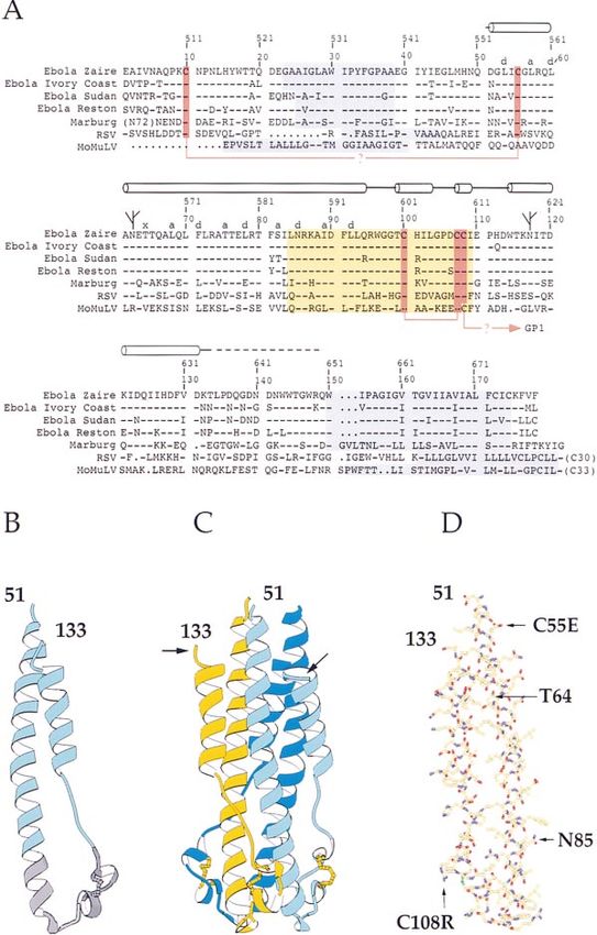
Molecular Cell
606

Figure 1. X-Ray Crystallographic Structure Determination
(A) Experimental electron density map calculated with single isomorphous replacement anomalous scattering (SIRAS) phases to 3.5 Å resolution
(Table 1B), showing GP2 Phe-71 packed at the core of the coiled coil.
(B) Density-modified electron density map calculated after iterative real-space NCS three-fold averaging, histogram matching, and solvent
flattening to improve and extend the phases to 3.0 Å resolution.
(C) Ribbon diagram of the GP2 (blue)/modified GCN4 (purple) hybrid trimer. The disulfide bond in GP2 is colored yellow. The N terminus of
the GCN4 trimer is at the far right, and the C terminus of GP2 is after the outer helical layer at the center. (A) and (B) were created with O
(Jones et al., 1991), (C) with Ribbons (Carson, 1991).

by X-ray crystallography. The structure shows that a                      at residues 55 and 108 were substituted with glutamic
disulfide-bonded loop, proposed in retroviruses to have                   acid and arginine, respectively, to minimize aggregation
immunosuppressive properties, connects an inner tri-                      and to promote crystallization. Arg-108 forms a crystal
meric core of N-terminal a helices to an outer layer                      lattice contact. Crystals contain two trimers of the GCN4-
composed of an antiparallel, linear strand-helix-strand                   GP2 chimera per asymmetric unit. To generate an iso-
segment (Figure 1). We compare the structure of GP2                       morphous heavy atom derivative, GCN4 zipper residue
to those of other viral and cellular membrane fusion                      Leu-12c, known to be on the outside surface of the
molecules and to a structure predicted for GP2 (Gal-                      trimer, was substituted with a cysteine that was reacted
laher, 1996). The comparison suggests that the ob-                        with p-hydroxy-mercuribenzoate before crystallization.
served structure may be a model for both a prefusion                      The structure was determined by single isomorphous
intermediate and the postfusion conformation of GP2.                      replacement with anomalous scattering (SIRAS) (Figure
                                                                          1A) and density modification including iterative real-
Results and Discussion                                                    space noncrystallographic averaging about the molecu-
                                                                          lar three-fold symmetry axes and solvent flipping (Figure
Structure Determination                                                   1B). (Data and phasing statistics are presented in Tables
Ebola Zaire GP2 is 179 residues long; it has a 16-residue                 1A and 1B and in the Experimental Procedures.) The
“internal fusion peptide,” residues 23–38, and a 25-resi-                 refined model consists of residues 3c–32c of the modi-
due membrane anchor, residues 150–175 (purple in Fig-                     fied GCN4 isoleucine zipper (purple in Figure 1C) and
ure 2A) (Feldmann et al., 1994; Sanchez et al., 1998a).                   residues 51–133 of one GP2 monomer (light blue in Fig-
Residues 51–149 were expressed in E. coli with a 32-                      ure 1C); the other monomers end at residues 131 and
residue modified GCN4 isoleucine zipper known to tri-                     132. Refinement statistics are presented in Table 1C.
merize (Harbury et al., 1993), replacing the N-terminal
fusion peptide loop residues 1–50 as described earlier                    Monomer Structure and a Conserved
(Weissenhorn et al., 1998a). Heptads in the GCN4 zipper                   Interhelical Linker
were placed in register with predicted heptads in the                     The GP2 ectodomain monomer is a helical hairpin about
N-terminal region of GP2 (Gallaher, 1996). Free cysteines                 65 Å long. A 44-residue N-terminal a helix (residues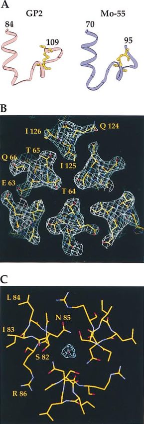
Structure of Ebola Virus GP2 Ectodomain
607

                                                                                   Figure 2. Sequence and Structure of Ebola
                                                                                   Virus Zaire GP2
                                                                                   (A) Sequence and secondary structure (cylin-
                                                                                   ders, helices; heptads, a, d, and x; dashed
                                                                                   lines, disordered) of Ebola Zaire GP2 aligned
                                                                                   with sequences of the other three Ebola sub-
                                                                                   types, Marburg virus GP2, and two retrovirus
                                                                                   Tms (RSV, Rous sarcoma virus; MoMulv, Mo-
                                                                                   loney murine leukemia virus). Cysteines are
                                                                                   marked red. The N-terminal fusion peptide
                                                                                   and C-terminal transmembrane anchors are
                                                                                   highlighted in purple, and the conserved “im-
                                                                                   munosuppressive motif” containing a disul-
                                                                                   fide-bonded loop and free cysteine in yellow.
                                                                                   The disulfide bonds between Cys-10 and
                                                                                   Cys-55 and Cys-108 and GP1 have not been
                                                                                   demonstrated biochemically or directly in the
                                                                                   current structure but have been suggested
                                                                                   previously (Sanchez et al., 1998b), and their
                                                                                   assignment is discussed in the text.
                                                                                   (B) Ribbon drawing of the Ebola GP2 mono-
                                                                                   mer. The N-terminal residue 51 and last struc-
                                                                                   tured residue 155 are labeled.
                                                                                   (C) Ribbon drawing of the trimer of Ebola GP2.
                                                                                   Arrows points to the residues 132–133, which
                                                                                   are ordered but slightly different in different
                                                                                   monomers (see text). The residues beyond
                                                                                   133 (to 149) are disordered. The disulfide
                                                                                   bonds are shown as ball and stick models
                                                                                   and colored yellow.
                                                                                   (D) All-atom model of the Ebola GP2 mono-
                                                                                   mer. Mutated residues C55E and C108R are
                                                                                   indicated with arrows. The side chain of Thr-
                                                                                   64, which packs irregularly in the core coiled
                                                                                   coil, and Asn-85, which is bound to the tenta-
                                                                                   tive chloride ion, are labeled.
                                                                                   (B), (C), and (D) were created with Ribbons
                                                                                   (Carson, 1991).

51–94) is followed by a 19-residue link to a short, antipar-     of the Murine Moloney Leukemia virus (MuMoLv) TM
allel a helix of 28 residues (residues 114–131) (Figure 2B).     subunit, Mo-55 (Figure 3A), despite the truncation of
Beyond the outer layer a helix, two monomers extend by           Mo-55 immediately C-terminal to the sequence for crys-
1 and 2 residues, respectively, while the remaining 16           tallization (Fass et al., 1996).
(17) residues are disordered (arrow in Figure 2C), as well          The structural conservation between filoviruses and
as all 18 residues of the third monomer.                         retroviruses of this complex motif suggests that it may
   The interhelical linker, residues 95–113, is part of a seg-   have been selected to serve a more complex function
ment, residues 84–109, containing a disulfide-bonded             than merely reversing the chain direction (Figure 3A).
loop (Cys-100 to Cys-107); the whole segment shares              It seems unlikely to have been conserved to have an
40%–50% sequence identity with an immunosuppres-                 immunosuppressive effect because that effect can be
sive sequence found in many retrovirus TM subunits               demonstrated using peptides homologous to the C ter-
(yellow in Figure 2A and gray in Figure 2B) (Volchkov            minus of the core helix, less than half the length of
et al., 1992). The conformation of this segment, which           the immunosuppressive motif (Cianciolo et al., 1985;
comprises the two C-terminal turns of the long a helix           Becker, 1996). Similarly, such an elaborate structure
and a four-residue turn followed by a short a helix that         also seems unnecessary simply to link two antiparallel
leads into an eight-residue disulfide-bonded loop (Fig-          a helices, a task that can be accomplished by four resi-
ure 3A), is very similar (1.5 Å rms) to that of the homolo-     dues. It may be that both the linker and disulfide-bonded
gous sequence in the structure of a 55-residue segment           loop were conserved to function as a hinge that can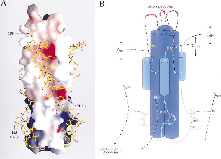
Molecular Cell
608

Table 1. Crystallographic Statistics
A. Data Collection
                                         Native                                           PHMBa
                                         20.0–3.0                3.09–3.0                 20.0–3.5                   3.6–3.5
Rmergeb                                  0.083                   0.29                     0.10                       0.23
Complete (%)                             99.3                    99.2                     99.3                       98.8
.3sI (%)                                 75.0                    48.2                     74.4                       56.1
No. reflections                          87,380                  4133                     66,460                     2745
No. unique reflections                   17,123                  1345                     10,870                     888
Average redundancy                       5.1                     3.07                     6.1                        3.0
Risoc                                                                                     0.18                       0.20
B. Phasing (SHARP)
                     Phasing Powerd                                                         Figure of Merit
Resolution           Acentric          Acentricanom       Centric           Resolution      Acentric          Centric
20–9.95              2.37              1.2                1.2               20–7.88         0.54              0.41
9.95–6.56            3.30              0.98               2.30              7.88–5.8        0.55              0.54
6.56–5.22            2.68              0.82               1.72              5.8–4.81        0.46              0.47
5.22–4.46            1.73              0.65               1.31              4.81–4.19       0.36              0.39
4.46–3.96            1.78              0.56               1.24              4.19–3.77       0.36              0.35
3.96–3.5             1.78              0.46               1.18              3.77–3.5        0.25              0.24
20–3.5               2.16              0.71               1.51              20–3.5          0.42              0.4
C. Refinement
R Values and Temperature Factors                              Model Geometry
No. reflections in working set         16,401                 Bond length rmsd from ideal              0.016 Å
Rcryste                                0.239 (0.332)f         Bond angles rmsd from ideal              1.548
Rfreee                                 0.256 (0.337)f         Ramachandran plot
                                                              % in most favored regiong                93.0
Average B                              38.7 Å2               % in additional allowed regionsg         6.7
a
  p-hydroxy-mercuribenzoate.
b
  Rmerge 5 ShSi| Ii(h) 2 ,I(h.)|/ShSiIi(h), where Ii(h) is the ith measurement, and ,I(h). is the weighted mean of all measurements of I(h).
c
  Riso 5 Sh | |FEMP(h)| 2 |Fnative(h)||/Sh|Fnative(h)|.
d
  Phasing power 5 ,|FH|./E, where ,|FH| is the rms structure factor amplitude for the heavy atom, and E is the estimated lack-of-closure
error.
e
  Rcryst and Rfree 5 Sh||F(h)obs | 2 | F(h)calc ||/Sh|F(h)obs| for reflections in the working and test sets, respectively.
f
  Numbers in parentheses are for final shell 3.07–3.00 Å.
g
  As defined in PROCHECK (Laskowski et al., 1993).

change the direction of the sequences C-terminal to it.                     the coiled coil, generally with a heptad (3-4) periodicity,
The hypothetical disulfide-bonded attachment of GP1                         where heptad positions are denoted a–g (Figure 2A). At
(and some retroviral Su subunits) to such a hinge (Gal-                     one position, fourth from the N terminus, an unusual
laher, 1996; Sanchez et al., 1998b) (Figure 5B) would                       3-4-4-3 periodicity is observed (Figures 2A and 2B),
provide a possible path for information to transfer from                    caused by the packing of Thr-64 directed at the three-
receptor binding by GP1 to trigger a conformational                         fold axis (“x-like” packing; Figure 3B) (see also Bullough
change in GP2. There is evidence for contacts between                       et al., 1994; Brown et al., 1996). Low-pH-treated HA2
gp120 and gp41 at the homologous loop in gp41 and                           has a similar 3-4-4-3 periodicity near the beginning of
for conformational changes of that region after receptor                    the central helix at Thr-59, a residue that is near the
and coreceptor binding (Sattentau and Moore, 1991;                          beginning of an interhelical loop in metastable HA but
Weissenhorn et al., 1996), but there are no studies to                      in the coiled coil in the low-pH-treated HA2 structure
date of the conformational changes that accompany                           (in Bullough et al., 1994; see Figures 3A and 5). It is
activation of filoviral membrane fusion.                                    possible by analogy that in a hypothetical metastable
                                                                            GP1-GP2 structure, GP2 may have a nonhelical segment
A Stutter in the Trimeric Coiled Coil Like                                  between a helices starting near Thr-59. (A similar irregu-
that in Low-pH-Induced HA2                                                  larity is found in the structure of HTLV-1 TM [B. Kobe,
The core of the trimeric structure is a triple-stranded                     R. J. Center, B. E. Kemp, and P. Poumbourious, personal
a-helical coiled coil formed by the 12-turn-long, N-termi-                  communication].)
nal a helix (residues 51–94) (Figure 2C). Near the N-ter-                      A practical consequence of this unanticipated break
minal end, the coil is underwound, at a stutter (Brown et                   in the 3-4 periodicity in GP2 is that we attached the
al., 1996), relative to classical knobs-into-holes packing                  GCN4 isoleucine zipper with its heptads out of phase
predicted by Crick (Crick, 1953). Seven hydrophobic and                     with the actual, as opposed to predicted (Gallaher,
four polar residues are packed in layers at the core of                     1996), heptads. (The remainder of the heptad prediction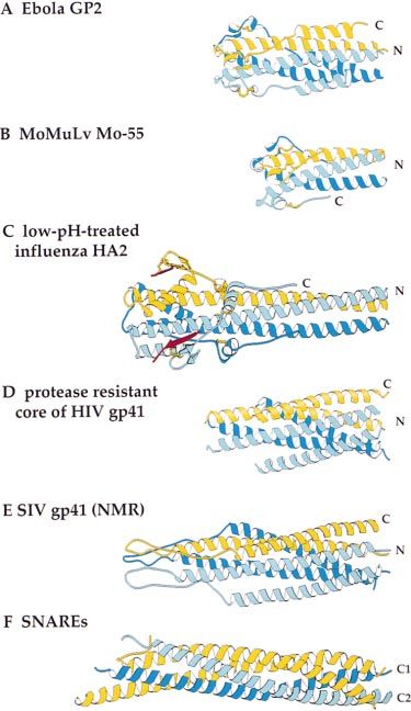
Structure of Ebola Virus GP2 Ectodomain
609

                                                                         for the N-terminal helix is correct [Gallaher, 1996].) In
                                                                         the crystals, we observe the GCN4 trimer axis to make
                                                                         an angle of a few degrees with the GP2 trimer axis
                                                                         (Figure 1C) in both copies of the molecule in the asym-
                                                                         metric unit. The independence of the structures of the
                                                                         GCN4 trimer and the GP2 trimer suggests that the zipper
                                                                         had its intended purpose of increasing the effective mo-
                                                                         larity of the GP2 chains so that they would fold stably
                                                                         and of helping to solubilize the molecule by replacing
                                                                         the fusion peptide (Weissenhorn et al., 1998a), without
                                                                         imposing the regular 3-4 periodicity of the GCN4 trimer.

                                                                         A Possible Chloride Ion Trapped
                                                                         in the Coiled Coil as in Mo-55
                                                                         Between the successive layers formed by Ser-82 and
                                                                         Asn-85 in the center of the coiled coil, we observe an
                                                                         electron dense peak from the solvent (5.9 s, B 5 39.5
                                                                         Å2) (Figure 3C). A similar feature was observed buried
                                                                         by the three Asn-71 amine groups of the Mo-55 trimer
                                                                         (Fass et al., 1996), which is the homolog of Asn-85 (Fig-
                                                                         ure 2D) in the sequence to GP2 and is conserved in a
                                                                         number of sequences of retroviruses (e.g., Figure 4 in
                                                                         Bukreyev et al. [1993]), suggesting that the trapped chlo-
                                                                         ride ion is a common feature in retroviral coiled coils.
                                                                         In the case of Mo-55, stability measurements of wild-
                                                                         type and an Asn-71 to Ile mutant in the presence of a
                                                                         series of anions indicated that the electron dense peak
                                                                         was due to a chloride ion. The crystalline GP2 has been
                                                                         maintained in buffers containing chloride from expres-
                                                                         sion through crystallization. Based on the conservation
                                                                         of the Asn in retroviral and filoviral sequences (Figure
                                                                         2A), and on the similarity of the electron density and
                                                                         coordination of the solvent peak in GP2 to that of the
                                                                         chloride in Mo-55, it is possible that GP2 also contains
                                                                         a chloride ion trapped at the homologous location near
                                                                         the C-terminal end of the central coiled coil. A solvent
                                                                         molecule thought to be an ion is also observed in the
                                                                         N-terminal part of the triple helical bundle at the core
                                                                         of the influenza HEF metastable conformation (Rosen-
                                                                         thal et al., 1998). It has been suggested that the chloride
                                                                         ion in Mo-55, like other buried polar interactions in coiled
                                                                         coils (Harbury et al., 1993; Lumb and Kim, 1995) that
                                                                         promote specificity in their folding, might prove impor-
                                                                         tant in MoMuLv (Fass et al., 1996). Since the chloride
                                                                         is located at the beginning of the sequence segment
Figure 3. A Conserved Motif, a Coiled Coil Stutter, and an Ion Bind-
                                                                         (84–109; yellow in Figure 2A) that is most conserved
ing Site in GP2
                                                                         between filoviral GP2s and retroviral TMs (see above),
(A) Comparison of the helix-turn-disulfide-bonded-loop motif of
                                                                         we speculate that it may be conserved for some func-
Ebola GP2 and MoMuLv Mo-55. Residues Leu-84 to Ile-109 of GP2
(yellow in Figure 2A) are shown in pink, Gln-70 to Phe-95 of Mo-55       tion, possibly during conformational switching.
in purple. The disulfide bond is drawn as a ball and stick model and
colored yellow.                                                          A Hydrophobic Core at the Base
(B) The “x-like” packing of Thr-64 pointing toward the three-fold        of the Coiled Coil
symmetry axis rather than like classical knobs-into-holes packing        Packed against the core coiled coil at its C terminus are
(e.g., Figure 1C). Electron density calculated with phases based on
                                                                         residues forming a turn and the eight-residue disulfide
the refined model (Table 1C) is superimposed on the model.
(C) Electron density thought to represent a chloride ion trapped at
                                                                         bond–containing loop (Figures 3A and 2C), which to-
the center of the coiled coil between Asn-85 and Ser 282. The            gether form a small hydrophobic core (residues F91,
distances between the amide nitrogen of Asn-85 and the tentative         L93, W96, I102, and I103) like that at the base of the
chloride ion average 3.5 Å, similar to the 3.25 Å distances observed   coiled coil in the low-pH-induced conformation of influ-
between a chloride ion and Asn-71 (the sequence homolog of GP2           enza HA (Figure 4C; Figure 4 in Bullough et al., [1994])
Asn-85) in Mo-55 (Fass et al., 1996).                                    and at the homologous location in Mo-55 (Fass et al.,
(A) was created with Ribbons (Carson, 1991), (B) and (C) with O
(Jones et al., 1991).
                                                                         1996) (Figures 4A and 4B). The interhelical loop is miss-
                                                                         ing from the X-ray structures of the proteolytically resis-
                                                                         tant core fragments of HIV-1 gp41 (Figure 4D). In an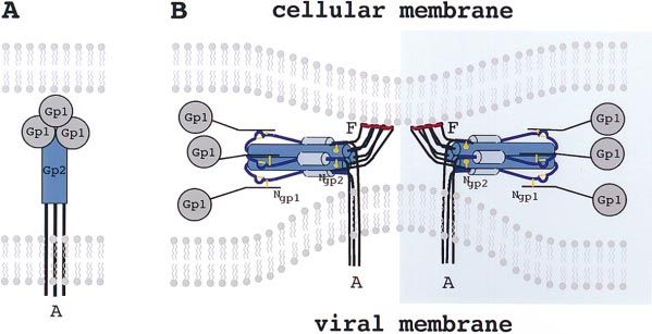
Molecular Cell
610

                                                                       packing is in phase with the predicted heptads (Gallaher,
                                                                       1996). (We cannot rule out the unlikely possibility that
                                                                       the GCN4 trimer which begins 13 Å further along the
                                                                       core coiled coil might have influenced the length of the
                                                                       outer layer a helix).

                                                                       A Flexible Link to the Transmembrane Anchor
                                                                       Of the 19 residues connecting the outer layer a helix to
                                                                       the transmembrane anchor, only the first one or two
                                                                       residues per monomer (Lys-132 and Thr-133 [built as
                                                                       Ala]; arrow in Figure 2C) are visible in the electron density
                                                                       maps. The two structured residues pack along the
                                                                       groove between the core a helices of the coiled coil
                                                                       (Figure 5A). The C-terminal-most structured segment of
                                                                       the low-pH-induced conformation of influenza HA2 is
                                                                       an extended strand of 11 residues, 152–162, which was
                                                                       also observed to adopt two conformations, ordered in
                                                                       two monomers and disordered in one monomer, in the
                                                                       crystallized trimer (at C label in Figure 4C) (Bullough
                                                                       et al., 1994). The observation that the final structured
                                                                       residues of GP2 and HA2 take different paths in the
                                                                       different copies of the monomers in crystals suggests
                                                                       that these residues can adopt alternate conformations
                                                                       of approximately the same energetic stability.
                                                                          One feature shared by the structures of GP2, low-pH-
                                                                       treated HA2, the proteolyticly resistant core of recombi-
                                                                       nant gp41, and the ectodomain of SIV gp41 is a long
                                                                       (13- to 18-residue) disordered segment between the last
                                                                       structured residue (labeled C in Figures 4A, 4C, 4D, and
                                                                       4E) and the transmembrane anchor sequence, either not
Figure 4. Comparison of GP2 with the Structures of Viral and Cellu-    visible, although present, in the crystals as in GP2 (15
lar Membrane Fusion Proteins                                           residues, 134–149) and HA2 (13 residues, 163–175 in
(A) Recombinant Ebola Zaire GP2.                                       crystal; anchor starts at 185), or inferred from its proteo-
(B) Recombinant Mo-55 from the TM subunit of MoMuLv (Fass et           lytic susceptibility in gp41 (18 residues, 155–172). In
al., 1996).                                                            HA2, these 23 disordered residues were proposed to
(C) Low-pH-treated HA2 from influenza virus (Bullough et al., 1994).   provide a flexible link from the rod-shaped molecule
(D) Recombinant, proteolysis-resistant core of HIV-1 gp41 (Weis-
                                                                       to the virus membrane that would allow the opposed
senhorn et al., 1997b).
(E) Recombinant SIV gp41, NMR structure (Caffrey et al., 1998).        membranes to approach more closely than the length
(F) Recombinant core coiled segments of the SNARES syntaxin 1-A        of the rod during fusion and allow HA2 to have both
(blue), synaptobrevin-II (light blue), and SNAP-25B (yellow) (Sutton   its fusion peptide and C-terminal anchor in the same
et al., 1998). This figure was created with RIBBONS (Carson, 1991).    membrane postfusion (Bullough et al., 1994). Immu-
                                                                       noelectron microscopy of low-pH-treated HA in viro-
                                                                       somes subsequently demonstrated that, in the absence
NMR structure of an ectodomain of SIV gp41, this loop
                                                                       of target membranes, association of HA rods with the
extends beyond the coiled coil, turning at the apex of
                                                                       same membrane by both the N-terminal fusion peptide
a disulfide-bonded loop before returning antiparallel to
                                                                       and the C-terminal anchor was permitted by the flexible
the outer layer a helix (Caffrey et al., 1998) (Figure 4E).
                                                                       segment at the C terminus of the HA2 ectodomain
                                                                       (Wharton et al., 1995).
An Outer Layer a Helix
The remaining length of the core coiled coil is stabilized
by the packing of a linear strand-helix-strand segment                 A Hypothetical Model for the Full-Length
into the groove between the core a helices (Figures 2C                 GP2 Ectodomain
and 5A). The first extended strand of this segment, Cys-               Based on the X-ray structure of the GP2 ectodomain
108 to Asp-113, makes nonpolar contacts into this                      without the fusion peptide, some aspects of the full-
groove with Ile-109 and Pro-111 and reaches to one                     length GP2 and its attachment to GP1 can be suggested.
core a helix with His-112 to make a salt bridge with Glu-              The structure establishes that two of the three cysteines,
77 (Figure 5A). A short, 4 1/2–turn a helix (residues Trp-             Cys-55 and Cys-108, are too far apart to form a disulfide
114 to Asp-131) then packs antiparallel into the groove                bond. This implies, as suggested previously but not es-
with nearly classical knobs-into-holes packing of the                  tablished biochemically (Gallaher, 1996; Sanchez et al.,
nonpolar side chains Trp-114, Ile-118, Ile-122, Ile-125,               1998a), that Cys-10, which is N-terminal to the fusion
and Phe-129 (Figure 5A). This outer layer a helix is two               peptide, will form a disulfide bond with Cys-55 near the
turns shorter in this structure of a GCN4-GP2 hybrid                   top of the N-terminal a helix (Figure 5B). These two
than predicted in a model of GP2, although the nonpolar                cysteines are conserved as a pair in filoviral and some
Structure of Ebola Virus GP2 Ectodomain 611 Figure 5. The Packing of the Outer Layer of the GP2 Ectodomain Trimer and a Hypothetical Model for the Whole Molecule (A) GP2 ectodomain with an electrostatic surface potential representation of the core coiled coil and with the outer layer drawn as an atomic model. Negative electrostatic potential, red; positive, blue. The position of His-112, which makes a salt bridge to Glu-77 of the inner core, and the position of Cys-108 (mutated to Arg), which forms a disulfide bond with Gp1, are indicated with arrows. The electrostatic surface was contoured between -30 kT/e and 130 kT/e. This figure was created with GRASP (Nicholls et al., 1991). (B) A hypothetical model of the full-length ectodomain of GP2 based on the portion of the structure determined here (X-ray structure shown in blue) showing the proposed location (dotted lines) of the fusion peptide in a hypothetical disulfide-bonded loop, the C-terminal disordered segments (Cgp2), and the proposed disulfide bond to GP1 (GP2 Cys-108 to GP1 Cys-19). Neither the Cys-10 to Cys-51 nor the Cys-108 to GP1 Cys-19 disulfide bonds have been demonstrated biochemically or structurally, but they are proposed based on sequence conservation and distances seen in the GP2 structure (see text). retroviral sequences (e.g., Figure 2A), or both are miss- considerations apply to the C-terminal regions of HA2 ing, suggesting that they are linked. This arrangement and gp41. would position the internal, nonpolar fusion peptide, res- GP1 is disulfide bonded to GP2, and the location of idues 23–38 (Figure 1A), as part of a disulfide-bonded the bond between GP2 Cys-108 and GP1 Cys-19 has loop at one end of the rod-shaped GP2 (Figure 5B). been proposed but not demonstrated biochemically Cysteine 55 (Figure 2D, C55E) is accessible on the outer (Gallaher, 1996; Sanchez et al., 1998a). The structure of surface of the core a helix in a groove between the outer GP2 reported here supports this proposal by locating layer helix-strand segments so that the returning part Cys-108 (Figure 2D, C108R) at the opposite end of the of the fusion peptide loop could form a second outer molecule from the other free cysteines of the fusion layer on the structure (Figure 5B). peptide loop (Figure 5B) (although strictly an internal Because they are disordered in the crystal, the loca- GP2 disulfide bond from Cys-10 to Cys-108 cannot be tion of the C-terminal 16 residues of the GP2 ectodomain ruled out). Sequence comparisons suggest that in some is unknown beyond the outer layer a helix (dotted lines retroviruses the cysteines homologous to GP2 Cys-108 in Figures 2A and 5B). In full-length GP2, these residues (Figure 5B) and GP1 Cys-19 could also form the in- are attached to the hydrophobic anchor in the viral mem- terchain disulfide bonds between the equivalent retrovi- brane, so that the orientation of the rod-shaped GP2 ral Su and Tm residues (e.g., RSV and ALSV [Herdandez relative to the viral membrane is determined by the struc- et al., 1997; but see also Pinter et al., 1997]). The GP2 ture of this apparently flexible segment. Seventeen resi- structure indicates that this tentative disulfide bond dues cannot extend the 62 Å back to the opposite end would attach the receptor-binding domain, GP1, at the of GP2, which suggests that in this form the GP2 rod end of the rod-shaped molecule distant from the N and will be oriented with the viral membrane near the same C termini (Figure 5B). This is very similar to the disulfide end of the rod as the fusion peptides. If, as an alterna- bond attachment of influenza HA1 to HA2, which is be- tive, the last two structured residues (residues 132–133) tween HA1 Cys-14, near the N-terminal of HA1, and HA2 of the ectodomain were to peel off the rod, this C-termi- Cys-137, located at the opposite end of the low-pH- nal region would be long enough to reach either end induced HA2 rod from the fusion peptide (yellow ball (hence two arrows at GP2 C termini in Figure 5B). Similar and stick in Figure 4C). In Figure 5B, the N and C termini
Molecular Cell
612

                                                                                    Figure 6. Hypothetical Model of Some Steps
                                                                                    in the Membrane Fusion Mechanism
                                                                                    (A) and (B) represent hypothetical intermedi-
                                                                                    ates and are based on Figure 3 of Weissen-
                                                                                    horn et al. (1997b) (see text). Fusion peptides
                                                                                    labeled F are colored red. GP2 transmembrane
                                                                                    anchors are labeled A. The GP2 outer layer a
                                                                                    helices are colored light blue; the N-terminal,
                                                                                    core coiled coil is dark blue; disulfide bonds
                                                                                    are yellow. Ngp2 and Ngp1 label the pro-
                                                                                    posed location of the N termini of those poly-
                                                                                    peptide chains. The gray spheres represent
                                                                                    the receptor-binding domain of GP1. The pic-
                                                                                    ture also incorporates suggestions that more
                                                                                    than one trimer might be involved in forming
                                                                                    an initial fusion pore (Danieli et al., 1996) and
                                                                                    that the bilayers may be distorted with mole-
                                                                                    cules entering at an angle (Ruigrok et al.,
                                                                                    1992; Tatulian et al., 1995), which in some
                                                                                    way results in distortions to the membrane
                                                                                    that favor membrane fusion (e.g., Cherno-
                                                                                    mordik et al., 1998).

of GP1 (dotted lines) are oriented by analogy to influenza      and a currently unknown stimulus in filoviruses); and a
HA, in which at low pH, the b strand containing HA1             stable, rod-like conformation (Figure 4) observed be-
Cys-14 is inverted projecting the receptor-binding do-          cause they are the states formed spontaneously by the
main away from the rod (red in Figure 4C); whereas              membrane fusion subunit when expressed alone (HA2,
in the metastable structure, the HA receptor-binding            gp41, and GP2) and in the case of influenza virus, when
domain points in the opposite direction (see Figure 3B          induced at the low pH of membrane fusion.
in Bullough et al. [1994]).                                        One possibility is that the rod-like conformation ob-
                                                                served here for GP2 ectodomain is the conformation of
Membrane Fusion                                                 both a prefusion intermediate (perhaps as a component
An intermediate in the membrane fusion mechanism like           of an oligomeric pore) and the postfusion state of the
that in Figure 6B was proposed (Weissenhorn et al.,             ectodomain. The distinction is that in the prefusion inter-
1997b, 1998b) to incorporate the observations that in           mediate the N and C termini are inserted in the cellular
both HA2 and gp41, the fusion peptide and membrane              and viral membranes, respectively, whereas in the post-
anchor sequences were projecting toward one end of              fusion state both sets of termini are inserted in the one
a thin rod-shaped molecule that could facilitate close          remaining, fused membrane. This possibility was sug-
apposition of the prefusion membranes. A similar model          gested for low-pH-induced HA2 based on the apparent
is suggested for GP2 based on its similarity in structure       flexibility between the molecular rod and both its termini
to gp41. In some strains of HIV-1, gp120 dissociates            and the observation that all of the mutations that altered
from gp41, but in influenza and, as we propose here, in         the pH of the fusion process were positioned in the
Ebola virus, the receptor-binding domain is attached by         structure at points of structural change leading to the
a disulfide bond at the opposite end of the fusion protein      rod-shaped conformation (Bullough et al., 1994). It was
rod, where it could be withdrawn from the site of fusion.       also suggested for gp41 because in the X-ray studies
                                                                both the membrane anchor and the fusion peptides
A Prefusion Intermediate and/or                                 were, more clearly than in the HA2 structure, at the same
a Postfusion Conformation?                                      end of the rod-shaped structure where they might draw
In the absence of direct studies of the membrane fusion–        two membranes together (Weissenhorn et al., 1997a).
active form of filovirus GP2, the similarity of the structure   (A corollary of the possibility that there is a single rod-
observed here to recombinant gp41 and low-pH-induced            like prefusion intermediate and postfusion state is that
HA2 provides evidence for the functional significance           whatever distortion or stress to the bilayers that might
of this structure. Only three conformational states of the      exist at the attachment positions of the prefusion inter-
membrane fusion subunits of viral glycoproteins have            mediate to the two membranes and might favor mem-
been defined that are not transient and that are suffi-         brane fusion [e.g., Sutton et al., 1998] would be relieved
ciently stable for biochemical and structural study (re-        postfusion at the site of attachment to the fused mem-
viewed in Skehel et al., 1995; Chen et al., 1998a): the         brane, extinguishing the membrane fusion activity.)
single-chain, precursor state (HA0, gp160, and GP) that            An alternative possibility, that GP2 mediates mem-
is not infectious because it cannot be triggered to a           brane fusion while its ectodomain is in a transient con-
membrane fusion–active conformation; the postransla-            formation, preceding the rod-shape structure observed
tionally cleaved, primed state (HA1/HA2, gp120/gp41,            here, is addressed by studies of inhibitors of membrane
and GP1/GP2) that is also membrane fusion inactive but          fusion of the similarly shaped gp41. Peptides corre-
can be triggered to undergo a conformational change             sponding to both the core coiled coil and the outer
by different events in different viruses (receptor and          layer a helices of gp41 inhibit viral infection by inhibiting
coreceptor binding in HIV-1, low pH for influenza virus,        membrane fusion (Wild et al., 1994). Structural studies
Structure of Ebola Virus GP2 Ectodomain
613
suggested that these peptides block fusion by blocking rod-shaped complex (Figure 4E); for viruses, the rod-
the refolding to the rod-shaped conformation (Lu et al., shaped complex is formed exclusively by the virus fu-
1995; Chan et al., 1997; Tan et al., 1997; Weissenhorn sion protein; its fusion peptide, by interacting with the
et al., 1997b; Malashkevich et al., 1998). Resistant muta- target membrane, substitutes for the membrane anchor
tions map to the coiled coil as expected from this pro- sequence of the cellular target membrane protein in
posed mechanism (Rimsky et al., 1998), and inhibition intracellular vesicle fusion.
only occurs when peptide is present at the time of fusion
when gp41 either becomes exposed and/or undergoes
Conclusions
a structural change (Furuta et al., 1998). Other studies
The structure of the Ebola virus GP2 ectodomain adds
suggest that peptides may inhibit by disrupting the rod-
to observations of similarities between virus fusion gly-
shaped conformation after it forms (Caffrey et al., 1998)
coproteins that have accumulated from analyses of their
or preventing it from assembling into an oligomeric fu-
amino acid sequences, their activities, and their biosyn-
sion pore (Munoz-Barroso et al., 1998).
thesis. For influenza, retro-, and filoviruses in particular,
there is a common requirement for proteolytic cleavage
Ancestral Viral Fusion Proteins of a biosynthetic precursor into distinct receptor-binding
The attachment of GP1 to GP2 by a disulfide bond near and membrane fusion domains, and common structural
the bottom of the GP2 stem, if confirmed, suggests that properties have been predicted from their sequences.
the N-terminal segment of GP1 may form part of the These similarities are now emphasized by the observa-
stem of GP1-GP2 in the conformation found on virus, tions that irrespective of either their initial structures or
in a similar way that the first 50 residues of HA1 form of differences in the processes involved in their activa-
part of the stem of influenza HA1-HA2. The recently tion for membrane fusion, the isolated fusion domains
determined structure of HEF, the hemagglutinin-ester- are rod-shaped coiled coils. This homology strongly
ase fusion glycoprotein of influenza C virus, suggested, suggests that the structures have a role in membrane
by comparison with the HA structure, that the stem of fusion and, for HA, it reinforces previous conclusions
HA, including N-terminal and C-terminal segments of that the changes in conformation required to form this
HA1 as well as all of HA2, constitute a single domain structure are necessary for fusion activity (Bullough et
derived from an ancestral fusion protein into which a al., 1994). Specific details of the Ebola GP2 structure,
receptor domain was inserted at a surface loop (Rosen- especially irregularities in the core coiled coil, a con-
thal et al., 1998; Zhang et al., submitted). served disulfide loop segment that may be a hinge, and
The attachment of GP1 to GP2 (and, e.g., RSV Su to the likely location of the site of attachment to GP1, sug-
Tm) and evidence that gp120 also contacts the disulfide- gest that filovirus fusion proteins might also be required
bonded loop at the base of gp41 (Sattentau and Moore, to refold in some way to mediate fusion.
1991; Weissenhorn et al., 1996) suggest the possibility
that retrovirus and filovirus glycoproteins may derive Experimental Procedures
from another ancestral stem-like fusion protein com-
posed of the membrane fusion subunit (gp41, TM, and Expression, Purification, and Crystallization
Hybrids of a modified 30-residue GCN4 trimeric coiled coil linked
GP2) and the N-terminal segment of the subunit preced-
to residues 51–149 of GP2 (residues 552–650 of GP) were expressed
ing the posttranslational cleavage site, and that most and purified in the same way as the construct pIIgp2(552–650) de-
of the remainder of the first subunit inserted into that scribed earlier (Weissenhorn et al., 1998a). To promote crystalliza-
ancestral fusion protein to provide receptor binding ac- tion, two residues, Cys-55 (Ser-565 in pIIgp2(552–650)) and Cys-108
tivity. Whether the retroviral and filoviral fusion protein (Ser-609 in pIIgp2(552–650)) were mutated to Glu and Arg, respec-
undergoes a complex refolding event to achieve the tively, by site-directed mutagenesis, following standard PCR proto-
cols. To make a heavy atom isomorphous derivate, Leu-12 of the
state observed for gp41 and GP2, or whether this confor-
GCN4 zipper was mutated to Cys, and the protein pIIGp2(51–149)cys
mation is simply exposed from a hidden location in the was reacted with p-hydroxy-mercuribenzoate before crystallization.
uninduced glycoprotein, is unknown. Crystals were grown by vapor diffusion in hanging drops by com-
bining 2 ml of protein solution (5 mg/ml in 20 mM Tris–HCl [pH 9.3])
with 1 ml of reservoir solution (100 mM Tris–HCl [pH 8.5], 30%–34%
Comparison with Intracellular Vesicle Fusion PEG 4000, 220 mM ammonium sulphate, 1.0% dioxane). Derivatized
pIIGp2(51–149)cys was crystallized by the same method and from
The fusion of intracellular transport vesicles involves the
the same reservoir buffers as the wild-type pIIGp2(51–149) but with
assembly of a four-stranded a-helical coil from three the addition of 5 mM lauryldimethylamine oxide (Hampton Research)
SNARE molecules, including one anchored in the vesicle to the crystallization drop.
and one anchored in the target membrane (Figure 4F)
(e.g., Brünger et al., 1998; Nicholson et al., 1998; Poirier
X-Ray Diffraction Data Collection
et al., 1998; Sutton et al., 1998). The C-terminal mem- Both proteins (wild-type and derivatized form) crystallize isomor-
brane anchors of both membrane proteins are at the phously in space group C2 with unit cell dimensions a 5 171.70 Å,
same end of the helical rod (Hanson et al., 1997; Lin and b 5 32.69 Å, c 5 168.86 Å, a 5 908, b 5 119.238, g 5 908 with two
Scheller, 1997; Poirier et al., 1998; Sutton et al., 1998), molecules per asymmetric unit. Diffraction data from both native
suggesting that a similarly shaped molecular complex and derivative crystals were measured at room temperature using
an 18 cm image plate scanner (MAR Research, Hamburg Germany)
is used for bringing membranes into apposition and
mounted on an Elliott GX-13 rotating anode source (Elliot, London)
mediating membrane fusion by both viruses and cells. with mirror optics. Oscillation images were processed using DENZO
In the case of cellular vesicle fusion, protein components (Otwinowski and Minor, 1997), and data reduction was carried out
of both membranes contribute to the formation of the using SCALEPACK (Otwinowski, 1993) (Table 1A). The CCP4 suiteMolecular Cell
614
of programs was used for further data processing and analysis metal atom and two waters) involved in crystal lattice interactions
(Collaborative Computational Project Number 4, 1994). and two chloride ions (inside the Ebola trimeric coiled coils) were
modeled in the electron density. The three tentatively assigned metal
Phasing atoms are 9.0 s, 5.0 s, and 4.8 s electron density peaks (in omit
The structure was determined by phasing with single isomorphous difference maps calculated with phases from the refined model) and
replacement anomalous scattering (SIRAS) and density modification refined to B factors of 42.3 Å2. The strongest peak is coordinated
using the p-hydroxy-mercuribenzoate derivative as the initial source between trimers in the crystal by two histidine residues (His-112B
of phase information. Six heavy atom positions (corresponding to and His-127D) with short (average bond distance 5 2.42 Å) in-
the GCN4 Cys-12 positions) were located in an isomorphous differ- teratomic distances to the side-chain nitrogens, characteristic of
ence Patterson map using RSPS (Collaborative Computational Proj- metal ions. The other two solvent peaks, one in each trimer in the
ect Number 4, 1994). Refinement of the heavy atom parameters was asymmetric unit, which may be metals or well-ordered waters, are
carried out with the program SHARP (Fortelle and Bricogne, 1997) at lattice contacts and are coordinated (average bond distance 5
(Table 1B). Three additional sites, spaced approximately 4 Å from 2.89 Å) between His-112 of GP2 and Gln-3 of the GCN4 sequence
the ones found initially, were located by a peak search using a log (from trimer chains C and E to chains F and A, respectively). The
likelihood gradient map after SIR phasing. These sites may corre- two chloride ions were modeled into 5.8 and 6.0 s electron density
spond to alternative cysteine conformations. Density modification, peaks and refined to a B factor of 39.5 Å2. They are assigned as
consisting of solvent flipping using an envelope determined by anal- chloride ions based on the close similarity of their locations to a
ysis of the local standard deviation of the SIRAS map, histogram chloride ion in Mo-55 (Fass et al., 1996) on sequence conservation
matching, and phase extension from 3.5 to 3.0 Å resolution, was of the ligands (Asn-85) (see text). Refinement was concluded using
carried out with the program SOLOMON (Collaborative Computa- CNS (Brünger et al., 1998) with restrained conjugate gradient minimi-
tional Project Number 4, 1994). The resulting electron density map zation and restrained B group refinement. A uniform bulk solvent
was of sufficient quality to fit the two GCN4 molecules (residues correction (B 5 41.57 Å2, k 5 0.29 e/Å3) and overall anisotropic
3–28), a Ca trace for the inner and outer layer helices of one Ebola thermal factor (B11 5 26.46 Å2 , B22 5 25.11 Å2, B33 5 11.546 Å2,
molecule, and three shorter fragments of the inner and outer layer B13 5 23.41 Å2) were applied. The final model contains residues
helices for the second Ebola molecule (molecule 2). Although some 3–131 for chains 1C and 2E; 3–132 for chains 1A, 1B, and 2D; and
electron density was evident for the linker loop region, no model 3–133 for chain 2F, two chloride and three zinc ions. The model has
could be built into it. From this model, it was clear that three-fold good stereochemistry and structural quality with an average bond
symmetry axes of the GCN4 trimers were slightly misaligned from length and bond angle deviation of 0.016 Å and 1.548, respectively,
the three-fold axes of the covalently linked Ebola GP2 trimers (for and no Ramachandran violations (Table 1C). Residues 32 and 51 of
the two GCN4 trimers, k 5 175.468, v 5 91.498, φ 5 81.258; for the both monomer A and F are built in poor electron density.
Ebola trimers, k 5 174.93, v 5 90.32, φ 5 90.76). The Rfree is 25.6%, and Rwork is 23.9% for the resolution range
The Ca coordinates were used to define four sets of three-fold 20–3.0 Å (Table 1C). Some noninterpreted electron density, enough
NCS matrices (two for the GCN4 and two for the Ebola trimers) with for 2 or 3 residues, can be observed at 0.5 s contour level in sA
LSQKAB (Collaborative Computational Project Number 4, 1994). The weighted electron density maps and may correspond to part of the
program NCSMASK (Collaborative Computational Project Number disordered C terminus of the Ebola molecule.
4, 1994) was used to generate four NCS masks that did not include
the GCN4–Ebola connections. Iterative solvent flattening, histogram Acknowledgments
matching, and three-fold NCS averaging with phase extension from
3.5 Å to 3.0 Å were performed with DM (Collaborative Computational We thank Gary Nabel and A. Sanchez for the DNA clone of the Ebola
Project Number 4, 1994) without phase combination to the previous virus glycoprotein, Dr. Bostjian Kobe for sharing unpublished data
cycle. The resulting experimental electron density map was of excel- on HTLV-1 gp21, Dr. M. Clore for the SIV coordinates, M. Pietras
lent quality and showed the location of most of the side chains. The for excellent technical assistance, and the members of the Harrison/
unweighted phase difference at 20–3.0 Å resolution between the Wiley laboratory for helpful discussions. W. W. was supported by
SIRAS phases and the phases after density modification was 748. the HHMI. A. C. is supported by a AFRT (Association Française pour
A partial model (residues 3–29 of GCN4 and 52–128 of Ebola GP2) la Recherche Therapeutique) postdoctoral fellowship. This work was
of one monomer was readily built using the program O (Jones et supported by a supplement to the NIAID grant (5RO1AI13654-20)
al., 1991), and the rest of the model was generated applying the for Expanded International Research on Emerging and Re-Emerging
NCS operators. A short stretch of disconnected electron density Diseases, the Medical Research Council (UK), and the Howard
(sufficient for approximately two or three residues) was observed Hughes Medical Institute. D. C. W. is an investigator of the HHMI.
in proximity of one monomer (F) of molecule 1 but could not be
interpreted. Received October 2, 1998; revised October 21, 1998.
Refinement References
Before refinement, 5% of the reflections were set aside for calcula-
tion of the free R factor (Brünger, 1992a). An overall B factor of 30 Becker, Y. (1996). Retrovirus and filovirus “immunosuppressive mo-
Å2 was applied to all atoms. The model was refined in X-PLOR tif” and the evolution of virus pathogenicity in HIV-1, HIV-2, and
(Brünger, 1992b) initially with tight restraints on both main and side Ebola viruses. Virus Genes 11, 191–195.
chains. Four-group rigid body fitting, followed by 300 cycles of Becker, S., Spiess, M., and Klenk, H.D. (1995). The asialoglycopro-
positional refinement and torsion angle dynamics simulating anneal- tein receptor is a potential liver-specific receptor for Marburg virus.
ing with tight restraints on main and side chains for the six mono- J. Gen. Virol. 76, 393–399.
mers, reduced the Rfree from 41.1% to 36.7% and the Rwork from Brown, J.H., Cohen, C., and Parry, D.A. (1996). Heptad breaks in
43.8% to 34.2% between 8.0 and 3.0 Å resolution. New phases and a-helical coiled coils: stutters and stammers. Proteins 26, 134–145.
figures of merit were calculated from the refined model using SFALL Brünger, A.T. (1992a). Free R value: a novel, statistical quantity for
and SIGMAA (Collaborative Computational Project Number 4, 1994) assessing the accuracy of crystal structures. Nature 355, 472–475.
and were used in DM for a new cycle of iterative NCS averaging.
Brünger, A.T. (1992). X-PLOR: A System for X-Ray Crystallography
Nonaveraged sA weighted electron density maps were used in sub-
and NMR. (New Haven, CT: Yale University).
sequent model-building stages during which the non-three-fold-
symmetric connections between GCN4 and Ebola GP2 (residues 29 Brünger, A.T., Adams, P.D., Clore, G.M., Gros, P., Grosse-Kuntsleve,
and 30 of GCN4 and 50 and 51 of GP2) were built. Three additional R.W., Jiang, J.-S., Kuszerski, J., Nilges, M., Pannu, N.S., and Read,
cycles of manual rebuilding and positional and B group refinement R.J. (1998). Crystallographic and NMR system (CNS): a new software
with tight restraints on the main chain (residues 5–28 and 55–129) system for macromolecular structure determination. ACTA Crys-
and weak restraints on side chains reduced the Rfree to 27.8% and tallogr. D 54, 905–921.
Rwork to 24.6%. At that stage, three metal ions (which may be one Bukreyev, A.A., Volchkov, V.E., Blinov, V.M., and Netesov, S.V.Structure of Ebola Virus GP2 Ectodomain
615
(1993). The GP-protein of Marburg virus contains the region similar Hanson, P.I., Roth, R., Morisaki, H., Jahn, R., and Heuser, J.E. (1997).
to the ‘immunosuppressive domain’ of oncogenic retrovirus P15E Structure and conformational changes in NSF and its membrane
proteins. FEBS Lett. 323, 183–187. receptor complexes visualized by quick-freeze/deep-etch electron
Bullough, P.A., Hughson, F.M., Skehel, J.J., and Wiley, D.C. (1994). microscopy. Cell 90, 523–535.
Structure of influenza haemagglutinin at the pH of membrane fusion. Harbury, P.B., Zhang, T., Kim, P.S., and Alber, T. (1993). A switch
Nature 371, 37–43. between two-, three-, and four-stranded coiled coils in GCN4 leucine
Caffrey, M., Cai, M., Kaufman, J., Stahl, S.J., Wingfield, P.T., Covell, zipper mutants. Science 262, 1401–1407.
D.G., Gronenborn, A., and Clore, G.M. (1998). Three-dimensional Herdandez, L.D., Peters, R.J., Delos, S.E., Young, J.A.T., Agard,
structure of the 44 kDa ectodomain of SIV gp41. EMBO J. 17, 4572– D.A., and White, J.M. (1997). Activation of a retroviral membrane
4584. fusion protein: soluble receptor-induced liposome binding of the
Carr, C.M., Chaudhry, C., and Kim, P.S. (1997). Influenza hemaggluti- ALSV envelope glycoprotein. J. Cell Biol. 139, 1455–1464.
nin is spring-loaded by a metastable native conformation. Proc. Hunter, E., and Swanstrom, R. (1990). Retrovirus envelope glycopro-
Natl. Acad. Sci. USA 94, 14306–14313. teins. Curr. Top. Microbiol. Immunol. 157, 187–253.
Carson, M. (1991). Ribbons 2.0. J. Appl. Crystallogr. 24, 958–961. Jones, T.A., Zou, J.Y., Cowan, S.W., and Kjeldgaard, M. (1991).
Centers for Disease Control and Prevention (1995). Update: out- Improved methods for binding protein models in electron density
break of Ebola viral hemorrhagic fever—Zaire, 1995. Morbid. Mortal. maps and the location of errors in these models. Acta Crystallogr.
Week. Rep. 44, 381–382. A 47, 110–119.
Chan, D.C., Fass, D., Berger, J.M., and Kim, P.S. (1997). Core struc- Joshi, S.B., Dutch, R.E., and Lamb, R.A. (1998). A core trimer of the
ture of gp41 from the HIV envelope glycoprotein. Cell 89, 263–273. paramyxovirus fusion protein: parallels to influenza virus haemag-
glutinin and HIV-1 gp41. Virology 248, 20–34.
Chen, J., Wharton, S.A., Weissenhorn, W., Calder, L.J., Hughson,
F.M., Skehel, J.J., and Wiley, D.C. (1995). A soluble domain of the Klenk, H.-D., Angliker, H., Garten, W., Hallenberger, S., Ohuchi, M.,
membrane-anchoring chain of influenza virus hemagglutinin (HA2) Ohuchi, R., Rott, R., Shaw, E., Stieneke-Grober, A., and Vey, M.
folds in Escherichia coli into the low-pH-induced conformation. (1993). Processing of viral glycoproteins by host proteases: struc-
Proc. Natl. Acad. Sci. USA 92, 12205–12209. tural aspects and functional consequences. In Virus Strategies, pp.
Chen, J., Lee, K.H., Steinhauer, D.A., Stevens, D.J., Skehel, J.J., 195–214 (Weinheim, Germany: Verlag Chemie).
and Wiley, D.C. (1998a). Structure of the hemagglutinin precursor Laskowski, R.A., MacArthur, M.W., Moss, D.S., and Thornton, J.M.
cleavage site, a determinant of influenza pathogenicity and the origin (1993). PROCHECK: a program to check the stereochemical quality
of the labile conformation. Cell 95, 409–417. of protein structures. J. Appl. Crystallogr. 26, 283–290.
Chen, J., Skehel, J.J., and Wiley, D.C. (1998b). A polar octapeptide Lin, R.C., and Scheller, R.H. (1997). Structural organization of the
fused to N-terminal fusion peptide solubilizes the influenza virus synaptic exocytosis core complex. Neuron 19, 1087–1094.
HA2 subunit ectodomain. Biochemistry 37, 13643–13649. Lu, M., Blacklow, S.C., and Kim, P.S. (1995). A trimeric structural
Chernomordik, L.V., Frolov, V.A., Leikina, E., Bronk, P., and Zim- domain of the HIV-1 transmembrane glycoprotein. Nat. Struct. Biol.
merberg, J. (1998). The pathway of membrane fusion catalyzed by 2, 1075–1082.
influenza hemagglutinin: restriction of lipids, hemifusion, and lipidic Lumb, K.J., and Kim, P.S. (1995). A buried polar interaction imparts
fusion pore formation. J. Cell Biol. 140, 1369–1382. structural uniqueness in a designed heterodimeric coiled coil. Bio-
Cianciolo, G.J., Copeland, T.D., Oroszlan, S., and Snyderman, R. chemistry 34, 8642–8648.
(1985). Inhibition of lymphocyte proliferation by a synthetic peptide Malashkevich, V.N., Chan, D.C., Chutkowski, C.T., and Kim, P.S.
homologous to retroviral envelope proteins. Science 230, 453–455. (1998). Crystal structure of the simian immunodeficiency virus (SIV)
Collaborative Computational Project Number 4 (1994). The CCP4 gp41 core: conserved helical interactions underlie the broad inhibi-
suite: programs for protein crystallography. Acta Crystallogr. D 50, tory activity of gp41 peptides. Proc. Natl. Acad. Sci. USA 95, 9134–
760–763. 9139.
Crick, F. (1953). The packing of alpha-helices: simple coiled-coils. Munoz-Barroso, I., Durell, S., Sakaguchi, K., Appella, E., and Blu-
Acta Crystallogr. 6, 689–697. menthal, R. (1998). Dilation of the human immunodeficiency virus-1
D’Souza, M.P., and Harden, V.A. (1996). Chemokines and HIV-1 envelope glycoprotein fusion pore revealed by the inhibitory action
second receptors. Nat. Med. 2, 1293. of a synthetic peptide from gp41. J. Cell Biol. 140, 315–323.
Danieli, T., Pelletier, S.L., Henis, Y.I., and White, J.M. (1996). Mem- Nicholls, A., Sharp, K.A., and Honig, B. (1991). Protein folding and
brane fusion mediated by the influenza virus hemagglutinin requires association: insights from the interfacial and thermodynamic prop-
the concerted action of at least three hemagglutinin trimers. J. Cell erties of hydrocarbons. Prot. Struct. Funct. Genet. 11, 281–296.
Biol. 133, 559–569. Nicholson, K.L., Munson, M., Miller, R.B., Filip, T.J., Fairman, R.,
Fass, D., and Kim, P.S. (1995). Dissection of a retrovirus envelope and Hughson, F.M. (1998). Regulation of SNARE complex assembly
protein reveals structural similarity to influenza hemagglutinin. Curr. by an N-terminal domain of the t-SNARE Sso1p. Nat. Struct. Biol.
Biol. 5, 1377–1383. 5, 793–802.
Fass, D., Harrison, S.C., and Kim, P.S. (1996). Retrovirus envelope Otwinowski, Z. (1993). Oscillation data reduction program. In Data
domain at 1.7 Å resolution. Nat. Struct. Biol. 3, 465–469. Collection and Processing, C.W. Carter, Jr., and R.M. Sweet, eds.
Feldmann, H., Will, C., Schikore, M., Slenczka, W., and Klenk, H.-D. (New York: Academic Press).
(1991). Glycosylation and oligomerization of the spike protein of Otwinowski, Z., and Minor, W. (1997). Processing of X-ray diffraction
Marburg virus. Virology 182, 353–356. data collected in oscillation mode. Methods Enzymol. 276, 307–326.
Feldmann, H., Nichol, S.T., Klenk, H.D., Peters, C.J., and Sanchez, Pinter, A., Kopelman, R., Li, Z., Kayman, S.C., and Sanders, D.A.
A. (1994). Characterization of filoviruses based on differences in (1997). Localization of the labile disulfide bond between SU and TM
structure and antigenicity of the virion glycoprotein. Virology 199, of the murine leukemia virus envelope protein complex to a highly
469–473. conserved CWLC motif in SU that resembles the active-site se-
Fortelle, E.d.L., and Bricogne, G. (1997). Maximum likelihood heavy quence of thiol-disulfide exchange enzymes. J. Virol. 71, 8073–8077.
atom refinement for multiple isomorphous replacement and multi Poirier, M.A., Xiao, W., Macosko, J.C., Chan, C., Shin, Y.-K., and
anomalous diffraction methods. Methods Enzymol. 276, 472–494. Bennett, M.K. (1998). The synaptic SNARE complex is a parallel
Furuta, R.A., Wild, C.T., Weng, Y., and Weiss, C.D. (1998). Capture four-stranded helical bundle. Nat. Struct. Biol. 5, 765–769.
of an early fusion-active conformation of HIV-1 gp41. Nat. Struct. Rimsky, L.T., Shugars, D.C., and Matthews, T.J. (1998). Determi-
Biol. 5, 276–279. nants of human immunodeficiency virus type 1 resistance to gp41-
Gallaher, W.R. (1996). Similar structural models of the transmem- derived inhibitory peptides. J. Virol. 72, 986–993.
brane proteins of Ebola and avian sarcoma viruses. Cell 85, 477–478. Rosenthal, P.B., Zhang, X., Formanowski, F., Fitz, W., Wong, C.-W.,You can also read