A dual flip-out mechanism for 5mC recognition by the Arabidopsis SUVH5 SRA domain and its impact on DNA methylation and H3K9 dimethylation in vivo
←
→
Page content transcription
If your browser does not render page correctly, please read the page content below
Downloaded from genesdev.cshlp.org on March 6, 2015 - Published by Cold Spring Harbor Laboratory Press

A dual flip-out mechanism for 5mC
recognition by the Arabidopsis SUVH5
SRA domain and its impact on
DNA methylation and H3K9
dimethylation in vivo
Eerappa Rajakumara,1,7 Julie A. Law,2,7 Dhirendra K. Simanshu,1 Philipp Voigt,3 Lianna M. Johnson,4
Danny Reinberg,3,5 Dinshaw J. Patel,1,9 and Steven E. Jacobsen2,6,8
1
 Structural Biology Program, Memorial Sloan-Kettering Cancer Center, New York, New York 10065, USA; 2Department
of Molecular Cell and Developmental Biology, University of California at Los Angeles, Los Angeles, California 90095, USA;
3
 Department of Biochemistry, New York University School of Medicine, New York, New York 10016, USA; 4Life Sciences Core
Curriculum, University of California at Los Angeles, Los Angeles, California 90095, USA; 5Howard Hughes Medical Institute,
New York University School of Medicine, New York, New York 10016, USA; 6Howard Hughes Medical Institute, University
of California at Los Angeles, Los Angeles, California 90095, USA

Cytosine DNA methylation is evolutionarily ancient, and in eukaryotes this epigenetic modification is associated
with gene silencing. Proteins with SRA (SET- or RING-associated) methyl-binding domains are required for the
establishment and/or maintenance of DNA methylation in both plants and mammals. The 5-methyl-cytosine
(5mC)-binding specificity of several SRA domains have been characterized, and each one has a preference for DNA
methylation in different sequence contexts. Here we demonstrate through mobility shift assays and calorimetric
measurements that the SU(VAR)3-9 HOMOLOG 5 (SUVH5) SRA domain differs from other SRA domains in that
it can bind methylated DNA in all contexts to similar extents. Crystal structures of the SUVH5 SRA domain
bound to 5mC-containing DNA in either the fully or hemimethylated CG context or the methylated CHH context
revealed a dual flip-out mechanism where both the 5mC and a base (5mC, C, or G, respectively) from the partner
strand are simultaneously extruded from the DNA duplex and positioned within binding pockets of individual
SRA domains. Our structure-based in vivo studies suggest that a functional SUVH5 SRA domain is required for
both DNA methylation and accumulation of the H3K9 dimethyl modification in vivo, suggesting a role for the
SRA domain in recruitment of SUVH5 to genomic loci.
[Keywords: DNA methylation; dual-base flip out; epigenetics; H3K9 methylation; SET domain; 5mC-binding pocket]
Supplemental material is available for this article.
Received August 11, 2010; revised version accepted November 24, 2010.

DNA methylation is associated with gene silencing in                        DNA methyltransferase 3 (DNMT3) family of de novo
most eukaryotic organisms. In mammals, DNA methyla-                         methyltransferases and is maintained by the DNMT1
tion is found predominantly in the symmetric CG context                     methyltransferase (Goll and Bestor 2005; Cheng and
(Ehrlich et al. 1982; Lister et al. 2009), while in plants DNA              Blumenthal 2008; Kim et al. 2009). In plants, DNA
methylation commonly occurs in all sequence contexts:                       methylation in all sequence contexts is established by
the symmetric CG and CHG contexts (where H = A, T, or                       DOMAINS REARRANGED METHYLTRANSFERASE
C) and the asymmetric CHH context (Zhang et al. 2006;                       2 (DRM2), a homolog of the DNMT3 family, and is
Henderson and Jacobsen 2007; Law and Jacobsen 2010).                        maintained by largely distinct pathways that use three
In mammals, DNA methylation is established by the                           different DNMTs (Law and Jacobsen 2010). DNA METH-
                                                                            YLTRANSFERASE 1 (MET1), a homolog of DNMT1,
                                                                            maintains CG methylation; CHROMOMETHYLASE 3
7
  These authors contributed equally to this work.                           (CMT3), a plant specific methyltransferase, maintains
Corresponding authors.                                                      CHG methylation; and DRM2 maintains CHH methyla-
8
  E-MAIL jacobsen@ucla.edu; FAX (310) 206-3987.
9
  E-MAIL pateld@mskcc.org; FAX (212) 717-3066.                              tion through persistent de novo methylation (Henderson
Article is online at http://www.genesdev.org/cgi/doi/10.1101/gad.1980311.   and Jacobsen 2007; Law and Jacobsen 2010).

          GENES & DEVELOPMENT 25:137–152 Ó 2011 by Cold Spring Harbor Laboratory Press ISSN 0890-9369/11; www.genesdev.org     137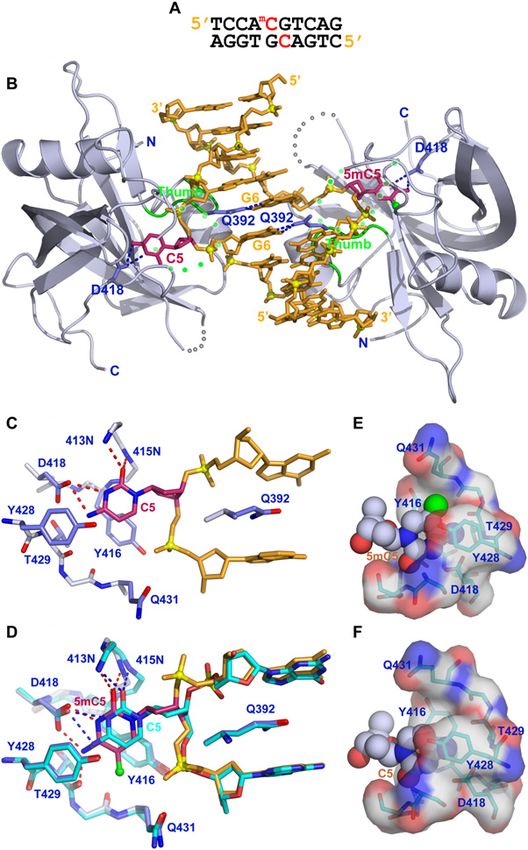
Downloaded from genesdev.cshlp.org on March 6, 2015 - Published by Cold Spring Harbor Laboratory Press

Rajakumara et al.

   Roles for proteins that contain SET- and RING-associ-               specific genomic loci (Ebbs et al. 2005; Ebbs and Bender
ated (SRA) domains have been demonstrated at the level                 2006). The mechanism governing the observed locus-
of establishment and/or maintenance of DNA methyla-                    specific contributions of SUVH4, SUVH5, and SUVH6
tion in both plants and animals. In Arabidopsis, de novo               are poorly understood. For SUVH4, a mutation in the
DNA methylation requires two (SET-associated) SRA                      SRA domain exhibited significantly reduced binding to
domain proteins, SU(VAR)3-9 HOMOLOG 2 (SUVH2)                          methylated DNA in vitro, and reduced both DNA meth-
and SUVH9, which function in a partially redundant                     ylation and H3K9me2 levels in vivo, suggesting a role for
manner and preferentially bind methylated DNA in the                   the SRA domain in the recruitment or retention of
CG and CHH contexts, respectively (Johnson et al. 2008).               SUVH4 to silenced loci (Johnson et al. 2007).
These proteins function late in the DRM2 pathway                          Despite the conservation in the SRA domain, the binding
and may aid in the recruitment or retention of DRM2 to                 specificities of previously characterized SRA domains vary
methylated loci. Another family of plant SRA domain pro-               greatly. Mechanistic insight into the ability of the SRA
teins, the RING-associated VARIANT IN METHYLATION                      domain to recognize methylated DNA was revealed by the
(VIM)/ORTHRUS (ORTHUS) family, is required to main-                    crystal structure of UHRF1 bound to hemimethylated CG
tain DNA methylation predominantly in the CG context                   DNA (Arita et al. 2008; Avvakumov et al. 2008; Hashimoto
(Woo et al. 2007, 2008; Kraft et al. 2008; Feng et al. 2010),          et al. 2008). In this structure, the UHRF1 SRA domain is
and the SRA domains of VIM1/ORTH2 and VIM3/ORTH1                       described as a hand grasping the DNA duplex, with two
have been shown to bind methylated CG sites (Johnson                   loops, termed the thumb and NKR finger (Avvakumov
et al. 2007; Woo et al. 2007). The mammalian homolog                   et al. 2008). The thumb loop contacts the DNA through the
of the VIM proteins, Ubiquitin-like PHD and RING finger                minor groove and the NKR finger enters the DNA duplex
domain (UHRF1), is also required to maintain DNA                       through the major groove and provides the arginine (Arg)
methylation in the CG context (Bostick et al. 2007; Sharif             residue that base-pairs with the orphaned guanine base
et al. 2007). The SRA domain of UHRF1 specifically binds               (Avvakumov et al. 2008). Consistent with the binding
hemimethylated CG sites (Bostick et al. 2007; Sharif                   of UHRF1 to hemimethylated DNA, an asparagine (Asn)
et al. 2007; Arita et al. 2008; Avvakumov et al. 2008;                 residue within the finger loop of the UHRF1 structure is
Hashimoto et al. 2008; Qian et al. 2008) and is necessary              predicted to clash sterically if a methylated cytosine base is
for the association of DNMT1 at chromatin (Bostick et al.              present on the opposite strand of DNA (Arita et al. 2008;
2007; Sharif et al. 2007), leading to a model in which                 Avvakumov et al. 2008; Hashimoto et al. 2008). However,
UHRF1 recruits DNMT1 to sites of hemimethylated                        there is no structural data available for the other known
DNA and thus facilitates the restoration of hemimethyl-                SRA domain proteins.
ated DNA to the fully methylated state. In Arabidopsis,                   In order to better understand the function and specific-
three other SRA domain proteins—SUVH4/KRYPTONITE,                      ity of SRA domains in general, and of SUVH5 in partic-
SUVH5, and SUVH6—are required to maintain DNA                          ular, the binding preferences of the SUVH5 SRA domain
methylation in the CHG context (Jackson et al. 2002;                   for DNA methylation in all contexts were determined,
Malagnac et al. 2002; Ebbs et al. 2005; Ebbs and Bender                and the structure of this domain bound to methylated
2006). The SRA domains of SUVH4 and SUVH6 have                         DNA oligomers in several sequence contexts was deter-
different binding preferences, with SUVH4 strongly prefer-             mined by X-ray crystallography. Our studies establish
ring CHG methylation over both CG and CHH methylation                  for the first time a dual flip-out mechanism on partner
and SUVH6 preferring both CHG and CHH methylation                      strands for 5-methyl-cytosine (5mC) recognition by the
strongly over CG methylation (Johnson et al. 2007).                    SUVH5 SRA domain that is independent of DNA se-
   In addition to their SRA domains, SUVH4, SUVH5, and                 quence context. The structural research was comple-
SUVH6 also contain histone methyltransferase (HMTase)                  mented by studying the impact of SUVH5 SRA mutants
domains and catalyze histone 3 Lys 9 dimethylation                     (involved in protein–DNA recognition) on the patterns
(H3K9me2) in vitro (Jackson et al. 2002, 2004; Ebbs and                of DNA methylation and H3K9 dimethylation in vivo.
Bender 2006), and are required for H3K9me2 methylation
in vivo (Johnson et al. 2002, 2007; Jackson et al. 2004;
Ebbs et al. 2005; Ebbs and Bender 2006). Genome-wide,                  Results
the repressive H3K9me2 and CHG methylation modifica-
                                                                       Electrophoretic mobility shift assay (EMSA) studies
tions are strongly correlated (Bernatavichute et al. 2008),
                                                                       of the 5mC-binding specificity of the SUVH5
and CHG methylation is thought to be maintained by
                                                                       SRA domain
a reinforcing loop of DNA methylation by CMT3 and
histone methylation by SUVH4, SUVH5, and SUVH6.                        The C-terminal half of SUVH5 contains the SRA and SET
However, these three HMTases do not contribute equally                 domains (Fig. 1A). Sequence alignments of the SRA do-
to the overall levels of H3K9me2 and DNA methylation in                mains of the SUVH and ORTH proteins, as well as of
vivo. SUVH4 is the predominant H3K9me2 HMTase, and                     UHRF1, are shown in Figure 1B and Supplemental Figure
mutation of this gene significantly reduces levels of DNA              S1. Two GST fusion constructs corresponding to amino
methylation (Jackson et al. 2002, 2004; Malagnac et al.                acids 299–522 and 362–528 of SUVH5 (Fig. 1A; Supple-
2002). Nonetheless, the levels of H3K9me2 and DNA                      mental Fig. S1) were used to determine the in vitro
methylation are further reduced in suvh4 suvh5 or suvh4                specificity of its SRA domain for 5mC sites using EMSAs.
suvh6 double and suvh4 suvh5 suvh6 triple mutants at                   DNA oligomers that contain multiple 5mC bases within

138      GENES & DEVELOPMENT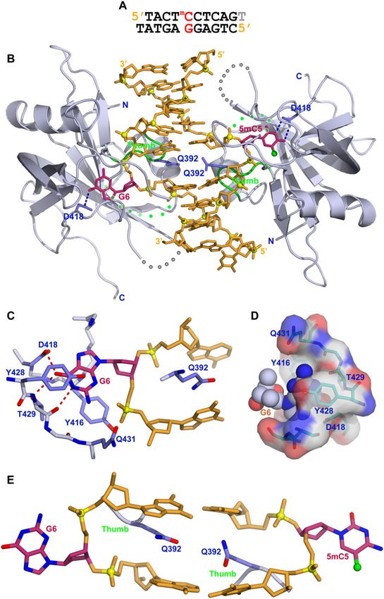
Downloaded from genesdev.cshlp.org on March 6, 2015 - Published by Cold Spring Harbor Laboratory Press

                                                                                            SUVH5 SRA shows a dual flip-out mechanism

Figure 1. Binding of the SUVH5 SRA domain to 5mC-containing DNA in different sequence contexts. (A) Schematic representation of
the domain architecture of SUVH5, with SRA and SET methyltransferase domains colored in blue and purple, respectively. (B) Multiple
sequence alignment of the SRA domains from the SET domain-associated SUVH family and from the ORTH1–4, ORTH-L, and UHRF1
RING domain-associated proteins in the region flanking the thumb and NKR finger. Sequence numbering of the SUVH family proteins
is based on SUVH5, whereas, for the RING domain-associated proteins, it is based on UHRF1. The secondary structural elements of the
SUVH5 SRA are indicated above the sequence (a helices are in green cylinders, b-strands are in blue arrows, and disordered regions are
represented by ##). Residues highlighted in a background color code correspond to conservation levels: fully conserved in red, and
conservative substitutions in yellow. The thumb and NKR finger corresponding to the UHRF1 are underlined in black at the bottom.
The residue that inserts into the duplex and displaces the 5mC residue in the SUVH5 SRA complex is indicated as an inverted red
triangle. Green and blue upright triangles correspond to residues that replace the looped-out 5mC and mask the unmodified C in the
UHRF1 SRA complex, respectively. Filled green circles designate residues that interact with the 5mC in the binding pocket of the
SUVH5 SRA and UHRF1 SRA complexes. Red and green stars designate DNA backbone-interacting residues in the SUVH5 SRA and
UHRF1 SRA complexes, respectively. (C) EMSAs using a GST-299–522 SUVH5 SRA domain fusion protein. The context and methylation
state of each radiolabeled dsDNA oligonucleotide is indicated above. (Un) Unmethylated; (FM) fully methylated; (HM) hemimethylated;
( ) no protein. (D) EMSA experiments using a GST-362–528 SUVH5 SRA domain fusion protein. In C and D, the first and second HMCG
oligonucleotides harbor methylated cytosines on the sense and antisense DNA strand, respectively. (E) ITC measurements of the binding
of the SUVH5 SRA domain to fully methylated CG DNA (diagram above). Experimental details are provided in the Materials and
Methods. The measured binding parameters are KD = 1.08 mM and N = 0.48. (F) ITC measurements of the binding of the SUVH5 SRA
domain to hemimethylated CG DNA. The measured binding parameters are KD = 5.0 mM and N = 0.58. (G) ITC measurements of the
binding of the SUVH5 SRA domain to methylated CHH DNA. The measured binding parameters are KD = 8.7 mM and N = 0.51.

                                                                                                    GENES & DEVELOPMENT           139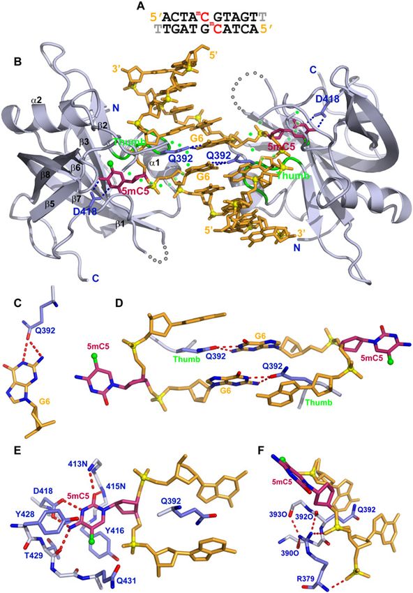
Downloaded from genesdev.cshlp.org on March 6, 2015 - Published by Cold Spring Harbor Laboratory Press
Rajakumara et al.
a single sequence context were used to assess the overall CG sequences, which is in sharp contrast to the preference
binding preferences of these SRA proteins. Both the longer of the UHRF1 SRA domain, which exhibits the reverse
and shorter SRA proteins bound DNA oligomers contain- preference; namely, a sevenfold preference for hemimethyl-
ing methylation in all sequence contexts (CG, CHG, and ated over fully methylated CG sequences (Bostick et al.
CHH) and in both the hemimethylated and fully methyl- 2007). Additionally, in the case of the SUVH5 SRA domain,
ated states preferentially over the corresponding unmethyl- the stoichiometry of binding is ;0.5, which reflects the
ated oligomers (Fig. 1C,D). Untagged SRA proteins of both ratio of 0.5 duplex bound per SRA or, equivalently, two
sizes that were used in the crystallographic analyses SRA domains bound per DNA duplex.
showed a similar specificity on these DNA substrates
(Supplemental Fig. S2A). To further quantify the binding
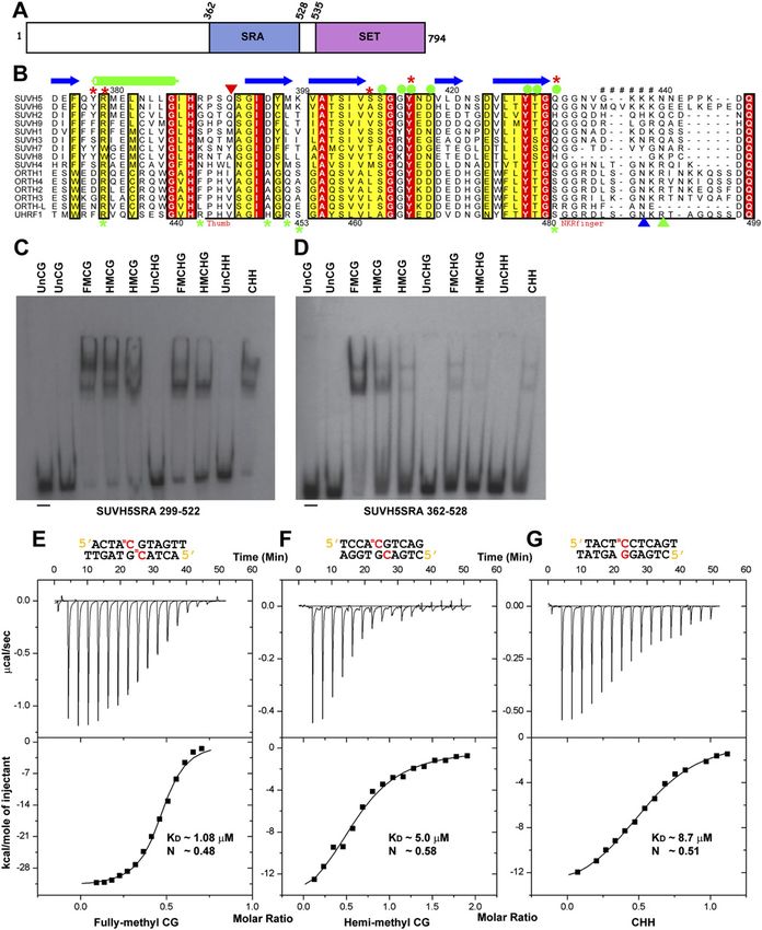
Structure of the SUVH5 SRA domain bound
preference of the GST-SRA fusion proteins, binding curves
to fully methylated CG DNA
were generated from EMSA experiments using the same
DNA oligomers and titrations of each GST-SRA protein We grew diffraction-quality crystals and solved the
(Supplemental Fig. S2B–D). The larger GST-SRA protein, structure of the SUVH5 SRA domain bound to a self-
which corresponds to the previously characterized SRA complementary 10-base-pair (bp) duplex (with a 39 thy-
domains of SUVH2, SUVH6, SUVH9, and UHRF1 (Bostick mine overhang) containing a central fully methylated CG
et al. 2007; Johnson et al. 2007, 2008), shows only minor step (Fig. 2A). The crystals of this complex (space group
preferences for DNA methylation in different sequence P42212) diffracted to 2.20 Å resolution, and the structure
contexts (Fig. 1C; Supplemental Fig. S2). The smaller GST- was solved using phases derived from multiwavelength
SRA protein shows a preference for methylation in the CG anomalous diffraction data collected from a crystal of the
context, but still binds methylation in other contexts over complex containing seleno–methionine-labeled protein.
unmethylated DNA, and shows the same order of prefer- The asymmetric unit is composed of one SRA domain
ence as the longer construct: fully or hemimethylated CG, bound to one strand of the fully methylated DNA (X-ray
then fully methylated CHG and methylated CHH, fol- statistics listed in Supplemental Table 1). A second struc-
lowed by hemimethylated CHG (Fig. 1C,D; Supplemental ture of this complex was solved in the space group P6122
Fig. S2). These findings demonstrate that the SRA domain at 2.65 Å resolution using the molecular replacement
of SUVH5 exhibits a different specificity from that shown method (Supplemental Table 1). The present study fo-
for other SRA domains (Bostick et al. 2007; Johnson et al. cuses on the higher-resolution 2.20 Å structure.
2007, 2008; Woo et al. 2007). Furthermore, our finding that The structure of the complex shown in Figure 2B con-
the SRA domain of SUVH5 binds to methylation in all tains two SRA molecules bound per DNA duplex, with
contexts and has little preference for a fully over a hemi- the 5mC residues on adjacent base pairs flipped out of the
methylated state suggests that this SRA domain might DNA helix and positioned in the binding pockets of two
either be recognizing only the methylated cytosine or be individual SRA domains. There is a crystallographic two-
able to accommodate both a methylated and an unmethyl- fold axis perpendicular to the DNA helical axis, and the
ated residue on the complementary DNA strand. two SRA molecules form no contacts with each other
in the complex. The observed stoichiometry of two SRA
molecules bound per duplex is consistent with an
Calorimetric studies of the 5mC-binding specificity
N-value of 0.5 (DNA duplex:SRA domain ratio) observed
of the SUVH5 SRA domain
in the ITC binding curve for complex formation of the
Although the substrate specificity of the SRA domains SUVH5 SRA domain with fully methylated DNA (Fig.
of several SUVH proteins have been reported (Johnson 1E). These findings support a model in which the stoi-
et al. 2007, 2008), no data on the affinity or stoichiometry chiometry represents the binding of two SRA domains to
of binding are available. Here, we used the isothermal individual 5mC residues rather than the dimerization of
titration calorimetric (ITC) approach to investigate these the two SRA domains with each other.
parameters for SUVH5 using the shorter SRA construct The flipping out of the 5mC residues in the fully
(362–528) and DNA oligomers containing methylation in methylated CG crystal structure introduces a gap in the
the fully and hemimethylated CG sequence contexts, the DNA duplex that is filled by the side chain of Gln392,
methylated CHH context, and the fully methylated CHG which resides within the thumb loop (residues 390–395)
context. The SUVH5 SRA domain binds to fully methyl- of the SUVH5 SRA domain. Gln392 is inserted through
ated CG with a dissociation constant (KD) of 1.08 mM the minor groove and is intercalated between flank-
(Fig. 1E). Consistent with the EMSA data using this SRA ing bases, forming intermolecular hydrogen bonds with
construct, the binding constant decreases by a factor of the Watson-Crick edge of the orphan guanine G6 (Fig.
4.6 (KD = 5.0 mM) (Fig. 1F) for hemimethylated CG, a 2C). The arrangement of the two G6–Gln392 pairs
factor of 8.1 (KD = 8.7 mM) (Fig. 1G) for methylated CHH, within the complex, which occur on both DNA strands,
and a factor of 6.0 (KD = 6.6 mM) (Supplemental Fig. S3) for are shown in Figure 2D. The NKR finger loop (resi-
fully methylated CHG sequence contexts. dues 433–444), which plays a critical role in the UHRF1
Together, these binding studies demonstrate that the SRA–hemimethylated DNA complex (Arita et al. 2008;
SUVH5 SRA domain recognizes 5mC in all sequence con- Avvakumov et al. 2008; Hashimoto et al. 2008), is
texts. Notably, the SRA domain of SUVH5 exhibits a 4.6- disordered in the SUVH5 SRA–fully methylated DNA
fold preference for fully methylated over hemimethylated complex. Despite the presence of two flipped-out 5mC
140 GENES & DEVELOPMENT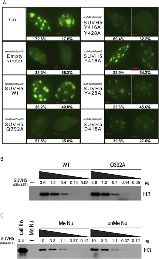
Downloaded from genesdev.cshlp.org on March 6, 2015 - Published by Cold Spring Harbor Laboratory Press

                                                                                         SUVH5 SRA shows a dual flip-out mechanism

residues on partner strands, the remaining bases in the            5mC. These functions are mediated through the intra-
DNA duplex are undistorted and essentially retain the reg-         and intermolecular hydrogen bond interactions depicted
ular B-form conformation.                                          in Figure 2F.
   The flipped out 5mC is positioned in a pocket within              The intermolecular contacts in the SUVH5 SRA complex
the SRA domain such that it is anchored in place via               are summarized in Supplemental Figure S4A. In essence,
stacking interactions with Tyr416 and Tyr428 and by                the key amino acids in the SUVH5 SRA domain that
intermolecular hydrogen bonds between its Watson-                  contribute to the molecular recognition of the flipped-out
Crick edge and the side chain of Asp418, the backbone              5mC and stabilization of the binding pocket are Gln392,
amide nitrogens, and a carbonyl group (Fig. 2E). The 5mC           Asp418, Tyr416, Tyr428, and Arg379. Hence, these residues
adopts an anti alignment around the glycosidic bond                constitute appropriate candidates for mutational studies.
(x = 119°), and there is sufficient room to accommodate
the methyl group at the cytosine 5 position, which is
                                                                   Structure of the SUVH5 SRA domain bound
stabilized by van der Waals contacts, most prominently
                                                                   to hemimethylated CG DNA
with the Ca and Cb atoms of Gln431 (Fig. 2E). It should be
noted that the N3 position of looped-out 5mC must be               We next solved the crystal structure of the SUVH5 SRA
protonated in order to pair with the carboxylate group of          domain bound to a complementary 10-bp duplex con-
Asp418 (Fig. 2E), which in turn would imply an unusual             taining a central hemimethylated CG step (Fig. 3A). The
ionization constant (pKa) for the looped-out 5mC. The              crystal belongs to the P42 space group, diffracts to 2.37 Å
same alignment of the Asp residue and the 5mC, charac-             resolution, and contains two SRA molecules and one
teristic of an unusual pKa for the looped-out 5mC, was             DNA duplex in the asymmetric unit (crystallographic
also observed in the published structures of the UHRF1             statistics listed in Supplemental Table 1).
SRA–DNA complexes (Arita et al. 2008; Avvakumov                       The structure of the complex is shown in Figure 3B, and
et al. 2008; Hashimoto et al. 2008).                               it contains two SUVH5 SRA domains bound per DNA
   Finally, Arg379 plays an important role in buttressing          duplex, consistent with the stoichiometry elucidated
the thumb loop, which provides the Gln392 residue that             from ITC binding data (Fig. 1F). Unexpectedly, both the
inserts into the DNA helix, as well as in anchoring the            5mC and the unmodified C from adjacent pairs on partner
DNA phosphodiester backbone flanking the flipped-out               strands simultaneously flip out of the duplex and are

                                                                   Figure 2. Crystal structure of the SUVH5 SRA domain bound to
                                                                   fully methylated CG DNA. (A) Sequence of the self-complementary
                                                                   fully methylated CG 10-mer (with a 39-T overhang) containing
                                                                   5mC-G steps on partner strands in the center of the duplex. (B)
                                                                   Stick (DNA) and ribbon (protein) representation of the 2.2 Å
                                                                   crystal structure of the 2:1 SUVH5 SRA–fully methylated CG
                                                                   DNA duplex complex. The DNA is colored in orange, except for
                                                                   5mC5, which is colored in purple. The 5-methyl group is shown
                                                                   as a small green sphere. Backbone phosphorus atoms are shown
                                                                   as yellow balls, and the 59 and 39 ends of the DNA are labeled.
                                                                   The SRA domain is colored in blue, with its secondary structural
                                                                   elements labeled with the same a/b numbering scheme as for the
                                                                   SRA domain of UHRF1 (Avvakumov et al. 2008). The thumb loop
                                                                   and disordered NKR loop segments are colored in green and
                                                                   dotted green, respectively. The 5mC5 residues on partner strands
                                                                   flip out through the minor groove and are positioned in binding
                                                                   pockets on individual SRA domains. The Watson-Crick edge of
                                                                   5mC5 is hydrogen-bonded with the side chain of Asp418. The side
                                                                   chain of Gln392 inserts into and fills the gap created by the
                                                                   flipped-out base and pairs with the Watson-Crick edge of the G6
                                                                   base. (C) Hydrogen bonding between the Watson-Crick edge of G6
                                                                   and the carbonyl group of the Gln392 side chain. (D) Relative
                                                                   alignments of stacked G6–Gln392 interactions in the structure of
                                                                   the complex. Note that the side chain of Gln392 is sandwiched
                                                                   between bases. (E) Interaction of the flipped-out 5mC5 with
                                                                   residues lining the binding pocket. The 5mC5 base is positioned
                                                                   between the aromatic rings of Tyr416 and Tyr428, with its
                                                                   Watson-Crick edge hydrogen-bonded to the protein backbone
                                                                   and side chain (Asp418) residues. (F) The side chain of Arg379
                                                                   forms a network of intramolecular hydrogen bonds with the
                                                                   backbone of the thumb segment and intermolecular hydrogen
                                                                   bonds with the phosphate backbone that buttresses the fold of the
                                                                   binding pocket.

                                                                                                  GENES & DEVELOPMENT           141
Downloaded from genesdev.cshlp.org on March 6, 2015 - Published by Cold Spring Harbor Laboratory Press

Rajakumara et al.

                                                                       filling representation and the binding pocket in a surface
                                                                       representation are shown accommodating the flipped-out
                                                                       5mC and C residues in Figure 3, E and F, respectively.
                                                                       Importantly, both the flipped-out C (x = 118°) and 5mC
                                                                       (x = 112°) residues adopt anti conformations about their
                                                                       glycosidic bonds (Fig. 3E,F).

                                                                       Structure of the SUVH5 SRA domain bound
                                                                       to methylated CHH DNA
                                                                       We also solved the crystal structure of the SUVH5 SRA
                                                                       domain bound to a complementary 10-bp duplex with a
                                                                       39 thymine overhang that contains a centrally positioned
                                                                       methylated CHH (where H = C, T, or A) step (Fig. 4A). The
                                                                       crystals belong to the P6122 space group, diffract to 2.75 Å
                                                                       resolution, and contain two SRA molecules and one DNA
                                                                       duplex in the asymmetric unit (crystallographic statistics
                                                                       listed in Supplemental Table 1).
                                                                          The structure of the complex is shown in Figure 4B and
                                                                       contains two SUVH5 SRA domains bound per DNA
                                                                       duplex, consistent with the stoichiometry elucidated
                                                                       from ITC binding data (Fig. 1G). To our surprise, both
                                                                       the 5mC and the G positioned opposite it on the comple-
                                                                       mentary strand are flipped out of the DNA duplex and are
                                                                       positioned in binding pockets within individual SRA
                                                                       domains. The flipped-out G adopts a syn conformation
                                                                       (x = 30°) about its glycosidic bond (Fig. 4C,D), allowing
                                                                       the flipped-out G to form the same distribution of in-
                                                                       termolecular hydrogen bonds (Fig. 4C) as are formed by
                                                                       the flipped-out anti 5mC (Fig. 2E). Since the flipped-out
                                                                       bases originate from the same base pair in the methylated
                                                                       CHH complex, the inserted Gln392 side chains are
                                                                       roughly in the same plane and are sandwiched between
Figure 3. Crystal structure of the SUVH5 SRA domain bound              flanking base pairs (Fig. 4E). The stark difference in the
to hemimethylated CG DNA. (A) Sequence of the complemen-               arrangement of the two Gln392 residues in this complex
tary hemimethylated CG 10-mer containing a 5mC-G step on               compared with that observed in the fully methylated CG
the top strand and an unmodified C-G step on the bottom strand
                                                                       complex, where the flipped-out bases on partner strands
in the center of the duplex. (B) Stick (DNA) and ribbon (protein)
                                                                       originate from flanking base pairs, can be appreciated by
representation of the 2.37 Å crystal structure of the 2:1 SUVH5
SRA–hemimethylated CG DNA duplex complex. The color                    comparing Figures 2D and 4E.
coding is the same as in Figure 2B. Note that the 5mC5 and the            Overall, the structures reveal that the SUVH5 SRA
C5 from adjacent base pairs on partner strands flip out through the    complexes with fully methylated CG, hemimethylated
minor groove and are positioned in binding pockets of individual       CG, and methylated CHH DNAs all involve the flipping
SRA domains. (C) Interaction of the looped-out C5 with residues        out of both the 5mC and a base—either a 5mC, a C, or a
lining the binding pocket in the structure of the complex. (D)         G, respectively—on the partner strand. The intermolec-
Superposition of the flipped-out C5 (in green) and the flipped-out     ular contacts in the above three SUVH5 SRA–DNA com-
5mC5 (in purple) within their respective binding pockets in the        plexes are summarized in Supplemental Figure S4, A–C,
structure of the complex. (E) Insertion of anti 5mC5 (space-filling
                                                                       respectively. One notable difference between the struc-
representation) into the binding pocket of the SRA domain (elec-
                                                                       tures is that the relative orientations of the two SRA
trostatic surface presentation). (F) Insertion of anti C5 (space-
filling representation) into the binding pocket of the SRA domain      domains are altered in the different complexes such that,
(electrostatic surface presentation).                                  in the fully methylated CG DNA and hemimethylated
                                                                       CG DNA complexes the two Gln392 residues lie in adjacent
                                                                       planes, while in the methylated CHH DNA complex the
                                                                       Gln392 residues lie in the same plane (Supplemental
positioned within binding pockets of individual SRA
                                                                       Fig. S4D,E).
domains.
   The alignment and intermolecular contacts of the
                                                                       Effects of SRA domain mutations on methyl-DNA
flipped-out unmodified C within its binding pocket are
                                                                       binding in vitro
shown in Figure 3C, and they compare favorably with that
of the flipped-out 5mC, as can be seen in the superposition            To test the importance of specific residues within the
of the flipped-out C and 5mC residues (Fig. 3D). Alternate             SUVH5 SRA domain for recognition of the flipped-out
representations showing the flipped-out base in a space-               base, we engineered point mutations within the SUVH5

142      GENES & DEVELOPMENT
Downloaded from genesdev.cshlp.org on March 6, 2015 - Published by Cold Spring Harbor Laboratory Press
SUVH5 SRA shows a dual flip-out mechanism
inserting into the duplex and taking the place of the
flipped-out base) to an alanine resulted in what appears
to be a complete loss of binding affinity (Supplemental
Fig. S5D). This finding strongly supports the crystallo-
graphic finding that the thumb loop rather than the NKR
finger facilitates the base-flipping mechanism in the SUVH5
SRA domain. Mutation of both Tyr residues (Tyr416Ala/
Tyr428Ala) that sandwich the flipped-out base also abro-
gates binding (Supplemental Fig. S5E). However, a single
Tyr416Ala mutation only reduced the binding affinity
(Supplemental Fig. S5C). Thus, both Tyr residues are im-
portant for positioning the 5mC in the binding pocket.
The Asp418Ala mutant, which is predicted to disrupt the
hydrogen bonds between the Asp side chain and the
Watson-Crick edge of the flipped-out 5mC in the structure
of the complex (Fig. 2E), exhibits a calculated KD = 21 mM
(Supplemental Fig. S5A), reflecting a 20-fold reduction in
binding affinity compared with wild type (Fig. 1E). This
structure–function analysis supports the underlying struc-
ture-based intermolecular interactions observed in the
crystal structure of the complex, where a combination of
specific hydrogen bonding, stacking, and hydrophobic
interactions contribute to the anchoring of the flipped-
out 5mC within its binding pocket in the SUVH5 SRA
domain.
Impact of guanine to inosine substitutions
and the presence of abasic sites
on binding to methylated DNA
To further investigate the interactions required for the
binding of the SUVH5 SRA domain to methylated DNA,
specifically those involving the DNA surrounding the
flipped-out 5mC, we carried out in vitro binding assays
Figure 4. Crystal structure of the SUVH5 SRA domain bound
using modified DNA duplexes. The guanine base present
to methylated CHH DNA. (A) Sequence of the complementary
opposite the 5mC forms hydrogen bonds with the Gln392
methylated CHH 10-mer (with a 39-T overhang) containing a
59-5mCCT step on the top strand and a 59-AGG step opposite residue of the SUVH5 SRA domain in both the fully and
it on the bottom strand toward the center of the duplex. In hemimethylated CG complexes, and mutation of Gln392
essence, a 5mC is positioned opposite a G. (B) Stick (DNA) and within SUVH5 ablates DNA binding, suggesting this in-
ribbon (protein) representation of the 2.75 Å crystal structure of teraction may be important. However, such an interaction
the 2:1 SUVH5 SRA–methylated CHH DNA duplex complex. is not observed in the complex containing methylation in
The color coding is the same as in Figure 2B. Note that the the CHH context, suggesting it may not be required.
5mC5 and the G6 from the same base pair simultaneously flip Thus, to investigate the relative importance of these
out of the DNA duplex through the minor groove and are guanine residues, binding assays were conducted using
positioned in binding pockets of individual SRA domains. (C)
substrates in which the guanine bases were replaced with
Interaction of the flipped-out G6 in a syn conformation with
inosine bases. Replacement of the guanine bases with
residues lining the binding pocket in the structure of the
complex. (D) Insertion of the syn G6 (space-filling representa- inosine bases in the fully methylated and hemimethyl-
tion) into the binding pocket of the SRA domain (electrostatic ated CG duplexes had no impact on either the KD or
surface presentation). (E) Relative alignments of the inserted N-values following complex formation with the SUVH5
Gln392 residues that are positioned opposite each other in the SRA domain (Supplemental Fig. S5F,G). Thus, given that
structure of the complex. Note that the side chain of the Gln392 the GC and IC pairs contain three and two hydrogen
residue is sandwiched between two bases. bonds, respectively, which are disrupted upon complex
formation, this finding demonstrates that reducing the
capacity of Gln392 to hydrogen-bond with the Watson-
SRA domain that we predicated might be important based Crick edge of guanine (Fig. 2E) does not have an impact on
on the crystallographic structures of the complexes. These the binding affinity.
mutant SRA domain proteins were assessed for their To investigate whether a dual base-flipping mechanism
ability to bind a fully methylated CG site by ITC exper- is required for binding to DNA methylation in the asym-
iments in vitro (Supplemental Fig. S5). Mutation of Gln392 metric CHH context, we replaced the guanine opposite
(the residue that base-pairs with the orphaned guanine by the 5mC with an abasic site (designated dS-mCHH) and
GENES & DEVELOPMENT 143Downloaded from genesdev.cshlp.org on March 6, 2015 - Published by Cold Spring Harbor Laboratory Press
Rajakumara et al.
monitored binding by the SUVH5 SRA domain by ITC
measurements. The observed KD of 2.7 mM and an N-value
of 1.1 (Supplemental Fig. S5H) imply that the SRA domain
still interacts with this type of substrate, and that flip-
ping out of the base opposing the 5mC is not required for
complex formation. Equally important, the N-value of
1.1 implies that a single SRA domain is bound to the dS-
mCHH DNA. These findings further support the hypoth-
esis that the SUVH5 SRA domain relies on the 5mC in the
absence of complementary guanine.
Effects of SRA domain mutations on DNA
methylation in vivo
To determine the in vivo significance of key residues
within the 5mC-binding pocket, the ability of an epitope-
tagged SUVH5 transgene (pSUVH5T3xFlag-SUVH5) car-
rying specific point mutations to restore methylation at
the Ta3 locus was assessed by Southern blotting follow-
ing digestion of genomic DNA with the methylation-
sensitive restriction enzyme MspI. DNA methylation at
Ta3 is redundantly controlled by SUVH4, SUVH5, and
SUVH6 such that, in a suvh4 suvh5 suvh6 triple mutant,
this locus is almost completely unmethylated (Ebbs and
Bender 2006), resulting in the presence of three main
bands (2.2 kb, 1.7 kb, and 0.7 kb in size) after Southern
blotting (Fig. 5A, lane 3). In a suvh5 single mutant,
methylation at this locus is indistinguishable from that
of wild-type plants (Fig. 5A, cf. lanes 1 and 2; Ebbs and
Bender 2006). However, in the sensitized suvh4 suvh5
suvh6 triple-mutant background, the addition of a func-
tional SUVH5 wild-type transgene results in an increase
in DNA methylation, which is visualized as the accumu- Figure 5. In vivo DNA methylation analysis. (A) Southern blot
lation of a 2.5-kb band to a similar intensity as observed showing the methylation status of the Ta3 locus. Genomic
for the 2.2-kb and 1.7-kb bands (Fig. 5A, lane 5). This DNA was extracted from the indicated Arabidopsis genotypes
pattern is comparable with that observed when muta- and digested with the methylation-sensitive MspI restriction
tions in SUVH4 and SUVH6 are present (Fig. 5A, lane 4). enzyme. pSUVH5T3xFlag-SUVH5 indicates the epitope-tagged
Mutant versions of SUVH5 were tested for their ability SUVH5 transgene, driven by its endogenous promoter, encoding
either the wild-type SUVH5 protein (wild type [Wt]) or the
to complement the suvh4 suvh5 suvh6 mutant pheno-
indicated mutant protein; the asterisk (*) indicates suvh2 and
type, and the expression of each mutant protein in vivo suvh9 mutations are also present in this background. Previous
was assessed by Western blotting using an antibody studies have shown these mutations do not affect methylation
against the Flag epitope (Fig. 5B). A SUVH5 transgene at Ta3. (un Me) DNA fragments generated by cleavage of un-
encoding a protein with mutations in both of the tyrosine methylated DNA by MspI; (Me) DNA fragment generated when
residues that form stacking interactions with the flipped- DNA methylation blocks cleavage by MspI. The ratio of the 2.5-
out 5mC (namely, Tyr416/Tyr428 in the crystal structure) kb band intensity to the 2.2-kb band intensity is shown below
to Ala failed to complement the methylation defect each lane number and was used to score the complementation
at Ta3 (Fig. 5A, lane 7), and a transgene encoding a muta- level of each SUVH5 transgene (either the wild-type genomic
tion in the glutamine that base-pairs with the orphaned sequence [lane 5] or the indicated point mutants [lanes 6–11]). A
schematic diagram of the Ta3 locus showing the MspI restric-
guanine base, Gln392, also significantly reduced the level
tion sites is shown below. (B) Western blot using an antibody
of complementation (Fig. 5A, lane 6). Mutation of either against the Flag epitope showing the expression of the wild-type
stacking tyrosine alone (Tyr416 or Tyr428); of Asp418, and mutant versions of the SUVH5 protein from the same T1
another residue that forms interactions within the plants characterized in A. Protein extracted from the nontrans-
methyl-cytosine-binding pocket; or of both Tyr416 and genic Colombia (Col) ecotype was used as a negative control. An
Asp418 was able to partially restore methylation at Ta3. unknown protein that cross-reacts with the Flag antibody and
These data confirm that residues in the thumb loop of serves as an internal loading control is shown in the bottom panel.
the SUVH5 SRA domain are important for the in vivo
function of SUVH5, and further support the crystallo-
graphic data showing that the residue that replaces the role is filled by an arginine that resides in the NKR finger
flipped-out 5mC and base-pairs with the orphaned G in loop (Arita et al. 2008; Avvakumov et al. 2008; Hashimoto
SUVH5 differs from that observed in UHRF1, where this et al. 2008).
144 GENES & DEVELOPMENTDownloaded from genesdev.cshlp.org on March 6, 2015 - Published by Cold Spring Harbor Laboratory Press
SUVH5 SRA shows a dual flip-out mechanism
Effects of SRA domain mutations on H3K9
dimethylation in vivo
To determine the effect of mutations within the SRA
domain of SUVH5 on H3K9 dimethylation levels in vivo,
immunofluorescence experiments were conducted using
nuclei isolated from the progeny of the transgenic SUVH5
lines characterized in Figure 5. Similar expression levels
of the various SUVH5 transgenes in this generation were
confirmed by Western blotting (Supplemental Fig. S6A).
As expected based on previous immunofluorescence
experiments in Arabidopsis (Johnson et al. 2008), nuclei
isolated from wild-type (Col ecotype) plants showed an
intense H3K9 dimethylation signal at chromocenters,
and nuclei isolated from a suvh4 suvh5 suvh6 triple
mutant transformed with an empty vector as a negative
control showed a weak and largely diffuse signal (Fig. 6A;
Supplemental Fig. S6). A strong H3K9 dimethylation sig-
nal at chromocenters was restored in nuclei isolated from
suvh4 suvh5 suvh6 mutant plants transformed with
a wild-type copy of SUVH5, demonstrating that this
transgene is able to complement both the DNA methyl-
ation and the histone methylation defects (Fig. 6A). Con-
sistent with the DNA methylation analysis at the Ta3
locus, suvh4 suvh5 suvh6 mutants transformed with a
SUVH5 transgene carrying alanine mutations at either
Gln392 or both Tyr416 and Tyr428 had the strongest
effect on global levels of H3K9 dimethylation (Fig. 6A).
Mutation of either tyrosine alone or of Asp418 had less
severe effects on H3K9 levels, with a portion of the nuclei
exhibiting a strong to moderate H3K9 dimethylation
signal at chromocenters (Fig. 6A). Together, these finding
suggest that, like what has been observed for SUVH4
(Johnson et al. 2007), a functional SRA domain is impor-
tant for the activity of SUVH5 at the level of either
recruitment and/or retention at genomic loci or HMTase Figure 6. Histone methylation analysis. (A, left) Immunofluo-
activation. rescence detection of H3K9 dimethylation in nuclei isolated
In order to differentiate between potential roles for the from the indicated genotype. Images representing the two most
SUVH5 SRA domain in targeting verses stimulation of its predominant classes of nuclei are shown, and percentages out of
200 nuclei are indicated below. See Supplemental Figure 6 for
H3K9 HMTase activity, the in vitro HMTase activity of
a full breakdown of classes and percentages for each mutant. (B)
SUVH5 proteins containing both the SRA and the SET HMTase assays using a wild-type or a Q392A mutant SUVH5
HMTase domains was assessed. To confirm that a func- SRA-SET protein (amino acids 362–794) and calf thymus his-
tional SRA domain is not required for the HMTase tones as a substrate. 3H-radiolabeled SAM was supplied as the
activity of SUVH5, and thus is unlikely to account for methyl donor. The concentration of SUVH5 protein used for each
the decreased level of H3K9 methylation observed in assay is indicated in micrograms, and the position of histone
vivo, the HMTase activity of a wild-type or Gln392Ala 3 (H3) is indicated (at the right). (C) HMTase assays using a wild-
mutant SUVH5 SRA-SET protein was assessed using calf type SUVH5 SRA-SET protein (amino acids 362–794) and either
thymus histones as a substrate (Fig. 6B). To determine calf thymus histones (Calf thy) or mononucleosomes assembled
whether the binding of the SRA domain to methylated using methylated (Me Nu) or unmethylated (unMe Nu) DNA.
The concentration of SUVH5 protein used for each assay is
DNA has a stimulatory effect on the HMTase activity of
indicated in micrograms, and the position of histone 3 (H3) is
SUVH5, the HMTase activity of wild-type SUVH5 was indicated (at the right).
assessed using mononucleosomes assembled with CG-
methylated or unmethylated DNA. If the presence of
DNA methylation results in an increase in the level of SRA domain is to recruit or retain SUVH5 at specific
HMTase activity, that may suggest that the binding of the genomic loci and thereby allow deposition of the H3K9
SRA domain to methylated DNA causes conformational dimethylation mark in a loci-specific manner. Under the
changes in the protein that allows more efficient HMTase conditions used, similar levels of HMTase activity were
activity. Alternatively, if the presence of DNA methyla- observed on methylated and unmethylated nucleosomes
tion does not have an effect on the level of HMTase (Fig. 6C). Nucleosomes that contain a lysine-to-alanine
activity, that may suggest that the function of the SUVH5 mutation at position 9 of the H3 tail (H3K9A) were used
GENES & DEVELOPMENT 145Downloaded from genesdev.cshlp.org on March 6, 2015 - Published by Cold Spring Harbor Laboratory Press
Rajakumara et al.
as a control to confirm the H3K9 specificity of SUVH5 in et al. 2008), where UHRF1 was found to bind with a
vitro (Supplemental Fig. S6C). These findings support the stoichiometry of one SRA domain bound per hemi-
hypothesis that the SRA domain of SUVH5 is important methylated CG DNA duplex and only the 5mC was flip-
for the recruitment of SUVH5, rather than activation of ped out and positioned in the SRA-binding pocket, while
its HMTase activity. the C on the adjacent base pair remained stacked within
the duplex.
To our knowledge, the structures of the fully and
Discussion hemimethylated complexes in Figures 2B and 3B, respec-
tively, represent the first examples of a dual-base flipping
SUVH5 is unusual among the SUVH family members in
from partner strands, thereby providing a novel mechanism
that its SRA domain efficiently binds methylated DNA in
for scanning both strands of the duplex simultaneously.
the fully and hemimethylated CG contexts (Fig. 1E,F,
The versatility of SRA domains in binding DNA as
respectively), as well as in the methylated CHH and fully
either a single molecule (UHRF1 complexes) or two mol-
methylated CHG contexts (Fig. 1G; Supplemental Fig. S3,
ecules (SUVH5 SRA complexes) is reminiscent of POU
respectively). In an attempt to understand the principles
domains, which flex to fit the DNA duplex such that the
underlying this broad specificity, we initiated a structure–
two-part DNA-binding domain (POUH and POUS) partially
function study aimed at solving the crystal structures
encircles the DNA, without base flipping, and the sub-
of the SUVH5 SRA in a complex with methylated DNA
domains adopt a DNA element-dependent range of confor-
in all of the above sequence contexts. We were successful
mations (Phillips and Luisi 2000).
in growing crystals and solving the structures of the first
three complexes, but were unable to grow diffraction-
quality crystals of SUVH5 SRA bound to fully methylated Dual flipping out of the 5mC/G bases positioned
CHG DNA. These structures identified the intermolec- opposite each other on partner strands
ular protein–DNA contacts within the complexes, allow- It was unclear what to expect as far as stoichiometry and
ing us to monitor the impact of specific SUVH5 SRA the number of flipped-out residues for the SUVH5 SRA
mutants on DNA methylation and H3K9 dimethylation domain bound to the asymmetrically methylated CHH
in vivo. DNA duplex (Fig. 4A). However, the ITC measurements
once again established that the stoichiometry was two
Dual flipping out of 5mC/C positioned SRA domains bound per duplex (Fig. 1G), as was also
on adjacent pairs on partner strands observed in the structure of the complex (Fig. 4B). In this
structure, both the 5mC and the G from the same pair are
Base flipping was first identified in the structures of the
flipping out of the DNA duplex and are positioned within
bacterial M.HhaI (Klimasauskas et al. 1994) and HaeIII
individual SRA-binding pockets (Fig. 4B). Thus, the dual
(Reinisch et al. 1995) methyltransferases bound to CG-
base-flipping mechanism exhibited by the SUVH5 SRA
containing DNA, and since then has been identified as
domain can involve bases located on either partner DNA
a conserved mechanism that is widely used by DNA- and
strands that are positioned on adjacent pairs (fully and
RNA-modifying and repair enzymes (Huffman et al.
hemimethylated CG DNA) or within the same base pair
2005). A central feature of these complexes involves the
(methylated CHH DNA).
flipping out of a single base, which is then inserted into an
active site pocket within the bound protein.
Packing of the SUVH5 SRA domain complexes
We anticipated that the structure of the SUVH5 SRA
in the crystal lattice
bound to fully methylated CG DNA (Fig. 2A) could
involve flipping out of either one or both 5mC residues of We questioned whether some aspect of crystal packing
the adjacent base pairs, depending on the number of SRA could contribute to the dual-base flipping observed in all
domains bound per DNA duplex. Both the ITC measure- three SUVH5 SRA–DNA complexes. We observed the
ments (Fig. 1E) and the structure of the complex (Fig. 2B) same crystal packing in all three complexes; namely, that
established a stoichiometry of two SRA domains bound the SRA domains from adjacent complexes (each of which
per fully methylated CG DNA duplex, with both 5mC contains two SUVH5 SRA domains bound per DNA du-
residues flipping out and being positioned within their plex) in the crystal lattice pack against each other, as is
individual SRA-binding pockets. shown for the complex of the SUVH5 SRA domain bound
Unexpectedly, ITC measurements established a stoichi- to fully methylated CG DNA (Supplemental Fig. S7A).
ometry of two SRA domains bound per hemimethylated There is an extensive interface (641 Å2 per monomer)
CG DNA duplex (Figs. 1F, 3A), despite the presence of involving hydrogen-bonding, hydrophobic, and salt bridge
only a single 5mC base. The crystallographic analysis of interactions formed between the interacting SRA do-
this complex revealed that both the 5mC and the C from mains (Supplemental Fig. S7B). It should be noted that
adjacent base pairs on the partner strands are looped out the packing interaction occurs on the opposite SRA face
and positioned within individual SRA-binding pockets (Supplemental Fig. S7A) from that which interacts with
(Fig. 3B), consistent with the stoichiometry inferred from the DNA. It is unlikely that crystal packing alone con-
the ITC measurements. This contrasts strikingly with tributes to the observed 2:1 (SUVH5 SRA:DNA duplex)
earlier structural studies of the UHRF1 SRA domain stoichiometry, since the same stoichiometry is also ob-
(Arita et al. 2008; Avvakumov et al. 2008; Hashimoto served from ITC studies (Fig. 1E–G; Supplemental Fig. S3)
146 GENES & DEVELOPMENTDownloaded from genesdev.cshlp.org on March 6, 2015 - Published by Cold Spring Harbor Laboratory Press
SUVH5 SRA shows a dual flip-out mechanism
in solution. Furthermore, we demonstrate using both gel orphaned guanine (Supplemental Fig. S8C). The methy-
filtration (Supplemental Fig. S7C) and tandem gel filtration lene side chain of this Arg also forms hydrophobic con-
multiangle light scattering (MALS) (Supplemental Fig. tacts with Val446 from the thumb domain (Supplemental
S7D,E) analyses that the SUVH5 SRA domain migrates Fig. S8D). Asn489 from the NKR finger appears to act as
as a monomer in the absence of DNA and as a complex a selectivity filter by preventing the symmetry-related C5
of two SRA molecules bound to DNA on complex forma- base from flipping out of the DNA duplex (Supplemen-
tion. Together, these data, along with the crystallographic tal Fig. S8D).
analyses, rule out protein-mediated dimerization for the In the SUVH5 SRA domain complex, the NKR finger is
SUVH5 SRA domain both in the free state and when disordered (in all three sequence contexts) and does not
bound to fully methylated CG DNA, and do not provide appear to play a role in DNA recognition. Rather, it is
support for models where an SRA dimer facilitates looping Gln392 from the thumb loop of each individual SRA
of methylated DNA. domain that inserts into the DNA duplex from the minor
groove and displaces the 5mC and C residues (Fig. 3B).
Unlike Arg491, the Gln392 residue interacts with the
Comparison of the SUVH5 and UHRF1 SRA domains
Watson-Crick edge of G6 (Fig. 2C,D). Thus, the SUVH5
bound to hemimethylated CG DNA
and UHRF1 SRA domains insert amino acids from
We can directly compare the structure of the UHRF1 SRA different loops into the DNA duplex in order to base-pair
domain bound to hemimethylated CG DNA (one SRA with the orphan guanines: UHRF1 inserts a residue from
bound per duplex) (Supplemental Fig. S8), which has been its NKR finger loop into the major groove and recognizes
published previously (Arita et al. 2008; Avvakumov et al. the Hoogsteen edge of the guanine base, while SUVH5
2008; Hashimoto et al. 2008), with our structure of the inserts a residue from its thumb loop into the minor groove
SUVH5 SRA domain bound to hemimethylated CG DNA and recognizes the Watson-Crick edge of the guanine base.
(two SRAs bound per duplex) (Fig. 3B). Despite these differences, the distribution of amino acids
Overall, the structures of the SUVH5 and UHRF1 SRA lining the binding pocket in the UHRF1 (Supplemental Fig.
domains in their DNA-bound complexes are similar S8E) and SUVH5 (Fig. 3D) complexes is similar.
(Arita et al. 2008; Avvakumov et al. 2008; Hashimoto
et al. 2008), with a root mean square deviation (r.m.s.d.) of Implications for the SUVH5 and UHRF1 SRA domains
1.33 Å (superposed structures of complexes are shown in bound to fully methylated CG DNA
Supplemental Fig. S9). Nevertheless, we observe several
Our structure of the SUVH5 SRA domain bound to fully
conformational features associated with the 5mC and
methylated CG DNA established that two SRA domains
C recognition of the SUVH5 SRA domain that differ in
can bind to the DNA duplex without generating steric
significant aspects from that reported earlier for the
clashes between the two bound SRA domains, allowing
UHRF1 SRA. Using the same secondary structural ele-
the two 5mC bases from the partner strands to be flipped
ment designations previously assigned to the UHRF1
out and positioned into the binding pockets of two
SRA domain structure, we observe that the loop that
individual SRA domains (Fig. 2B). This does not appear
connects a2 and b6 is absent (Supplemental Fig. S1), and
to be the case for the UHRF1 SRA domain, since our
two additional loops—one that connects b6 and b7 (474–
modeling of a second SRA domain onto the structure of
483), and another that connects b5 and a2 (435–441),
the single SRA bound to a hemimethylated CG DNA
termed the NKR finger—are disordered in the structure of
duplex resulted in steric clashes between the NKR fingers
the SUVH5 SRA–DNA complex (Fig. 2B). In the UHRF1
of the individual SRA domains. Furthermore, it appears
SRA complex, the NKR finger projects into the major
that Asn489 blocks access of the stacked C on the
groove of the hemimethylated CG DNA and provides
complementary strand from flipping out of the duplex
two key residues: one that is involved in the flipping out
in UHRF1 complexes, while no such block exists for the
of the 5mC, and another that is involved in discriminat-
SUVH5 complexes, allowing for the dual base-flipping
ing against the presence of a methylated C on the comple-
mechanism observed for the SUVH5 SRA domain.
mentary strand (Supplemental Fig. S8B, loops are shown in
red; Arita et al. 2008; Avvakumov et al. 2008; Hashimoto
Preferential recognition of the 5mC-containing
et al. 2008).
DNA by the SUVH5 SRA domain
In addition to using two SRA domains to recognize the
DNA duplex in the SUVH5 SRA domain complex, the Our ITC and EMSA binding studies establish that the
residues involved in the 5mC recognition also differ SUVH5 SRA domain binds to unmethylated CG-containing
between the structures of the SUVH5 and UHRF1 SRA DNA very poorly. Thus, there is a requirement for a 5mC
domain complexes. In the UHRF1 SRA complex, the in all three DNA sequence (fully methylated CG, hemi-
thumb loop (444–449 residues) and the NKR finger loop methylated CG, and methylated CHH) contexts. This
(483–496 residues) have been described as a hand gasp- could reflect initial specific recognition of the methyl
ing the DNA such that the former enters the minor group of the 5mC during scanning by the SUVH5 SRA
groove and the latter intrudes through the major groove domain, similar to what has been proposed as a recogni-
(Avvakumov et al. 2008). Specifically, the NKR finger tion mechanism for damaged DNA. Thus, although 5mC
provides the Arg491 residue that replaces the 5mC in the and C show only small differences in their positioning in
DNA duplex and pairs with the Hoogsteen edge of the the SUVH5 SRA-binding pocket (Fig. 3D), it appears that
GENES & DEVELOPMENT 147Downloaded from genesdev.cshlp.org on March 6, 2015 - Published by Cold Spring Harbor Laboratory Press
Rajakumara et al.
van der Waals contacts involving the methyl group must CHG methylation over both CG and CHH methylation
contribute to binding affinity. (Johnson et al. 2007), and SUVH6 prefers methylated
It is conceivable that the SUVH5 SRA recognizes a CHG and CHH over CG sites (Johnson et al. 2007).
looped-out 5mC and, in doing so, thermodynamically Finally, this study shows that the SUVH5 can bind to
destabilizes the surrounding segment of the DNA duplex, methylated DNA in all sequence contexts to similar
thereby facilitating looping out of an adjacent 5mC or extents. Such plasticity makes it difficult to predict the
C on the partner strand for fully methylated and hemi- sequence specificity of specific SRA domains on the basis
methylated CG duplexes, or an opposing G on the partner of either primary sequence or their unliganded structure.
strand in the methylated CHH duplex. There is some However, based on the structure of the SUVH5 and
precedent for this in the literature in the structure of the UHRF1 SRA domains and their sequence comparisons,
Rad4 nucleotide excision repair protein bound to dam- it is conceivable that the different specificities observed
aged DNA (Min and Pavletich 2007). In this example, for the Arabidopsis SET-associated SUVH SRA domains
Rad4 flips both the damaged cyclobutane pyrimidine might be attributed to the high degree of sequence and
dimer, and the two tandem mismatched thymine bases length variation within their thumb and NKR finger loop
on the complementary strand out of the duplex. motifs (Fig. 1B; Supplemental Fig. S1).
The SRA domains of SUVH5, SUVH6, SUVH2, and
Comparison with other proteins that recognize SUVH9—all of which have been shown to bind methyl-
5mC bases present in DNA duplexes ated DNA—possess residues within their predicted thumb
loops (like Gln) that could perform a base replacement
The structures of the SUVH5 SRA domain bound to
function analogous to the role observed for Gln392 of the
methylated DNA reported in this study mark the first
SUVH5 SRA domain. For SUVH1, SUVH3, and SUVH8,
structures of a SET-associated SRA domain bound to
there is no obvious residue in the predicted thumb loop to
5mC-containing DNA in different sequence contexts. In
perform the base replacement function, and the NKR
addition to the RING-associated SRA domain protein
finger loops are much shorter in comparison with the
UHRF1 (Supplemental Fig. S8B; Avvakumov et al. 2008),
UHRF1 structure, suggesting that, if these proteins ac-
structures have also been reported for the 5mC-binding
tively bind 5mC sites, the base replacement residue
domains (MBD) of human MBD1 (Ohki et al. 2001) and
might come from the shorter NKR finger loop or else-
MeCP2 (Ho et al. 2008) in complexes with methylated
where in the molecule.
DNA in the fully methylated CG context. Both MBD1
The SUVH5 SRA exhibits plasticity in its substrate
and MeCP2 recognize fully methylated CG DNA with
recognition such that it can recognize and flip out non-
a stoichiometry of one protein bound per duplex, and this
cognate cytosine and guanine bases, which is reminiscent
recognition does not require base flipping on either strand
of HhaI cytosine methyltransferase. HhaI binds to non-
(structure of MeCP2–DNA complex shown in Supple-
cognate mismatch target bases (G • A, G • U, G • T, and
mental Fig. S10A). In the MBD1–DNA complex, recogni-
G • A) in the presence of the complementary 59-GCGC-39
tion of the methyl group of 5mC is facilitated by a hydro-
recognition sequence, and even exhibits methyltransfer-
phobic patch (Ohki et al. 2001), and, in the MeCP2–DNA,
ase activity by transferring a methyl group as long as a
it is facilitated by a predominantly hydrophilic surface
G • U mismatch is present (Klimasauskas and Roberts
that includes tightly bound water molecules (Supplemen-
1995). The structure of HhaI in a complex with a mis-
tal Fig. S10B; Ho et al. 2008). Thus, for the UHRF1 SRA
matched recognition sequence has clearly demonstrated
domain, as well as the MBD domains of MBD1 and
that noncognate base (G • A, G • U, and G • abasic) flipping
MeCP2, recognition of the 5mC-containing DNA involves
and recognition by the active site residues can occur in
a single domain that is able to recognize both strands of the
the presence of a cognate complementary recognition se-
methylated CG DNA, whereas, in the SUVH5 complex,
quence (O’Gara et al. 1998). For the SUVH5 SRA domain,
two SRA domains bind independently to each strand of the
the noncognate base flipping occurs only when there is
DNA duplex at either a fully or hemimethylated CG site
a cognate 5mC present on the complementary strand.
(Figs. 2B, 3B) or a methylated CHH site (Fig. 4B).
However, the mechanism of the noncognate base flip-
ping, its potential presence in vivo, and its biological role
Plasticity in substrate recognition by the SUVH
require further investigation.
family of SRA domain proteins
In contrast to the SET-associated SUVH family of SRA
Cytosine and H3K9 methylation marks play a decisive domain proteins, the ‘‘GNKR’’ motif within the NKR
role in determining the epigenetic repression of eukary- finger is conserved in all RING-associated SRA do-
otic genes. Animals contain only RING-associated SRA main proteins, including UHRF1/ICBP90 in mammals
domain proteins, while plants—including Arabidopsis, (Avvakumov et al. 2008; Hashimoto et al. 2008) and its
Oryza sativa, and Zea mays—contain both SET (SUVH)- paralogs, the VIM/ORTHUS family of proteins (except in
and RING (VIM)-associated SRA domain proteins. The uncharacterized ORTH-L) in Arabidopsis (Fig. 1B; Johnson
SRA domains of the SUVH family of proteins show a et al. 2007; Woo et al. 2007, 2008). In UHRF1, the ‘‘GNKR’’
range of plasticity in 5mC recognition in different se- motif provides both the base replacement residue (Arg491)
quence contexts: SUVH9 preferentially binds methylated and the complementary strand C/5mC-masking residue
CHH sites (Johnson et al. 2008), SUVH2 prefers meth- (Asn489). These residues are conserved not only in the
ylated CG sites (Johnson et al. 2008), SUVH4 prefers UHRF1 family of proteins in animals, but also in their
148 GENES & DEVELOPMENTYou can also read