Activation of Mitochondrial Protein Phosphatase SLP2 by MIA40 Regulates Seed Germination1 OPEN
←
→
Page content transcription
If your browser does not render page correctly, please read the page content below
Activation of Mitochondrial Protein Phosphatase
SLP2 by MIA40 Regulates Seed Germination1[OPEN]
R. Glen Uhrig, Anne-Marie Labandera, Lay-Yin Tang, Nicolas A. Sieben, Marilyn Goudreault,
Edward Yeung, Anne-Claude Gingras, Marcus A. Samuel, and Greg B.G. Moorhead*
Department of Biological Sciences, University of Calgary, Calgary, Alberta T2N 1N4, Canada (R.G.U., A.-M.L.,
L.-Y.T., N.A.S., E.Y., M.A.S., G.B.G.M.); Group of Plant Biotechnology, Department of Biology, Swiss Federal
Institute of Technology, 8092 Zurich, Switzerland (R.G.U.); and Samuel Lunenfeld Research Institute at Mount
Sinai Hospital, Toronto, Ontario M5G 1X5, Canada (M.G., A.-C.G.)
ORCID IDs: 0000-0003-2773-4381 (R.G.U.); 0000-0002-0593-4166 (A.-M.L.); 0000-0002-7981-7827 (N.A.S.); 0000-0003-3282-7590 (G.B.G.M.).

Reversible protein phosphorylation catalyzed by protein kinases and phosphatases represents the most prolific and well-
characterized posttranslational modification known. Here, we demonstrate that Arabidopsis (Arabidopsis thaliana) Shewanella-like
protein phosphatase 2 (AtSLP2) is a bona fide Ser/Thr protein phosphatase that is targeted to the mitochondrial intermembrane
space (IMS) where it interacts with the mitochondrial oxidoreductase import and assembly protein 40 (AtMIA40), forming a
protein complex. Interaction with AtMIA40 is necessary for the phosphatase activity of AtSLP2 and is dependent on the
formation of disulfide bridges on AtSLP2. Furthermore, by utilizing atslp2 null mutant, AtSLP2 complemented and AtSLP2
overexpressing plants, we identify a function for the AtSLP2-AtMIA40 complex in negatively regulating gibberellic acid-related
processes during seed germination. Results presented here characterize a mitochondrial IMS-localized protein phosphatase
identified in photosynthetic eukaryotes as well as a protein phosphatase target of the highly conserved eukaryotic MIA40
IMS oxidoreductase.

  Reversible protein phosphorylation is a key regula-                          prokaryotic-like PPP-family phosphatases called the
tory mechanism controlling nearly all biological pro-                          Shewanella-like protein (SLP) phosphatases (Andreeva
cesses, with at least 75% of all eukaryotic proteins                           and Kutuzov, 2004; Uhrig and Moorhead, 2011; Uhrig
suggested to be phosphorylated (Sharma et al., 2014).                          et al., 2013a), which was found to further divide into
This regulatory mechanism is governed by the oppos-                            two distinct groups called SLP1 and SLP2, differing in
ing actions of protein kinases and protein phospha-                            both subcellular localization and spatial expression
tases. In humans and Arabidopsis (Arabidopsis thaliana)                        across the plant (Uhrig and Moorhead, 2011). Charac-
the canonical eukaryotic PPP Ser/Thr protein phos-                             terization of SLP phosphatases from Arabidopsis (At)
phatase complement is encoded by 13 and 26 catalytic                           showed that AtSLP1 is chloroplast targeted and
subunits respectively, most of which require interaction                       expressed exclusively in photosynthetic tissues, while
with regulatory proteins to direct their specificity                            AtSLP2 is likely cytosolic and highly expressed in
within the cell (Moorhead et al., 2009; Heroes et al.,                         nonphotosynthetic tissues (Uhrig and Moorhead,
2013; Uhrig et al., 2013b). In Arabidopsis and all other                       2011).
plants, we have uncovered a unique subfamily of                                   With 95% of mitochondrial targeted proteins being
                                                                               nuclear encoded and in need of import from the cytosol,
   1
     This work was funded by the National Science and Engineering
                                                                               a diversity of protein import mechanisms have evolved
Research Council of Canada, Alberta Ingenuity Technology Futures,              across eukaryotes (Hentchel and Escalante-Semerena,
and the Killam Trusts.                                                         2015). Of recent interest is a unique mitochondrial tar-
   * Address correspondence to moorhead@ucalgary.ca.                           geting peptide independent protein import mechanism
   The author responsible for distribution of materials integral to the        responsible for the import and assembly of mitochon-
findings presented in this article in accordance with the policy de-            drial intermembrane space (IMS) proteins. This mech-
scribed in the Instructions for Authors (www.plantphysiol.org) is:             anism consists of two proteins, Essential for Respiration
Greg B.G. Moorhead (moorhead@ucalgary.ca).                                     and Vegetative Growth 1 and Mitochondrial Import
   R.G.U. and G.B.G.M. conceived the study and wrote the manu-                 and Assembly 40 (ERV1, MIA40; Herrmann and
script with assistance from A.-M.L.; experimental work was per-
                                                                               Riemer, 2012). These two proteins form a redox relay
formed by R.G.U., A.-M.L., L.-Y.T., and N.A.S.; M.G. and A.-C.G.
performed all protein mass spectrometry sample analysis; E.Y. iden-
                                                                               with cytochrome c, with MIA40 being responsible for
tified the atslp2-2 seed phenotype; M.A.S. assisted in plant phenotype          catalyzing disulfide bridge formation on target pro-
assessment, conceptualizing pharmacological seed experimentation,              teins, trapping them in the IMS (Banci et al., 2009;
and confocal microscopy.                                                       Sideris et al., 2009).
   [OPEN]
          Articles can be viewed without a subscription.                          Seed germination is a process largely governed by
   www.plantphysiol.org/cgi/doi/10.1104/pp.16.01641                            the opposing effects of two key phytohormones,
956      Plant PhysiologyÒ, February 2017, Vol. 173, pp. 956–969, www.plantphysiol.org Ó 2017 American Society of Plant Biologists. All Rights Reserved.

                                     Downloaded on March 8, 2021. - Published by https://plantphysiol.org
                                   Copyright (c) 2020 American Society of Plant Biologists. All rights reserved.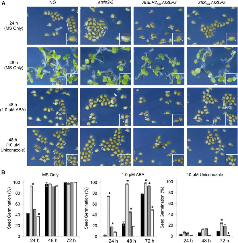
Mitochondrial SLP2-MIA40 Protein Interaction

abscisic acid (ABA) and gibberellic acid (GA; Kucera                           AtSLP2 was found to uniquely reside in the mito-
et al., 2005). ABA functions to establish dormancy in                          chondrial IMS. Here, it specifically interacts with
developing seeds while maintaining a negative regu-                            mitochondrial IMS import and assembly protein 40
latory role during seed germination by preserving seed                         (AtMIA40) yielding intramolecular disulfide bonds
dormancy (Müller et al., 2006). Conversely, GA posi-                           that activate the protein phosphatase to negatively
tively regulates seed germination by stimulating seed                          regulate seed germination by inhibiting GA related
swelling, endosperm rupture, and testa cracking to fa-                         processes.
cilitate radicle emergence and seedling establishment
(Holdsworth et al., 2008; Piskurewicz et al., 2008).
Emphasizing the interdependence of ABA and GA in                               RESULTS
seed germinative processes is the GA biosynthetic                              AtSLP2 Interacts with Mitochondrial Redox Relay
mutant ga1, which also exhibits enhanced dormancy                              Protein AtMIA40
and requires exogenous GA to germinate, while
DELLA transcription factor signaling mutant rgl2                                  AtSLP2 was found to be specifically expressed in
demonstrates reduced seed dormancy resulting from                              dark-grown Arabidopsis cell culture, roots, and seeds,
enhanced GA-induced gene expression (Lee et al., 2002;                         while its paralog AtSLP1 was not found in any of these
Cao et al., 2005; Kucera et al., 2005). A loss of both GA                      tissues but exclusively in photosynthetic tissue types
biosynthesis (ga1-3) and signaling through crosses                             (Supplemental Fig. S1; Uhrig and Moorhead, 2011).
with higher order rga1/rgl2/gai mutants results in                             In order to identify potential protein interactors of
GA-independent germination (Cao et al., 2005).                                 AtSLP2, Arabidopsis roots and cell culture constitu-
   Through a combination of biochemistry, cell biology,                        tively expressing tandem affinity purification (TAP)-
and pharmacological data, the involvement of a Ser/Thr                         tagged AtSLP2 (35S pro ::AtSLP2-TAP; AtSLP2-TAP)
protein phosphatase in regulating GA-related pro-                              were used (Fig. 1A). AtSLP2-TAP was overexpressed
cesses during seed germination has been shown.                                 in both wild-type Arabidopsis cell culture and plants

          Figure 1. AtSLP2-TAP pull-downs isolate AtSLP2-specific protein interactors. A, Cartoon depiction of the C-terminal TAP tag in
          frame with either AtSLP2 or GFP. The dashed line represents a human rhinovirus 3C protease cleavage site. B, Representative
          silver-stained SDS-PAGE and anti-Myc immunoblot of TAP pull-downs from stably transfected dark-grown Arabidopsis cell
          culture. GFP-TAP and AtSLP1-TAP pull-downs offered a control for nonspecific protein interactors (Supplemental Table S1 and
          raw data deposited at massive.ucsd.edu). Lanes contain 30 mL (silver) and 5 mL (western) of TAP-purified protein from 50 g of dark-
          grown Arabidopsis cell culture. C, Reciprocal AtMIA40-TAP pull-downs verify specific interaction between AtSLP2 and AtMIA40.
          AtMIA40-TAP was stably transfected into dark-grown Arabidopsis cell culture and used as bait to pull-down endogenous partners,
          which included AtSLP2. AtSLP2 was absent from in-parallel GFP-TAP pull-downs. D, Immunoprecipitation of endogenous
          AtMIA40-AtSLP2 protein complex using affinity-purified anti-AtMIA40 IgG and preimmune serum IgG (PIS). Immunoblotting was
          performed using 0.5 mg/mL anti-Myc (ICL), 1:1,000 anti-AtMIA40, and 1.2 mg/mL affinity-purified anti-AtSLP2 (Uhrig and
          Moorhead, 2011).

Plant Physiol. Vol. 173, 2017                                                                                                                   957

                                  Downloaded on March 8, 2021. - Published by https://plantphysiol.org
                                Copyright (c) 2020 American Society of Plant Biologists. All rights reserved.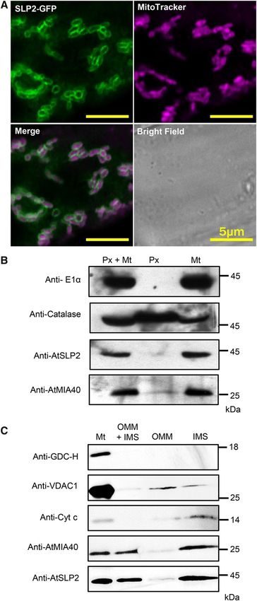
Uhrig et al.
alongside controls GFP-TAP (35S pro ::GFP-TAP) in 2015). Using Vicia faba leaves transiently expressing a
Arabidopsis cell culture and AtSLP1-TAP (35Spro::AtSLP1- peroxisome-targeted GFP and AtSLP2-RFP, AtSLP2
TAP) in Arabidopsis plants and cell culture. AtSLP2- was demonstrated to not be peroxisomal (Supplemental
and control-TAP pull-downs were performed in Fig. S5C). As a further control, V. faba plants transiently
parallel, with each isolated TAP-tagged protein expressing mitochondrial-targeted GFP (Mito-GFP;
detected by immunoblot using anti-Myc anti- Supplemental Fig. S5, D–F) determined that within the
bodies (Fig. 1B). Figure 1B depicts the typical MitoTracker Deep Red FM channel, chloroplasts
AtSLP2- and GFP-TAP interactome isolated using autofluoresce, but that the Deep Red FM dye is mito-
TAP constructs stably expressed in cell culture. The chondrial specific.
highly enriched ;25-kD polypeptide in the AtSLP2- To support our imaging data, we isolated intact mi-
TAP lane was excised and identified by mass spec- tochondria and peroxisomes from dark-grown wild-
trometry as Cox19-like CHCH family MIA40 protein type Arabidopsis cell culture (Fig. 2B). Immunoblotting
(AtMIA40). Peptide coverage was 57.4%, and no with AtMIA40- (Supplemental Figs. S3 and S4) and
AtMIA40 peptides were recovered in a digest of the AtSLP2-specific antibodies demonstrated that endog-
equivalent gel piece in the control lane. In parallel, a enous AtMIA40 resides in both organelles, while
subtractive mass spectrometry approach involving AtSLP2 is mitochondrion specific (Fig. 2B). Additional
the direct comparison of on-bead digested AtSLP2-, immunoblotting of both the purified mitochondria
AtSLP1-, and control-TAP pull-downs was performed and peroxisome fractions with antibodies to mito-
to identify additional AtSLP2 protein interactors chondrial pyruvate dehydrogenase complex protein
(Supplemental Table S1). AtSLP2-specific binding E1a (Szurmak et al., 2003) confirmed that the peroxi-
partners found in both dark-grown Arabidopsis cell somal fraction was completely free of mitochon-
culture and roots included AtMIA40. Other potential dria. Given that AtMIA40 proteins reside in the IMS
interacting proteins, although enriched compared to (Hentchel and Escalante-Semerena, 2015), we further
the control-TAP pull-downs, do not reside in the same fractionated isolated mitochondria into IMS and outer
compartment as AtSLP2 and were also recovered with mitochondrial membrane (OMM) and looked for
the SLP1-TAP, which is chloroplastic, suggesting they AtSLP2 localization (Fig. 2C). Consistent with form-
are likely not real AtSLP2 partners. For this reason, ing a complex, both AtMIA40 and AtSLP2 were highly
we chose to explore the association of AtSLP2 with enriched in the IMS fraction with the IMS marker
AtMIA40. cytochrome c.
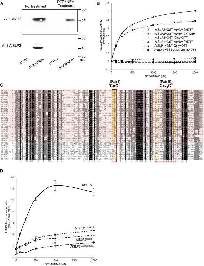
Verification of a specific AtSLP2-AtMIA40 protein
complex was supported through reciprocal TAP pull-
downs using AtMIA40-TAP (35Spro::AtMIA40-TAP) AtMIA40 Activates the Ser/Thr Phosphatase Activity of
stably expressed in dark-grown Arabidopsis cell cul- AtSLP2 through Disulfide Bond Formation
ture (Fig. 1C). Control-TAP pull-downs from the same
tissue failed to retrieve AtMIA40, corroborating the MIA40 proteins are oxidoreductases that aid the
specificity of the observed AtSLP2-AtMIA40 interac- formation of disulfides on target proteins by first
tion (Supplemental Table S1). Further support for an forming intermolecular disulfides between MIA40 and
AtSLP2-AtMIA40 protein complex was observed by its partner (Banci et al., 2009). To determine if AtMIA40
coimmunoprecipitation of endogenous AtSLP2 and associates with AtSLP2 in the absence of disulfide
AtMIA40 using affinity-purified anti-AtMIA40 IgG bonds, we performed anti-AtMIA40 immunoprecipi-
(Fig. 1D; Supplemental Figs. S2–S4). tations using extracts treated sequentially with or
without dithiothreitol (DTT) and the irreversible thiol-
modifying agent N-ethylmaleimide (NEM). This
AtSLP2 and AtMIA40 Colocalize to Mitochondria revealed that the association of AtMIA40 with AtSLP2
is dependent upon disulfide bond formation (Fig. 3A).
As in other eukaryotes, Arabidopsis MIA40 is a Activity assays employing the phosphatase substrate
mitochondrially targeted IMS oxidoreductase (Hentchel pNPP and recombinant AtSLP2 exhibited an AtMIA40
and Escalante-Semerena, 2015). Utilizing plants sta- concentration-dependent increase in phosphatase ac-
bly overexpressing AtSLP2-GFP, we found colocali- tivity (Fig. 3B) and that activation of HIS6-AtSLP2 by
zation with MitoTracker Deep Red FM fluorescent GST-AtMIA40 was dependent on the presence of re-
staining, which specifically visualizes mitochondria ductant (Fig. 3B). Importantly, GST-AtMIA40 did not
(Fig. 2A; Supplemental Fig. S5A). Consistent with activate the AtSLP2 paralog, AtSLP1.
this finding, overlapping signals from Arabidopsis Yeast and human MIA40s contain a specific CPC Cys
stably expressing AtSLP2-GFP (35Spro:AtSLP2-GFP) pair that forms sequential disulfide bonds on its partner
and AtMIA40-RFP (35Spro:AtMIA40-RFP) confirmed protein to yield the mature oxidized, properly folded
AtSLP2 colocalization with AtMIA40 in the mito- IMS protein (Mordas and Tokatlidis, 2015). To date,
chondria (Supplemental Fig. S5B). Plant MIA40 has MIA40 proteins have been observed to form two di-
been reported to also reside in peroxisomes (Hell, sulfide bonds on Cys pairs of client proteins that are
2008; Banci et al., 2009; Sideris et al., 2009; Herrmann typically separated by three or nine amino acids, al-
and Riemer, 2012; Hentchel and Escalante-Semerena, though many variants of this spacing pattern have
958 Plant Physiol. Vol. 173, 2017
Downloaded on March 8, 2021. - Published by https://plantphysiol.org
Copyright (c) 2020 American Society of Plant Biologists. All rights reserved.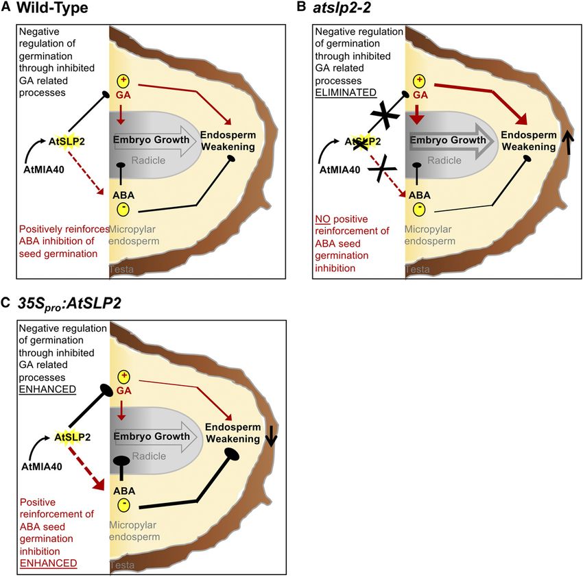
Mitochondrial SLP2-MIA40 Protein Interaction

                                                                           recently emerged (Mordas and Tokatlidis, 2015). Given
                                                                           that AtSLP1 cannot be activated by AtMIA40, we
                                                                           aligned AtSLP1 with AtSLP2 orthologs and searched
                                                                           for unique Cys pairs in AtSLP2 that may form disul-
                                                                           fides. This analysis revealed CxC and Cx 14 C pairs
                                                                           unique to SLP2 phosphatases (Fig. 3C). Individual
                                                                           mutation of one Cys from each pair (single mutant) or
                                                                           concurrent mutation of one Cys from each pair (double
                                                                           mutant) almost completely abolished AtSLP2 activa-
                                                                           tion by AtMIA40, indicating these conserved Cys pairs
                                                                           form disulfide bridges to mature AtSLP2 in the IMS
                                                                           (Fig. 3D; Supplemental Fig. S6).
                                                                              Bacterially expressed and purified HIS6-AtSLP2 was
                                                                           also found to possess an AtMIA40-dependent increase
                                                                           in activity toward pSer, pThr, pTyr and pThr/pTyr
                                                                           phosphorylated peptides (Fig. 4A; Supplemental Table
                                                                           S2). Despite an almost 20-fold activation in HIS6-
                                                                           AtSLP2 activity, AtMIA40 did not alter the specificity
                                                                           of AtSLP2 toward any one substrate peptide (Fig. 4A)
                                                                           and also suggested that AtSLP2 was a potential dual
                                                                           specificity protein phosphatase. However, assays con-
                                                                           ducted using AtSLP2-TAP isolated from dark-grown
                                                                           Arabidopsis cell culture showed that AtSLP2 dis-
                                                                           played a clear preference for a pThr-containing peptide
                                                                           substrate over a pTyr-containing peptide (Fig. 4B).
                                                                           Collectively, these results indicate that AtMIA40 func-
                                                                           tions to dramatically enhance AtSLP2 activity, but not
                                                                           alter its phosphoprotein specificity. Furthermore, data
                                                                           indicate that this is accomplished through the forma-
                                                                           tion of AtMIA40 catalyzed disulfide bridges as previ-
                                                                           ously described for other MIA40 orthologs (Banci et al.,
                                                                           2009; Sideris et al., 2009).

                                                                           AtSLP2 and AtMIA40 Function to Negatively Regulate
                                                                           Seed Germination

                                                                             To elucidate the biological function of the protein
                                                                           phosphatase AtSLP2, an insertional knockout (atslp2-2)
                                                                           mutant was screened for growth phenotypes. Despite
                                                                           the availability of other atslp2 insertional mutant
                                                                           plants, exhaustive screening failed to identify a second
                                                                           knockout where AtSLP2 protein levels were di-
                                                                           minished. This lead us to complement atslp2-2 us-
                                                                           ing an endogenous promoter-driven AtSLP2 construct
                                                                           (AtSLP2pro::AtSLP2), in addition to generating constitutive

                                                                           pyruvate dehydrogenase complex E1a (anti-E1a; mitochondrial
                                                                           matrix) and 1:200 anticatalase (peroxisome) IgG. Each lane con-
                                                                           tains 15 mg of total protein. C, Isolated mitochondria (Mt) were
                                                                           further subfractionated into OMM and IMS and immunoblotted
                                                                           using 1:5000 anti-H protein of Gly decarboxylase complex (anti-
Figure 2. AtSLP2 and AtMIA40 are located in the mitochondrial IMS.         GDC-H; mitochondria matrix marker), 1:5,000 antivoltage-
A, Stably transfected Arabidopsis roots expressing 35Spro::AtSLP2-         dependent anion-selective channel protein 1 (anti-VDAC1; OMM
GFP (green) are stained with MitoTracker Deep Red FM (purple) to           marker) and 2 mg/mL anticytochrome c (anticyt c; IMS marker)
illuminate mitochondria. Representative individual, merged, and            IgG. Each lane contains 10 mg of total protein. Immunoblotting
bright field images are shown. B, Intact mitochondria and peroxi-          was performed in conjunction with affinity-purified anti-AtSLP2
somes were isolated from dark-grown wild-type Arabidopsis cell             IgG (Uhrig and Moorhead, 2011) and anti-AtMIA40 crude im-
culture. Immunoblotting of the purified mitochondrial (Mt) and per-        mune serum (Supplemental Fig. S4) used at 1.2 mg/mL and a 1:1,000
oxisomal (Px) fractions were performed using 1:100 antimitochondrial       dilution, respectively.

Plant Physiol. Vol. 173, 2017                                                                                                            959

                                  Downloaded on March 8, 2021. - Published by https://plantphysiol.org
                                Copyright (c) 2020 American Society of Plant Biologists. All rights reserved.
Uhrig et al.
Figure 3. AtSLP2 is redox activated by AtMIA40. A, Immunoprecipitation of endogenous AtSLP2-AtMIA40 complex is redox
dependent. Treatment involved sequential reduction (20 mM DTT) and alkylation (110 mM NEM) of a dark-grown wild-type
Arabidopsis cell culture extract prior to immunoprecipitation (PIS, preimmune serum IgG). B, HIS6-AtSLP2 (250 ng) incubated
with increasing amounts of GST-AtMIA40 in the presence of 5 mM DTT or TCEP activates AtSLP2, while failing to activate AtSLP1.
Fold-change in AtSLP2 activity was derived from AtSLP2 assays without AtMIA40. C, Alignment of SLP1 and SLP2 (pink shading)
960 Plant Physiol. Vol. 173, 2017
Downloaded on March 8, 2021. - Published by https://plantphysiol.org
Copyright (c) 2020 American Society of Plant Biologists. All rights reserved.Mitochondrial SLP2-MIA40 Protein Interaction
Further examination of AtSLP2 and AtMIA40 protein
levels during imbibition and germination was per-
formed (Supplemental Fig. S8). Here “seed imbibition”
refers to seed water uptake and swelling at 4°C in the
dark, while “seed germination” refers to seeds exposed
to 24 h light at room temperature after imbibition. These
two processes were explicitly separated to fully resolve
when AtSLP2 and AtMIA40 protein levels were maxi-
mal (Supplemental Fig. S8). AtSLP2 and AtMIA40
protein levels were found to largely parallel each other
over the course of imbibition and germination with
protein levels peaking early in imbibition (3–6 h).
AtSLP2-AtMIA40 Protein Complex Negatively Regulates
GA Biosynthesis
The observed early germination phenotype in
atslp2-2 was suggestive of an alternation in hormone-
related processes. As seed germination is governed by
complex hormone-linked metabolism and signaling
mechanisms centering around the antagonistic in-
teraction displayed by ABA and GA, enhanced GA
biosynthesis/signaling or ABA insensitivity could
lead to rapid germination. To explore this hypothesis,
a pharmacological approach was undertaken to test
the ability of atslp2-2 to exhibit accelerated germina-
tion phenotypes in the presence of either Uniconazole
(GA biosynthetic inhibitor) or ABA (to ascertain ABA
insensitivity). We performed germination assays over
a range of Uniconazole (0–100 mM) and ABA (0–5 mM)
concentrations (Supplemental Fig. S9) with a detailed
analysis of the effect of 10 mM and 1 mM Uniconazole
Figure 4. Substrate specificity of AtSLP2-AtMIA40 complex. A, A panel and ABA, respectively (Fig. 5). Uniconazole (10 mM)
of phospho-peptides was screened using 250 ng of bacterially expressed almost completely inhibited the observed germina-
and purified HIS6-AtSLP2 with (gray bars) and without (black bars) the tion phenotype of atslp2-2, while 1 mM ABA delayed,
addition of HIS6-AtMIA40 to assess the influence of AtMIA40 on the but did not inhibit, atslp2-2 germination (Fig. 5).
substrate specificity of AtSLP2. Phospho-peptides depicted here repre- These observations implicate endogenous GA levels
sent pThr (RRP1B, BRCA1), pSer (Ki67, B56), pThr/pTyr (p38, p38beta)
as responsible for the observed germination pheno-
and pTyr (Rat SAPK3), containing peptides. B, AtSLP2-TAP is pSer/pThr
specific. AtSLP2-TAP activity was assessed using BRCA1 (pThr) and Rat
type of atslp2-2, suggesting that there are increased
SAPK3 (pTyr) peptide substrates. Maximal AtSLP2-TAP activity was GA levels in the absence of AtSLP2.
achieved using exogenous HIS6-AtMIA40. All assays were conducted at We further analyzed publicly available microarray
30°C for 1 h, with enzymatic activity deduced with malachite green. data and found that AtSLP2 transcripts are down-
Error bars represent 6 SE (n = 3). regulated in response to ABA (Supplemental Fig. S10A)
and up-regulated in response to GA (Supplemental
Fig. S10B) during seed imbibition. GA-induced expres-
AtSLP2 (35Spro::AtSLP2) overexpression plant lines. As sion of AtSLP2 negatively correlated with Arabidopsis
shown in Supplemental Figure S7, western blotting con- GA3 oxidase (GA3OX; At5g25900) and GID1A (At3g05120),
firmed atslp2-2 as a complete knockout, which could be while positively correlating with DELLA transcrip-
complemented (AtSLP2pro::AtSLP2). Examination of atslp2-2 tion factors RGA1 (At1g14920) and RGL2 (At3g03450)
seedlings found they displayed accelerated germination, that act downstream of GA production. Furthermore,
while 35Spro::AtSLP2 seedlings were delayed in ger- wild-type Arabidopsis seeds germinated under con-
mination (Fig. 5). Complementation of atslp2-2 with trol conditions revealed that AtSLP2 transcript main-
AtSLP2pro::AtSLP2 rescued the early germination phenotype. tains an in-parallel decrease in expression with GA3OX,
Figure 3. (Continued.)
orthologs from across photosynthetic eukaryotes revealed conserved SLP2 Cys pairs. Alignment was created using MAFFT (http://
mafft.cbrc.jp/alignment/software/). Asterisks denote cysteines (yellow) targeted for site-directed mutagenesis. Protein name, gene
identifier, organism and sequences are presented in Supplemental Table S4. D, Site-directed mutagenesis of identified SLP2-
specific cysteines in AtSLP2 results in reduced AtSLP2 activation by AtMIA40. Error bars represent 6 SE (n = 3).
Plant Physiol. Vol. 173, 2017 961
Downloaded on March 8, 2021. - Published by https://plantphysiol.org
Copyright (c) 2020 American Society of Plant Biologists. All rights reserved.Uhrig et al.
Figure 5. Analysis of seed germination on 0.53 MS-agar plates containing either ABA or Uniconazole. Control plates consisted
of 0.53 MS only. A, Seed germination exhibited by wild-type NÖ, atslp2-2, and AtSLP2pro::AtSLP2 complemented atslp2-2 and
35Spro::AtSLP2 seeds. B, Quantitative analysis of AtSLP2 seed germination. Plant lines wild-type NÖ (black; far left), atslp2-2 (light
gray; middle left); AtSLP2pro::AtSLP2 complemented atslp2-2 (dark gray, middle right) and 35Spro::AtSLP2 seeds (white; right).
Application of 1.0 mM ABA slowed, but did not abolish, seed germination in atslp2-2 seeds, while nearly all seeds were equally
inhibited by the application of 10 mM Uniconazole. A seed germination time-course examining multiple ABA and Uniconazole
concentrations over 72 h is also shown (Supplemental Fig. S10). All seeds were imbibed at 4°C for 48 h on each plate prior to
germination under 16 h light:8 h dark at 23°C. Error bars represent 6 SE (n = 3 plates; average of 80–120 seeds per plate). Asterisks
denote two-tailed Student’s t test (P , 0.05) when atslp2-2, AtSLP2pro::AtSLP2, and 35Spro::AtSLP2 seeds were compared to wild-
type NÖ. F-test indicated normal data distribution.
GID1A, RGL2, and RGA1 (Supplemental Fig. S11A), Subsequent assessment of Arabidopsis MIA40,
while also being slightly down-regulated in germinat- GA3OX, GID1, RGA1, and RGL2 transcript levels
ing seeds grown on an ABA supplemented medium in atslp2-2, AtSLP2 pro ::AtSLP2, wild-type NÖ and
(Supplemental Fig. S11B). Taken together with our 35S pro ::AtSLP2 seeds over a 12 h imbibition time
pharmacological data, these datasets indicate AtSLP2 course by qRT-PCR (Fig. 6) further corroborated
functions to negatively regulate GA-related cellular available microarray data of germinating Arabidopsis
processes. seeds (Supplemental Figs. S10 and S11). Consistent
962 Plant Physiol. Vol. 173, 2017
Downloaded on March 8, 2021. - Published by https://plantphysiol.org
Copyright (c) 2020 American Society of Plant Biologists. All rights reserved.Mitochondrial SLP2-MIA40 Protein Interaction
with the presence, absence, or overabundance of RQ fold-change. Changes in AtSLP2 transcript abun-
AtSLP2 protein, AtSLP2 transcript levels changed as dance were paralleled by a corresponding change in
expected (Fig. 6A). AtMIA40 transcript levels largely AtSLP2 protein levels (Supplemental Fig. S8), while peak
mirrored these transcriptional changes in AtSLP2, AtSLP2 and AtMIA40 transcript levels (;6 h imbibition)
with moderately decreased expression in atslp2-2 corresponded to peak AtSLP2 protein (3–6 h) during
and slightly increased expression in 35S pro ::AtSLP2 wild-type NÖ imbibition (Fig. 6; Supplemental Fig. S8).
(Fig. 6B). GA biosynthetic and signaling machinery
transcript levels similarly exhibited changes in
transcript levels mirroring AtSLP2 (Fig. 6, C–F). This DISCUSSION
was most well resolved in atslp2-2, where decreases AtSLP2 Phosphatase Represents a Novel IMS Client of
in both GA biosynthetic and signaling machinery Redox Relay Protein AtMIA40
were observed. The largest changes in transcript levels
were observed for AtSLP2 in the 35Spro::AtSLP2 and SLP2 represents a novel interaction partner of MIA40
AtSLP2pro::AtSLP2 plant lines (4–5 RQ fold increase), in photosynthetic eukaryotes, as SLP2 phosphatases are
while overall changes in GA-related transcript levels absent in metazoans (Uhrig and Moorhead, 2011).
were relatively moderate, exhibiting only a 60.5 to 1.0 Previous examination of atmia40 insertional mutant
Figure 6. Quantitative PCR analysis of GA biosynthetic and signaling protein genes from imbibed atslp2-2, wild-type
NÖ, AtSLP2 pro ::AtSLP2, and 35S pro ::AtSLP2 seeds. Seeds were imbibed for 0 h, 6 h, and 12 h in water at 4°C in the dark
followed by snap freezing. A to F, Transcriptional expression of Arabidopsis SLP2, MIA40, GA3 oxidase (GA3OX ),
GA-insensitive dwarf 1a (GID1A), regulator of GA1 (RGA1), and regulator of GA-like 2 (RGL2) was assessed. Error bars
represent 6 SE (n = 3).
Plant Physiol. Vol. 173, 2017 963
Downloaded on March 8, 2021. - Published by https://plantphysiol.org
Copyright (c) 2020 American Society of Plant Biologists. All rights reserved.Uhrig et al.
plants found AtMIA40 responsible for importing mitochondrial IMS reversible protein phosphorylation
the chaperone for Mn-superoxide dismutase (CCS1) also represents a conserved phenomenon.
and Cu/Zn superoxide dismutase 1 into the mito-
chondrial IMS, as has been observed in other eukary-
otes (Kawamata and Manfredi, 2010), as well as Cu/Zn Negative Regulation of Seed Germination through
superoxide dismutase 3 into the plant peroxisome GA Metabolism
(Hentchel and Escalante-Semerena, 2015). In addition
to these proteins, research in nonphotosynthetic eu- In exploring the biological function of AtSLP2,
karyotes has identified COX17, translocase of inner atslp2-2 seeds exhibited accelerated seed germination
membrane proteins TIM22, and a number of other mi- compared to NÖ seeds, while 35S pro ::AtSLP2 seeds
tochondrial IMS proteins to be directly regulated by displayed delayed seed germination (Fig. 5). This in-
MIA40 (Banci et al., 2009; Reddehase et al., 2009; Sideris dicated that AtSLP2 was a regulator of seed germi-
et al., 2009; Darshi et al., 2012; Weckbecker et al., 2012). nation. With germinative processes predominantly
However, AtSLP2 represents the first protein phos- governed by ABA and GA, this suggested that atslp2-2
phatase identified to interact with MIA40. seeds were either ABA insensitive/underproducing or
MIA40 proteins have been found to catalyze the GA sensitive/overproducing. Lack of dramatic ABA
formation of disulfide bridges on target protein sub- insensitivity and maintenance of atslp2-2-accelerated
strates through their oxidoreductase activity, trapping germination in the presence of ABA indicated that
the MIA40 substrate in the mitochondrial IMS (Banci the observed seed germination phenotype is likely
et al., 2009; Sideris et al., 2009). This is achieved via the due to enhanced GA production or altered GA sig-
key cysteines of MIA40’s active site (the CPC motif), naling. If atslp2-2 seeds were ABA insensitive, appli-
which first forms disulfide bridge intermediates with cation of 1 mM ABA should not have induced the
the target protein substrate, followed by the formation concentration-dependent effect on seed germination
of disulfide bridges on the target substrate and the re- that was documented previously (Koornneef et al.,
lease of reduced MIA40. The association of AtSLP2 with 1989). Furthermore, Uniconazole almost completely
AtMIA40 appears to be disulfide bond dependent as abrogated the accelerated germination of atslp2-2
thiol modification of reduced cysteines blocked their seeds, indicating GA is required for the observed
association (Fig. 3A). Through sequence comparison germination phenotypes. Given GA signaling (nega-
with its closely related paralog SLP1, we deduced and tive regulators) mutants such as rgl2 and other DELLA
then confirmed which Cys pairs form disulfide bonds loss-of-function mutants can germinate in the presence
and are necessary for AtMIA40 activation of AtSLP2 of GA inhibitors and confer GA phenotypes in the
(Fig. 3, C and D). Collectively, these findings support absence of bioactive GA (Sun and Kamiya, 1994; Lee
AtMIA40 activation of AtSLP2 through the catalyzed et al., 2002; Cao et al., 2005), the AtSLP2-AtMIA40
formation of disulfide bridges. Like other MIA40 sub- complex likely functions to negatively regulate GA
strates, this may function to trap AtSLP2 in the IMS biosynthesis.
by providing correct tertiary structure (Fischer and If GA biosynthesis were up-regulated in atslp2-2
Riemer, 2013). seeds, we would expect a corresponding increase in
The mitochondrial localization of AtSLP2 (Fig. 2), GA-induced degradation of negative regulators RGL2
combined with its 4- (pNPP) to 20- (phosphorylated and RGA1 (Xu et al., 2014). In accordance with
peptides) fold increase in activity upon interaction with this hypothesis, expression of DELLA protein RGL2,
IMS-targeted AtMIA40 and a phosphorylated substrate the major determinant of seed germination, was
preference indicative of Ser/Thr PPP-family protein down-regulated in atslp2-2 (Fig. 6; Lee et al., 2002). In
phosphatases (Fig. 4) suggest reversible protein phos- 35S pro ::AtSLP2 seeds, this pattern of GA-negative reg-
phorylation plays a regulatory role within the mito- ulator down-regulation was inverse. 35Spro::AtSLP2
chondrial IMS. No studies to date have specifically seeds maintained elevated RGL2 transcript levels rel-
examined the role of reversible protein phosphoryla- ative to wild-type Arabidopsis following imbibition
tion in the mitochondrial IMS of any organism despite (Fig. 6).
targeted proteomics studies revealing the presence of a
number of protein kinases and protein phosphatases in AtSLP2-AtMIA40 Protein Complex Functions to
this subcompartment (Salvi et al., 2005; Pagliarini and Negatively Regulate GA Biosynthesis
Dixon, 2006; Vögtle et al., 2012; Duncan et al., 2013).
To date, 10 protein kinases have been localized to the When compared to atslp2-2 seeds, atmia40 seeds
plant mitchondrion, but none specifically to the IMS exhibited a moderate accelerated germination phenotype
(Havelund et al., 2013). Furthermore, recent elucidation (Supplemental Fig. S12). This suggests that AtSLP2 likely
of the IMS proteome of yeast identified a number of represents the functional driver of the AtSLP2-AtMIA40
IMS proteins that have been previously documented complex in negatively regulating GA-related cellular
in whole mitochondria phosphoproteomics studies to processes (Fig. 7). Reinforcing this hypothesis is the
be phosphorylated (Vögtle et al., 2012). Given the bacterially expressed and purified AtSLP2, which
prevalence of regulatory protein phosphorylation maintained detectable protein phosphatase activity
across eukaryotes, it is reasonable to predict that even in the absence of AtMIA40 (Uhrig and Moorhead,
964 Plant Physiol. Vol. 173, 2017
Downloaded on March 8, 2021. - Published by https://plantphysiol.org
Copyright (c) 2020 American Society of Plant Biologists. All rights reserved.Mitochondrial SLP2-MIA40 Protein Interaction
2011), indicating that atmia40 seeds likely possess negatively regulate seed germination via GA-related
some residual AtSLP2 protein phosphatase activity, processes, there exists a pressing need to identify its
which mitigates, to a certain extent, the absence of substrate(s). Albeit challenging, substrate identification
AtMIA40 and its requirement to fully activate AtSLP2. will better resolve the working hypothesis presented
With seed phenotype data supporting a role for here by shedding light on the connection between mi-
mitochondrially targeted AtSLP2 in regulating cyto- tochondrial targeted AtSLP2 and its negative reg-
solic GA-related processes, there remains a need to ulation of cytosolic/nuclear-localized GA-related
resolve the intermediate mechanisms connecting the processes. The work presented here outlines an excit-
mitochondria to cytosolic GA biosynthesis. ing new connection between the mitochondria and
With the discovery that AtSLP2 protein phosphatase cytosol through plant cell signaling, in addition to provid-
interacts with IMS redox relay protein AtMIA40 to ing precedence for reversible protein phosphorylation
Figure 7. Model of AtSLP2-AtMIA40 protein complex function during seed germination. A, Under wild-type conditions, AtMIA40-
activated AtSLP2 functions to negatively regulate GA biosynthesis and in doing so positively reinforces ABA inhibition of seed
germination. B, Absence of AtSLP2 (atslp2-2) results in unimpeded GA-related cellular processes leading to an accelerated seed
germination phenotype. C, Overexpression of AtSLP2 (35Spro::AtSLP2) leads to enhanced inhibition of GA-related cellular pro-
cesses, which concomitantly enhances ABA inhibition of seed germination. This leads to a delayed seed germination phenotype.
Effects of the presence or absence of an AtSLP2-AtMIA40 protein complex over seed germinative processes is described by a
combination of arrow width and color. Arrow width denotes enhanced (thick) versus reduced (thin) pathway flux/phytohormone
effect, respectively. Solid black lines versus solid red lines depict negative or positive influence over seed germination, respectively.
Dashed red lines represent indirect AtSLP2-AtMIA40 protein complex influence over ABA-directed seed germination processes.
Plant Physiol. Vol. 173, 2017 965
Downloaded on March 8, 2021. - Published by https://plantphysiol.org
Copyright (c) 2020 American Society of Plant Biologists. All rights reserved.Uhrig et al.
in the mitochondrial IMS. Future studies will focus Complementation and Screening of AtSLP2 Mutant Plants
on elucidating the intermediate steps and compo- Complementation of atslp2-2 knockout plants was performed by the am-
nents of how AtSLP2 negatively regulates GA-related plification of AtSLP2 native promoter and gene from genomic DNA of wild-
processes. type ecotype NÖ Arabidopsis leaves using the Gateway-compatible primers
59-GGGGACAAGTTTGTACAAAAAAGCAGGCTTCCTATC TCTCTTA-
TAACGAAGAAA-39 and 59-GGGGACCACTTTGTACAAGAAAGCT
GGGTCAGCTTTGACTTCAACTTGCTTAG-39. The 1.581-kb promoter region
MATERIALS AND METHODS was determined based on the Arabidopsis Gene Regulatory Information Server.
The fragment was inserted into pDONR221 and cloned into the destination
Plant Growth Conditions vector pEarleyGate 304 (Earley et al., 2006). The binary vector was transformed
into atslp2-2 mutant as above. Basta (BaR)-resistant plants were genotyped by
Standard Arabidopsis (Arabidopsis thaliana) growth conditions involved dry PCR and expression of AtSLP2 assessed by western blotting.
sterilization and stratification at 4°C for 2 d in the dark on 0.53 Murashige and
Skoog (MS) agar plates. Plants were germinated under 16 h light and 8 h dark at
22°C at a light level of 125 mmol m22s21. Five-day-old seedlings were trans- Arabidopsis Cell Culture Transfection and
ferred to soil supplemented with 0.5 g/L 20:20:20 fertilizer and grown under a TAP Pull-Downs
12-h-light/12-h-dark cycle regiment at 22°C at a light level of 125 mmol m22s21.
Plant roots were isolated from germinating Arabidopsis seedlings grown Wild-type light-grown Arabidopsis cell culture was individually transfected
as above in Magenta boxes for 21 d. Magenta box growth media consisted of by A. tumefaciens carrying AtSLP2, AtSLP1, AtMIA40, and GFP-TAP constructs
as previously described (Uhrig and Moorhead, 2011). Positive transformants
0.53 MS media supplemented with 1 mM potassium phosphate. Seedlings
were screened via immunoblotting with anti-Myc IgG (ICL). Arabidopsis cell
subjected to ABA and Uniconazole (LKT Laboratories) were sterilized and
culture was subsequently transitioned from light to dark over a 2 week period,
stratified as above on 0.53 MS agar followed by germination under 16 h light
with subculturing performed every 7 d. TAP pull-downs from dark cell culture
and 8 h dark at 22°C for 4 d. For seed germination assays, statistical significance
were performed as previously described (Templeton et al., 2011) using an ex-
between mutant and corresponding wild-type seed germination was per-
tract from 50 g of cells per pull-down, while pull-downs from roots were per-
formed using a two-tailed Student’s t test. Sample variance was assessed using formed using 5 g of tissue. Subsequent washing of tagged protein bound
an F-test. No statistically significant variance was observed. Ni-NTA with 10 mL of triethylammonium bicarbonate (pH 8.5) was performed
prior to on-bead, overnight (18 h) proteolysis using 750 ng of trypsin (Promega)
Insertional atslp2 and atmia40 Plant Genotyping at 30°C. Peptides were eluted using a 50% acetonitrile: 2.5% formic acid solution
and dried prior to analysis by mass spectrometry. TAP pull-downs subjected
Insertional mutants atslp2-2 (RATM13-2055-1) and atmia40 (SALK_044358) to immunoblot analysis were eluted from Ni-NTA using a minimal volume of
Arabidopsis were screened by PCR using the following gene primers: 13 sodium dodecyl sulfate polyacrylamide gel electrophoresis (SDS-PAGE)
59-TGGTTGCAATTCTGACACAAC, 39-CAAGAATCTCTTTGCAATCGG sample buffer and boiling for 5 min.
(atslp2-2), and 59-TCGTACAGATGCGATCATCTG, 39-AAGGGACACA-
AAACGTAAACG (atmia40). The T-DNA insert primers used were:
Mass Spectrometry
39-GTATTTATCCCGTTCGTTTTCGT (atslp2-2) and 59-TGGTTCACG-
TAGTGGGCCATCG (atmia40). Insertional mutant plant lines were Sample digestion, processing, mass spectrometry, and data analysis were
obtained from the Riken Bioresource Center Experimental Plant Division performed as in Templeton et al. (2011). In addition to GFP-TAP pull-downs
(RIKEN; http://epd.brc.riken.jp/en/) and the Arabidopsis Information Re- controlling for experimentation involving cell culture, AtSLP1 offered a control
source (www.arabidopsis.org; atmia40), respectively. for AtSLP2 pull-downs from root tissue, as AtSLP1 is not expressed in nongreen
tissues and is the closest ortholog of AtSLP2 (Uhrig and Moorhead, 2011). Bona
fide binding partners were only considered if they were not found in the ma-
Molecular Cloning, Expression, and Recombinant jority of independent negative controls: 63 GFP-TAP and 73 AtSLP1-TAP pull-
Protein Purification downs. Raw data from the excised band and on-bead digested samples have
been deposited at “MassIVE” (massive.ucsd.edu).
Fluorescent and TAP constructs were created using full-length At1g18480
(AtSLP2), At1g07010 (AtSLP1), and At5g23395 (AtMIA40) clones obtained
from the Arabidopsis Information Resource (http://www.arabidopsis.org/). AtMIA40 Immunoprecipitation
PCR amplification of each respective cDNA was performed using Gateway-
compatible primers and inserted into pDONR221 (Invitrogen). Fluorescent Affinity-purified anti-AtMIA40 IgG and rabbit preimmune serum IgG
(10 mg) were coupled to Protein A-Sepharose 4B beads (Life Technologies) as
constructs were subsequently created using the binary vectors pB7RWG2
described in Ulke-Lemée et al. (2007). AtMIA40 immunoprecipitation was
(cRFP) and pK7RWG2 (cGFP; https://gateway.psb.ugent.be/), while TAP
performed with 50 mg protein extract isolated from 10-d-old dark-grown
clones were created using pYL436 (Rubio et al., 2005). Transformation of each
Arabidopsis cell culture as in Uhrig et al. (2016). Protein extract was pre-
construct into Agrobacterium tumefaciens strain GV3101 (A. tumefaciens)
treated with 20 mM DTT for 45 min at 4°C followed by alkylation with 110 mM
was performed to facilitate stable integration of each construct into either
NEM for 1 h at 4°C. A control without reducing/alkylating agents was per-
Arabidopsis plants (Zhang et al., 2006) or cell culture (Templeton et al., 2011). formed in parallel. Immunoprecipitations were performed three times with
At5g23395 was cloned into pDEST15 (N-terminal GST-tag) and pDEST17 three separate plant samples.
(N-terminal HIS6-tag) for expression in and purification from Escherichia coli,
while AtSLP1 and 2 were cloned into pET-48b(+) (Novagen) and purified via
Ni-NTA as previously described (Uhrig and Moorhead, 2011). GST-only (pGEX4T-1; Transient Vicia faba Expression and Imaging
Qiagen) was also expressed and purified for use as a control in enzyme and in vitro
Transient expression of 35Spro::AtSLP2-RFP and 35Spro::Peroxisome-GFP or
interaction analyses. GST-AtMIA40 and GST-only expression utilized BL21 (DE3)
35Spro::Mito-GFP (Nelson et al., 2007) in V. faba leaves was achieved by
Codon(+)-RIL (Stratagene) E. coli grown at 37°C to an OD600 of 0.4 to 0.5 followed by
particle bombardment (PDS-1000 system; Bio-Rad). Plasmid DNA (5 mg each
induction with 0.1 mM isopropyl b-D-1-thiogalactopyranoside overnight
for cobombardment or 10 mg for single bombardment) was coupled to 1 mg
(18 h) at room temperature (23°C). Protein purification for use in enzymatic of gold microcarriers, washed, spotted on a macrocarrier, and accelerated
assays and protein interaction experimentation was performed as per Uhrig onto the leaves. Stably transfected plants expressing 35Spro::AtSLP2-GFP
and Moorhead (2011) using 500 mL of settled glutathione Sepharose (GE were stained with 10 mM MitoTracker Deep Red FM for 10 min and rinsed
Healthcare). Protein elution was performed using 10 mM GSH in 50 mM HEPES- with water. All stably transfected 35Spro::AtMIA40-RFP/35Spro::AtSLP2-GFP
KOH, pH 7.8, 150 mM NaCl and 5% glycerol. Eluates were concentrated to a small Arabidopsis tissues were mounted on a microscope slide in 50% (v/v) glyc-
volume using a 10,000 D cutoff concentrator (Amicon), followed by dialysis erol and visualized at 633 magnification using a confocal microscope (Leica
against elution buffer lacking GSH prior to snap freezing and storage at 280°C. TCS SP5). The following excitation/emission (Ex/Em) settings were used:
966 Plant Physiol. Vol. 173, 2017
Downloaded on March 8, 2021. - Published by https://plantphysiol.org
Copyright (c) 2020 American Society of Plant Biologists. All rights reserved.Mitochondrial SLP2-MIA40 Protein Interaction
RFP, 543/580 to 682 nm; GFP, 488/497 to 538 nm; MitoTracker Deep Red 7.5, 150 mM NaCl, 5% (v/v) glycerol, 1% (v/v) Tween 20, 1 mM PMSF, and 1 mM
FM, 633/700 to 756 nm. benzamidine. Seed and root lysates were clarified by centrifugation at 4°C and
14,000g.
Isolation of Arabidopsis Cell Culture Mitochondrial
Fractions and Peroxisomes Western Blotting
Seven-day-old, dark-grown Arabidopsis culture (120 g) was filtered through All antibodies were validated within this manuscript (MIA40) or as
one layer of Miracloth to remove growth medium and homogenized batchwise noted below in published articles or in the datasheets for the commercial
40 g at a time in a precooled 4°C mortar and pestle. Grinding of each 40 g antibodies. Affinity-purified polyclonal rabbit anti-AtSLP1 and 2 anti-
was performed in 100 mL homogenization buffer consisting of 0.3 M mannitol, bodies previously generated (Uhrig and Moorhead, 2011) were used at
50 mM MOPS-KOH, pH 7.8, 5 mM EDTA, 0.5% (w/v) BSA, 1.0% (w/v) 1.5 and 1.2 mg/mL, respectively. Rabbit anti-AtMIA40 crude immune se-
polyvinlypyrrolidone, and 10 mM DTT added just before use. Isolation of rum was used at a 1:1000 dilution and validated in Supplemental Figs. S3
mitochondria plus peroxisome was obtained by Percoll density gradient and S4. Mitochondria and peroxisomes were probed using 1:100 anti-
centrifugation as previously described (Eubel et al., 2007). Separation of mitochondrial pyruvate dehydrogenase complex E1a (mitochondrial
mitochondria from peroxisome was achieved by resuspending the fraction matrix [Szurmak et al., 2003]) and 1:200 anticatalase (peroxisome) IgG
in 1 mL Suc buffer (0.3 M Suc, 10 mM TES-NaOH, pH 7.5, 0.1% [w/v] BSA) (Chuong et al., 2005). Mitochondrial subcompartments were blotted using
and overlaying on 28% (v/v) Percoll in Suc buffer followed by centrifugation 1:5000 anti-H protein of Gly decarboxylase complex (anti-GDC-H; mito-
at 41,000g for 30 min at 4°C (Struglics et al., 1993). Mitochondria (upper layer) chondria matrix marker; Agrisera/AS05 074), 1:5000 antivoltage-dependent
and peroxisome (lower layer) fractions were collected separately, washed in anion-selective channel protein 1 (anti-VDAC1; OMM marker; Agrisera) and
mannitol buffer (20 mM MOPS-KOH, pH 7.5, 0.3 M mannitol, 0.2% [w/v] BSA) 2 mg/mL anticytochrome c (anticyt c [Sweetlove et al., 2001]) IgG. Com-
and centrifuged at 18,675g for 15 min at 4°C. Isolated mitochondria and mercially available rabbit anti-Myc (ICL/RMYC-45A) and mouse anti-GFP
peroxisomes were lysed with 50 mM MOPS-KOH, pH 7.5, 150 mM NaCl, (Roche/11814460001) antibodies were used at 0.2 mg/mL and 0.4 mg/mL,
0.2% (v/v) Triton X-100 and boiled in SDS-cocktail. Mitochondrial sub- respectively.
compartments were isolated as in Sweetlove et al. (2001). Organelle isolations
were performed three times from three separate plant samples and a repre-
qRT-PCR Analysis
sentative experiment is shown.
Total RNA from the various Arabidopsis seed lines were isolated using a
modified TRIzol (Invitrogen) protocol (Meng and Feldman, 2010). First-
Enzymatic Analysis strand cDNA was synthesized from 1.67 mg total RNA using oligo(dT)12-18
Enzyme assays utilizing pNPP (Sigma-Aldrich) were conducted using primer and SuperScript II Reverse Transcriptase (Invitrogen) following the
Ni-NTA-purified HIS6-AtSLP2 and glutathione Sepharose-purified GST-AtMIA40 manufacturer’s instructions. The qPCR was performed using StepOnePlus
or HIS6-AtMIA40 (Supplemental Fig. S2). All assays were done in parallel Real-Time PCR System (Applied Biosystems). Primer pairs are listed
with Ni-NTA-purified eluates generated from nontransformed BL21 (DE3) (Supplemental Table S3). Each PCR reaction contained 13 Fast SYBR Green
Codon(+)-RIL E. coli to account for potential background pNPP cleavage from Master Mix (Applied Biosystems), 200 nM of each primer, and 0.25 mL cDNA
nonspecific copurifying proteins (Supplemental Fig. S2). Phosphatase activity was in a final volume of 20 mL. PCR amplification was performed for 40 cycles at
calculated relative to assays conducted with AtSLP2 only under previously deter- 95°C for 3 s and 60°C for 30 s with a preceding initial enzyme activation of 20 s
mined conditions giving highest activity (Uhrig and Moorhead, 2011). All pNPP at 95°C. Relative expression levels were calculated by D-DCt method, and all
assays were conducted as previously described (Uhrig and Moorhead, 2011) using quantifications were normalized using At1g13440 mRNA as an internal
either 5 mM DTT, Tris(2-carboxyethyl)phosphine or reduced glutathione as a re- control (Dekkers et al., 2012). For each target gene, the reactions were carried
ductant. Expressed and purified GST only was used as an assay control. out in triplicate.
Malachite green (Sigma-Aldrich) assays were performed as previously de-
scribed (Baykov et al., 1988), alternatively employing a 10% (w/v) ammonium Accession Numbers
molybdate solution. Each assay used 1 mg of recombinant phosphatase protein
incubated for 1 h at 30°C in 160 mL of 13 dilution buffer containing each re- Sequence data from this article can be found in the GenBank/EMBL data
spective experimental phosphopeptide substrate (Supplemental Table S2). libraries under accession numbers At5g23395, At1g18480, At1g07010,
Quenched assays were left at room temperature (23°C) for 10 min prior to At5g25900, At3g05120, At1g14920, At3g03450, AT3G13860, AT1G72730,
spectrophotometric assessment at 630 nm. Several phosphorylated peptide and AT5G37600.
substrates were the kind gift of Dr. D. Alessi of the Protein Phosphorylation
Unit (Dundee, Scotland).
Assays performed using TAP isolated AtSLP2 employed 65 mL of protein Supplemental Data
phosphatase bound to Ni-NTA that was isolated from 20 g of Arabidopsis rosette
The following supplemental materials are available.
tissue as outlined above. Each aliquot of coupled Ni-NTA matrix was resuspended
in 235 mL of 13 dilution buffer containing 500 mM of the respective phosphor- Supplemental Figure S1. Immunoblot analysis of dark-grown, wild-type
ylated peptide and 1 mM HIS6-AtMIA40. Each assay was incubated for 1 h in a Arabidopsis cell culture.
30°C water bath, followed by pelleting of Ni-NTA matrix. Reaction mix was
Supplemental Figure S2. Colloidal blue-stained purified fractions of bac-
removed (160 mL) and quenched with 40 mL of malachite green solution.
terially expressed and purified Arabidopsis proteins used during in vitro
experimentation.
Anti-AtMIA40 Antibody Production Supplemental Figure S3. Purification of 6 M urea extracted HIS6-AtMIA40.
HIS6-AtMIA40 was expressed in BL21 (DE3) Codon(+)-RIL E. coli at 37°C, Supplemental Figure S4. Analysis of anti-AtMIA40 crude immune serum.
induced with 0.5 mM IPTG for 4 h and purified from inclusion bodies using
Ni-NTA according to the manufacturer’s instructions (Qiagen). Purified protein Supplemental Figure S5. Confocal imaging of AtSLP2 and AtMIA40 in
was dialyzed against water and used for polyclonal antibody production in a New plant cells.
Zealand White rabbit as described (Tran et al., 2004). AtMIA40 antigen purity and Supplemental Figure S6. SDS-PAGE analysis of purified HIS6-AtSLP2 and
crude immune serum specificity are shown (Supplemental Figs. S3 and S4). AtMIA40.
Supplemental Figure S7. Immunoblot analysis of atslp2-2, wild-type NÖ,
Seed and Root Protein Extraction AtSLP2pro:AtSLP2 complemented atslp2-2 and 35Spro:AtSLP2 Arabidopsis
seed and root tissue.
Seeds (50 mg) were extracted by mortar and pestle in a solution consisting of
100 mM Tris-HCl, pH 9.5, 150 mM NaCl, 1% (w/v) sarkosyl, and 5 mM DTT. Root Supplemental Figure S8. Western blot analysis of AtSLP2 and AtMIA40
proteins from 10-d-old seedlings were extracted with 50 mM HEPES-NaOH, pH expression in imbibed and germinating seeds.
Plant Physiol. Vol. 173, 2017 967
Downloaded on March 8, 2021. - Published by https://plantphysiol.org
Copyright (c) 2020 American Society of Plant Biologists. All rights reserved.Uhrig et al.
Supplemental Figure S9. Quantitative time-course measurement of seed Hell K (2008) The Erv1-Mia40 disulfide relay system in the intermembrane
germination on 0.53 MS-agar plates containing ABA or Uniconazole. space of mitochondria. Biochim Biophys Acta 1783: 601–609
Hentchel KL, Escalante-Semerena JC (2015) Acylation of biomolecules in
Supplemental Figure S10. Relative transcriptional expression of
prokaryotes: a widespread strategy for the control of biological function
GA-related biosynthetic and signaling proteins from Biological
and metabolic stress. Microbiol Mol Biol Rev 79: 321–346
Arabidopsis Resource.
Heroes E, Lesage B, Görnemann J, Beullens M, Van Meervelt L, Bollen M
Supplemental Figure S11. Relative transcriptional changes in GA-related (2013) The PP1 binding code: a molecular-lego strategy that governs
biosynthetic and signaling proteins as well as AtSLP2 from Genevestigator. specificity. FEBS J 280: 584–595
Herrmann JM, Riemer J (2012) Mitochondrial disulfide relay: redox-
Supplemental Figure S12. Quantitative measurement of seed germination regulated protein import into the intermembrane space. J Biol Chem
on 0.53 MS-agar plates containing ABA or Uniconazole. 287: 4426–4433
Supplemental Table S1. AtSLP2-specific protein interactors recovered Holdsworth MJ, Bentsink L, Soppe WJ (2008) Molecular networks regu-
from cell culture and root tissue. lating Arabidopsis seed maturation, after-ripening, dormancy and ger-
mination. New Phytol 179: 33–54
Supplemental Table S2. Phosphorylated peptide substrates used in Kawamata H, Manfredi G (2010) Import, maturation, and function of
AtSLP2 assays. SOD1 and its copper chaperone CCS in the mitochondrial intermem-
Supplemental Table S3. Primers for qPCR analysis of GA-related genes. brane space. Antioxid Redox Signal 13: 1375–1384
Koornneef M, Hanhart CJ, Hilhorst HW, Karssen CM (1989) In vivo in-
Supplemental Table S4. SLP1 and SLP2 sequence data used for MAFFT hibition of seed development and reserve protein accumulation in re-
alignment. combinants of abscisic acid biosynthesis and responsiveness mutants in
Arabidopsis thaliana. Plant Physiol 90: 463–469
Kucera B, Cohn MA, Leubner-Metzger G (2005) Plant hormone interac-
ACKNOWLEDGMENTS tions during seed dormancy release and germination. Seed Sci Res 15:
281–307
The authors would like to thank Dr. Douglas Randall (University of Lee S, Cheng H, King KE, Wang W, He Y, Hussain A, Lo J, Harberd NP,
Missouri) for his kind provision of mitochondrial PDC E1a antibodies. Thanks Peng J (2002) Gibberellin regulates Arabidopsis seed germination via
also go to Dr. D. Alessi of the Protein Phosphorylation Unit (Dundee, Scotland) RGL2, a GAI/RGA-like gene whose expression is up-regulated follow-
for the provision of phosphorylated peptides and Siyu Liang for helpful dis- ing imbibition. Genes Dev 16: 646–658
cussions regarding qRT-PCR. Finally, thanks to Dr. Doug Muench and Dr. Meng L, Feldman L (2010) A rapid TRIzol-based two-step method for
Howard Ceri (University of Calgary) for providing fluorescent DNA constructs DNA-free RNA extraction from Arabidopsis siliques and dry seeds.
and confocal microscope facilities. Biotechnol J 5: 183–186
Received October 24, 2016; accepted December 1, 2016; published December 6, Moorhead GB, De Wever V, Templeton G, Kerk D (2009) Evolution of
2016. protein phosphatases in plants and animals. Biochem J 417: 401–409
Mordas A, Tokatlidis K (2015) The MIA pathway: a key regulator of mi-
tochondrial oxidative protein folding and biogenesis. Acc Chem Res 48:
LITERATURE CITED 2191–2199
Müller K, Tintelnot S, Leubner-Metzger G (2006) Endosperm-limited
Andreeva AV, Kutuzov MA (2004) Widespread presence of “bacterial-like” Brassicaceae seed germ ination: Abscisic acid in hi bits em bryo-
PPP phosphatases in eukaryotes. BMC Evol Biol 4: 47 induced endosperm weakening of Lepidium sativum (cress) and endo-
Banci L, Bertini I, Cefaro C, Ciofi-Baffoni S, Gallo A, Martinelli M, sperm rupture of cress and Arabidopsis thaliana. Plant Cell Physiol 47:
Sideris DP, Katrakili N, Tokatlidis K (2009) MIA40 is an oxidoreduc- 864–877
tase that catalyzes oxidative protein folding in mitochondria. Nat Struct Nelson BK, Cai X, Nebenfuhr A (2007) A multicolored set of in vivo or-
Mol Biol 16: 198–206 ganelle markers for co-localization studies in Arabidopsis and other
Baykov AA, Evtushenko OA, Avaeva SM (1988) A malachite green procedure plants. Plant J 51: 1126–1136
for orthophosphate determination and its use in alkaline phosphatase-based Pagliarini DJ, Dixon JE (2006) Mitochondrial modulation: Reversible
enzyme immunoassay. Anal Biochem 171: 266–270 phosphorylation takes center stage? Trends Biochem Sci 31: 26–34
Cao D, Hussain A, Cheng H, Peng J (2005) Loss of function of four DELLA Piskurewicz U, Jikumaru Y, Kinoshita N, Nambara E, Kamiya Y, Lopez-
genes leads to light- and gibberellin-independent seed germination in Molina L (2008) The gibberellic acid signaling repressor RGL2 inhibits
Arabidopsis. Planta 223: 105–113 Arabidopsis seed germination by stimulating abscisic acid synthesis and
Chuong SD, Park NI, Freeman MC, Mullen RT, Muench DG (2005) The ABI5 activity. Plant Cell 20: 2729–2745
peroxisomal multifunctional protein interacts with cortical microtubules Reddehase S, Grumbt B, Neupert W, Hell K (2009) The disulfide relay
in plant cells. BMC Cell Biol 6: 40 system of mitochondria is required for the biogenesis of mitochondrial
Darshi M, Trinh KN, Murphy AN, Taylor SS (2012) Targeting and import Ccs1 and Sod1. J Mol Biol 385: 331–338
mechanism of coiled-coil helix coiled-coil helix domain-containing Rubio V, Shen Y, Saijo Y, Liu Y, Gusmaroli G, Dinesh-Kumar SP, Deng
protein 3 (ChChd3) into the mitochondrial intermembrane space. XW (2005) An alternative tandem affinity purification strategy applied
J Biol Chem 287: 39480–39491 to Arabidopsis protein complex isolation. Plant J 41: 767–778
Dekkers BJ, Willems L, Bassel GW, van Bolderen-Veldkamp RP, Salvi M, Brunati AM, Toninello A (2005) Tyrosine phosphorylation in
Ligterink W, Hilhorst HW, Bentsink L (2012) Identification of ref- mitochondria: a new frontier in mitochondrial signaling. Free Radic Biol
erence genes for RT-qPCR expression analysis in Arabidopsis and Med 38: 1267–1277
tomato seeds. Plant Cell Physiol 53: 28–37 Sharma K, D’Souza RC, Tyanova S, Schaab C, Wisniewski JR, Cox J,
Duncan MR, Fullerton M, Chaudhuri M (2013) Tim50 in Trypanosoma Mann M (2014) Ultradeep human phosphoproteome reveals a distinct
brucei possesses a dual specificity phosphatase activity and is critical for regulatory nature of Tyr and Ser/Thr-based signaling. Cell Reports 8:
mitochondrial protein import. J Biol Chem 288: 3184–3197 1583–1594
Earley KW, Haag JR, Pontes O, Opper K, Juehne T, Song K, Pikaard CS Sideris DP, Petrakis N, Katrakili N, Mikropoulou D, Gallo A, Ciofi-
(2006) Gateway-compatible vectors for plant functional genomics and Baffoni S, Banci L, Bertini I, Tokatlidis K (2009) A novel intermembrane
proteomics. Plant J 45: 616–629 space-targeting signal docks cysteines onto Mia40 during mitochondrial
Eubel H, Heazlewood JL, Millar AH (2007) Isolation and subfractionation of oxidative folding. J Cell Biol 187: 1007–1022
plant mitochondria for proteomic analysis. Methods Mol Biol 355: 49–62 Struglics A, Fredlund KM, Rasmusson AG, Moller IM (1993) The pres-
Fischer M, Riemer J (2013) The mitochondrial disulfide relay system: roles ence of a short redox chain in the membrane of intact potato tuber
in oxidative protein folding and beyond. Int J Cell Biol 2013: 742923 peroxisomes and the association of malate dehydrogenase with the
Havelund JF, Thelen JJ, Møller IM (2013) Biochemistry, proteomics, and peroxisomal membrane. Physiol Plant 88: 19–28
phosphoproteomics of plant mitochondria from non-photosynthetic Sun TP, Kamiya Y (1994) The Arabidopsis GA1 locus encodes the cyclase ent-
cells. Front Plant Sci 4: 51 kaurene synthetase A of gibberellin biosynthesis. Plant Cell 6: 1509–1518
968 Plant Physiol. Vol. 173, 2017
Downloaded on March 8, 2021. - Published by https://plantphysiol.org
Copyright (c) 2020 American Society of Plant Biologists. All rights reserved.You can also read