An RNA aptamer with potent affinity for a toxic dimer of amyloid b42 has potential utility for histochemical studies of Alzheimer's disease
←
→
Page content transcription
If your browser does not render page correctly, please read the page content below
JBC Papers in Press. Published on March 2, 2020 as Manuscript RA119.010955
          The latest version is at https://www.jbc.org/cgi/doi/10.1074/jbc.RA119.010955

      An RNA aptamer with potent affinity for a toxic dimer of amyloid b42 has potential utility
                       for histochemical studies of Alzheimer’s disease*

          Kazuma Murakami,1,* Yayoi Obata,1 Asa Sekikawa,1 Haruka Ueda,1 Naotaka Izuo,2,#
              Tatsuya Awano,3 Keiji Takabe,3 Takahiko Shimizu,2,§ and Kazuhiro Irie1,†

    From 1Division of Food Science and Biotechnology, Graduate School of Agriculture, Kyoto University,
    Kyoto 606-8502, Japan; 2Department of Endocrinology, Hematology and Gerontology, Graduate School
    of Medicine, Chiba University, Chiba, 260-8670, Japan; 3Division of Forest and Biomaterials Sciences,
                  Graduate School of Agriculture, Kyoto University, Kyoto 606-8502, Japan

                          *Running title: RNA aptamers targeting Ab42 toxic dimer
#
  Department of Pharmaceutical Therapy and Neuropharmacology, Faculty of Pharmaceutical Sciences,
University of Toyama, Toyama, 930-0194, Japan
§
  Present address: Department of Mechanism of Aging, National Center for Geriatrics and Gerontology,
Obu, 474-8511, Japan

                                                                                                            Downloaded from http://www.jbc.org/ by guest on October 11, 2020
*
  To whom correspondence should be addressed: Kazuma Murakami, Ph.D., Kitashirakawa Oiwake-cho,
Sakyo-ku, Kyoto 606-8502, Japan. Tel.: +81-75-753-6282, Fax: +81-75-753-6284, E-mail:
alzkazu@kais.kyoto-u.ac.jp (ORCiD: 0000-0003-3152-1784)
†
  To whom correspondence should be addressed: Kazuhiro Irie, Ph.D., Kitashirakawa Oiwake-cho,
Sakyo-ku, Kyoto 606-8502, Japan. Tel.: +81-75-753-6281, Fax: +81-75-753-6284, E-mail:
irie@kais.kyoto-u.ac.jp (ORCiD: 0000-0001-7109-8568)

Keywords: Alzheimer's disease, amyloid b, aggregation, oligomer, RNA, aptamer, circular dichroism
(CD), G-quadruplex, immunohistochemistry, transgenic mice

ABSTRACT                                                 less-toxic Ab40 aggregates. Comparison of
    Oligomers of amyloid b42 (Ab42) rather               circular dichroism data from the full-length
than fibrils, drive the pathogenesis of                  and random regions of E22P-AbD43 suggested
Alzheimer’s disease (AD). In particular, toxic           that the preferential binding of E22P-AbD43
oligomeric species called protofibrils (PFs)             toward the dimer might be related to the
have attracted significant attention. Herein, we         formation of a G-quadruplex structure.
report RNA aptamers with higher affinity                 E22P-AbD43 significantly inhibited the
toward PFs derived from a toxic Ab42 dimer               nucleation phase of the dimer and its
than toward fibrils produced from wild-type              associated neurotoxicity in SH-SY5Y human
Ab42 or from a toxic, conformationally                   neuroblastoma cells. Of note, E22P-AbD43 also
constrained Aβ42 variant, E22P-Aβ42. We                  significantly    protected      against      the
obtained these RNA aptamers by using the                 neurotoxicity of wild-type Aβ42 and
pre-incubated dimer model of E22P-Aβ42,                  E22P-Aβ42. Furthermore, in an AD mouse
which dimerized via a linker located at Val-40,          model, E22P-AbD43 preferentially recognized
as the target of in vitro selection. This dimer          diffuse aggregates, which likely originated
formed PFs during incubation. Several                    from PFs or higher-order oligomers with
physicochemical characteristics of an identified         curvilinear structures, compared with senile
aptamer,     E22P-AbD43,       suggested    that         plaques formed from fibrils. We conclude that
preferential affinity of this aptamer toward             the E22P-AbD43 aptamer is a promising
PFs is due to its higher affinity for the toxic          research and diagnostic tool for further studies
dimer unit (KD = 20 ± 6.0 nM) of Ab42 than for           of AD etiology.

                                                     1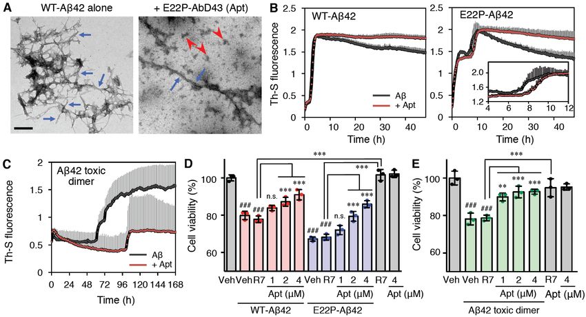
Amyloid fibrils of senile plaques in                Aβ42 has a turn at Gly25 and Ser26 (Fig. 1A:
Alzheimer’s disease (AD) consist mainly of the           “toxic conformation theory”) (17,18). The E22P
40-mer or 42-mer amyloid b-proteins (Ab40 and            mutation induces the toxic conformation of Aβ42
Ab42), which are produced from the Ab precursor          and Aβ40, which tends to stabilize oligomer
protein (1,2). The ability of Ab42 to aggregate          formation and promotes the neurotoxicity (Fig.
and its neurotoxicity is higher than that of Ab40        1A) (18,19). We recently developed a covalently
(3). Cumulative evidence shows that metastable           linked dimer model of E22P-Aβ42 using an
oligomers of Ab42 can induce neuronal death and          L,L-2,6-diaminopimelic acid (DAP) linker at
cognitive dysfunction, which is in contrast to           Val40 in the C-terminal hydrophobic core (Fig.
end-stage mature fibrils whose contribution to AD        1A, E22P-V40DAP-Aβ42 dimer), which plays an
pathology is lower (4). Notably, these toxic             important role in the formation of toxic oligomers.
oligomers include protofibrils (PFs) or                  This dimer model produced quasi-stable PFs,
higher-order oligomers with a curvilinear                which were toxic oligomers, and had a neurotoxic
structure (5).       Although some less toxic            effect on SH-SY5Y human neuroblastoma cells
higher-order oligomers exist, most of the                following incubation (20). The current report
higher-order oligomers such as PFs (6,7), the            describes a comprehensive study on the
                                                         development of RNA aptamers with potent
Ab-derived      diffusible   ligands    (8),   and

                                                                                                               Downloaded from http://www.jbc.org/ by guest on October 11, 2020
                                                         affinity for the toxic dimer model of Aβ42
amylospheroid (9) are potently neurotoxic. “Toxic
                                                         (E22P-V40DAP-Aβ42 dimer), and we discuss
oligomers” are defined in this work as the
                                                         their application to therapeutics and diagnostics
constituents of these higher-order oligomers (24–
                                                         based on experiments using an AD mouse brain.
700-mer) that are neurotoxic. The development
of oligomer-specific inhibitors is imperative for        RESULTS
making meaningful progress toward developing
                                                         Isolation of RNA Aptamers Targeting the
AD therapies without adverse effects.
                                                         Pre-incubated E22P-V40DAP-Aβ42 Dimer and
     Aptamers are single-stranded DNA or RNA
                                                         Their Recognition toward PFs
oligonucleotides used for molecular recognition of            As a model target molecule for the
diverse targets for diagnostic and therapeutic
                                                         development of an aptamer toward PFs, the
applications (10). However, current application of
                                                         E22P-V40DAP-Aβ42 dimer (9 kDa) was initially
aptamers to the field of Aβ research remains             used as a model target for selection of candidate
limited. There are several published aptamers
                                                         aptamers. The selection was performed using a
targeting Aβ including RNA aptamers targeting
                                                         column where an E22P-V40DAP-Aβ42 dimer
Aβ40 fibrils (11,12), RNA aptamers targeting
                                                         biotinylated at the N-terminus (termed the
other amyloid fibrils and Aβ40 fibrils (13), DNA         biotin-E22P-V40DAP-Aβ42            dimer)     was
aptamers developed against a-synuclein but               immobilized, and the binding rate of RNA
recognizing Aβ40 oligomers (14) and DNA                  nucleotides to Aβ was calculated by real-time
aptamers targeting Aβ40 oligomers (15). Because          PCR. However, RNA pools failed to be enriched
more aggregative Aβ42 exists in a more dynamic           even after several rounds of SELEX (data not
conformational equilibrium of monomeric Aβ42             shown). Pre-incubation (37°C, 24 h) with the
(16), it is difficult to isolate and characterize        E22P-V40DAP-Aβ42 dimer that generates a
aptamers targeting the short-lived, metastable           lower molecular weight oligomer of Aβ42 than
structures of toxic Aβ42 oligomers, unlike               PFs was also unsuccessful (data not shown).
less-toxic Aβ40 oligomers, for in vitro selection             To further enlarge the molecular weight of
(known as the systematic evolution of ligands by         the target for potential selection, an extended
exponential enrichment, SELEX).                          pre-incubation        period        with       the
    Based on solid-state nuclear magnetic                E22P-V40DAP-Aβ42 dimer (37°C, 48 h) was
resonance (NMR) and systematic proline                   next used for the selection process in combination
replacement, Irie and colleagues identified the          with the method of membrane filtering (molecular
toxic conformer of Aβ42 to have a turn at Glu22          weight cut-off: 50 kDa). As reported previously
and Asp23, whereas the non-toxic conformer of

                                                     2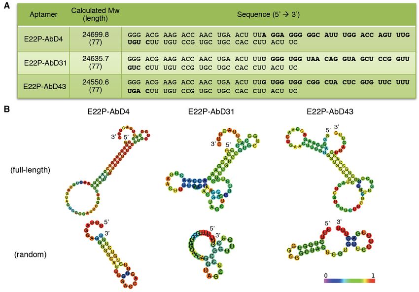
(20), the biotin-E22P-V40DAP-Aβ42 dimer                  (22) (Fig. S3A) and Multiple EM for Motif
formed PFs after incubation for 48 h, as                 Elucidation (MEME) (23) (Fig. S3B).
determined by transmission electron microscopy                Aptamers can fold into various secondary
(TEM) analysis (Fig. S1A). The RNA-binding               structures that exhibit high binding capacity, such
ratio gradually increased after seven rounds of          as stem-loop, G-quadruplex and three-way
selection, meaning that the RNA pool (ca. 26             junction structures (10). Given the slightly higher
kDa) was effectively enriched (Fig. 1B). Given           content of guanine in the obtained aptamers (Fig.
unsuccessful enrichment in the two trials stated         2A: E22P-AbD4, 29%; E22P-AbD31, 27%;
above using the Aβ-immobilized column, it                E22P-AbD43, 26%), parallel, anti-parallel and
should be noted that the use of membrane filtering       hybrid G-quadruplex structures are predicted for
(Fig. S2) may be more effective for the selection        these aptamers (24,25). Based on circular
of aptamers targeting oligomeric assemblies              dichroism (CD) spectra used to analyze their
including ordered structures such as PFs. The            secondary structure, E22P-AbD4, -AbD31 and
column matrix may have prevented association of          -AbD43 exhibited a negative peak around 240 nm
dimer model, which could be required for the             and a positive peak around 265 nm, suggesting
enrichment of aptamers. Alternatively, steric            the RNA aptamers formed G-quadruplexes (Fig.
hindrance of PFs at the N-terminal region may            3A), where four guanines are held in a square

                                                                                                                Downloaded from http://www.jbc.org/ by guest on October 11, 2020
have affected the selection process.                     planar G-tetrad arrangement through Hoogsteen
     After cloning of the enriched RNA pools             hydrogen bonding (25).
after round seven, 36 identical monoclones were               The random region sequence of each aptamer
obtained. This was followed by binding tests             was prepared to elucidate the structure of these
based on the membrane filter method. As shown            RNA aptamers. Although the ellipticity of each
in Fig. 1C, only three monoclones (E22P-AbD4,            random region sequence at the maximum
-AbD31 and -AbD43) showed higher affinity                absorption wavelength (~265 nm) decreased, the
toward the PFs than the enriched RNA pool after          difference in the ellipticity of E22P-AbD43
round seven (R7). The binding of the initial             between the full-length sequence and the random
RNA pool (R0) to the PFs (i.e., before selection)        region sequence was relatively small when
was confirmed to be negligible.                          compared with the results for the other two
     Since the Aβ42 monomer can partially form           aptamers (Fig. 3A).
the toxic conformation (18), there was the                    Next,     bio-layer    interferometry    (BLI)
possibility that the obtained monoclones would           measurements were performed to determine the
also bind to fibrils produced from wild-type Aβ42        kinetics and affinity of the interaction between the
or E22P-Aβ42. Using a membrane filter, all three         RNA aptamer and Aβ. For this purpose, the
aptamers (E22P-AbD4, -AbD31 and -AbD43)                  5'-biotinylated RNA clones were immobilized
were shown to exhibit significant preferential           onto a streptavidin biosensor and Aβ was used as
binding toward PFs when compared with these              the analyte. We observed highly stable complexes
two types of fibrils (Fig. 1D).                          for all three aptamers with PFs, characterized by
                                                         dissociation constants (KD) in the nanomolar
G-quadruplex Structure of RNA Aptamers is                range using the appropriate fitting of the 2:1
Involved in Their Binding to PFs                         heterogeneous ligand binding model of the
     CentroidFold was used to predict the folding        full-length sequence (77 nt) contingent on the
of the RNA aptamers (21). As shown in Fig. 2B,           lower chi-square coefficient (Fig. S4A). Table S1
the 77-nt RNA sequences (Fig. 2A) of                     provides a summary of the binding kinetics
E22P-AbD4, -AbD31 and -AbD43 folded to                   between the three aptamers and PFs.
primarily form stem-loop intramolecular base                  The dissociation constant (KD) of the random
pairing as a hairpin structure. Alignment search,        region sequence (29 nt) of E22P-AbD4, -AbD31
classification and local supermotif analysis did         and -AbD43 for Aβ as E22P-AbD4(Ran29),
not find a specific common domain among these            -AbD31(Ran29) and -AbD43(Ran29) was derived
aptamers based on analysis with Clustal Omega            from curve fitting of the data with a 1:1

                                                     3
interaction contingent on the lower chi-square          wild-type Aβ42 fibril (Fig. 3B). These results
coefficient. The truncation of E22P-AbD4 and            suggest the preferable binding of E22P-AbD43 to
-AbD31 moderately enhanced the affinity,                toxic Aβ42 aggregates rather than less-toxic Aβ40
whereas the affinity of E22P-AbD43(Ran29) was           aggregates.
lower than that of full-length E22P-AbD43 (Fig.              Additional binding assay using fluorescence
S4A, Fig. S5 and Table S1). Notably, the constant       polarization for E22P-AbD43 showed the similar
regions may assist the binding of the                   tendency (Fig. S7; KD = 54 ± 1.1 nM for Aβ42
E22P-AbD43 aptamer. An alternative explanation          toxic dimer, KD = 160 ± 1.4 nM for wild-type
might be that the constant regions are occluding        Aβ42, KD = 120 ± 1.2 nM for E22P-Aβ42).
the random fragment region of E22P-AbD4 and             E22P-AbD43 did not bind to wild-type Aβ42
E22P-AbD31 but not that of E22P-AbD43.                  fibril. The slight difference of KD values between
Comparing the CD absorption data for the                BLI and fluorescence polarization experiments
full-length and random region of E22P-AbD43             might be ascribed to the presence or absence of
(Fig. 3A), the preferential binding of                  immobilization of one side to the support. The
E22P-AbD43 toward PFs might be partly related           immobilization of analytes/ligands might help
to the formation of a G-quadruplex structure.           stabilize their conformation to modulate the
Attenuated total reflection-Fourier transform           binding. The advantages and limitations of these

                                                                                                             Downloaded from http://www.jbc.org/ by guest on October 11, 2020
infrared spectroscopy (ATR-FTIR) (Fig. S6) of           methods were compared by Rossi, A. M. and
E22P-AbD43 showed absorptions at 1649                   Taylor, C. W. (29).
(guanine carbonyl group) cm-1, which is
suggestive of the possible presence of a                E22P-AbD43 Prevents the Formation of Aβ42
G-quadruplex structure according to previous            Aggregates and their Neurotoxicity
combination studies with vibrational CD (26)                 Because the binding affinity of E22P-AbD43
using anti-Aβ aptamer forming G-quadruplex as a         toward PFs (KD = 150 ± 11 nM) was the most
reference (14,27).                                      potent among the three aptamers examined (Fig.
                                                        S4A and Table S1), we selected full-length
Characterization of E22P-AbD43 for Its                  E22P-AbD43 for the following studies on anti-Aβ
Preferential Recognition Unit of PFs                    properties. The full-length sequence, which
     To obtain information on how E22P-AbD43            should be more resistant to degradation, is also
can preferentially recognize PFs, BLI tests were        more appropriate for cell culture tests than the
conducted using wild-type Aβ42, E22P-Aβ42 and           corresponding random region. We next
the E22P-V40DAP-Aβ42 dimer as analytes. As              determined whether E22P-AbD43 could prevent
shown in Fig. 3B, E22P-AbD43 demonstrated 2–8           the aggregation of wild-type Aβ42. Using TEM,
fold stronger binding affinity toward the dimer         wild-type Aβ42 was shown to form typical long
model (KD1 = 20 ± 6.0 nM) than toward both              amyloid fibrils, whereas in the presence of
monomers of wild-type Aβ42 (KD1 = 80 ± 8.3              E22P-AbD43 wild-type Aβ42 formed short or
nM) or E22P-Aβ42 (KD1 =50 ± 5.0 nM),                    globular (curvilinear) aggregates (Fig. 4A).
indicating that a minimum unit of recognition of             Naiki    et     al.   have      proposed   a
PFs by E22P-AbD43 is the dimer. Table S1                nucleation-dependent polymerization model for
provides a summary of the binding kinetics              Aβ42 that is composed of nucleation and
between the E22P-AbD43 and each Aβ42.                   elongation phases. During the nucleation phase,
     Furthermore, the binding affinity of               the Aβ42 monomer gradually forms low
E22P-AbD43 to wild-type Aβ40 and PFs prepared           molecular weight oligomers (called “nuclei”),
from wild-type Aβ40 basically according to the          followed by the elongation phase where each
developed protocol (28) was weaker than                 nucleus acts as a template and associates with
wild-type Aβ42 and PFs model produced from the          monomers for polymerization and the subsequent
E22P-V40DAP-Aβ42 dimer, respectively (Fig.              formation of higher-order oligomers and fibrils
S4B). In addition, E22P-AbD43 did not react with        (30). The thioflavin-T (Th-T) assay (31) is often
wild-type Aβ40 fibril (Fig. S4B) similar to             used to quantify the inhibition of Aβ42

                                                    4
aggregation by test compounds, but RNA evaluation of Aβ oligomers using E22P-AbD43
nucleotides themselves enhance the fluorescence based on the previous study (20), the
of the Th-T reagent (32). The thioflavin-S (Th-S) neurotoxicity caused by the E22P-V40DAP-Aβ42
can be used instead of Th-T because Th-S dimer was significantly reduced by E22P-AbD43
exhibits almost no non-specific interaction with (1, 2, and 4 µM) in a dose-dependent manner (Fig.
RNA. 4E). The aptamer itself at the maximum
In the Th-S fluorescence assay performed in concentration (4 µM) was inactive. E22P-AbD43
the present study, the peptides were used at 10 was also significantly protective against the
µM effective concentration of the monomer (i.e., neurotoxicity of wild-type Aβ42 and E22P-Aβ42
the monomer was used at 10 µM and the dimer after a 16-h incubation (Fig. 4D). The R7 pool of
was used at 5 µM). Both wild-type Aβ42 (0–2 h RNA exhibited almost no alternation on the
and 2–6 h) and E22P-Aβ42 (0–4 h and 4–12 h) cytotoxicity of all Aβ samples tested (wild-type
exhibited two sigmoid-like curve points in the Aβ42, E22P-Aβ42, and E22P-V40DAP-Aβ42
process of Th-S fluorescence. These curves may dimer) (Fig. 4D–E). These findings indicate that
reflect a conformational transition or equilibrium E22P-AbD43 can preferentially delay the onset of
during the nucleation of Aβ. E22P-AbD43 nuclei-driven toxic oligomerization in the
moderately retarded the nucleation phase (4–12 h) pathogenesis of AD.
Downloaded from http://www.jbc.org/ by guest on October 11, 2020
of E22P-Aβ42 but it did not significantly slow the
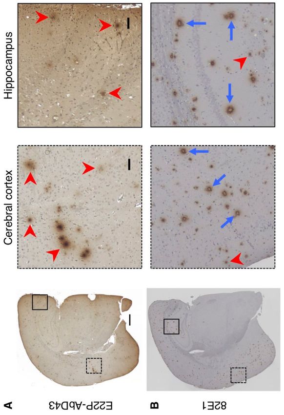
nucleation of wild-type Aβ42 (Fig. 4B). E22P-AbD43 Recognizes Diffuse Aggregates
Incidentally, a known drawback of the Th-T or Rather Than Senile Plaques in an AD Mouse
Th-S method is the gradual decrease in Th-T or We next performed histochemical analysis of
Th-S signal during the later stage of the E22P-AbD43 using an AD mouse brain
elongation phase of Aβ, possibly because of the (Tg2576/PS2) model (33) at the age of 6 months.
limited surface area of mature fibrils with a high E22P-AbD43 recognized diffuse aggregates of Aβ
core density to which Th-T or Th-S can bind. This mainly in the cerebral cortex and hippocampus
may explain why the signals from the elongation regions (Fig. 5A), whereas an anti-Aβ-N-terminus
phase of both wild-type Aβ42 and E22P-Aβ42 in antibody (82E1) (34) mainly stained senile
the presence of E22P-AbD43 did not decrease plaques with dense cores (Fig. 5B). Given that the
(Fig. 4B). Furthermore, to evaluate the effect of diffuse halo surrounding the plaque cores contains
E22P-AbD43 on the nucleation phase of Aβ42, immature or oligomeric aggregates (35), the
the E22P-V40DAP-Aβ42 dimer was used. diffuse aggregates detected by E22P-AbD43
Notably, E22P-AbD43 delayed the nucleation of might have originated from PFs or higher-order
the E22P-V40DAP-Aβ42 dimer by almost 2 days oligomers with curvilinear structures derived
(Fig. 4C). These findings suggest that from Aβ, as likely observed in a previous report
E22P-AbD43 may prevent the formation of PFs (36). Interestingly, 82E1 reacted with diffuse
by interacting with the E22P-V40DAP-Aβ42 aggregates and senile plaques, possibly because
dimer. the N-termini of the PFs were exposed.
Because E22P-AbD43 may interact with PFs Noteworthy, E22P-AbD43 exclusively bound to
during the nucleation phase, we investigated the diffuse aggregates both in the cerebral cortex and
effect of E22P-AbD43 on the neurotoxicity of the hippocampus based on counting the numbers of
E22P-V40DAP-Aβ42 dimer as well as wild-type diffuse aggregates and senile plaques (Fig. S8).
Aβ42 and E22P-Aβ42. For this purpose, we used Using multiphoton microscopy, Farrar et al. (37)
cultures of SH-SY5Y human neuroblastoma cells demonstrated that the β55 aptamer developed by
that are a typical model of neuronal cells (Fig. Ylera et al. (11) simultaneously stained the
4D–E). The peptides were added to the culture diffuse halos of the amyloid of angiopathic
medium at 1 µM effective concentration of the lesions as well as dense-core Aβ plaques in
monomer (i.e., the monomer was used at 1 µM Tg2576/PS1 mice.
and the dimer was used at 0.5 µM). After
incubation for 16 h, which is appropriate for the
5DISCUSSION histochemical analysis that indicate that senile
In the field of AD research, antibodies plaques composed of amyloid fibrils inherently
against Aβ have entered clinical trials as potential contain mRNA (41,42). To conclude, the present
therapeutics and diagnostics. Irie and colleagues findings demonstrate the development of RNA
recently developed 11A1 (38) and 24B3 (20) aptamers against PFs prepared from the
antibodies, which target the toxic conformation E22P-V40DAP-Aβ42 dimer with a toxic
and toxic oligomers of Aβ42, respectively. Both conformation as the smallest unit of PFs, and their
of these toxic moieties of Aβ42 include a turn possible application as therapeutic and diagnostic
structure at Glu22 and Asp23. Aβ40 can also form tools for AD was evaluated. The relatively
toxic oligomers derived from a toxic stronger recognition by E22P-AbD43 of the Aβ42
conformation, as exemplified by the work of Irie dimer model rather than PFs (Fig. 3B, Fig. S4A
et al. (39), and 11A1 also binds to Aβ40, albeit at and Table S1) indicates that Aβ42 dimers
a slightly lower affinity toward Aβ42 (38). dissociate at equilibrium in PFs, because the
Despite various advances in antibody research, a formation of soluble PFs is a reversible reaction
weakness of antibodies and protein-based drugs is (5). This is the first report of RNA aptamers that
their potential inherent immunogenicity because target the toxic dimer of Aβ42. The affinity (KD =
of their chronic usage in animals and humans. 20 ± 6.0 nM) of E22P-AbD43 toward Aβ42 toxic
Downloaded from http://www.jbc.org/ by guest on October 11, 2020
Aptamers are a suitable alternative with dimer may not still be sufficient for the
comparable or potentially even superior affinity therapeutic application; however, this KD value is
and selectivity when compared with that of extensively lower than any previously reported
antibodies, and RNA and DNA aptamers can be anti-Aβ aptamers (e.g. KD values of anti-Aβ40
synthesized on a larger scale at a lower cost. aptamers (12) are 10–20 µM). Further trials to
Recently, Kageyama et al. demonstrated the enhance the stability of toxic Aβ oligomers are
presence of an Aβ toxic conformer in the human currently underway.
inferior parietal cortex several decades before the Tsukakoshi et al. reported that a
onset of AD using the 11A1 antibody (40). Since telomestatin derivative increased the binding
an RNA aptamer (β55) recognizing Aβ40 fibrils capacity of G-quadruplex-forming aptamers by
(11) stained the diffuse halos of an amyloid in enhancing p-p stacking on the G-tetrad plane (27).
addition to dense-core Aβ plaques in Tg2576/PS1 Since the planarity derives from a,b-unsaturated
mice (37), E22P-AbD43 targeting more carbonyl groups in not only curcumin but also
pathogenic Aβ42 may represent an ultra-early non-catechol-type flavonoids (e.g., morin and
diagnostic tool of AD. Moreover, the strategy of datiscetin), targeting His13,14 and Phe19,20
stabilizing unstable oligomers of Aβ42 by through p-p stacking for intercalation into b-sheet
covalent bonding within the C-terminal region regions could contribute to the suppression of
followed by pre-incubation was an effective Aβ42 oligomerization (43,44). We expect the
approach to enrich RNA pools, and such a guanine bases in E22P-AbD43 could generate a
strategy may be helpful for the discovery of scaffold like a flat G-quadruplex structure (Fig.
“unstable” target-based drugs in other fields of 3A) that may target similar residues to these
amyloid research. The changes of the SELEX phytochemicals. BLI studies including the
target from a dimer-immobilized bead to analysis for the percentage contributions from KD1
non-immobilized PFs may have contributed to the and KD2 implied a 2:1 heterogeneous ligand
more efficient aptamer enrichment program used binding model as the binding mode of full-length
herein and the selection of E22P-AbD43. E22P-AbD43 to Aβ42 dimers (Table S1 and Table
Unlike RNA pools, DNA pools failed to be S2), which possibly suggests the existence of two
enriched by using pre-incubated PFs despite independent binding sites within E22P-AbD43
several trials (data not shown). Although it and the generation of complexes comprising an
remains controversial whether RNA or DNA Aβ42 dimer:E22P-AbD43 ratio of 2:1 or 1:1.
aptamers are more useful for development as Structural analysis of these complexes by
anti-Aβ aptamers, there are reports based on crystallographic analysis is now underway.
6Additionally, the formation of trimer-induced Purification of the
oligomers, which E22P-AbD43 may not readily biotin-E22P-V40DAP-Aβ42 dimer was carried
recognize, may also have an impact on out using a YMC-Pack ODS-A column (20 mm
Aβ42-induced neurotoxicity. i.d. x 150 mm; YMC) with elution at 8.0 mL/min
This report also describes the unprecedented and a 80 min exponential gradient (curve 7 in
application of RNA aptamers to histochemical Waters model 600E program) of 30–60% CH3CN
staining of AD mouse brains. Considering the containing 0.1% TFA. Subsequent purification
preferential recognition by E22P-AbD43 of was performed using an x-Bridge Peptide BEH
pre-matured aggregates of Aβ in AD mice (Fig. C18 OBDTM Prep Column (300Å, 5 µm, 19 mm
5A), the application of this aptamer in early i.d. x 150 mm; Waters, Milford, MA; USA) with
diagnosis or positron emission tomography (PET) elution at 8.0 mL/min and a 80 min exponential
imaging of amyloidogenesis for determining the gradient (curve 7 in Waters model 600E program)
progression of AD has potential. Further of 10–50% CH3CN containing 0.1% NH4OH. The
experiments are required to shorten the length of pre-incubation of the biotinylated dimer model at
the RNA aptamers to facilitate their passage across 37°C for 48 h led to the production of quasi-stable
the blood–brain barrier and their applications in PFs, and the presence of PFs was confirmed by
AD therapy. TEM (Fig. S1A). PFs were also prepared from
Downloaded from http://www.jbc.org/ by guest on October 11, 2020
wild-type Aβ40 by incubation at 37°C for 48 h
Experimental procedures basically according to the developed protocol (28)
Preparation of PFs using a Millipore Amicon Ultra-0.5 centrifugal
The E22P-V40DAP-Aβ42 dimer or filter unit (molecular weight cut off: 50 kDa,
biotin-E22P-V40DAP-Aβ42 dimer, in which Millipore). The presence of PFs from wild-type
Val40 of Aβ42 was replaced with DAP as a cross Aβ40 was also confirmed by TEM (Fig. S1B), in
linker, was synthesized using a previously which the morphology was similar to that of
described procedure (20) with a slight Teplow’s work (28). The slight difference
modification to the HPLC condition required for between Fig. S1A and S1B might be due to the
purification. Briefly, the E22P-V40DAP-Aβ42 difference of Aβ used for preparation (i.e. Fig.
dimer was purified using a YMC-Pack ODS-A S1A: biotin-E22P-V40DAP-Aβ42 dimer, Fig.
column (20 mm i.d. x 150 mm; YMC, Kyoto; S1B: wild-type Aβ40).
Japan) attached to an HPLC instrument (Waters For preparation of fibrils of wild-type Aβ40
model 600E with a 2487 UV detector) with and wild-type Aβ42, the pellets were obtained by
elution at 8.0 mL/min and a 80 min exponential centrifugation at 20,130 x g at 4°C for 1 h after
gradient (curve 7 in Waters model 600E program) each Aβ solution was incubated for 2 weeks at
of 30–60% CH3CN containing 0.1% 37°C.
trifluoroacetic acid (TFA). Subsequent serial
purification was performed using a YMC-Pack HFIP Treatment of Aβ
PROTEIN RP column (20 mm i.d. x 150 mm; For treatment with
YMC) with elution at 8.0 mL/min using an 80 1,1,1,3,3,3-hexafluoro-2-propanol (HFIP; Wako,
min linear gradient (curve 6 in Waters model Osaka; Japan), each Aβ was dissolved in HFIP at
600E program) of 20–60% CH3CN containing 1 mg/mL. After incubation at room temperature
0.1% TFA and an x-Bridge Peptide BEH C18 for 30 min, the solution was sonicated for 5 min,
OBDTM Prep Column (300Å, 5 µm, 19 mm i.d. x and dried in vacuo. The resultant Aβ film was
150 mm; Waters, Milford, MA; USA) with stored at −80°C until use.
elution at 8.0 mL/min using a 60 min linear
gradient (curve 6 in Waters model 600E program) Preparation of the Initial RNA Pool
of 15–40% CH3CN containing 0.1% NH4OH. A DNA library consisting of a randomized
Pre-incubation of the dimer model at 37°C for 48 region of 29 nucleotides (A:T:G:C =
h led to the production of quasi-stable PFs, as 25:25:25:25%), 5'- and 3'-primer regions and
observed previously (20). thymine linkers of three nucleotides between the
7randomized region and two primer regions (biotin-E22P-V40DAP-Aβ42 dimer) treated with
(library: 5'-TAG AGA TAA TAC GAC TCA HFIP was dissolved in 0.1% NH4OH at 250 µM,
CTA TAG GGA CGA AGA CCA ACT GAA C followed by 10-fold dilution with phosphate
-ttt-N29-ttt- GTC CGT GCT GCC ACC TTA CTT buffered saline (PBS; 50 mM sodium phosphate,
C-3') was designed. DNA library and both and 100 mM NaCl, pH 7.4) to a final
primers were purchased from either concentration of 25 µM. To avoid excessive
ThermoFisher or Eurofins (Tokyo; Japan). PCR aggregation of Aβ42, dimers without
was performed to amplify with the 5’-primer pre-incubation were incubated with Dynabeads
(5’-TAG AGA TAA TAC GAC TCA CTA TAG 280-M streptavidin (Veritas, Tokyo; Japan) in
GGA CGA AGA CCA ACT GAA C-3’) and PBS at 4°C for 1 h. The renatured RNA pool was
3’-primer (5’-GAA GTA AGG TGG CAG CAC incubated with the biotin-dimer-immobilized
GGA C-3’) under the following conditions: 20 streptavidin beads (RNA pool:E22P-Aβ42 dimer
cycles at 98°C for 10 s (denaturing), 53°C for 30 s = 100:400 pmol) in PBS at 4°C for 2 h. After
(annealing), and 72°C for 5 s (extension) using removing unbound RNA by washing with PBS,
the TaKaRa Ex Taq Hot Start Version kit on a Aβ was denatured by a sodium dodecyl sulfate
TaKaRa PCR Thermal Cycler Dice mini buffer (SDS buffer: 3.5 mM sodium dodecyl
(TaKaRa). Following purification of the double sulfate, 0.5 M ammonium acetate, 10 mM
Downloaded from http://www.jbc.org/ by guest on October 11, 2020
stranded, PCR-amplified DNA template with the magnesium acetate, 1 mM EDTA) at 70°C for 10
QIAquick PCR Purification Kit (QIAGEN), min. Bound RNA was purified as described above.
RiboMAX Large-Scale RNA Production Purified RNA was reverse-transcribed and
System-T7 (Promega, Madison, WI; USA) was amplified by PCR. The negative selection of the
used to generate the RNA pool for selection. RNA pool against streptavidin beads was
After phenol-chloroform extraction and desalting performed before round eight to remove
using Illustra MicroSpin G-25 columns (GE non-specific RNAs that bind the beads. Although
Healthcare), the integrity of RNA was confirmed nine rounds were used, significant enrichment
by electrophoresis using 6% Tris–borate– was not observed (data not shown).
ethylenediaminetetraacetic acid (EDTA)–urea
acrylamide gels (Invitrogen) and stained by (2) Selection using the pre-incubated Aβ42
SYBR Green (TaKaRa). RNA quantification was toxic dimer as target–The biotin-dimer treated
performed using a UV BioPhotometer with HFIP was dissolved in 0.1% NH4OH at 250
(Eppendorf). In the SELEX or binding assays (Fig. µM, followed by 10-fold dilution with PBS to a
1B–D), the ratio of bound RNA was calculated by final concentration of 25 µM.
real-time PCR (Real-time PCR CFX96, Bio-Rad) Initially, the renatured RNA pool was
using the PrimeScriptTM RT reagent Kit (Perfect incubated with the pre-incubated dimer model
Real Time, TaKaRa) and TB Green™ Premix Ex (biotin-E22P-V40DAP-Aβ42 dimer)
TaqTM II (Tli RNaseH Plus, TaKaRa). The fibrils (RNA:E22P-Aβ42 dimer = 250:1000 pmol) in
induced from wild-type Aβ42 and E22P-Aβ42 PBS at 37°C for 24 h. The resulting mixture
were synthesized as reported previously (45), and was incubated with Dynabeads M-280
were prepared so that each Aβ dissolved in 0.1% streptavidin beads (Veritas, Tokyo; Japan) in PBS
NH4OH at 250 µM, followed by 10-fold dilution at 25°C for 1 h. After removing unbound RNA by
with PBS to a final concentration of 25 µM, and washing with PBS, Aβ was denatured by the SDS
then subjected to incubation at 37°C for 48 h. The buffer at 70°C for 10 min. Bound RNA was
RNA pool or the aptamer was denatured at 90°C purified by extraction with phenol–chloroform–
for 10 min and renatured rapidly on ice for 10 min isoamyl alcohol and chloroform-isoamyl alcohol,
for refolding before use in the following studies. followed by ethanol precipitation. Purified RNA
was reverse-transcribed into cDNA by the
SELEX for RNA Aptamers Using PFs as Targets ImProm-II Reverse Transcription System
(1) Selection using Aβ42 toxic dimer as (Promega) and amplified by PCR. The negative
target–The biotin-dimer selection of RNA pool against streptavidin beads
8was performed before round three to remove containing 0.05% Tween-20 (IBL, Gunma; Japan)
non-specific RNAs that bind the beads. including various concentrations of HFIP-treated
Although five rounds were used, the significant Aβ (200–1,600 nM) with agitation under the
enrichment was not observed (data not shown). condition of association for 180 s and dissociation
for 180 s.
(3) Selection using PFs as target–The dimer The sensorgrams were better fitted globally
(E22P-V40DAP-Aβ42 dimer) treated with HFIP to a 2:1 heterogeneous ligand binding model or a
was dissolved in 0.1% NH4OH at 250 µM, 1:1 binding model contingent on the lower
followed by 10-fold dilution with PBS to a final chi-square coefficient, and the association rate kon
concentration of 25 µM. To enlarge the molecular (kon1 and kon2) the dissociation rate koff (koff1 and
weight of the oligomer, the dimers were further koff2) and KD (KD1 and KD2) were obtained by
pre-incubated at 37°C for 48 h. The formation of fitting the data through Data Analysis Software
PFs was confirmed by TEM analysis (Fig. S1A). (ForteBio) (Table S1). For a reference, the kinetic
The renatured RNA pool was incubated with parameters for kon, koff, and KD in the
mixture of PFs (RNA pool:E22P-Aβ42 dimer = corresponding binding model contingent on the
100:400 pmol) in PBS at 25°C for 1 h. By using a higher chi-square coefficient are shown in Table
Millipore Amicon Ultra-0.5 centrifugal filter unit S2. The percent contributions from KD1 and KD2
Downloaded from http://www.jbc.org/ by guest on October 11, 2020
(molecular weight cut off: 50 kDa, Millipore), were shown in the case of the 2:1 heterogeneous
bound RNA with PFs were collected. After ligand model. The contribution of KD1 were larger
washing with PBS, Aβ was denatured by the SDS than that of KD2 (Table S1 and S2). Because the
buffer at 70°C for 10 min. Bound RNA was association signal shifts of aptamer for wild-type
purified as described above. Purified RNA was Aβ42 fibril and wild-type Aβ40 fibril were below
reverse-transcribed and was amplified by PCR. the signal-to-noise, the curve fitting was not
Over the course of SELEX, the ratio of RNA:Aβ performed, and kinetic parameters were not able
was increased gradually from 1:4 to 1:1 to 2:1 in to be calculated (Table S1 and S2). In general, the
the first five rounds, next round, and final round, 2:1 heterogeneous ligand binding model simply
respectively. After seven rounds, a significant assumes binding with analyte at two independent
enrichment was observed (Fig. 1B). ligand sites of the 1:1 binding model. Each ligand
site binds the analyte independently with a
BLI Measurements different rate constant with an additional
BLI experiments were conducted at 30 °C parameter to account for percentage of binding
with an OctetRED96 (ForteBio, Menlo Park, CA; contributed by each interaction. Chi-square
USA). The biotinylation of RNA aptamers were coefficient is simply the square of the sum of
conducted by the 5’ EndTag Nucleic Acid deviations between the data and the fit curve.
Labeling System (Vector) using Other plausible and equally feasible synergistic
biotin-PEG6-maleimide (TCI, Tokyo; Japan) and 2:1 heterogeneous ligand binding models were
according to the manufacture’s protocol. The not considered to analyze the BLI binding data.
RNA oligonucleotide of the random region
sequence of E22P-AbD43 was purchased from Fluorescence Polarization
FASMAC (Kanagawa, Japan). Biotinylated RNA The fluorescence polarization binding assays
aptamers were immobilized on a streptavidin between FITC-labeled aptamer (20 nM) and
biosensor that was subjected to 10 min of HFIP-treated Aβ (10-9.2–10-6.5 M) were performed
rehydration in PBS buffer before carrying out the with a Multilabel Plate Reader ARVO X5 (Perkin
binding experiments. The immobilization of the Elmer, Waltham, MA; USA). FITC-labeled
biotinylated aptamers to the sensor was performed aptamer was prepared by the 5’ EndTag Nucleic
with 200 µL of 1 µM biotinylated aptamers in Acid Labeling System (Vector) using
96-well black plate for 840 s followed by a 180 s fluorescein-5-maleimide (TCI, Tokyo; Japan) and
incubation of the sensor in PBS buffer. The according to the manufacture’s protocol. The
binding reaction occurred in 200 µL PBS buffer samples in 200 µL of phosphatase buffered saline
9plus potassium (PBS-K; 10 mM sodium
phosphate, 140 mM NaCl, pH 7.4) were TEM
incubated at room temperature for 1 h in 96-well The aggregates of each Aβ solution (25 µM)
black plate. The excitation and emission in PBS after 24 or 48 h incubation in the absence
wavelengths were 485 and 538 nm, respcetively, or presence of E22P-AbD43 (50 µM) were
and then fluorescence polarization was calculated. examined under a TEM (JEM-1400, JEOL,
The resulting millipolarization (mP) values were Tokyo; Japan). After each Aβ aggregate was
plotted against concentration using GraphPad centrifuged (4°C, 17,900 g, 10 min), the
Prism version 6 (GraphPad Software, San Diego, supernatant was removed from the pellet. The
CA, USA). resultant pellet was gently resuspended in water
(100 µL) using a vortex for 1 min just before
Sequencing and Prediction of Secondary TEM analysis. The sample suspension (15 µL)
Structure of the RNAs was applied to a 400-mesh carbon–coated copper
After enrichment of bound RNA, the grid (thickness: 20–25 nm; Veco, Eerbeek,
individual aptamer clones were obtained by Netherlands) and incubated for 5 min before
ligation of the PCR products using the TOPO TA being negatively stained twice with 2% uranyl
cloning kit (Invitrogen). Cloned DNA was acetate. Stained samples were subjected to TEM.
Downloaded from http://www.jbc.org/ by guest on October 11, 2020
sequenced at Eurofins. Alignment search,
classification and local super-motif analysis were Th-S Aggregation Assay
performed based on sequences of 36 unique The aggregation of each Aβ in the presence
aptamers by Clustal Omega (22) (Fig. S3A) and of RNA aptamers was evaluated by a Th-S
MEME (23) (Fig. S3B). To identify common (Sigma-Aldrich, St. Louis, MO; USA)
secondary structural motifs, RNA sequences were fluorescence assay based on the Th-T assay
folded using the energy minimization algorithm developed by Naiki and Gejyo (31) with
of CentroidFold (21) (Fig. 2B). modifications, because the inherent non-specific
binding of Th-S to nucleic acids is much lower
CD Spectopolarimetry than that of Th-T (32). The procedure has been
CD spectra were measured on a JASCO described previously (43). In brief, wild-type
J-805 spectopolarimeter using a 0.1 mm Aβ42 and E22P-Aβ42 in addition to the
pathlength quartz cell, as described previously E22P-V40DAP-Aβ42 dimer were dissolved in
(20). After dilution of the RNA solution (final 0.1% NH4OH at 100 or 50 µM, and E22P-AbD43
concentration: 10 µM) with PBS containing 1 mM was dissolved in RNase-free water (Invitrogen) at
EDTA, an aliquot (200 µL) was loaded into the 200 µM, followed by dilution with PBS to the
quartz cell, and the CD spectrum was recorded desired concentration (wild-type Aβ42 and
over the wavelength range of 220–320 nm. The E22P-Aβ42, 10 µM; E22P-V40DAP-Aβ42 dimer,
spectra are shown after subtraction of the 5 µM; E22P-AbD43, 40 µM). The fluorescence
spectrum of the vehicle alone. intensity was measured by excitation at 430 nm
and emission at 485 nm using a microplate reader
ATR-FTIR (Fluoroskan Ascent, ThermoFisher, Rockford, IL;
FTIR were measured using ATR method as USA).
described previously (19). A droplet (5~10 µg) of
aptamer solution with PBS containing 1 mM MTT Assay
EDTA was loaded and then air-dried on an ATR SH-SY5Y cells (ATCC, Manassas, VA;
unit in the FTIR 470plus spectrometer (Jasco, USA) maintained in a mixed medium containing
Tokyo; Japan). To improve the signal-to-noise equal amounts of Eagle’s minimal essential
ratio, 128 scans were accumulated, and the medium (EMEM; Wako) and Ham’s F12 medium
spectra were recorded at a resolution of 4.0 cm-1. (Wako) containing 10% fetal bovine serum
The spectra are shown after suppression of water (Biological Industries), were used as a typical
vapor signal. neuronal cell model to estimate the neurotoxicity
10of each Aβ (wild-type Aβ42, E22P-Aβ42, and the temperature for 30 min. After washing with
E22P-V40DAP-Aβ42 dimer) with slight ice-cold PBS-K containing 0.02% Tween-20
modifications to the described method (20). (PBS-T), blocking was performed in a blocking
Briefly, Aβ, E22P-AbD43 or R7 were dissolved buffer, PBS-T with 10 µg/ml bovine serum
in 0.1% NH4OH or in RNase-free water to make a albumin (Nacalai) and 10 µg/ml yeast tRNA
12× stock before being diluted with culture (Nacalai, Kyoto; Japan) at room temperature for
medium to the desired final concentration. After 60 min. Biotinylated E22P-AbD43 (400 nM)
pre-incubating 120 µL of Aβ with E22P-AbD43 diluted by RNase-free water (Invitrogen) with 1
or R7 for 30 min at room temperature, the culture mM EDTA was applied at room temperature for
medium used on near-confluent cells (1 × 10⁴ 60 min, followed by reaction with horseradish
cells/well) for overnight adaptation was peroxidase (HRP)-conjugated avidin by the
exchanged with the pre-incubated solution (120 VECTORSTAIN ABC HRP Kit (Vector) for 30
µL). After incubation for 16 h at 37 °C, 15 min at room temperature. Alternatively, the
µL/well of Dye solution from the CellTiter 96 sections were treated with 82E1 (1 µg/mL)
Non-Radioactive Cell Proliferation Assay kit diluted with PBS-T at room temperature for 60
(Promega, Madison, WI; USA) was added to the min, followed by reaction with the biotinylated
cells, followed by incubation for 4 h at 37 °C. The secondary antibody for 30 min at room
Downloaded from http://www.jbc.org/ by guest on October 11, 2020
solubilization/stop solution (100 µL/well) was temperature before incubation with
subsequently added to the cells. The cell lysate HRP-conjugated avidin by the VECTORSTAIN
was incubated overnight in the dark at room ABC HRP Kit (Vector) for 30 min at room
temperature before performing measurements at temperature. To visualize the signals, brain
570 nm with a microplate reader (Multiskan FC; sections were treated with 3,3′-diaminobenzidine
ThermoFisher). The absorbance obtained by (Dojindo, Kumamoto; Japan) at 37°C for 12 h
adding the vehicle (0.1% NH4OH + RNase-free (E22P-AbD43) or at room temperature for 12 min
water) was taken as 100%. (82E1). Nuclei were stained with hematoxylin
reagent (Wako). After dehydration and soaking in
Histochemical Staining xylene, the brain sections were mounted with
All experimental procedures were performed Permount (FALMA).
in accordance with specified guidelines for the
care and use of laboratory animals, and were Statistical Analysis
approved by the Animal Care and Use Committee Statistical analysis was performed using the
of Chiba University. scientific data analysis software GraphPad Prism
Five micrometer-thick coronal version 6 (GraphPad Software) with one-way
paraffin-embedded sections were prepared from analysis of variance (ANOVA) followed by
4% paraformaldehyde-fixed brain hemispheres of Tukey’s test. Statistical significance is indicated
an AD mouse brain (Tg2576/PS2) (33) at the age in figure legends as ###p < 0.001 versus vehicle
of 6 months. After deparaffinization and alone, *p < 0.05, **p < 0.01 and ***p < 0.001.
hydration, the slices were autoclaved at 120°C for
20 min for antigen activation. To inactivate the
endogenous peroxidase, the brain sections were
soaked in methanol with 0.1% H2O2 at room
Acknowledgements: We thank Prof. Hirohide Saito at CiRA, Kyoto University for helpful discussions of
aptamer selection, Prof. Masaya Nagao and Dr. Taiho Kambe at the Graduate School of Biostudies, Kyoto
University for accessibility to real-time PCR, and Mr. Takeshi Seguchi at Primetech (Tokyo, Japan) for
technical assistance of BLI measurement. We thank Edanz Group (www.edanzediting.com/ac) for editing
a draft of this manuscript.
11Conflicts of interest: The authors declare that they have no conflicts of interest with the contents of this
article.
Author contributions: K.M. performed the experiments of RNA aptamers and DNA aptamers, designed
the study, and wrote the manuscript. Y.O. performed the experiments of RNA aptamers. A.S. and H.U.
performed the experiments of DNA aptamers. N.I. and T.S. performed the histochemistry. T.A. and
K.T. contributed to the experiment of TEM. K.I. designed the study and wrote the manuscript.
Downloaded from http://www.jbc.org/ by guest on October 11, 2020
12References
1. Glenner, G. G., and Wong, C. W. (1984) Alzheimer's disease: initial report of the purification and
characterization of a novel cerebrovascular amyloid protein. Biochem Biophys Res Commun 120,
885-890
2. Masters, C. L., Simms, G., Weinman, N. A., Multhaup, G., McDonald, B. L., and Beyreuther, K.
(1985) Amyloid plaque core protein in Alzheimer disease and Down syndrome. Proc Natl Acad
Sci U S A 82, 4245-4249
3. Haass, C., and Selkoe, D. J. (2007) Soluble protein oligomers in neurodegeneration: lessons from
the Alzheimer's amyloid β-peptide. Nat Rev Mol Cell Biol 8, 101-112
4. Roychaudhuri, R., Yang, M., Hoshi, M. M., and Teplow, D. B. (2009) Amyloid β-protein
assembly and Alzheimer disease. J Biol Chem 284, 4749-4753
5. Watanabe-Nakayama, T., Ono, K., Itami, M., Takahashi, R., Teplow, D. B., and Yamada, M.
(2016) High-speed atomic force microscopy reveals structural dynamics of amyloid β1-42
aggregates. Proc Natl Acad Sci U S A 113, 5835-5840
6. Harper, J. D., Wong, S. S., Lieber, C. M., and Lansbury, P. T. (1997) Observation of metastable
Aβ amyloid protofibrils by atomic force microscopy. Chem Biol 4, 119-125
7. Walsh, D. M., Lomakin, A., Benedek, G. B., Condron, M. M., and Teplow, D. B. (1997) Amyloid
Downloaded from http://www.jbc.org/ by guest on October 11, 2020
β-protein fibrillogenesis. Detection of a protofibrillar intermediate. J. Biol. Chem. 272,
22364-22372
8. Lambert, M. P., Barlow, A. K., Chromy, B. A., Edwards, C., Freed, R., Liosatos, M., Morgan, T.
E., Rozovsky, I., Trommer, B., Viola, K. L., Wals, P., Zhang, C., Finch, C. E., Krafft, G. A., and
Klein, W. L. (1998) Diffusible, nonfibrillar ligands derived from Aβ1-42 are potent central
nervous system neurotoxins. Proc Natl Acad Sci U S A 95, 6448-6453
9. Hoshi, M., Sato, M., Matsumoto, S., Noguchi, A., Yasutake, K., Yoshida, N., and Sato, K. (2003)
Spherical aggregates of β-amyloid (amylospheroid) show high neurotoxicity and activate tau
protein kinase I/glycogen synthase kinase-3β. Proc Natl Acad Sci U S A 100, 6370-6375
10. Zhou, J., and Rossi, J. (2017) Aptamers as targeted therapeutics: current potential and challenges.
Nat Rev Drug Discov 16, 181-202
11. Ylera, F., Lurz, R., Erdmann, V. A., and Furste, J. P. (2002) Selection of RNA aptamers to the
Alzheimer's disease amyloid peptide. Biochem Biophys Res Commun 290, 1583-1588
12. Takahashi, T., Tada, K., and Mihara, H. (2009) RNA aptamers selected against amyloid β-peptide
(Aβ) inhibit the aggregation of Aβ. Mol Biosyst 5, 986-991
13. Rahimi, F., Murakami, K., Summers, J. L., Chen, C. H., and Bitan, G. (2009) RNA aptamers
generated against oligomeric Aβ40 recognize common amyloid aptatopes with low specificity but
high sensitivity. PLoS One 4, e7694
14. Tsukakoshi, K., Abe, K., Sode, K., and Ikebukuro, K. (2012) Selection of DNA aptamers that
recognize a-synuclein oligomers using a competitive screening method. Anal Chem 84,
5542-5547
15. Chakravarthy, M., AlShamaileh, H., Huang, H., Tannenberg, R. K., Chen, S., Worrall, S., Dodd, P.
R., and Veedu, R. N. (2018) Development of DNA aptamers targeting low-molecular-weight
amyloid-β peptide aggregates in vitro. Chem Commun 54, 4593-4596
16. Irie, K., Murakami, K., Masuda, Y., Morimoto, A., Ohigashi, H., Hara, H., Ohashi, R., Takegoshi,
K., Fukuda, H., Nagao, M., Shimizu, T., and Shirasawa, T. (2007) The toxic conformation of the
42-residue amyloid β peptide and its relevance to oxidative stress in Alzheimer's disease.
Mini-Rev Med Chem 7, 1001-1008
17. Morimoto, A., Irie, K., Murakami, K., Masuda, Y., Ohigashi, H., Nagao, M., Fukuda, H., Shimizu,
T., and Shirasawa, T. (2004) Analysis of the secondary structure of β-amyloid (Aβ42) fibrils by
systematic proline replacement. J Biol Chem 279, 52781-52788
1318. Masuda, Y., Uemura, S., Ohashi, R., Nakanishi, A., Takegoshi, K., Shimizu, T., Shirasawa, T., and
Irie, K. (2009) Identification of physiological and toxic conformations in Aβ42 aggregates.
ChemBioChem 10, 287-295
19. Murakami, K., Irie, K., Morimoto, A., Ohigashi, H., Shindo, M., Nagao, M., Shimizu, T., and
Shirasawa, T. (2003) Neurotoxicity and physicochemical properties of Aβ mutant peptides from
cerebral amyloid angiopathy: implication for the pathogenesis of cerebral amyloid angiopathy
and Alzheimer's disease. J Biol Chem 278, 46179-46187
20. Murakami, K., Tokuda, M., Suzuki, T., Irie, Y., Hanaki, M., Izuo, N., Monobe, Y., Akagi, K., Ishii,
R., Tatebe, H., Tokuda, T., Maeda, M., Kume, T., Shimizu, T., and Irie, K. (2016) Monoclonal
antibody with conformational specificity for a toxic conformer of amyloid β42 and its application
toward the Alzheimer's disease diagnosis. Sci Rep 6, 29038
21. Sato, K., Hamada, M., Asai, K., and Mituyama, T. (2009) CENTROIDFOLD: a web server for
RNA secondary structure prediction. Nucleic Acids Res 37, W277-280
22. Larkin, M. A., Blackshields, G., Brown, N. P., Chenna, R., McGettigan, P. A., McWilliam, H.,
Valentin, F., Wallace, I. M., Wilm, A., Lopez, R., Thompson, J. D., Gibson, T. J., and Higgins, D.
G. (2007) Clustal W and Clustal X version 2.0. Bioinformatics 23, 2947-2948
23. Bailey, T. L., and Elkan, C. (1994) Fitting a mixture model by expectation maximization to
Downloaded from http://www.jbc.org/ by guest on October 11, 2020
discover motifs in biopolymers. Proc Int Conf Intell Syst Mol Biol 2, 28-36
24. Ambrus, A., Chen, D., Dai, J., Bialis, T., Jones, R. A., and Yang, D. (2006) Human telomeric
sequence forms a hybrid-type intramolecular G-quadruplex structure with mixed
parallel/antiparallel strands in potassium solution. Nucleic Acids Res 34, 2723-2735
25. Burge, S., Parkinson, G. N., Hazel, P., Todd, A. K., and Neidle, S. (2006) Quadruplex DNA:
sequence, topology and structure. Nucleic Acids Res 34, 5402-5415
26. Andrushchenko, V., Tsankov, D., Krasteva, M., Wieser, H., and Bour, P. (2011) Spectroscopic
detection of DNA quadruplexes by vibrational circular dichroism. J Am Chem Soc 133,
15055-15064
27. Tsukakoshi, K., Ikuta, Y., Abe, K., Yoshida, W., Iida, K., Ma, Y., Nagasawa, K., Sode, K., and
Ikebukuro, K. (2016) Structural regulation by a G-quadruplex ligand increases binding abilities of
G-quadruplex-forming aptamers. Chem Commun 52, 12646-12649
28. Ono, K., Condron, M. M., Ho, L., Wang, J., Zhao, W., Pasinetti, G. M., and Teplow, D. B. (2008)
Effects of grape seed-derived polyphenols on amyloid β-protein self-assembly and cytotoxicity. J
Biol Chem 283, 32176-32187
29. Rossi, A. M., and Taylor, C. W. (2011) Analysis of protein-ligand interactions by fluorescence
polarization. Nat Protoc 6, 365-387
30. Serio, T. R., Cashikar, A. G., Kowal, A. S., Sawicki, G. J., Moslehi, J. J., Serpell, L., Arnsdorf, M.
F., and Lindquist, S. L. (2000) Nucleated conformational conversion and the replication of
conformational information by a prion determinant. Science 289, 1317-1321
31. Naiki, H., and Gejyo, F. (1999) Kinetic analysis of amyloid fibril formation. Methods Enzymol
309, 305-318
32. Sugimoto, S., Arita-Morioka, K., Mizunoe, Y., Yamanaka, K., and Ogura, T. (2015) Thioflavin T
as a fluorescence probe for monitoring RNA metabolism at molecular and cellular levels. Nucleic
Acids Res 43, e92
33. Toda, T., Noda, Y., Ito, G., Maeda, M., and Shimizu, T. (2011) Presenilin-2 mutation causes early
amyloid accumulation and memory impairment in a transgenic mouse model of Alzheimer's
disease. J Biomed Biotechnol 2011, 617974
34. Horikoshi, Y., Mori, T., Maeda, M., Kinoshita, N., Sato, K., and Yamaguchi, H. (2004) Aβ
N-terminal-end specific antibody reduced β-amyloid in Alzheimer-model mice. Biochem Biophys
Res Commun 325, 384-387
1435. Koffie, R. M., Meyer-Luehmann, M., Hashimoto, T., Adams, K. W., Mielke, M. L., Garcia-Alloza,
M., Micheva, K. D., Smith, S. J., Kim, M. L., Lee, V. M., Hyman, B. T., and Spires-Jones, T. L.
(2009) Oligomeric amyloid β associates with postsynaptic densities and correlates with excitatory
synapse loss near senile plaques. Proc Natl Acad Sci U S A 106, 4012-4017
36. Kayed, R., Head, E., Thompson, J. L., McIntire, T. M., Milton, S. C., Cotman, C. W., and Glabe,
C. G. (2003) Common structure of soluble amyloid oligomers implies common mechanism of
pathogenesis. Science 300, 486-489
37. Farrar, C. T., William, C. M., Hudry, E., Hashimoto, T., and Hyman, B. T. (2014) RNA aptamer
probes as optical imaging agents for the detection of amyloid plaques. PloS one 9, e89901
38. Murakami, K., Horikoshi-Sakuraba, Y., Murata, N., Noda, Y., Masuda, Y., Kinoshita, N., Hatsuta,
H., Murayama, S., Shirasawa, T., Shimizu, T., and Irie, K. (2010) Monoclonal antibody against
the turn of the 42-residue amyloid β protein at positions 22 and 23. ACS Chem Neurosci 1,
747-756
39. Irie, Y., Murakami, K., Hanaki, M., Hanaki, Y., Suzuki, T., Monobe, Y., Takai, T., Akagi, K. I.,
Kawase, T., Hirose, K., and Irie, K. (2017) Synthetic models of quasi-stable amyloid β40
oligomers with significant neurotoxicity. ACS Chem Neurosci 8, 807-816
40. Kageyama, Y., Saito, A., Pletnikova, O., Rudow, G. L., Irie, Y., An, Y., Murakami, K., Irie, K.,
Downloaded from http://www.jbc.org/ by guest on October 11, 2020
Resnick, S. M., Fowler, D. R., Martin, L. J., and Troncoso, J. C. (2018) Amyloid β toxic
conformer has dynamic localization in the human inferior parietal cortex in absence of amyloid
plaques. Sci Rep 8, 16895
41. Ginsberg, S. D., Galvin, J. E., Chiu, T. S., Lee, V. M., Masliah, E., and Trojanowski, J. Q. (1998)
RNA sequestration to pathological lesions of neurodegenerative diseases. Acta Neuropathol 96,
487-494
42. Ginsberg, S. D., Crino, P. B., Hemby, S. E., Weingarten, J. A., Lee, V. M., Eberwine, J. H., and
Trojanowski, J. Q. (1999) Predominance of neuronal mRNAs in individual Alzheimer's disease
senile plaques. Ann Neurol 45, 174-181
43. Sato, M., Murakami, K., Uno, M., Nakagawa, Y., Katayama, S., Akagi, K., Masuda, Y., Takegoshi,
K., and Irie, K. (2013) Site-specific inhibitory mechanism for amyloid β42 aggregation by
catechol-type flavonoids targeting the Lys residues. J Biol Chem 288, 23212-23224
44. Hanaki, M., Murakami, K., Akagi, K., and Irie, K. (2016) Structural insights into mechanisms for
inhibiting amyloid β42 aggregation by non-catechol-type flavonoids. Bioorg Med Chem 24,
304-313
45. Murakami, K., Irie, K., Ohigashi, H., Hara, H., Nagao, M., Shimizu, T., and Shirasawa, T. (2005)
Formation and stabilization model of the 42-mer Aβ radical: implications for the long-lasting
oxidative stress in Alzheimer's disease. J Am Chem Soc 127, 15168-15174
FOOTNOTES
This study was supported in part by the JSPS KAKENHI, grant number 26221202 to K.I. and K.M., and
22603006 to K.M.
The abbreviations used are: Aβ, amyloid β-protein; AD, Alzheimer's disease; ATR-FTIR, attenuated total
reflection-Fourier transform infrared spectroscopy; BLI, bio-layer interferometry; CD, circular dichroism;
DAP, L,L-2,6-diaminopimelic acid; EDTA, ethylenediaminetetraacetic acid; HFIP,
1,1,1,3,3,3-hexafluoro-2-propanol; HRP, horseradish peroxidase; PBS, phosphate-buffered saline; PFs,
protofibrils; SELEX, systematic evolution of ligands by exponential enrichment; TEM, transmission
electron microscopy; TFA, trifluoroacetic acid; Th-S, thioflavin-S; Th-T, thioflavin-T.
15You can also read