The vomeronasal system of the newborn capybara: a morphological and immunohistochemical study - Nature
←
→
Page content transcription
If your browser does not render page correctly, please read the page content below
www.nature.com/scientificreports
OPEN The vomeronasal
system of the newborn
capybara: a morphological
and immunohistochemical study
Mateo V. Torres 1, Irene Ortiz‑Leal 1,3, Paula R. Villamayor 1,3
, Andrea Ferreiro2,
José Luis Rois2 & Pablo Sanchez‑Quinteiro 1*
The vomeronasal system (VNS) is responsible for the perception mainly of pheromones and
kairomones. Primarily studied in laboratory rodents, it plays a crucial role in their socio-sexual
behaviour. As a wild rodent, the capybara offers a more objective and representative perspective to
understand the significance of the system in the Rodentia, avoiding the risk of extrapolating from
laboratory rodent strains, exposed to high levels of artificial selection pressure. We have studied the
main morphological and immunohistochemical features of the capybara vomeronasal organ (VNO)
and accessory olfactory bulb (AOB). The study was done in newborn individuals to investigate the
maturity of the system at this early stage. We used techniques such as histological stains, lectins-
labelling and immunohistochemical characterization of a range of proteins, including G proteins
(Gαi2, Gαo) and olfactory marking protein. As a result, we conclude that the VNS of the capybara at
birth is capable of establishing the same function as that of the adult, and that it presents unique
features as the high degree of differentiation of the AOB and the active cellular migration in the
vomeronasal epithelium. All together makes the capybara a promising model for the study of chemical
communication in the first days of life.
The vomeronasal system (VNS) is the sensorial system responsible in most vertebrates for the detection of
chemosensory signals linked to innate socio-sexual behaviours1,2. In mammals, the VNS presents a high
morphofunctional3 and genomic4 diversity among different species. The vomeronasal organ (VNO) specialises
in detecting pheromones for the purpose of reproductive behaviours such as maternal aggression and sexual
attraction5. The VNS is also involved in the recognition of major histocompatibility complex (MHC) associated
peptides6, kairomones7 and aversive m olecules8.
By performing an in-depth study of the macroscopic and microscopic morphological characteristics of the
vomeronasal system in the newborn capybara, we aimed to achieve two objectives. On the one hand, we aimed
to obtain general information regarding the vomeronasal system in a rodent model that is distinct from most
studied laboratory rodents. On the other hand, because the capybara is a precocial animal species, we aimed
to determine the degree to which the capybara vomeronasal system morphology at birth has adapted to the
requirements of a demanding socio-cognitive environment.
Most studies of the VNS have been done on laboratory rodent strains, exposed to artificial selection pressure
that do not reflect the selection pressure present in the wild. Therefore, these laboratory strains present significant
genetic and behavioural differences compared with wild rodent models9. The laboratory mouse (Mus musculus)
and rat (Rattus norvegicus) may not be representative of all animals that make up this family.
A remarkable differential feature among rodents is the altricial character of mice and rats, compared with
the precocial character presented by hystricomorphic rodents, such as the guinea pig or capybara10. Altricial
neonates lack fully developed senses and require extensive parental care, such as brooding or food provisioning.
In contrast, precocial species are born with more developed senses, require limited parental care, and can feed
1
Department of Anatomy, Animal Production, and Clinical Veterinary Sciences. Faculty of Veterinary, University of
Santiago de Compostela, Av Carballo Calero s/n, 27002 Lugo, Spain. 2Marcelle Nature Park, Outeiro de Rei, Lugo,
Spain. *These authors contributed equally: Irene Ortiz-Leal and Paula R. Villamayor. *email: pablo.sanchez@
usc.es
Scientific Reports | (2020) 10:13304 | https://doi.org/10.1038/s41598-020-69994-w 1
Vol.:(0123456789)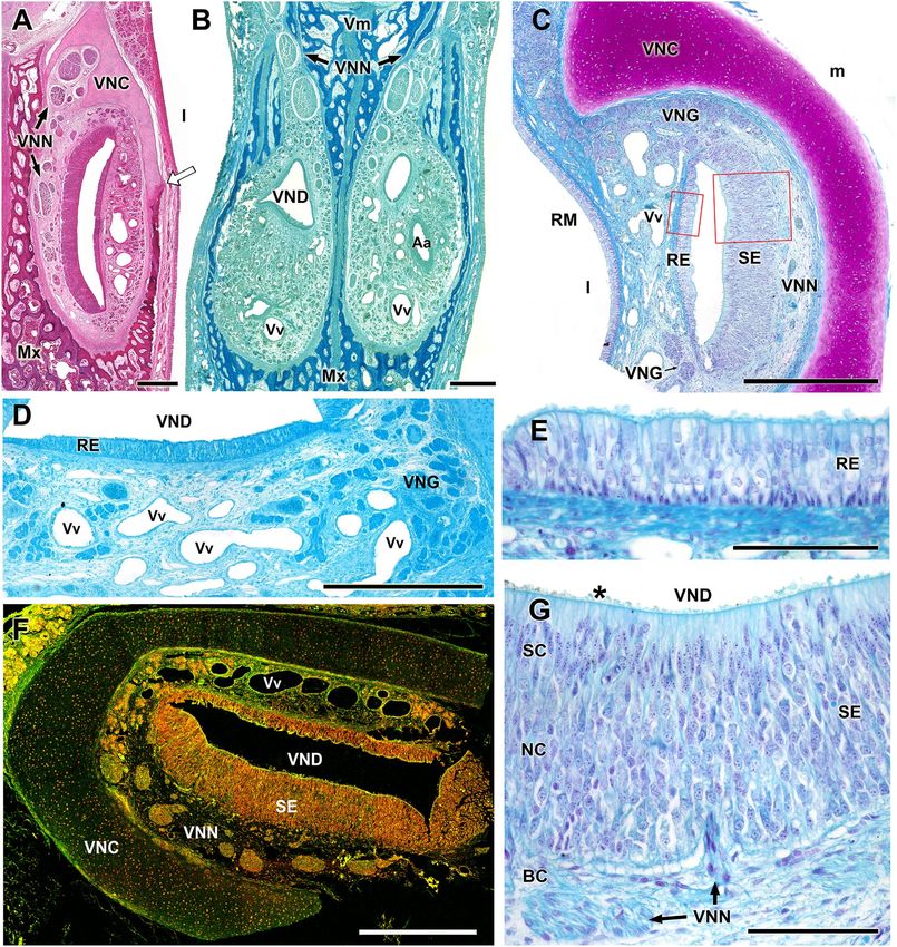
www.nature.com/scientificreports/
self-sufficiently, early on, while still being nursed11. Differences in the maturation of sensory systems between
altricial and precocial species may provide insight regarding behavioral development patterns, in both groups.
Both the main olfactory and vomeronasal systems act synergistically and constantly interact during
development12. Although the age at which the main olfactory system acquires adult-like morphology has been
well-established to demonstrate a more-organized system at an earlier postnatal age among precocial species
compared with altricial rodents13,14, less information is available regarding the VNS during the early postnatal
development of different rodent species. The development of the VNS has been exclusively assessed in imma-
ture altricial species, including the mouse and r at15,however, no studies have examined the morphological and
functional maturity of the VNS in precocial rodents during the perinatal period.
Hystricognathi has become a model group for the study of the anatomical diversity of the VNS, as shown by
the studies in c hinchillas16,17, guinea pigs18, degus19,20, and mole rats21,22. Suárez et al.23 paid special attention to the
organisation of the first integrative centre of the VNS, the accessory olfactory bulb (AOB) in capybaras Hydroch-
oerus hydrochaeris, particularly to the morphometry of the anteroposterior zonation, which is determined by the
expression of the G proteins. These authors showed how the Gαo-positive AOB caudal subdomain in capybaras
is larger than the rostral subdomain, which differs from the Octodon degus AOB, which presents a larger Gαi2
anterior region. Whereas capybaras are semi-aquatic mammals, whose chemocommunication relies mostly on
the oily secretions associated with male-to-male pheromonal communications, the degus lives in semiarid spaces
and prevalently establishes male–female interactions. Therefore, this study suggests that ecological specialisations
may play important roles in shaping the AOB.
The present study describes the anatomy, histology, and histochemical and immunohistochemical features of
the VNS of the newborn capybara and discusses its functional status at birth. We employed dissection, microdis-
section, histological staining and immunohistochemical techniques. Three lectins were studied: Ulex europaeus
agglutinin (UEA), specific for the canid vomeronasal s ystem24,Bandeiraea simplicifolia isolectin B 4 (BSI-B4),
which marks the VNS in both rats25 and opossums26,and Lycopersicon esculentum agglutinin (LEA), a specific
marker for both olfactory systems.
The immunohistochemical study covered a large number of antibodies, which provides useful information
on VNS function. Antibodies against Gαi2 and Gαo, were used to determine which pheromone receptor fami-
lies—V1R27 or V2R28, respectively—are expressed in the VNS. The mitral cells, primary neural elements of the
AOB, were labelled with antibodies against microtubule-associated protein 2 (MAP-2). The neuronal growth,
especially important during the first stages of life, was studied by employing anti-growth-associated protein 43
(GAP-43) and anti-Luteinizing hormone-releasing hormone (LHRH). The maturity of the system was determined
using anti-olfactory marker protein (OMP). The calcium-binding proteins calbindin (CB) and calretinin (CR)
were used to identify neuroactive substances. Astrocytes and ensheathing cells were recognised by an antibody
against glial fibrillary acidic protein (GFAP).
Our study aimed to address current gaps in our understanding of the rodent vomeronasal development,
through ontogeny, by providing essential information regarding the newborn capybara VNS, showing that this
species presents an advanced stage of structural maturity during the first days of life. The macroscopic, histo-
logical and immunohistochemical peculiarities and differences from the VNS of mice and rats demonstrate the
wide diversity of the VNS between even closely related species, supporting the necessity of studying each species
individually to avoid making incorrect extrapolations.
Material and methods
Through a collaboration with Marcelle Nature Park (Outeiro de Rei, Spain), we were provided with three one-
day-old capybaras (Hydrochoerus hydrochaeris) for use in this study. The animals died by perinatal causes, and
they were two males and one female.
The heads were separated and introduced into the fixative after removing the jaws and extracting the skin,
muscular plane and other structures such as the tongue and eyes. A window was opened dorsally in the skull in
the proximity of the olfactory bulbs to facilitate the penetration of the fixative. The fixatives used were 10% formol
and freshly prepared Bouin’s fixative. The latter is especially suitable for the study of the nervous system due to
its superior penetration capacity and because it lends consistency to the tissues, thus facilitating its subsequent
processing. After 24 h, the samples were transferred into 70% ethanol.
Sample extraction. We focused the extraction of the samples on the following anatomical structures: the
nasal cavity (NC), vomeronasal organ (VNO), vomeronasal nerves, and olfactory bulbs (OBs).
Nasal cavity The entire NC was separated by a transverse incision made rostrally to the ethmoidal fossa to
prevent damage to the olfactory bulbs. The resulting sample was used to study the macroscopic and microscopic
changes in the topography of the VNO throughout the NC.
Vomeronasal organ and nerves After opening the NC using a rotating saw, the dorsal and ventral turbinates
were removed. This allowed the visualisation of the nasal septum in its entirety, over which the vomeronasal
nerves were dissected. Once the VNOs were identified on both sides of the base of the anterior portion of the
nasal septum—and because of their small size and the close contact they have with the vomer bone—it was
necessary to extract them with the help of a surgical microscope (Zeiss OPMI 1 Ent).
Main and accessory olfactory bulbs The complete removal of the cranial vault was performed using a gouge
forceps. It was begun caudally to take advantage of the lower resistance presented by the bone at this level. Spe-
cial care was taken when approaching the OBs, located deep in the ethmoidal fossa, since they are extremely
delicate. To access the OBs, the bony orbital fossa, which laterally covers the bulbs was removed. Finally, using
a scalpel, the dura mater and the olfactory nerves were dissected together since both structures hold the bulbs
against the ethmoidal cribriform plate.
Scientific Reports | (2020) 10:13304 | https://doi.org/10.1038/s41598-020-69994-w 2
Vol:.(1234567890)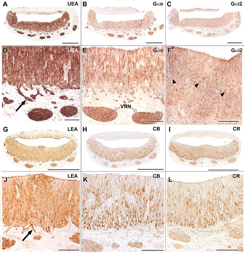
www.nature.com/scientificreports/
Ab/Lectin* 1st Ab species/dilution 1st Ab Catalogue number 2nd Ab species/dilution (Catalogue number)
Anti-Gαo Rabbit 1:100 MBL 551 ImmPRESS VR HRP Anti-Rabbit IgG Reagent MP-6401-15
Sta Cruz Biotechnology
Anti-Gαi2 Rabbit 1:100 ImmPRESS VR HRP Anti-Rabbit IgG Reagent MP-6401-15
SC-7276
Anti-OMP Goat 1:400 Wako S44-10001 Horse 1:250 Vector BA-9500
Anti-MAP2 Mouse 1:200 Sigma M4403 ImmPRESS VR HRP Anti-Mouse IgG Reagent MP-6402-15
Anti-GAP43 Mouse 1:800 Sigma G9264 ImmPRESS VR HRP Anti-Mouse IgG Reagent MP-6402-15
Anti-GFAP Rabbit 1:400 Dako Z0334 ImmPRESS VR HRP Anti-Rabbit IgG Reagent MP-6401-15
Anti-Calbindin Rabbit 1:5000 Swant CB38 ImmPRESS VR HRP Anti-Rabbit IgG Reagent MP-6401-15
Anti-Calretinin Rabbit 1:5000 Swant 7697 ImmPRESS VR HRP Anti-Rabbit IgG Reagent MP-6401-15
Anti-LHRH Rabbit 1:500 Fisher Scientific A235481 ImmPRESS VR HRP Anti-Rabbit IgG Reagent MP-6401-15
UEA-I* 1:10 Vector L-1060 Rabbit 1:50 DAKO P289
LEA* 20 µg/ml Vector B-1175 Vectastain ABC reagent PK-4000
BSI-B4* 100 µg/ml Sigma L-2140 Vectastain ABC reagent PK-4000
Table 1. Antibodies and lectins used, with species of elaboration, dilution, manufacturer, and catalogue
number. Abbreviations Gαo: Subunit αo of G protein; Gαi2: Subunit αi2 of G protein; OMP: olfactory marker
protein; MAP-2: microtubule associated protein-2; GAP-43: growth-associated protein 43; GFAP: glial
fibrillary acidic protein; CB: calbindin; CR: calretinin; LHRH: luteinizing hormone-releasing hormone; UEA:
Ulex europaeus agglutinin; LEA: Lycopersicum esculentum agglutinin; BSI-B4: Bandeiraea simplicifolia isolectin
B4; HRP: horseradish peroxidase; IgG: Immunoglobulin G; ABC: avidin–biotin-complex.
Sample processing for histological study. Paraffin embedding was used to perform the histological
processing of all samples (VNOs and OBs). In one of the individuals, the complete NC was pre-decalcified; it was
immersed in a decalcifying solution (Shandon TBD-1 Decalcifier, Thermo, Pittsburgh, PA, USA) and continu-
ously stirred for thirty hours. The samples were then washed under running water for two hours, and were cut
into several blocks which were serially cut from the incisor papilla to the caudal end of the vomeronasal cartilage
in order to obtain information on the changes in the VNO throughout its length. Following this process, all
blocks were paraffin embedded.
Cutting The samples were cut with a Leica Reichert Jung microtome with a thickness of 4–8 μm, depending
on the tissue to be processed. We opted for thinner cuts in the study of the VNO and thicker cuts in the study of
the AOB, as these allow a better visualisation of the nerve and glial processes.
General histological staining. In order to highlight the different tissue components, we used the follow-
ing stainings: Haematoxylin–Eosin (HE) as a general staining, periodic acid-Schiff (PAS) and Alcian Blue (AB)
for neutral and acid mucopolysaccharides, respectively, and Nissl staining for the brain tissue. Additionally, two
specific stainings, Gallego’s Trichrome and Tolivia, were performed.
Gallego’s trichrome This stain allows for the differentiation of components of the connective tissue. In non-
decalcified samples, it stains erythrocytes green, muscle fibres and collagen light blue, epithelium and glandular
tissue red, bone dark blue and cartilage purple. In decalcified samples, the pattern varies slightly; the glandular
tissue appears light blue, the muscle fibres and the epithelia are stained green29. The protocol used was as fol-
lows: sections were deparaffinised and rehydrated to stain with Ziehl acetic fuchsin for 2 min (10 drops Ziehl
fuchsin, 1 drop acetic acid, 10 cc distilled water). After washing them with distilled water, they were introduced
into formalin–acetic acid solution for 5 min (2 drops formalin, 2 drops glacial acetic acid, 10 cc distilled water).
After two more washes, the sections were finally introduced into picroindigocarmine for 3–5 min (one part 1%
indigocarmine aqueous solution, two parts saturated aqueous picric acid solution).
Tolivia This technique stains the myelinated nerve fibres a black colour and the neuronal somas a pink colour.
To favour the fixing of the dyes, the sections were introduced into 2.5% FeNH4(SO4)2 for 1 h. The myelin stain
solution was prepared freshly as follows: 5 ml of 20% haematoxylin plus 10 ml of 1% L i2CO3 in 50 ml of 50%
ethanol. The samples were in this solution for 2.5 h. After three washes in tap water for 5 min, the slides were
bathed for 5 min in a solution of 0.2% pyronine in 20% formaldehyde. Finally, the samples were dehydrated,
cleared and m ounted30.
Histochemical and immunohistochemical staining. Histochemical labelling (HQ) with lectins This
technique is based on the binding between certain proteins (lectins) and carbohydrates, which can be found free,
in oligosaccharides or in glycoproteins. For our study, we used (1) a lectin that comes from the gorse, the Ulex
europaeus agglutinin (UEA), which recognises α-L-fucose, (2) the α-galactose-specific BSI-B4 that comes from
Bandeiraea simplicifolia, and (3) Lycopersicon esculentum agglutinin (LEA), a lectin coming from tomato with a
high affinity for N-acetyl-β-D-glucosamine oligomers (Table 1). These stains selectively recognise the different
components of the olfactory and vomeronasal pathways in some species. They have been used in both VNO and
AOB sections.
The protocol for the UEA is as follows. It begins by (i) blocking the endogenous peroxidase activity of the
sample, avoiding possible interference with the developing solution. To do this, the sample is incubated in 3%
H2O2 solution for 10 min and then (ii) incubated for 30 min in 2% bovine serum albumin (BSA), which prevents
Scientific Reports | (2020) 10:13304 | https://doi.org/10.1038/s41598-020-69994-w 3
Vol.:(0123456789)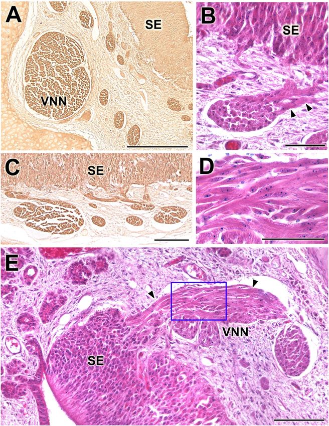
www.nature.com/scientificreports/
nonspecific binding. The next step is (iii) incubation with the UEA lectin for 1 h to visualise the lectin-carbo-
hydrate junction followed by (iv) 3 × 5 min washes in 0.1 M phosphate buffer (PB, pH 7.2), and (v) incubating
for 12 h in a peroxidase-conjugated immunoglobulin against the UEA. Finally, (vi) the sections were washed
with PB and developed by (vii) incubation of the sections in a solution of 0.05% diaminobenzidine (DAB) and
0.003% H2O2 for 5 min.
The protocol for the LEA and BSI-B4 begins with the same two steps. Next, we (iii) incubated the sections
overnight in biotinylated lectins diluted in 0.5% BSA. The next day, the samples were (iv) incubated for 1.5 h in
Vectastain ABC reagent (Vector Laboratories, Burlingame, CA, USA). The samples were finally (v) developed
by incubation in the same DAB solution as the UEA.
Immunohistochemistry (IHQ) techniques After deparaffinisation and rehydration, this protocol also began by
(i) blocking the activity of endogenous peroxidase. Later, (ii) the non-specific binding was blocked with 2.5%
horse normal serum from the ImmPRESS reagent kit Anti-mouse IgG/Anti-rabbit IgG (Vector Laboratories,
CA, USA) for 30 min. (iii) The primary antibody was then added at the corresponding dilution (Table 1) and
allowed to incubate overnight. The next day, (iv) the samples were incubated for 20 min with the corresponding
ImmPRESS VR Polymer HRP Anti-Rabbit IgG Reagent. (v) After rinsing in Tris-buffer (pH 7.61) for 10 min,
(vi) the samples were finally developed using DAB as a chromogen in the same way as for the lectins.
All immunohistochemical protocols were checked with the appropriate controls. In the absence of a posi-
tive control specific to capybaras, we replicated the entire histochemical procedure with mouse tissues known
to express the proteins of interest. Samples for which the primary antibody was omitted were used as negative
controls.
Acquisition of images and digital treatment. Digital images were taken using the Karl Zeiss Axi-
ocam MRc5 digital camera coupled to a Zeiss Axiophot microscope. Adobe Photoshop CS4 (Adobe Systems,
San Jose, CA, USA) was used as needed to adjust parameters such as brightness or contrast, balance light levels,
and crop or resize images for presentation in this work. Some photomicrographs were formed as a mosaic of
several photographs merged with an image-stitching software (PTGui Pro, New House Internet Services BV, The
Netherlands).
Ethical approval. All the animals employed in this study dead by natural causes.
Informed consent. No human subject was used in this study.
Results
The main macroscopic features of the capybara VNO are depicted in Figs. 1,2. A series of cross sections were
made along the previously decalcified nasal cavity (Fig. 1). By this method, we identified the organ located in
the central levels where the cavity is T-shaped and dorsally occupied by the turbinates (Fig. 1A). Ventrally, a
recess, formed by the presence of the roots of the incisors, is occupied by the prominent dorsal projection of the
palatine process of the incisive bone (Pp), on which both VNOs rest.
The VNO was dissected in the other two heads. After removing the roof, side walls and floor of the nasal
cavity, there were no recognisable landmarks of the organ, which is hidden under a triangular elevation in the
mucosa (Fig. 2C). A transverse section confirmed the identity of the organ (Fig. 2B). The opening of the VNO was
not visualised macroscopically, but the dorsal location of the organ suggested that it communicates directly to the
nasal cavity, and not with the incisive duct (ID) as in some other non-rodent species. This was subsequently con-
firmed by serial histological sections. The incisive duct communicates the oral cavity with the nasal cavity through
the incisive papilla. The presence of a wide palatine fissure in the skull allows this communication (Fig. 2E,F).
The brain was removed to appreciate the degree of development and the topography of the olfactory por-
tion, mainly formed by the OBs, the lateral olfactory tract and the piriform lobes (Fig. 3B). The main olfactory
bulbs (MOB) were then sectioned at the peduncle level and the anterior frontal lobe was removed to visualise
the AOB more easily. It corresponded to an oval elevation in the dorsocaudal area of the OB (Fig. 3A,C). This
was delimited by blood vessels and the arrival of the vomeronasal nerve through its medial face was visualised.
The VNO in the newborn capybaras (P0) presents a capsule, a vomeronasal duct and the parenchyma.
Capsule Well-developed, it completely envelopes the organ in its central part (Fig. 4A–C). Anteriorly, related
to the opening of the vomeronasal duct, the lateral part of the capsule is absent (Fig. 4B). Caudally, it presents
a dorsal groove for the exit of the nerves (Fig. 5B). The capsule is cartilaginous in nature but, in the caudal
third it is progressively replaced from ventrodorsally by a bone capsule, which is noticeable in the adult skull
(Fig. 2A,D). Ventrally, this bone capsule is an extension of the maxillary bone, and dorsally it is completed by
the vomer bone (Fig. 5A,B).
Vomeronasal duct The duct is semicircular in section and is laterally covered by a respiratory epithelium and
medially by a neuroreceptor epithelium, which in comparison has a significantly higher degree of development
than the former (Fig. 5C–G). In the medial epithelium, three cell strata are distinguished: the sustentacular cell
layer (SC) in the apical part, the neuroreceptor cell layer (NC) in the centre, and the basal cell layer (BC). The
duct opens into the nasal cavity directly at its most anterior end through a small hole only visible in the histo-
logical series (Fig. 4B). Ventrally, the vomeronasal duct is related to the incisive duct (Fig. 4C). The ID ends in
the incisive papilla, communicating in this way both the nasal and oral cavities. Additionally, the ID presents a
cartilaginous envelope that prevents its collapse (Fig. 4D).
Parenchyma Its main components are vessels (mostly veins and sinuses), nerves, glands and loose connective
tissue. The axons of the neuroreceptor cells converge, forming nerve fascicles (Fig. 6A) in the dorsomedial part
of the parenchyma. They leave the parenchyma through the dorsal fissure of the capsule (Fig. 5B). At higher
Scientific Reports | (2020) 10:13304 | https://doi.org/10.1038/s41598-020-69994-w 4
Vol:.(1234567890)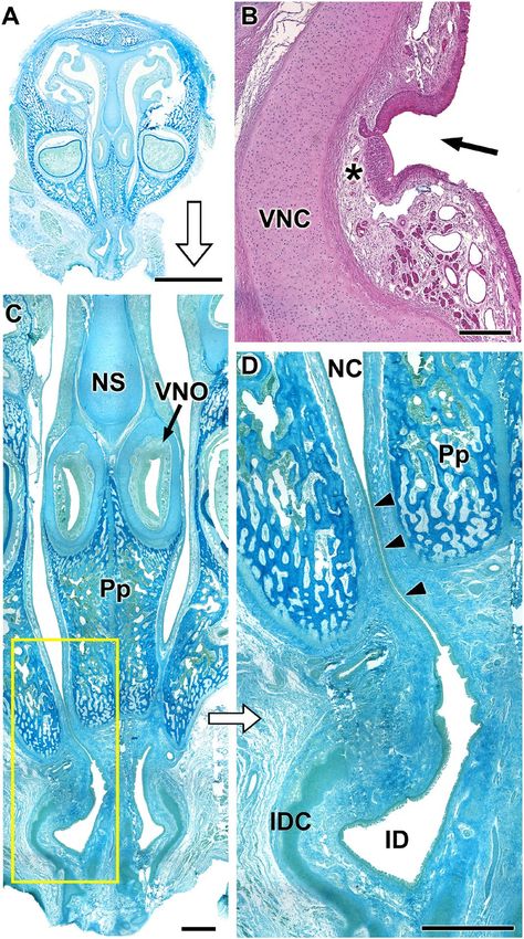
www.nature.com/scientificreports/

                            Figure 1.  Cross sections of the nasal cavity in seven levels (C) ordered from rostral (1) to caudal (7). The levels
                            of the sections are represented in the lateral view of the skull (B). The central part of the VNO is located at
                            level 4 and can be seen at greater magnification in (A). In the same image, we can see the incisive papilla (IP)
                            and the roots of the incisors (IR). At higher magnifications (D) two slits are observed that correspond to the
                            vomeronasal ducts, associated with their cartilaginous capsule (arrow). Both organs are located in the central
                            part of the nasal septum, dorsal to the incisive bone (Pp) and ventral to the cartilage of the nasal septum (NSC).
                            Scale bars: (A,C) 1 cm; (D) 0.5 cm.

                            magnifications, individual nucleated cells can be seen leaving the sensory epithelium (SE) towards the nerve
                            bundles, constituting a striking migratory stream (Fig. 6B–E). They are immunolabelled with anti-GAP-43
                            (Fig. 6A,C), whereas the labelling with anti-LHRH gave a negative result. The vasculature of the organ is poorly
                            developed, presenting veins of moderate calibre and arteries of small calibre, which only become evident in the
                            caudal zone, suggesting that the erectile character of the parenchyma is moderate (Fig. 5F). The vomeronasal
                            glands, acinar, tubular or tubuloacinar, concentrated in the dorsal area (Fig. 5D). Caudally, the ends of the organs
                            present a greater development of the glandular component (Fig. 5B). The morphology of the acini corresponds
                            to the serous type, and its secretion is AB positive and very weakly PAS positive (Fig. 5A,D).
                                The microscopic study of the AOB confirms its dorsocaudal location with respect to the MOB (Fig. 3C).
                            Through sagittal sections, the characteristic laminar structure of the AOB is identified, differentiating the follow-
                            ing strata: nervous, glomerular, external plexiform with a high degree of development, internal plexiform, a layer
                            comprising numerous mitral cells between both plexiform layers named as mitral cell layer, and a deeper granular
                            layer (Fig. 7A,B). The horizontal sections provide more information about the topography of the AOB and, in
                            them, the arrival of the vomeronasal nerve can be seen through the medial surface of the OB (Fig. 7D). Tolivia
                            staining allows the identification of abundant black myelinated fibres in the AOB granular stratum that converge
                            in the LOT. In this staining, the mitral cells are stained red and show their polyhedric structure (Fig. 7C). The
                            glomeruli in the AOB are smaller and less numerous than those in the MOB (Fig. 7E,F).

                            Lectin histochemical and immunohistochemical study. Both the UEA and the LEA lectins produce
                            an intense label in the sensory epithelium (SE) of the VNO but are negative in the respiratory epithelium (RE)

Scientific Reports |   (2020) 10:13304 |               https://doi.org/10.1038/s41598-020-69994-w                                              5

                                                                                                                                          Vol.:(0123456789)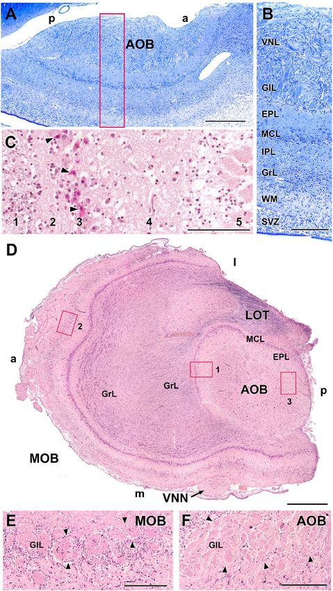
www.nature.com/scientificreports/
Figure 2. Dissection of the VNO and the incisive papilla. (A–D) The adult capybara skull gives us the first
information on the topographic features of the VNO. (D) The dorsorostral view of the skull shows the bony
structures that support the caudal third of both organs. The inset is shown in (A). (B) VNO cross section after
its extraction, bounded by the blue circle. Arrow: VNO capsule. (C) Dissection of the deep plane of the left nasal
cavity. The VNO corresponds to the triangular area in the anteroventral part of the nasal cavity. (E) Ventral view
of an adult capybara skull showing the palatine fissures (PF). (F) Roof of the oral cavity of the neonate capybara
showing the incisive papilla (IP). a: Anterior, p: posterior, d: dorsal, l: lateral, m: medial, v: ventral; In: Incisive
bone; IT: Incisor teeth; Mx: Maxillary bone; NS: Nasal septum. Scale bars: (B) 2 mm; (C) 1 cm.
(Fig. 8A,G). The UEA lectin labels all of the cells, but the LEA lectin only labels a portion of the cell population
(Fig. 8D,J). The vomeronasal nerves (VNNs) and the vomeronasal glands (VNGs) are also positive with both
markers. Anti-Gαo and anti- Gαi2 produce immunolabelling in both the SE and in the VNN, (Fig. 8B,C,E,F).
Calbindin and calretinin markers label the SE and the VNNs (Fig. 8H–I, K,L).
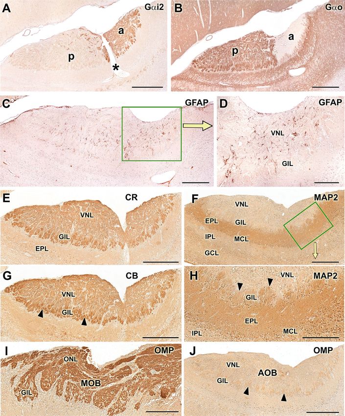
In the AOB, the anti-Gαi2 and anti-Gαo immunohistochemical labellings followed an intense and comple-
mentary pattern. The first sharply marks the nervous and glomerular layers in the anterior area of the AOB. The
anti-Gαo immunostains the same layers in the posterior area of the AOB as well as the rest of nervous tissue
(Fig. 9A,B). Immunolabelling with anti-GFAP produces a more prominent diffuse pattern in the nervous and
glomerular part of the AOB. It allows identification of the ensheathing glia of the nervous layer of the AOB, and
a significant number of astrocytes (Fig. 9C,D). Calbindin and calretinin produce a complete labelling of the AOB
without discriminating a zonation and specifically, CR revealed immunopositive mitral cell bodies (Fig. 9E,G).
Anti-MAP-2 labelling focused on both the external plexiform and glomerular layers, but the latter is labelled
with less intensity (Fig. 9F,H). The anti-OMP was strongly positive in the MOB, but in the AOB only marked
the glomerular layer (Fig. 9I,J).
UEA lectin is positive in the nervous and glomerular strata of both the AOB and MOB. In the AOB, it pro-
duces a more intense marking in the anterior area (Fig. 10A–C). The LEA lectin completely marks the AOB
without differentiating zones (Fig. 10D). Finally, the lectin BSI-B4 staining did not result in positive labelling,
in either the VNO or the AOB.
Scientific Reports | (2020) 10:13304 | https://doi.org/10.1038/s41598-020-69994-w 6
Vol:.(1234567890)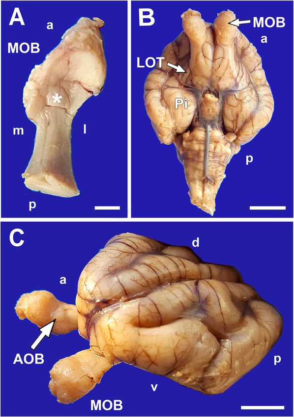
www.nature.com/scientificreports/

                            Figure 3.  Olfactory bulbs of the neonate capybara. (A) Dorsal view of the right olfactory bulb showing the
                            location of the AOB (asterisk). (B) Ventral view of the brain showing the topography of the olfactory pathway.
                            MOB: Main olfactory bulb; LOT: Lateral olfactory tract; Pi: Piriform lobe. (C) Rostrolateral view of the brain
                            where the MOB and the AOB (arrow) are differentiated. a: Anterior; p: posterior; d: dorsal; v: ventral. Scale bars:
                            (A) 2.5 mm; (B,C) 1 cm.

Scientific Reports |   (2020) 10:13304 |               https://doi.org/10.1038/s41598-020-69994-w                                             7

                                                                                                                                        Vol.:(0123456789)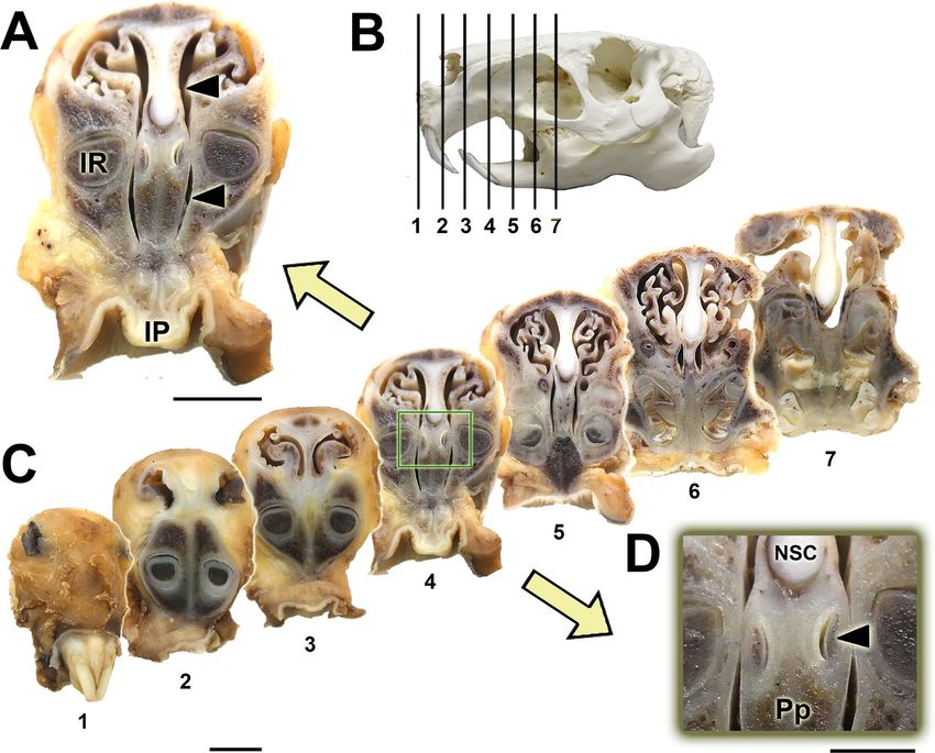
www.nature.com/scientificreports/
Figure 4. Histological images of the whole nasal cavity of the P0 capybara showing the topographical
relationships of the VNO. (A,C,D) Successive magnifications showing the location of the VNO and the
communication (arrowheads) established between the nasal and oral cavities through the incisive duct (ID).
(B) Mouth of the vomeronasal duct in the nasal cavity (arrow). At this level, the receptor epithelium (asterisk)
is already present. IDC: Incisive duct cartilage; NC: Nasal cavity; NS: Nasal septum; Pp: Palatal process of the
incisive bone; VNC: Vomeronasal cartilage; VNO: Vomeronasal organ. Stainings: (A,C,D) Gallego’s trichrome;
(B) Hematoxylin–Eosin. Scale bars: (A) 1 cm; (B) 250 µm; (C, D) 1 mm.
Discussion
There are significant structural, phylogenetic, and species-specific variations of the VNS between s pecies31. Rela-
tive to the olfactory system, the VNS has at genomic level a greater diversity4. Comparative sequence analyses
have demonstrated that MOE receptor gene sequences are well-conserved, whereas VNO receptors are associ-
ated with a wide variety of genes, suggesting a more dynamic e volution32. However, we must not forget that
relationships between these systems reflect their common history of ecological adaptations during e volution12.
In addition, pheromones are, by definition, specific for each species, and different species have different behav-
ioural and reproductive s trategies33,34. Given this huge diversity more morphofunctional studies of the VNS are
needed to understand the basis of this genetic and behavioural multiplicity.
Currently, the most studied and representative VNS models among mammals are the mouse and the rat—spe-
cies in which the VNS presents great morphofunctional development. However, these species comprise only a
Scientific Reports | (2020) 10:13304 | https://doi.org/10.1038/s41598-020-69994-w 8
Vol:.(1234567890)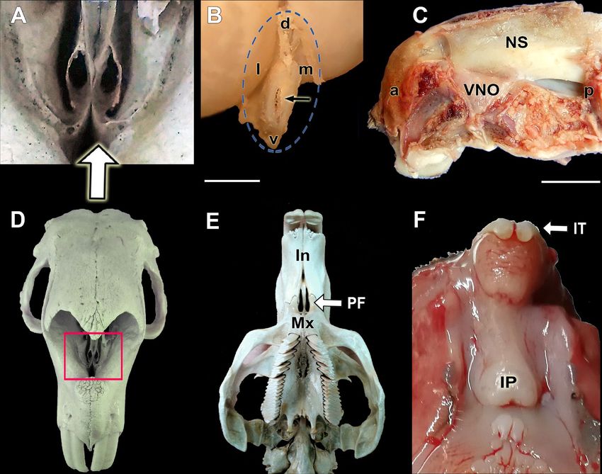
www.nature.com/scientificreports/

                            Figure 5.  Histological sections of the capybara VNO showing its main components. (A,B) Transverse sections
                            of the nasal septum exposing the nature of the vomeronasal capsule. (A) corresponds to the central level, where
                            the cartilage is replaced ventrally by the dorsal projection of the maxillary bone (white arrow). (B) corresponds
                            to a caudal level where the bone capsule fully encapsulates both VNOs. (C) Cross section of the VNO showing
                            the main components in the parenchyma: Vomeronasal duct (VND) lined medially by sensory epithelium
                            (SE) and laterally by respiratory epithelium (RE), vomeronasal glands (VNG), vomeronasal nerves (VNN),
                            vomeronasal cartilage (VNC) and veins (Vv). Both epithelia, RE and SE, insets are magnified in figures (E) and
                            (G) respectively. In the SE, the three cellular strata can be seen: SC, sustentacular cells; NC, neuroreceptor cells;
                            and BC, basal cells. The microvilli (asterisk) contact with the lumen of the vomeronasal duct. (D) Enlargement
                            of the dorsolateral area of the VNO showing the serous and AB + nature of the vomeronasal glands. (F) Study
                            of the VNO irrigation by confocal microscopy showing veins along the lateral part of parenchyma. Elastin
                            autofluorescence of a transversal section. Nuclear counterstaining with TO-PRO-3. Aa: Artery; Mx: Maxillary
                            bone; MR: Respiratory mucosa of the nasal cavity: Vm: Vomer bone; l: lateral; m: medial. Stainings: (A)
                            Hematoxylin–Eosin; (B,C,E,G) Gallego’s trichrome; (D) Alcian blue. Scale bars: (A–D) 500 µm; (E–G) 100 µm.

Scientific Reports |   (2020) 10:13304 |               https://doi.org/10.1038/s41598-020-69994-w                                                   9

                                                                                                                                           Vol.:(0123456789)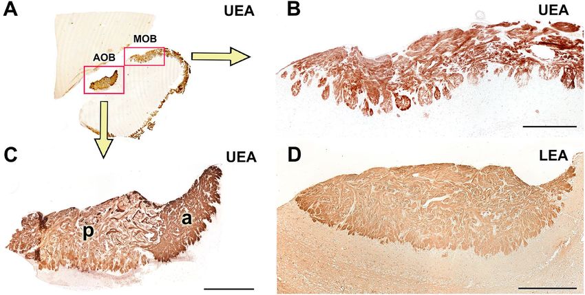
www.nature.com/scientificreports/
Figure 6. Histological study of the capybara vomeronasal nerves (VNN). (A,C) Large branches of the VNN in
the dorsomedial (A) and medial (C) areas of the VNO immunostained by anti-GAP43. (B,E) Migratory stream
of cells departing from the sensory epithelium (arrowheads). (D) Higher magnification of the inset showed in
E. Stainings: (B,D,E) Hematoxylin–eosin. SE: sensory epithelium; VNN: Vomeronasal nerves. Scale bars: (A)
250 µm; (B,D) 50 µm; (C,E) 100 µm.
small fraction of the Rodentia family; therefore, we opted to study another rodent, the capybara, to determine
whether the VNS of the mouse and rat are representative of the entire rodent family or whether significant dif-
ferences exist in other species. Moreover, most studied rodents derive from laboratory strains, that omit natural
conditions35. Studying a rodent species that has not undergone artificial selection by humans was another goal
of this study. Finally, because the capybara is a precocial species, the use of newborn individuals allowed us to
determine whether and how the VNS morphology had adapted to the requirements of a challenging environ-
ment, at birth.
Although valuable information on the expression of G proteins in the AOB of the adult capybara is available23,
we have expanded these observations, addressing the main morphological and neurochemical features of both
the AOB and the VNO. Our immunohistochemical study paid as well special attention to G proteins because new
information on their perinatal expression could be relevant to understanding the role of the VNS at this stage of
life. Adult capybara express the two families of vomeronasal receptors, V1R and V2R, which can be identified
Scientific Reports | (2020) 10:13304 | https://doi.org/10.1038/s41598-020-69994-w 10
Vol:.(1234567890)www.nature.com/scientificreports/
Figure 7. Capybara AOB microscopic study. (A) Sagittal section stained by Nissl. The laminar organisation
is visible and showed at higher magnifications in (B): Inset from (A) Vomeronasal nervous layer (VNL),
glomerular layer (GlL), external plexiform layer (EPL), mitral cell layer (MCL), internal plexiform layer (IPL),
granular cell layer (GCL), white matter (WM) and subventricular zone (SVZ). (C) Higher magnification of the
inset 1 in (D). The Tolivia staining shows the polyhedric morphology of mitral cells (arrowheads). (1) GCL;
(2) IPL; (3) MCL; (4) EPL; (5) GlL. (D) Horizontal section of the complete olfactory bulb stained with Tolivia
to identify the convergence of myelinic fibres in the lateral olfactory tract (LOT). The differences in size and
lamination of MOB and AOB and the arrival of the vomeronasal nerve (VNN) from the medial side of the
olfactory bulb are noticeable. (E) Higher magnification of the inset 2 in (D) showing the GlL of the MOB (F)
Higher magnification of the inset 3 in (D) showing the GlL of the AOB. a: Anterior; p: posterior; l: lateral; m:
medial. Scale bars: (A) 500 µm; (B,C,E,F) 250 µm; (D) 1 mm.
immunohistochemically by studying G protein e xpression23, as V1R neuroreceptor cells specifically possess the
αi2 subunit of the G proteins in their sensory transduction chain, whereas V2R cells express the αo subunit.
As G proteins play a critical role in the pheromonal signal transduction chain, their immunopositivity along
the neuroepithelium of the VNO, the VNN, and the nervous and glomerular layers of the P0 capybara AOB
points to the existence of full maturity of the VNS in the newborn capybara (Fig. 8E,F). This fact is consistent
irth36. A comparable pattern of early prenatal development
with the high degree of maturity of the offspring at b
has been found in the VNS of other precocial mammals such as pigs37 and sheep38, although in neither case was
the expression of G proteins in the VNS evaluated. The expression of G proteins observed in the AOB of the
newborn capybara (Fig. 9A,B) establish a clear anteroposterior zonation, identical to that observed in the adult
Scientific Reports | (2020) 10:13304 | https://doi.org/10.1038/s41598-020-69994-w 11
Vol.:(0123456789)www.nature.com/scientificreports/
Figure 8. Capybara VNO histochemical and immunohistochemical labelling. (A,D) UEA lectin strongly marks
both the entire sensory epithelium and vomeronasal nerves. It also allows the identification of the migratory
current (arrow). (B,E) IHC labelling with anti-Gαo stains the vomeronasal nerves (white arrows) and produces
a focally diffuse pattern in the neuroepithelium. (C,F) IHC labelling with anti-Gαi2 stains the nerve component
and marks isolated receptor cells (arrowheads). (G,J) The LEA lectin produces a label similar to the UEA
lectin, but not as thoroughly. A major part of the sensory epithelium and the vomeronasal nerves are marked.
Migration is also identified (arrow). (H,K) Anti-Calbindin (CB) produces a cellular labelling distributed in
the central and basal areas of the epithelium. (I,L) Anti-Calretinin (CR) produces a cellular labelling mainly
concentrated in the basal area of the epithelium. Scale bars: (A–C, G–I) 500 µm; (D–F) 100 µm.
OB23: the Gαi2 protein is expressed in the anterior area of the AOB and the Gαo protein in the posterior area.
A
This zonation is also discriminated by the UEA lectin (Fig. 10C), which stained the anterior part more intensely.
This pattern coincides with that observed in mice15,39. However, a later exhaustive study of UEA labelling in the
mouse olfactory system40 could only identify such AOB zonation in 3 of the 16 mice studied. The authors con-
cluded that these individual differences may be caused by the presence or absence of cues that activate receptor
cells or may reflect the differences between wild and laboratory rodents. The presence of the UEA anteroposterior
Scientific Reports | (2020) 10:13304 | https://doi.org/10.1038/s41598-020-69994-w 12
Vol:.(1234567890)www.nature.com/scientificreports/
Figure 9. Immunohistochemical labelling in the capybara AOB. (A) The IHC labelling with anti-Gαi2 stains
the nervous and glomerular layers of the anterior area of the AOB. (B) On the other hand, the anti-Gαo marks
all of the nervous tissue except the anterior part of the AOB, resulting in a complementary expression pattern of
both G proteins. (C,D) Marking with anti-GFAP produces a more prominent diffuse pattern in the nervous and
glomerular part of the AOB. (E,G) In the AOB, both anti-Calretinin (CR) and anti-Calbindin (CB) produce a
complete label more intense in the glomerular layer (arrowheads). (F,H) MAP2 labelling focuses on the external
plexiform and in the glomerular layers (arrowheads). (I,J) The anti-OMP is immunopositive in the MOB,
marking intensely the nervous and glomerular layers (GlL) whereas in the AOB the labelling is very faint, except
in the GlL (arrowheads). a: Anterior; p: posterior. Scale bars: (A–C,E–G and J) 500 µm; (D,H and I) 250 µm.
Scientific Reports | (2020) 10:13304 | https://doi.org/10.1038/s41598-020-69994-w 13
Vol.:(0123456789)www.nature.com/scientificreports/
Figure 10. Lectin histochemical labelling in the capybara AOB. (A–C) UEA lectin is positive in both the
nervous and glomerular strata of the entire olfactory bulb. In the AOB, it produces a slightly more intense
labelling in the anterior area. (D) LEA lectin is positive in the entire AOB without differentiating zones. a:
Anterior; p: posterior. Scale bars: (B) 250 µm; (C,D) 500 µm.
zonation in the capybara appears to confirm this theory regarding the differences between wild and laboratory
animals. LEA lectin (Fig. 10D) stained equally both zones, being an useful marker of both olfactory systems.
Although the small number of available individuals available prevented the realization of a rigorous statisti-
cal study, all of our newborn specimens showed a caudal portion that was larger than the rostral portion, which
demonstrated that starting at the perinatal stage, the capybara shows the bias toward a more prominent caudal
AOB that was previously described in adult individuals by Suarez et al.23,41 These results, combined with the
recent observations regarding the morphometrical parameters of the AOB, in two closely related degus species
with contrasting social h abits20, invite the hypothesis that some structural features of the AOB reflect the species
lifestyle and arise during an early stage of the ontogeny.
We undertook further immunohistochemical and morphological studies that confirmed the functionality
of the VNO and AOB at this early stage. In the VNO, the calcium-binding-proteins, calretinin and calbindin
markers (Fig. 8K,L) stained the parenchymal nerve bundles and almost all of the sensory neuroepithelial cells,
although they labelled different cell populations. Something similar happens in the shrew, where calretinin
produces an intense labelling in almost all receptor cells and in the VNNs in prenatal individuals42. Both mark-
ers produced a similar labelling on the AOB, labelling the VNL and GlL layers, although the second one more
intensely. In addition, calretinin produced a labelling of mitral cells as seen in prenatal studies in the shrew42
and adult hedgehog43.
The antibody to glial fibrillary acidic protein (Fig. 9C,D) labelled a significant number of astrocytes in the
nerve and glomerular layers of the AOB. This fact is striking when compared with other species, since in most
studies the astrocytic development at this early stage is hardly noticeable. In the case of the opossum, there is
hardly any labelling with GFAP in a P0 individual44. Only studies in P0 mice show an astrocytic development
close to that of the capybara, but it occurs in the intermediate superficial zone, which corresponds to a primitive
stage of the internal plexiform layer. Therefore, the early labelling of astrocytes in P0 capybaras reinforces the
idea that it presents a VNS with a high degree of maturity at birth.
Anti-microtubule-associated protein 2 (MAP-2) is an invaluable marker for the dendritic trees of mitral c ells46.
In the AOB of P0 capybara (Fig. 9F-H), the MAP-2 antibody produces diffuse labelling which is consistent with
that seen in other species at birth such as rats, mice and o possum44–48. Anti-olfactory-marker-protein (OMP)
produces an intense pattern of labelling in the nervous and glomerular layers of the MOB, but hardly produces
labelling in the AOB, immunostaining only the glomerular layer weakly (Fig. 9I,J). It has been observed in other
species that the nerve and glomerular layers of both bulbs are positive for the OMP marker after birth. However,
it has also been seen that OMP marking has a considerable difference in the degree of intensity between AOB
and MOB, being more intense in the case of MOB. This also happens in rodents such as the mouse or rat49 or in
marsupials such as the opossum50.
In addition to this neurochemical findings, our study in the P0 capybara has provided evidence for certain
morphological and immunohistochemical features unique to this species—for instance, the nature of the capsule
that protects both VNOs, their dorsal location in the nasal cavity over the palatal process of the incisive bone,
Scientific Reports | (2020) 10:13304 | https://doi.org/10.1038/s41598-020-69994-w 14
Vol:.(1234567890)www.nature.com/scientificreports/
the high degree of morphological differentiation of the AOB at that early stage, and finally the presence of a
migratory stream from the neuroepithelium of the VNO to the VNNs.
Macroscopically, the dorsal location of both VNOs is remarkable, as they rest on the palatal process of the
incisive bone and not on the vomer bone, as is the case in most species (Figs. 1and 4). This topography is only
comparable to that described in r abbits51, where the organs rest over a prominence of the vomer bone. Two pat-
terns of communication of the vomeronasal duct with the outside have been described. In the first, the vome-
ronasal duct opens directly into the incisive duct, as is the case in most domestic mammals such as cows, dogs,
horses and p igs52 as well as in lemurs53. But in the second, the VNO flows directly to the ventral meatus of the
nasal cavity, in its rostral area. The latter is the case for all r odents54 and lagomorphs51 studied to date. Accord-
ingly, in the capybara, both vomeronasal ducts open directly into the nasal cavity. Though in most species, the
nature of the vomeronasal capsule is exclusively cartilaginous, there are species such as the mouse in which the
capsule is formed by a thin bone s heet39 or doubly composed of cartilage and bone, as in the lagomorphs51. In
the newborn capybara, we have observed a different pattern thus far unpublished in the literature,the capsule is
initially cartilaginous, but from the central area of the organ it is progressively replaced from ventral to dorsal
by a bone sheet that ends up completely enveloping the VNO in its caudal area (Figs.2A and 5A,B).
The development and maturity of the receptor neuroepithelium in newborn individuals (Fig. 5E) resembles
the highly differentiated epithelium typical of another rodent, the r at54,55. The parenchyma presents moderate
development of blood vessels (Fig. 5F). In this way, the capybara differs from other species such as cats56 or
rabbits51, in which the development of the vascular pump is very sophisticated. The vomeronasal pump is the
physiological mechanism by which pheromones dissolved in fluids are transported to the vomeronasal d uct57,58.
The secretion of the vomeronasal glands to the duct of the organ plays a very important role in vomeronasal
perireceptor processes59,60. The newborn capybara VNO shows a significant development of the glandular tis-
sue, especially in the caudal end of the organ (Fig. 5B). However, in the central part of the duct, the presence
of glands is more moderate. This pattern is analogous to that found in other rodents such as rats, guinea pigs61
and mice62,63. However, histochemically, their character in the capybara is both PAS + and AB + , which contrasts
with that found in most rodents studied, which only express PAS + glandular secretion (rat and guinea pig64,
mouse62,65, vole66, hamster67, chinchilla16). This notorious variation in gland characteristics within the same
order may reflect an adaptation in capybaras to the aquatic nature of their habitat, which might require a specific
pheromone-receptor interaction milieu.
All previous studies on the Rodentia vomeronasal glands (VNG) applying conventional histochemical tech-
niques were done in adult specimens. There is less information regarding the prenatal and perinatal stages. We
found in the newborn capybara that the VNGs are reactive to UEA-I, a lectin specific for mucous secretions68
and to LEA, which has been linked to the presence of sialic acid and galactosamine units in the g lands25,69, and
negative for the α-galactose-specific BSI-B4 lectin. These results agree with those found in newborn mice39,
although in the latter case, the glandular development was less notable. Apart from rodents, precocial mammals
show a remarkable prenatal development of the VNG, as Salazar et al.38 found LEA + glands in sheep foetus in
the second trimester of pregnancy and in pig foetus in the fourth week of p regnancy37,70.
In the parenchyma of the newborn capybara, the VNNs are concentrated in the medial area of the organ,
forming nervous fascicles that leave the parenchyma dorsally. An exceptional point about which there is no
information in the literature is the profuse cellular migration that occurs from the sensory epithelium to the
nerve fascicles (Fig. 6). This cell migration was positive for immunohistochemical labelling with anti-GAP-43 and
histochemical labelling with both the UEA and LEA lectins, but it was negative when labelled with anti-LHRH;
this indicates that this migration is not analogous to that described in rat and mouse prenatal foetuses, which
originates from the vomeronasal part of the olfactory placode to the hypothalamus and is mostly comprised of
immunopositive LHRH cells71. It is difficult to hypothesise about the significance of these cells since this is an
unprecedented finding in both the olfactory and vomeronasal nerves. Although further studies should clarify
the nature and fate of these cells, the immunopositivity for GAP-43 suggests to their neuronal nature.
With regard to the morphology of the AOB, our observations in the newborn capybara show that from
birth, the macroscopic and microscopic organisation of the AOB are already defined, showing that the AOB of
the capybara has a differentiated laminar pattern (Fig. 7), similar to that found in other r odents39, lagomorphs,
marsupials and prosimians72–74. From the macroscopic point of view, it is remarkable that in the specimens
studied by Suárez et al.23, the arrival of the VNN to the AOB is described as occurring from the lateral side of
the bulb. Our dissections and histological series show that the arrival takes place through the medial side of the
bulb (Fig. 7D), as happens in other rodents such as rats and mice and in most of the mammals studied, even
belonging to other f amilies75,76. However, as McCotter himself describes in the guinea p ig75, a Hystricognathi
rodent like the capybara constitutes an exception to this pattern, since in this species the arrival of the nerve takes
place through the ventrolateral part of the bulb. Although we did not have access to adult individuals that could
help us to elucidate this aspect, access to the Comparative Mammalian Brain Collections from the Universities
of Wisconsin and Michigan State (https://brainmuseum.org/) allowed us to study a Nissl cross section of the
adult capybara brain (Specimen 62-621, section 160). It clearly shows the arrival of the VNN to the AOB from
the medial side of the left hemi-brain. Future studies should clarify this issue.
In summary, our observations show that after birth, the capybara VNS does possess: (1) A VNO that com-
municates directly with the nasal cavity, and indirectly with the oral cavity; (2) A VNO and an AOB that are
morphologically similar to those of the adult; (3) Active secretory vomeronasal glands; (4) The same Gαo and
Gαi2 sensitivity of the neurosensory epithelium, VNNs, and nervous and glomerular AOB layers as has been
described in adult capybara; and (5) Almost all of the neurochemical markers employed show an activity typical
of adult animals.
On the basis of these findings, the general conclusion may be drawn that the VNS of the capybara at birth is
capable of establishing the same function as that of the adult animal. If we add to this our observations on the
Scientific Reports | (2020) 10:13304 | https://doi.org/10.1038/s41598-020-69994-w 15
Vol.:(0123456789)www.nature.com/scientificreports/
degree of differentiation of the AOB and the active cellular migration that occurs in the epithelium of the VNO,
which is not described in any other mammalian species, we can conclude by considering the capybara as an
excellent and promising model for the study of chemical communication in the first days of life.
Received: 11 April 2020; Accepted: 22 July 2020
References
1. Keverne, E. B. Pheromones, vomeronasal function, and gender-specific behavior. Cell 108, 735–738 (2002).
2. Kelliher, K. R. The combined role of the main olfactory and vomeronasal systems in social communication in mammals. Horm.
Behav. 52, 561–570 (2007).
3. Meisami, E. & Bhatnagar, K. P. Structure and diversity in mammalian accessory olfactory bulb. Microsc. Res. Tech. 43, 476–499
(1998).
4. Grus, W. E. & Zhang, J. Rapid turnover and species-specificity of vomeronasal pheromone receptor genes in mice and rats. Gene
340, 303–312 (2004).
5. Martín-Sánchez, A. et al. From sexual attraction to maternal aggression: when pheromones change their behavioural significance.
Horm. Behav. 68, 65–76 (2015).
6. Leinders-Zufall, T. et al. A family of nonclassical class I MHC genes contributes to ultrasensitive chemodetection by mouse vome-
ronasal sensory neurons. J. Neurosci. 34, 5121–5133 (2014).
7. Papes, F., Logan, D. W. & Stowers, L. The vomeronasal organ mediates interspecies defensive behaviors through detection of protein
pheromone homologs. Cell 141, 692–703 (2010).
8. Bufe, B. et al. Bacterial MgrB peptide activates chemoreceptor Fpr3 in mouse accessory olfactory system and drives avoidance
behaviour. Nat. Commun. 10, 4889 (2019).
9. Smith, J., Hurst, J. L. & Barnard, C. J. Comparing behaviour in wild and laboratory strains of the house mouse: levels of comparison
and functional inference. Behav Process. 32, 79–86 (1994).
10. Derrickson, E. M. Comparative reproductive strategies of altricial and precocial eutherian mammals. Funct. Ecol. 6, 57–65 (1992).
11. Scheiber, I. B. R., Weiß, B. M., Kingma, S. A. & Komdeur, J. The importance of the altricial – precocial spectrum for social com-
plexity in mammals and birds – a review. Front. Zool. 14, 3 (2017).
12. Suárez, R., García-González, D. & de Castro, F. Mutual differences between the main olfactory and vomeronasal systems in devel-
opment and evolution. Front. Neuroanat. 6, 1–14 (2012).
13. Brunjes, P. C. Olfactory bulb maturation in Acomys cahirinus: is neural growth similar in precocial and altricial murids?. Brain
Res. 284, 335–341 (1983).
14. Leon, M., Coopersmith, R., Ulibarri, C., Porter, R. H. & Powers, J. B. Development of olfactory bulb organization in precocial and
altricial rodents. Brain Res. 314, 45–53 (1984).
15. Salazar, I. & Sánchez-Quinteiro, P. Differential development of binding sites for four lectins in the vomeronasal system of juvenile
mouse: from the sensory transduction site to the first relay stage. Brain Res. 979, 15–26 (2003).
16. Oikawa, T., Shimamura, K., Saito, T. R. & Taniguchi, K. Fine structure of the vomeronasal organ in the chinchilla (Chinchilla
laniger). Jikken Dobutsu 43, 487–497 (1994).
17. Jurcisek, J. A., Durbin, J. E., Kusewitt, D. F. & Bakaletz, L. O. Anatomy of the nasal cavity in the chinchilla. Cells Tissues Org. 174,
136–152 (2003).
18. Sugai, T., Sugitani, M. & Onoda, N. Subdivisions of the guinea-pig accessory olfactory bulb revealed by the combined method
with immunohistochemistry, electrophysiological, and optical recordings. Neuroscience 79, 871–885 (1997).
19. Suárez, R. & Mpodozis, J. Heterogeneities of size and sexual dimorphism between the subdomains of the lateral-innervated acces-
sory olfactory bulb (AOB) of Octodon degus (Rodentia: Hystricognathi). Behav. Brain Res. 198, 306–312 (2009).
20. Fernández-Aburto, P., Delgado, S. E., Sobrero, R. & Mpodozis, J. Can social behaviour drive accessory olfactory bulb asymmetries?
Sister species of caviomorph rodents as a case in point. J. Anat. 236, 612–621 (2020).
21. Dennis, J. C. et al. Is the mole rat vomeronasal organ functional?. Anat. Rec. 303, 318–329 (2020).
22. Browe, B. M., Vice, E. N. & Park, T. J. Naked mole-rats: blind, naked, and feeling no pain. Anat. Rec. 303, 77–88 (2020).
23. Suárez, R. et al. Share and differential traits in the accessory olfactory bulb of caviomorph rodents with particular reference to the
semiaquatic capybara. J. Anat. 218, 558–565 (2011).
24. Salazar, I., Cifuentes, J. M. & Sanchez-Quinteiro, P. Morphological and immunohistochemical features of the vomeronasal system
in dogs. Anat. Rec. 296, 146–155 (2013).
25. Salazar, I. & Sánchez-Quinteiro, P. Lectin binding patterns in the vomeronasal organ and accessory olfactory bulb of the rat. Anat.
Embryol. 198, 331–339 (1998).
26. Shapiro, L. S., Ee, P. L. & Halpern, M. Lectin histochemical identification of carbohydrate moieties in opossum chemosensory
systems during development, with special emphasis on VVA-identified subdivisions in the accessory olfactory bulb. J. Morphol.
224, 331–349 (1995).
27. Shinohara, H., Asano, T. & Kato, K. Differential localization of G-proteins Gi and Go in the accessory olfactory bulb of the rat. J.
Neurosci. 12, 1275–1279 (1992).
28. Jia, C. & Halpern, M. Subclasses of vomeronasal receptor neurons: differential expression of G proteins (Gi alpha 2 and G (o alpha))
and segregated projections to the accessory olfactory bulb. Brain Res. 719, 117–128 (1996).
29. Ortiz-Hidalgo, C. Abelardo Gallego (1879–1930) and his contributions to histotechnology: the Gallego stains. Acta Histochem.
113, 189–193 (2011).
30. Tolivia, J., Tolivia, D. & Navarro, A. New technique for differential staining of myelinated fibers and nerve cells on paraffin sections.
Anat. Rec. 222, 437–440 (1998).
31. Salazar, I. & Sánchez-Quinteiro, P. The risk of extrapolation in neuroanatomy: The case of the mammalian vomeronasal system.
Front. Neuroanat. 3, 22 (2009).
32. D’Oliveira Albanus, R., Siqueira Dalmolin R.J., Rybarczyk-Filho, J.L., Alves Castro, M.A. & Fonseca Moreira, J.C. Differential
evolutionary constraints in the evolution of chemoreceptors: a murine and human case study. Sci. World J. 23, 696485 (2014).
33. Keverne, E. B. Odor here, odor there: chemosensation and reproductive function. Nat. Neurosci. 8, 1637–1638 (2005).
34. Brennan, P. A. & Zufall, F. Pheromonal communication in vertebrates. Nature 444, 308–315 (2006).
35. Harr, B. et al. Genomic resources for wild populations of the house mouse, Mus musculus and its close relative Mus spretus. Sci.
Data 3, 160075 (2016).
36. Miglino, M. A., Dos Santos, T. C., Kanashiro, C. & Dos Santos Ferraz, R. H. Morphology and reproductive physiology of female
capybaras. In Capybara: Biology, Use and Conservation of an Exceptional Neotropical Species (eds Moreira, J. et al.) 131–146
(Springer, New York, NY, 2013).
37. Salazar, I., Sánchez-Quinteiro, P., Lombardero, M., Alemañ, N. & Fernández, P. The prenatal maturity of the accessory olfactory
bulb in pigs. Chem. Senses 29, 3–11 (2004).
Scientific Reports | (2020) 10:13304 | https://doi.org/10.1038/s41598-020-69994-w 16
Vol:.(1234567890)You can also read