Characterization of a green Stentor with symbiotic algae growing in an extremely oligotrophic environment and storing large amounts of starch ...
←
→
Page content transcription
If your browser does not render page correctly, please read the page content below
www.nature.com/scientificreports
OPEN Characterization of a green Stentor
with symbiotic algae growing
in an extremely oligotrophic
environment and storing large
amounts of starch granules in its
cytoplasm
Ryo Hoshina1*, Yuuji Tsukii2, Terue Harumoto3 & Toshinobu Suzaki4
The genus Stentor is a relatively well-known ciliate owing to its lucid trumpet shape. Stentor pyriformis
represents a green, short, and fat Stentor, but it is a little-known species. We investigated 124 ponds
and wetlands in Japan and confirmed the presence of S. pyriformis at 23 locations. All these ponds
were noticeably oligotrophic. With the improvement of oligotrophic culture conditions, we succeeded
in long-term cultivation of three strains of S. pyriformis. The cytoplasm of S. piriformis contains a large
number of 1–3 μm refractive granules that turn brown by Lugol’s staining. The granules also show
a typical Maltese-cross pattern by polarization microscopy, strongly suggesting that the granules
are made of amylopectin-rich starch. By analyzing the algal rDNA, it was found that all S. pyriformis
symbionts investigated in this study were Chlorella variabilis. This species is known as the symbiont
of Paramecium bursaria and is physiologically specialized for endosymbiosis. Genetic discrepancies
between C. variabilis of S. pyriformis and P. bursaria may indicate that algal sharing was an old
incident. Having symbiotic algae and storing carbohydrate granules in the cytoplasm is considered a
powerful strategy for this ciliate to withstand oligotrophic and cold winter environments in highland
bogs.
Mixotrophic protists are reported to live in a wide range of environments1, even in highly oligotrophic environ-
ments where other photoautotrophic and heterotrophic organisms cannot survive2,3. Possible reasons why these
protists are adapted to such a harsh environment are (1) there are few large predator animals in such p onds3,
(2) high UV resistance due to symbiosis shading e ffect4, and (3) mixotrophy allows adaptation to harsh envi-
ronmental conditions by optimizing the combination of heterotrophic and photoautotrophic organisms in the
same organism1.
Mixotrophic protists such as Stentor pyriformis (algae-retaining ciliate) and Mayorella viridis (algae-retaining
amoeba) are frequently observed and documented as the dominant protist species in highland wetlands in
Tohoku district, Japan, where average winter temperatures remain below freezing for a few months5. Even in
such harsh conditions, these protists survive in non-freezing locations at the bottom of the pond, but it remains
unclear how survival strategies of such protists are related to mixotrophy.
The genus Stentor (family Stentoridae, order Heterotrichida) is a relatively well-known ciliate characterized
by its lucid trumpet shape. S. pyriformis is a poorly described species, although S. pyriformis is clearly distin-
guishable from other Stentor species (Table S1). The species was first described in 1 8936 and then appeared in
a microbiota report in 19087. However, its next appearance was not until 1994, in the study on revision of the
genus8. As described in the original literature, difficulties in the cultivation of this s pecies6 may have hindered the
1
Nagahama Institute of Bio-Science and Technology, Tamura 1266, Nagahama, Shiga 526‑0829, Japan. 2Laboratory
of Biological Science, Hosei University, 2‑17‑1 Fujimi, Chiyoda‑ku, Tokyo 102‑8160, Japan. 3Research Group of
Biological Sciences, Division of Natural Sciences, Nara Women’s University, Kitauoya‑Nishimachi, Nara 630‑8506,
Japan. 4Department of Biology, Graduate School of Science, Kobe University, 1‑1 Rokkodai‑cho, Nada‑ku,
Kobe 657‑8501, Japan. *email: wwhoseena@hotmail.com
Scientific Reports | (2021) 11:2865 | https://doi.org/10.1038/s41598-021-82416-9 1
Vol.:(0123456789)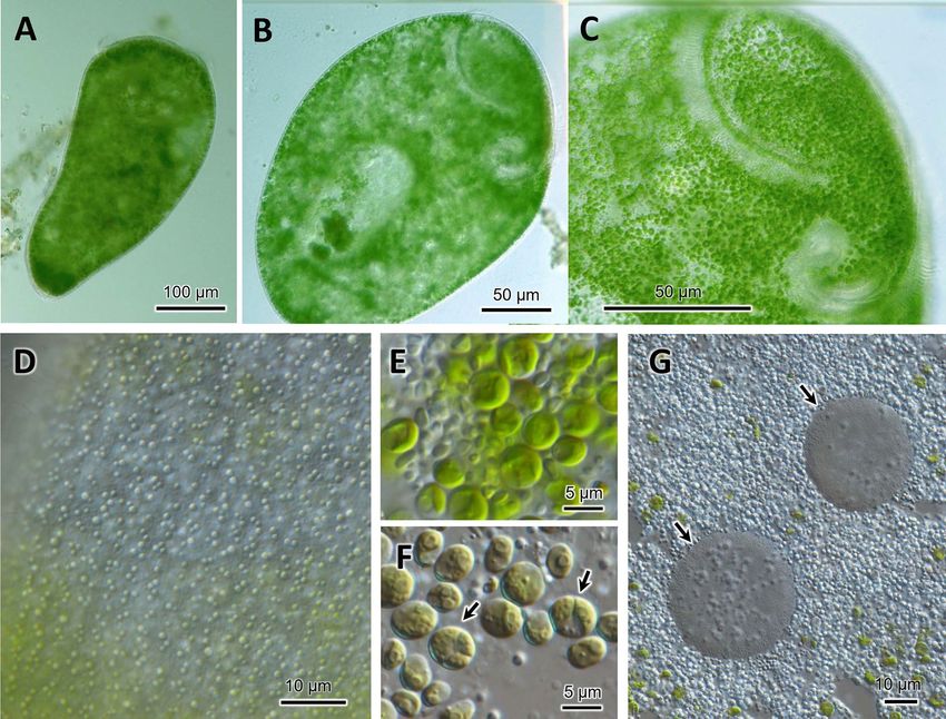
www.nature.com/scientificreports/
research on this species. In Japan, S. pyriformis can be found only in highland highly oligotrophic moors, sug-
gesting that intracellular symbiotic algae would help this species of Stentor survive in such a harsh environment.
In this study, we introduce some unique cell morphology of S. pyriformis and the characteristics of symbiotic
algae in relation to its life strategy.
Methods
Sampling. Water containing dead leaves, twigs, or the remnants of submerged plants was sampled from
ponds in Japan. The water sample was brought back to the laboratory at Tokyo and was crudely cultured in Petri
dishes. A few days later, Stentor cells containing green coccoids within their bodies were often observed. If the
green Stentor was visible, it was directly collected using Komagome pipette or cup attached to the tip of the rod.
We measured hydrogen ion concentration (pH) in some pond samples using URCERI Digital PH Meter (Shenz-
henshi Huanhui Dianzishangwu, Shenzhen, China) and electric conductivity (EC) using AquaPro Water Quality
Tester AP-2 (HM Digital, CA, USA).
Culture. Strains of S. pyriformis were cultured in 2% KCM medium (160 µg/L KCl, 260 µg/L C aCl2, 500 µg/L
MgSO4 · 7H2O; pH 6.9) in Petri dishes (diameter, 9 cm; height, 2 cm) under fluorescent (64 W; height, 20 cm)
(12L:12D) or LED light conditions at 25 °C. After multiple trials, S. pyriformis was successfully cultured only
in low EC medium such as 2% KCM. Its EC was identified to be 1.5 µS/cm. The culture medium was changed
once a week; half the volume of culture medium (10–15 mL) was discarded and fresh medium was compensated
for the shortfall. S. pyriformis were fed a non-photosynthetic cryptophyte, Chilomonas paramecium, cultured
on Euglena medium (2 g/L tryptone, 1 g/L proteose peptone, 2 g/L yeast extract, 1 g/L sodium acetate, 0.01 g/L
CaCl2) in a 50 mL polypropylene tube until stationary phase, which was centrifuged and washed with pure water
or by 2% KCM before feeding.
Cytological observations. For electron microscopy, cells were chemically fixed with glutaraldehyde and
osmium tetroxide or by metal contact quick freezing as described previously9,10. After thin sectioning, sam-
ples on the grid mesh were stained with a lead citrate s tain11 and threefold diluted EM Stainer (a lanthanoid
salts-based stain, Nisshin EM, Tokyo12). The presence of α-1, 4-linked glucose in the cytoplasm of the host S.
pyriformis and in the chloroplast of the symbiont was tested using Lugol’s iodine solution (3% iodine (wt/v), 2%
(wt/v) potassium iodine, and 73.4% (v/v) ethanol). Polarized light microscopy using a light microscope (Nikon
Eclipse Ni, Nikon, Tokyo) with a set of orthogonal polarizing filters (Nikon) on both the condenser lens and
the CCD camera was used for imaging. For Lugol’s iodine staining, 1-μm-thick sections of chemically fixed,
Spurr’s resin-embedded samples were stained with Lugol’s iodine solution for 1 min and examined under a light
microscope. For comparison, potato starch was stained with Lugol’s iodine solution for 1 min and photographed
under the same conditions. For electron microscopy of the iodine reaction, sections were first stained with lead
citrate and EM Stainer and then photographed. The sections were further treated with Lugol’s iodine solution
for 30 s, and the same field of view of the same sample was photographed again under an electron microscope.
Reinfection experiment. We investigated whether endosymbiotic algal cells isolated from S. pyriformis
could also be infected with Paramecium bursaria (strain PbKb1) and coexist in the cytoplasm. The reinfec-
tion experiment was conducted according to Omura et al.13. Aposymbiotic P. bursaria was prepared using the
method described by Higuchi et al.14. When endosymbiotic Chlorella variabilis cells isolated from P. bursaria is
mixed with aposymbiotic P. bursaria, they re-establish symbiosis within a few days. Therefore, symbiotic algal
cells were isolated from S. pyriformis and fed to aposymbiotic P. bursaria. After 30 days, microscopic observation
was performed to confirm whether P. bursaria accepted the alga as a symbiont. P. bursaria was fed with Chloro-
gonium capillatum (NIES-3374) once every 3 to 4 days as food.
DNA extraction, amplification, and sequencing. Stentor cells in the fresh sample from Toriko-Daira
(the day after the collection) were isolated under a stereoscopic microscope, and each was transferred into a
depression slide filled with pure water. Each ciliate was washed through the tip of a micropipette and transferred
into another depression, with this process being repeated twice. Before DNA extraction, we cultured these ‘clean’
(without algae outside) ciliates for 2 days. The aim of this short-term culture was to prompt the ciliates to digest
the algae, which they had taken as food, not as symbionts. Thereafter, the isolated individuals were washed twice,
and then their DNA was extracted. For the cultured Stentor, we isolated individuals and washed twice, and then
their DNA was extracted. For each strain, about 20 individuals were collected into one sample.
DNA extraction was performed using NucleoSpin Plant II kit (Macherey–Nagel, Düren, Germany) with modi-
fied cell fracturing. Stentor cells, each containing many algal cells, were incubated for 5 min in 400 µL Buffer PL1
with 10 µL RNase A at 65 °C. After adding 400 µL of glass beads (ø 0.1 mm), each sample was homogenized in
BeadSmash 12 (WakenBTech, Kyoto, Japan) at 5,000 rpm for 30 s. The homogenization was repeated five times,
and then each sample was again incubated for 10 min at 65 °C. The subsequent procedures were performed
according to the manufacturer’s instructions.
PCR was performed to amplify Stentor SSU to internal transcribed spacer (ITS) rDNA region using KOD FX
Neo (Toyobo, Osaka, Japan) with the primer pair of SR-115 (5′ SSU)/Hits5 (5′ LSU; –GGT TCR CTC GCC GTT
ACT A–). The PCR conditions were as follows. An initial denaturation step at 94 °C for 2 min was followed by 45
cycles of the following conditions: 10 s at 98 °C, 30 s at 52 °C, and 90 s at 68 °C. The amplification was completed
with a final step of 68 °C for 1 min. The PCR products were verified by agarose gel electrophoresis, cutting out
the shorter band (due to shorter ITSs, a general trend in ciliate, and being intron-less) from the gel and puri-
fied using NucleoSpin Gel and PCR Clean-up kit (Macherey–Nagel). The above primer pair amplifies ciliate
Scientific Reports | (2021) 11:2865 | https://doi.org/10.1038/s41598-021-82416-9 2
Vol:.(1234567890)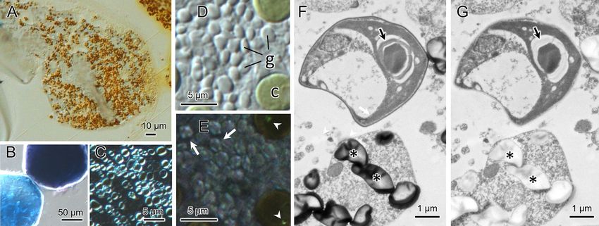
www.nature.com/scientificreports/
DNA well but not algal DNA as it is very thin. Therefore, algae-targeted PCR was separately performed with the
primer pairs of SR-1/INT-5R16 (3′ SSU) and INT-4F16 (3′ SSU)/HLR3R17 (5′ LSU). The PCR conditions were the
same as those for Stentor. The PCR products were verified by agarose gel electrophoresis and purified using the
NucleoSpin Gel and PCR Clean-up kit. The PCR products for both ciliate and algae were directly sequenced.
Phylogenetic analyses of S. pyriformis and their symbiotic algae. SSU rDNA sequences for the
Stentor species were obtained by searching the keywords [stentor + ssu] and [stentor + 18 s] from the NCBI
database. After rough alignment using Clustal X 218, the shorter sequences, and sequences including several ‘N’
were removed. Recent phylogenetic analyses including that of Stentor species have indicated stable relationships
between the Stentor species and its sister c lades19–22. Therefore, the Stentor sequences were aligned with a lim-
ited number of outgroup taxa. A bootstrap tree was constructed using the neighbor-joining (NJ) method with
default setting in Clustal X2 and examined using 1000 bootstrap replicates. For maximum likelihood (ML) and
Bayesian inference (BI) analyses, the best nucleotide substitution model for the data set was analyzed using the
Akaike information criterion (AIC) via MEGA X23, and the GTR + G + I model was selected. ML analyses were
performed with MEGA X using the nearest-neighbor interchange (NNI) branch-swapping algorithm and 1,000
bootstrap replicates were used to estimate node support values. BI analyses were conducted using the Markov
chain Monte Carlo (MCMC) method implemented in MrBayes v3.2.624. MCMC was run for 107 generations
with four chains, and trees were sampled every 1000th generation. The fixed number of samples (25,000) was
discarded as burn-in, and convergence was checked by Tracer v1.649.
The SSU rDNA sequences of S. pyriformis algae were first checked for group I intron insertions, following the
method described by H oshina25. The joined exons were then submitted to BLASTN (NCBI), which indicated
that the algae are closely related to species of Chlorella clade (Chlorellaceae). The alignment data of chlorellacean
SSU + ITS2 rDNA sequences have been published by Heeg and W olf26. Based on this, we added several recently
described species, symbionts of some protozoa and sequences obtained here, and then re-aligned them. Tree
construction methods (and selected models) were identical to those for the host ciliate, except for MCMC run-
ning for 1 08 generations.
Results
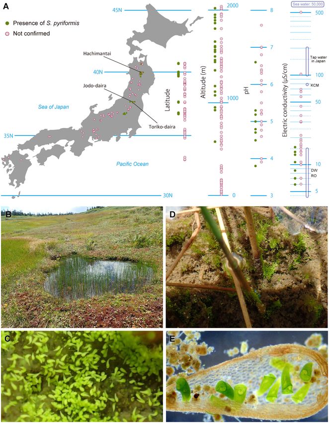
Distribution and environment. We investigated 124 ponds and wetlands in Japan and confirmed the
presence of S. pyriformis at 23 locations (Fig. 1A). Distribution areas were somewhat biased into four areas of
the middle to Northern part of Japan, which are located at 550 to 2020 m altitude. Water conditions were slightly
acidic with a pH of 3.8 to 6.6 but showed extremely low values of electric conductivity (EC): 6–16 µS/cm. These
EC values overlap with those of distilled water or reverse osmosis water (Fig. 1A). Bogs where S. pyriformis was
detected were usually located near the mountain peak or along the ridge (Fig. 1B). Frequently, we encountered
blooming of S. pyriformis on the bottom of the bogs (Fig. 1C). Other times, they were almost all attached to plant
stalks or plant debris (Fig. 1D,E).
Light microscopy. Cells of S. pyriformis were broadly trumpet-shaped, usually 220–500 × 120–300 µm.
This length–width ratio did not change significantly between the cells attached to something and swimming
(Figs. 1E, 2A). The cells were colored green due to their endosymbiotic green algae that were distributed along
the whole body (Fig. 2B,C). A large number of transparent vesicles were present along the ciliary rows imme-
diately under the cell surface (Fig. 2D). To see the contents, the crushed cells were observed. Symbiotic algae
appeared to be typical Chlorella-like algae, but no dividing alga was observed (Fig. 2E). The algal cells appeared
more vividly green when compared to those in P. bursaria, suggesting that they are richer in photosynthetic
pigments (Fig. 2E,F, and Table S2). The symbiotic algae in S. pyriformis had the same size (Table S2, Fig. S1) and
morphology as those in P. bursaria, but their biological properties were slightly different. As shown in Table S2,
S. pyriformis’s algae did not grow on agar plates, but could only be cultured in well-aerated liquid media. Reinfec-
tion experiments showed that S. pyriformis’s algae failed to re-infect the aposymbiotic strain of P. bursaria, but
those isolated from P. bursaria easily re-infected aposymbiotic P. bursaria. Macronuclei were, in general, large
and spherical (ø 20–35 µm, Fig. 2G). The average number of macronuclei was 6.1 (range 4–10, n = 9) for freshly
obtained samples, whereas four-year cultured cells (Table 1) contained only one or two. Micronuclei could not
be identified.
Cellular structure of S. pyriformis. The ultrastructural observations were performed on samples col-
lected on Oct. 28, 2019 at a small pond in Toriko-daira, Japan (37°42′17″N 140°14′53″E). First, the chemically
fixed S. pyriformis was observed with an electron microscope. Large vacuoles were found inside the cells, and the
symbiotic algal cells were inside the vacuoles. The symbionts were found uncovered by individual symbiosome
membranes (Fig. 3A,B). Many dark gray stained granules were found in the cytoplasm (asterisks in Fig. 3B).
Granules were spherical or oval. The dyeability was not uniform, and the periphery was dyed more intensely.
When the same sample was observed by the quick-freezing and freeze substitution method, the appearance in
the cytoplasm was observed quite differently (Fig. 3C). Large intracellular vacuoles as observed under chemical
fixation were not seen. In addition, individual symbiotic algae were covered by a single symbiosome membrane
(Fig. 3C,D). The distance between the symbiosome membrane and the cell wall of alga was extremely close
(20–50 nm). Fluffy projections were observed on the cell wall of the symbiotic algae (arrows in Fig. 3D). Pyr-
enoids were observed in the chloroplasts of the symbiotic algae, through which thylakoid membranes penetrated
(arrow in Fig. 3C). Many multi-vesicular bodies were observed in the cytoplasm (mv in Fig. 3C,E). The multi-
vesicular bodies were not observed at all in the samples prepared by chemical fixation, suggesting that this struc-
ture is very fragile and chemical treatment disintegrates it completely. The number of multi-vesicular bodies per
Scientific Reports | (2021) 11:2865 | https://doi.org/10.1038/s41598-021-82416-9 3
Vol.:(0123456789)
www.nature.com/scientificreports/

          Scientific Reports |   (2021) 11:2865 |   https://doi.org/10.1038/s41598-021-82416-9   4

Vol:.(1234567890)
www.nature.com/scientificreports/
◂Figure 1. Geographical distribution and habitat of Stentor pyriformis. (A). Latitude with simple map, altitude,
pH, and Electric conductivity (EC) of investigation sites are shown. pH and EC were measured only on 29 sites.
For pH and EC, there are some extended notations (e.g., EC: 11–14) in “Tsukii note,” which are spotted as two
points of those largest and smallest (e.g., 11 and 14). Reference data of EC values for some general waters were
quoted from websites of water companies, Merck Millipore (https://www.merckmillipore.com/), Kurita Water
Industries (https://kcr.kurita.co.jp/), and Japan Society of Refrigerating and Air Conditioning Engineers (https
://www.jsrae.or.jp/). KCM: 1 × KCM medium (see “Methods”). DW: distilled water. RO: reverse osmosis water.
The map data was obtained from Silhouette Design (https://kage-design.com/) and simplified using Adobe
Illustrator CS5.B. A bog in Hachimantai area (see Fig. 1). (C). Blooming of S. pyriformis on the bottom of the
bog. (D). Stentor pyriformis gathering to plant stalks. (E). Living S. pyriformis gathered in plant debris. A movie
is available online showing many Stentors on the bottom of the bog at https://1drv.ms/v/s!Aia81H4VPPEYgct
oYoHSdbRPYBkvyg?e=KXnnJa/.
cell was not clear, but there were several granules in each cell. The maximum size of multi-vesicular bodies was
about 1 µm, and the size of small vesicles was 100–400 nm in diameter (Fig. S2).
Cytoplasmic granules. Cytoplasmic granules are colored brown by Lugol staining, indicating that the
granules contained glucans composed of α-1, 4-linked glucose (Fig. 4A). As summarized in Table 2, the stored
carbohydrate granules with α-1,4-linked glucose as a backbone are classified into three types based on their
physical and chemical properties, amylose-type starch, amylopectin-type starch, and glycogen. The brown color
of the intracellular granules of S. pyriformis suggests that these granules are rich in amylopectin. For comparison,
potato starch, which is an amylose-rich starch, was stained with Lugol under the same condition and turned blue
(Fig. 4B). This indicates that this glucan is of the amylopectin-glycogen type. Observation of the isolated gran-
ules with a differential interference contrast (DIC) microscope revealed that the granules had a strong refractive
index (Fig. 4C). Figure 4D,E shows DIC (D) and polarization (E) microscopy of the cytoplasmic granules of S.
pyriformis. In the crossed polarizer orientation, each cytoplasmic granule showed a Maltese cross pattern char-
acteristic of starch granules. An ultra-thin section of chemically fixed S. pyriformis showed that the granules were
stained with heavy metals including osmium, lead, and lanthanoid ions (Fig. 4F, asterisks). The starch sheath in
the pyrenoid of the symbiont was also well stained, as shown in Fig. 4F (arrow). After taking the micrograph,
the section shown in Fig. 4F was treated with Lugol’s iodine solution, as shown in Fig. 4G. The stain of both the
cytoplasmic granules and the starch sheath was removed by iodine treatment, suggesting that the glucan gran-
ules and the starch in symbiotic algae share the same affinity to heavy metals.
Host rDNA sequence and phylogeny. SSU, ITS1, 5.8S, ITS2, and 5′ LSU rDNA sequences of four S.
pyriformis strains were obtained (Table 1). There were 2049 nucleotides, and all four sequences were completely
identical, including one C/T mixture (Y) at the tetraloop of helix E 23_1227 in the SSU rRNA structure (data not
shown).
Stentor SSU rDNA were collected and aligned with those of Blepharisma and several outgroup taxa. The phy-
logenetic tree (Fig. 5A) clearly shows the monophyly of the genus Stentor (the only genus of family Stentoridae).
The monophyly of each species was supported by values of Bayesian posterior probabilities (PP) = 0.99–1 and
bootstrap values (BV) = 96–100.
For the branching pattern of the relationships of the Stentor species, BI, ML, and NJ analyses showed some-
what different topologies. Here, we provide the species relationships reflecting the differences in these three
analyses with iconic morphological characters (Fig. 5B). The monophyletic relationship of S. roeseli and S. muel-
leri was perfectly supported. S. polymorphus, S. igneus, and S. cf. katashimai (see Thamm et al.19) made a clade,
which was placed as a sister to S. coeruleus. S. multiformis and S. elegans made a clade. S. pyriformis was clustered
with S. amethystinus in all analyses, although supporting values were not high (PP/MLBV/NJBV = 0.85/74/ < 50).
Sequence differences between S. pyriformis and S. amethystinus were 32 substitutions and 5 indels (Y is counted
as one substitution).
Symbiotic algal rDNA sequence and phylogeny. Algal sequences covering SSU, ITS1, 5.8S, ITS2, and
5′ LSU rDNA for four S. pyriformis strains were obtained (Table 1). All sequences contained group I introns at
positions S943, S1367, S1512, and L200 (corresponding to the Escherichia coli SSU and LSU rRNA). Because of
this, each sequence reached more than 3,900 bases (L200 introns were not completely determined). The algal
sequences among Jodo-daira 1436, Toriko-daira 1256, and fresh samples from Toriko-daira were identical, even
including the introns and the fast evolving ITS rDNA. The algal sequence of Hachimantai 1204 had only one
different site from the others. It was at the bulge loop of helix P1 of S1512 intron28, where, Hachimantai 1204 had
A/T mixture (W), whereas the others had A.
Search for matching sequences using combined SSU rDNA showed they were closely related to the member
of Chlorella clade (sensu Krienitz et al.29), Chlorellaceae (Trebouxiophyceae). Using SSU + ITS2 rDNA of the
member of Chlorella clade, phylogenetic analyses were performed. All tree analyses (only ML tree is shown)
indicated the symbiotic algae of S. pyriformis are clustered with C. variabilis, with which monophyletic relation-
ships were fully supported (Fig. S3).
Scientific Reports | (2021) 11:2865 | https://doi.org/10.1038/s41598-021-82416-9 5
Vol.:(0123456789)www.nature.com/scientificreports/
Figure 2. Light micrographs of living and crushed cells of Stentor pyriformis. (A). A swimming cell showing
short and fat body. (B). A slightly squeezed cell under coverslip. (C). Buccal cavity. (D): Surface vesicles
immediately under the cell surface. (E): Symbiotic Chlorella cells as seen in the crushed cytoplasm of S.
pyriformis with starch granules. The Chlorella cells appear vividly green, and dividing cells were rarely seen.
(F). Symbiotic Chlorella variabilis cells in Paramecium bursaria for comparison. The picture was taken under
the same photographic conditions as (E). Cells are pale green and many dividing Chlorella cells are observed
(arrows). The cell size variation was larger than that in S. pyriformis. G. Spherical macronuclei (arrows) found
with abundant starch granules in the cytoplasm crushed between the slide and the coverslip.
Strains Collection site* Collection date Host rDNA Symbiont rDNA
Hachimantai 1204 Hachimantai Sep. 24, 2015 LC533384 LC533388
Jodo-daira 1436 Jodo-daira Oct. 10, 2015 LC533385 LC533389
Toriko-daira 1256 Toriko-daira Oct. 10, 2015 LC533386 LC533390
(Fresh sample) Toriko-daira Oct. 28, 2019 LC533387 LC533391
Table 1. Stentor pyriformis strains and GenBank Accession numbers for the host and symbiont rDNA. *See
Fig. 1.
Discussion
Distribution of Stentor pyriformis in Japan and its optimal culture conditions. S. pyriformis was
described by Johnson in 18936. This algae-bearing Stentor has separated spherical macronuclei without pig-
mentation, which certainly differentiates it from other Stentor species (see Table S1, Fig. 5B). While the most
common algae-bearing Stentor, S. polymorphus assumes a slender trumpet shape (often shortened), S. pyriformis
never resembles such a slender trumpet, but assumes a pear or short conical shape, even when it is swimming6.
Presence or absence of colored pigmentation is also a prominent characteristic for separating Stentor species.
Among algae-bearing Stentor spp., S. polymorphus and S. pyriformis only are considered colorless species,
whereas colored species are S. amethystinus, S. fuliginosus, S. araucanus, and S. tartari8 (Table S1). Therefore,
S. pyriformis is a clearly discernible species; however, it remains underexplored. Indeed, we could only find
one paper on the new habitats of S. pyriformis7, with the exception of the paper of species consolidation of this
genus8. We confirmed the presence of S. pyriformis at 23 locations (Fig. 1A). This indicates that S. pyriformis is
Scientific Reports | (2021) 11:2865 | https://doi.org/10.1038/s41598-021-82416-9 6
Vol:.(1234567890)www.nature.com/scientificreports/
Figure 3. (A) and (B). Chemically-fixed specimen of S. pyriformis. The cytoplasm was observed as highly
vacuolated, in which symbiotic Chlorella cells (Ch) were observed to be scattered in a large vacuole without
being covered by individual surrounding membranes. Chlorella at the lower left is a rarely seen dividing
organism. Many small electron-dense granules of 1–2 μm in size were present in the cytoplasm. (C)–(E)
S. Quick-frozen and freeze substituted specimen of S. pyriformis. The cytoplasm was not vacuolated, and
each symbiotic Chlorella was surrounded by a symbiosome membrane (arrowheads in D). The symbiosome
membrane was closely opposed to the cell wall surface by a distance of less than 50 nm. The cell wall surface
of the symbiotic Chlorella was ornamented by fluffy filaments (arrow). In the symbiotic Chlorella, the pyrenoid
structure was penetrated by thylakoid membranes (arrow in C), characteristic of the genus Chlorella. (D) is an
enlarged view of the area indicated by the rectangle in (C). In the cytoplasm, many multi-vesicular bodies of
about 1 μm in diameter with unknown function were observed (mv in C and also in E).
by no means a rare organism. We assume one of the reasons why S. pyriformis has been poorly studied is the dif-
ficulty of cultivation. In fact, Johnson6 noted that he could not keep them more than a month and never observed
any cells in fission. In addition, after five years of failure, it was finally possible to culture S. pyriformis for more
than several months. Because of objectively unfounded data that we could not include in the distribution data
(Fig. 1A), we noticed the wetlands where we found S. pyriformis were limited to small ponds or bogs locating
near the mountain peak or along the ridge (Fig. 1B). That is, the ponds depending on rainfall without inflowing
rivers. Because there is no nutrient flowing in, waters in these ponds showed noticeable oligotrophic tendency,
i.e., extremely low electric conductivity (Fig. 1A), which gave us some clues on culture.
The most important point of culture for S. pyriformis was keeping the medium lower electric conductivity.
We use the KCM medium diluted by 2% with Milli-Q water, and changed medium once a week. A non-photo-
synthetic cryptophyte, Chilomonas paramecium was selected for food. We selected the food so that it would not
itself grow in the culture medium. Growing organisms, like photosynthetic algae, seemed to cause damage to S.
pyriformis. Using this culture method, S. pyriformis can be maintained for more than four years (see Table 1). For
the organisms not easy to grow in culture, Professor Michael Melkonian mentioned no protist is ‘uncultivable’,
there is just human f ailure30. Here, it just became possible to culture S. pyriformis 120 years after its discovery;
however, this method does not always work. S. pyriformis appears to be extremely fragile and disintegrates when
any variables are unintentionally altered, that is, the culture is still unstable. When its condition deteriorates, the
cells divide unevenly in such a way that a part of the cell is broken. When this happens, the cells become spheri-
cal, and the drug drops to the bottom of the dish. It retains this shape for more than a month, but eventually
disappears. The doubling time of S. pyriformis remains 3 to 4 weeks, even under favorable conditions (data not
shown). We occasionally encountered the blooming of S. pyriformis all over the bottom of the ponds (Fig. 1C).
S. pyriformis, therefore, does not seem to be a particularly slow growing species, but our culture method appears
to be far from the optimal culture conditions for them. Three S. pyriformis strains used in this study are available
from the authors upon request.
Ultrastructure. In this study, we compared the conventional chemical fixation method with the rapid-freez-
ing fixation method for electron microscopic observation. As a result, large vacuoles were observed in the cyto-
plasm when chemical fixation was used, but not by rapid freezing. Instead, many multi-vesicular bodies were
observed in the cytoplasm. The quick-freezing and freeze-substitution method is considered superior in that it
can prevent deformation of the intracellular structure compared to chemical fixation31. Therefore, it is possible
that the originally existing multi-vesicular bodies were artificially disintegrated by chemical fixation, and the
constituent biological membranes fused together, eventually forming large vacuoles. To the authors’ knowledge,
Scientific Reports | (2021) 11:2865 | https://doi.org/10.1038/s41598-021-82416-9 7
Vol.:(0123456789)www.nature.com/scientificreports/
Figure 4. Histochemical localization of starch in Stentor pyriformis. (A). Light micrograph of a thick section
of a Spurr’s resin-embedded cell stained with Lugol’s solution. Cytoplasmic granules stained brown, suggesting
high content of α-1, 6-linked glucose branch. (B): Potato starch granules stained with Lugol’s solution shown
as comparison. Potato starch stains blue due to its high amylose content. (C): A DIC image of the freshly
isolated cytoplasmic granules observed under the crossed polarizer condition, showing that the granules
have a high refractile index. (D) and (E). Polarization microscopy of starch granules in S. pyriformis. (D).
DIC image of compressed and disrupted cytoplasm of S. pyriformis. Symbiotic Chlorella (c) and cytoplasmic
granules (g) are shown. (E). Polarization micrograph of the same area as shown in D under the cross-nicol
condition. In the crossed polarizer orientation, birefringent anisotropic specimens such as plant starch grains
show a characteristic “Maltese cross” pattern (see Olympus website: https://www.olympus-lifescience.com/en/
microscope-resource/primer/techniques/dic/dicphasecomparison/). The cytoplasmic granules of S. pyriformis
show a Maltese cross pattern (arrows), indicating that the granules have birefringent properties like plant
starch. Arrowheads show birefringence signals of the starch sheath in symbiotic Chlorella cells. (F) and (G):
Transmission electron micrographs of a section of S. pyriformis chemically fixed and stained with lead and
lanthanoid. Photographs of the same section were taken before (F) and after (G) treatment with Lugol’s iodine
solution. Arrows indicate the pyrenoid starch sheath, which was stained with heavy metals in F, but the stain
was removed by iodine treatment. Granules in the ciliate cytoplasm (asterisks) were also destained by iodine
treatment.
Granule storage substances Starch (amylose) Starch (amylopectin) Glycogen
α-1,6-linked glucose branch None At every 20–25 glucose units61 At every 10–14 glucose units61
62 62 61
Iodine coloration Blue Red–purple or Dull-red Reddish brown61
From less than 1 μm to more than
Particle size 10–300 nm (α-particle)64
100 μm63
Maltese cross Yes65 Yes65 No66
Table 2. Properties of storage glucans with α-1, 4-linked glucose backbone.
no intracellular structure similar to the multi-vesicular body in S. pyriformis has been reported in protists. As
multi-vesicular bodies of S. pyriformis could only be observed using the freeze-substitution method, similar
granules may also be found in other protists if the same technique is used for electron microscopy. In animals,
on the other hand, aggregates of secretory vesicles resembling the multi-vesicular bodies of S. pyriformis are pre-
sent in cardiac telocytes32. The extracellular vesicles form multi-vesicular structures of about 1 μm in diameter
and contain materials for intercellular communication that are involved in cardiac physiology and regeneration.
Because S. pyriformis cells often form aggregates at the bottom of the pond, some chemicals may be released
from the multi-vesicular body, attracting nearby cells and forming aggregates.
Observation by the freeze-substitution method revealed that the symbiosome membrane was in close contact
with the symbiotic chlorella. Furthermore, fluffy projections were observed on the cell wall of the symbiotic
chlorella. These characteristics were consistent with those of C. variabilis, which is symbiotic in the cells of P.
bursaria9. The only difference was that in S. pyriformis, the symbiotic chlorella cells were scattered in the cyto-
plasm, whereas the symbiotic Chlorella in P. bursaria were anchored directly below the cell surface.
Storage granules. The iodine in Lugol’s solution selectively binds to α-1, 4-linked glucose found in poly-
saccharides, such as starch33 and glycogen34. The color stained with Lugol’s solution reflects the type of glucose
polymer. Starches with high amylose content stain blue-violet (cf. Fig. 4B), high amylopectin stains red–purple,
Scientific Reports | (2021) 11:2865 | https://doi.org/10.1038/s41598-021-82416-9 8
Vol:.(1234567890)www.nature.com/scientificreports/
B FN659825 Stentor roeseli
s
1/99/100
n
leu
BI
tio
FN659824 Stentor roeseli
1/91/86
Pi nuc
Sy nta
nt
LT628481 Stentor roeseli
ML
bio
e
ro
KP970247 Stentor roeseli
gm
NJ
m
1/99/99 1/99/99
ac
AF357913 Stentor roeseli
M
KJ651826 Stentor roeseli
Stentor roeseli V 1/100/100 1/100/100 KP970248 Stentor roeseli
1/99/ FN659819 Stentor muelleri
Stentor muelleri M 100 KJ651824 Stentor muelleri
.99/96/96
FN659820 Stentor muelleri
Stentor coeruleus M KM222111 Stentor sp. FG-2015
FN659814 Stentor coeruleus
Stentor cf. katashimai M FN659815 Stentor coeruleus
DQ136037 Stentor coeruleus
Stentor polymorphus M KP970244 Stentor coeruleus
.99/50/-
KJ651823 Stentor coeruleus
Stentor igneus B 1/99/98 KJ651825 Stentor coeruleus
AF357145 Stentor coeruleus
Stentor multiformis B FN659816 Stentor coeruleus
LT628482 Stentor coeruleus
Stentor elegans B LN869963 Stentor cf. coeruleus
.99/ FN659813 Stentor coeruleus
65/66
Stentor amethystinus B DQ132978 Stentor coeruleus
JQ282899 Stentor coeruleus
Stentor pyriformis KF639913 Stentor sp.
B
FN659812 Stentor coeruleus
.97/69/82
.98/64/63 KP970243 Stentor coeruleus
FN659811 Stentor coeruleus
.99/61/67 FN659810 Stentor coeruleus
FN659809 Stentor coeruleus
AM713189 Stentor coeruleus
1/100/100 LN869971 Stentor sp. aTTo1
FN659818 Stentor cf. katashimai
.99/-/99 AM713190 Stentor polymorphus
KP970246 Stentor polymorphus
.99/52/61 1/98
JQ282898 Stentor polymorphus
/99
FN659823 Stentor polymorphus
Stentor spp. .99/ AF357144 Stentor polymorphus
95/88
a few 1/100/100
1/100/100 JQ282900 Stentor sp. NMF-2012
Vermiform Moniliform Beads KP970245 Stentor igneus
FN659821 Stentor multiformis
Macronucleus types .97/61
/71 FN659817 Stentor elegans
.95/-/65
KJ651828 Stentor sp. SUAS-2014
AM713191 Stentor amethystinus
1/100/100 AY775566 Stentor amethystinus
FN659808 Stentor amethystinus
.99/83/93
FN659807 Stentor amethystinus
-/74/- Stentor pyriformis Jodo-Daira 1436
Stentor pyriformis Hachimantai 1204
1/100/100 Stentor pyriformis Torigo-Daira 1256
Stentor pyriformis Torigo-Daira (Fresh)
AM713182 Blepharisma americanum
1/99/100
.99/-/- AM713183 Blepharisma undulans
1/96/97
A
-/-/64 AM713185 Blepharisma japonicum
AM713186 Blepharisma elongatum
1/100/100 1/81/82 AM713184 Blepharisma hyalinum
AM713187 Blepharisma steini
1/95/99 U47620 Eufolliculina uhligi
1/97/99 EU583992 Folliculina sp. WWS-2008
AY630405 Maristentor dinoferus
1/100/100 KJ651817 Fabrea salina
DQ168805 Fabrea salina
KJ651818 Gruberia sp. SUAS-2014
Figure 5. Phylogenetic relationships of Stentor species. (A). Bayesian inference tree for Stentor species
based on SSU rDNA sequences. The tree was rooted with Gruberia sp. SUAS-2014. Numbers at the branches
correspond to MrBayes posterior probabilities (PP)/maximum likelihood/neighbor-joining bootstrap values
(BVs). Hyphens correspond to PP values below 0.70 and BVs below 50%. (B). Summary of the interspecific
relationships and their iconic morphocharacters (shapes of macronucleus, presence or absence of cortical
pigmentation and symbiotic algae). The figure explaining the varieties of macronuclei was modified from
Foissner and Wölfl8.
and glycogen stains reddish brown (Table 2). The granules in the cytoplasm of S. pyriformis stained reddish
brown with Lugol’s solution (Fig. 4A), suggesting that these granules are composed of α-1,4-linked glucans
with high number of α-1,6-linked branch points, either amylopectin-rich starch or glycogen. The pyrenoid of
Chlorella spp. is surrounded by a starch sheath of two large p
lates35. As shown in Fig. 4F,G, the image contrast
Scientific Reports | (2021) 11:2865 | https://doi.org/10.1038/s41598-021-82416-9 9
Vol.:(0123456789)www.nature.com/scientificreports/
formed by electron staining of the starch granule in the chloroplast (arrow) was lost by treatment with Lugol’s
solution. Although the detailed mechanism is unknown, this observation suggests that electron-stained heavy
metals (osmium, lead, and lanthanoid ions) bound to the granules may have been eliminated by iodine in Lugol’s
solution. The cytoplasmic granules of S. pyriformis showed the same staining properties as the starch granules
in the chloroplasts of symbiotic chlorella, suggesting that both types of granules share chemical characteristics
as polysaccharides.
Alveolates make up one of the most diverse and largest groups of protists. They include three major taxa:
dinoflagellates, ciliates, and apicomplexan protozoa. All three alveolate lineages store glucose in an α-1,4-linked
glucose chain with α-1, 6 branches. Ciliates are known to synthesize glycogen granules. For example, Tetrahymena
has glycogen granules between 35 and 40 nm in diameter, each granule being a collection of small γ-granules
of 2–3 nm in size36. Dinoflagellates and apicomplexans typically produce more complex and larger spherical
starch particles, usually greater than 1 μm in s ize37,38. Amylopectin-rich starch and glycogen are very similar
polysaccharides, but they differ in granule size and birefringence (Table 2). Starch granules are large, birefringent,
and have a high refractive index, but glycogen does not exhibit birefringence, and its granules generally have a
size of 300 nm or less. When observed with a polarizing microscope, the starch granules show a Maltese cross
pattern. This pattern is derived from the radial arrangement of amylose and amylopectin molecules in granules
and is one of the criteria for starch identification. Since the cytoplasmic granules of S. pyriformis are large in
size (1–3 μm) and show a typical Maltese cross pattern as shown in Fig. 4E, these granules are likely to be starch
granules rich in amylopectin.
Phylogeny of S. pyriformis and its morphology. Relationships of Stentor species were not clearly
resolved. BI and ML analyses indicated basal diverging of the S. pyriformis + S. amethystinus clade from oth-
ers, but NJ analysis did not indicate so (Fig. 5). Recent phylogenetic analyses inclusive of Stentor species also
indicated basal diverging of S. amethystinus from the others; however, the monophyly of the others is not highly
supported21,22. Therefore, the one thing that can be said is that S. pyriformis is closely related to S. amethystinus.
For the identification of Stentor species, the shape of macronucleus, presence or absence of cortical pigmen-
tation, and symbiotic algae are very important and iconic characteristics8,19. S. pyriformis and S. amethystinus
share beaded macronuclei and the presence of symbiotic chlorella (Table S1, Fig. 5B). Pigmentation is present in
S. amethystinus, but not in S. pyriformis. Pigmentation is a noticeable characteristic, which tinctures the whole
body of Stentor cells. The pigment is thought to function as a defense against p redators39. However, the kind of
pigment compound depends on the species40, and the relationship between pigment possession and phylogeny is
poor (Fig. 5). Of note, colorless vesicles exist in S. pyriformis (Fig. 2D). The short and fat shape is also a common
characteristic for S. pyriformis and S. amethystinus, in this genus with many elongated trumpet shape s pecies6,8.
Symbiotic algae in S. pyriformis. Algae-targeted PCR products from whole cells of S. pyriformis were
sequenced directly, and clear peaks were obtained for each. This shows that all or nearly all of the algal sym-
bionts in each Stentor cell are unified, regardless of samples under long-term culture or nature. In addition, all
symbionts were closely related to C. variabilis (Fig. S3), which has been known as a representative symbiont of
P. bursaria (Oligohymenophorea), the model organism of multi-algae retaining protists ( MARP41) style sym-
bioses. For the chlorellacean species, the diversity of ITS2 sequence comparisons has often been adopted. For
two organisms to compare, ITS2 sequence differences (gaps are counted as a fifth character) usually fall either
less than 2% for single species or more than 10% for different s pecies42,43. This characteristic simply encourages
a species concept. The ITS2 sequences of S. pyriformis algae differ only by one nucleotide site out of 248 sites
from those of P. bursaria algae (Fig. 6A), which strongly suggests the symbiotic chlorella of S. pyriformis are also
C. variabilis. Several Stentor species retain coccoid green a lgae8 (Table S1), but only three algal sequences have
been published. Two algal sequences from S. polymorphus belonged to different clades from C hlorellaceae44,45.
As for the other algal sequence of S. amethystinus, the symbiont may belong to C hlorellaceae46. This sequence
(EF589816) is short (991 bp) and only covers a part of SSU rDNA; therefore, it was not included in our phylo-
genetic analyses (Fig. S3). The sequence differs from C. variabilis with 10 base changes and 3 indels, indicating
that it is not C. variabilis.
In the case of P. bursaria-C. variabilis symbiosis, C. variabilis has been shown to be vastly different from other
free-living species. C. variabilis demands organic nitrogen c ompounds47 and leaks nearly half of the photosyn-
thate to outside algal c ells48,49. Furthermore, they are sensitive to the C. variabilis virus (CvV; so-called ‘NC64A
virus’), which is abundant in natural freshwater50–52. Therefore, C. variabilis should be considered an already
evolved species that is unable to survive without the protection of the host c ell53.
Four C. variabilis rDNA sequences obtained from S. pyriformis were identical, with the exception of a nucleo-
tide position in the S1512 intron. Here, the regions without group I introns, i.e., SSU, ITS1, 5.8S, and ITS2 rDNA,
are compared among C. variabilis sequences of S. pyriformis and of P. bursaria. Several published sequences
cover the above SSU-ITS region, of which varieties are shown as P. bursaria symbiont genotype (PbS-gt) 1 to 3
(Fig. 6A). Due to the small number of sequences, it is still unknown whether these genotypes depend on (or are
related to) their living regions. Genotype 1 was from USA and Japan, genotype 2 was from China, and genotype
3 was from Australia. All available sequences for S. pyriformis symbionts were obtained in this study, and they
were all from Japan. As a result, all sequences of S. pyriformis symbionts were aggregated into a single genotype
SpS, which was distantly related to all P. bursaria symbionts, including those from Japan (Fig. 6B). Five variable
sites are found in SSU rDNA among C. variabilis genotypes, of which four are concentrated to that of the sym-
bionts of S. pyriformis (SpS) (Fig. 6A). C/T substitution at alignment position 656 will be a hemi-compensatory
base change (hemi-CBC) at the E23_2 helix of SSU rRNA structure (Fig. 6C), whereas the other four sites are at
single strand regions (data not shown). Mutations (1821–1828) including comparatively large indels were seen
Scientific Reports | (2021) 11:2865 | https://doi.org/10.1038/s41598-021-82416-9 10
Vol:.(1234567890)www.nature.com/scientificreports/
Figure 6. Sequence differences of SSU, ITS1, 5.8S and ITS2 rRNA gene (without group I introns) among
Chlorella variabilis. “PbS-gt” indicates Paramecium bursaria symbiont genotypes. Genotype 1 includes SAG
211-6, ATCC 50258 (NC64A), NIES-2541, and some other US and Japanese strains. Genotype 2 is the alga of
Chinese P. bursaria strain Cs2, and genotype 3 is the alga of Australian P. bursaria strain MRBG1. For further
information, see Hoshina et al.53. “SpS” indicates the algal sequence of Stentor pyriformis strains collected from
Japan. (A). Different positions. Numerals represent the nucleotide number in aligned sequences (2462 aligned
sites). (B). Distance tree of above four types of sequences. (C). E23_2 helix of SSU rRNA structure that includes
hemi-CBC at the alignment position 656. (D). Deformation of ITS1 Helix 1 associated with the mutations
including several nucleotide insertions.
in ITS1 region (Fig. 6A). It was found that all these mutations are assembled in helix 1 (for chlorellacean ITS1
structure, see Bock et al.54,55). Thermodynamic analysis via Mfold56,57 predicted that PbS sequences form linear
helix 1 similar to the other chlorellacean species, but SpS sequences including the additional nucleotides may
form a dichotomous branching of helix 1 (Fig. 6D).
The group I introns inserted in SSU rDNA of S. pyriformis symbionts are identical to those of P. bursaria
symbionts28,58 in terms of numbers (three introns) and insertion sites (S943, S1367 and S1512). The sequences of
S943 and S1512 introns are matched more than 99%. However, with respect to the S1367 intron, a large length
gap was found (168 nucleotides) at the tip of P8 (Fig. S4). This section has been indicated as a homing endonu-
clease gene r emnant28, and those of S. pyriformis symbionts are presumed to be a more degenerated form than
those of P. bursaria symbionts.
At any rate, the symbiotic algae of S. pyriformis were found to be C. variabilis. Because S. pyriformis never
lost the symbiotic algae in four years of culture, and all four algae had nearly identical genetic characteristics,
the symbiotic relationships between S. pyriformis and C. variabilis can be regarded as stable, or permanent.
Although S. pyriformis and P. bursaria share C. variabilis as their endosymbionts, considering the genetic differ-
ences depending on their host species, the sharing event has not happened recently. Symbiont sharing among
various host species has also been known for some c iliates41,59 (Carolibrandtia ciliaticola in Fig. S3), and a script
to spread a particular algal symbiont has been s uggested41. Given the physiological characters of C. variabilis
(mentioned above), this algal species might be an ideal algal symbiont, and it will be no surprise if the other
protists also retained C. variabilis as their algal partners. Research on the symbiotic algae that other Stentor spp.
have and on host and regional dependencies are awaited.
Adaptation of S. pyriformis to oligotrophic environment in highland marsh. In Japan, S. pyri-
formis lives only in alpine ponds (Fig. 1), where the winter is cold, and the surface of the pond is always covered
with ice. The water in these ponds has low electrical conductivity (~ 10 μS/cm), and there are few living organ-
isms except S. pyriformis, meaning that only little food is available in wintertime. The reason this ciliate is rich
in stored carbohydrate granules may be due to its need for nutrients to survive such harsh winter environments.
Preliminary studies suggest that many protists, especially ciliates, may make starch. Large amounts of cyto-
plasmic granules that show a Maltese cross were observed in chlorella-bearing ciliates such as P. bursaria, while
only a small amount of such granules was observed in Euplotes aediculatus, Paramecium caudatum, Blepharisma
japonicum, and Tetrahymena pyriformis. Protists with symbiotic algae seem to produce particularly large amounts
Scientific Reports | (2021) 11:2865 | https://doi.org/10.1038/s41598-021-82416-9 11
Vol.:(0123456789)www.nature.com/scientificreports/
of stored carbohydrate granules in the cytoplasm, but the mechanism of starch synthesis may be widely shared
by ciliates.
P. bursaria has been shown to be more resistant to starvation conditions than the aposymbiotic strain of
the same s pecies13. Under food-deprived conditions, P. bursaria was interpreted to have survived by digesting
symbiotic algae. Resting cyst formation and cannibalism are known as other strategies for protozoans to survive
starvation conditions60. This study suggests that the use of carbohydrate granules stored in cells may be another
possible strategy for ciliates to survive harsh environments such as highland oligotrophic bogs.
Received: 19 May 2020; Accepted: 30 December 2020
References
1. Selosse, M.-A., Charpin, M. & Not, F. Mixotrophy everywhere on land and in water: The grand écart hypothesis. Ecol. Lett. 20,
246–263 (2017).
2. Sonntag, B., Posch, T., Klammer, S., Teubner, K. & Psenner, R. Phagotrophic ciliates and flagellates in an oligotrophic, deep, alpine
lake: Contrasting variability with seasons and depths. Aquat. Microb. Ecol. 43, 193–207 (2006).
3. Woelfl, S., Garcia, P. & Duarte, C. Chlorella-bearing ciliates (Stentor, Ophrydium) dominate in an oligotrophic, deep North Patagon-
ian lake (Lake Caburgua, Chile). Limnologica 40, 134–139 (2010).
4. Sonntag, B., Summerer, M. & Sommaruga, R. Are freshwater mixotrophic ciliates less sensitive to solar ultraviolet radiation than
heterotrophic ones?. J. Eukaryot. Microbiol. 58, 196–202 (2011).
5. Tsukii, Y. Protist Information Server. http://protist.i.hosei.ac.jp (2017).
6. Johnson, H. P. A contribution to the morphology and biology of the stentors. J. Morph. 8, 467–563 (1893).
7. Walker, E. R. Observations on the micro-fauna of an Oregon pond. Trans. Am. Microsc. Soc. 28, 75–84 (1908).
8. Foissner, W. & Wölfl, S. Revision of the genus Stentor Oken (Protozoa, Ciliophora) and description of S. araucanusnov. spec. from
South American lakes. J. Plankton Res. 16, 255–289 (1994).
9. Song, C., Murata, K. & Suzaki, T. Intracellular symbiosis of algae with possible involvement of mitochondrial dynamics. Sci. Rep.
7, 1221. https://doi.org/10.1038/s41598-017-01331-0 (2017).
10. Hoshina, R., Hayakawa, M. M., Kobayashi, M., Higuchi, R. & Suzaki, T. Pediludiella daitoensis gen. et sp. nov. (Scenedesmaceae,
Chlorophyceae), a large coccoid green alga isolated from a Loxodes ciliate. Sci. Rep. 10, 628. https://doi.org/10.1038/s41598-020-
57423-x (2020).
11. Reynolds, E. S. The use of lead citrate at high pH as an electron-opaque stain in electron microscopy. J. Cell. Biol. 17, 208–212
(1963).
12. Hosogi, N., Nishioka, H. & Nakakoshi, M. Evaluation of lanthanide salts as alternative stains to uranyl acetate. Microscopy 64,
429–435 (2015).
13. Omura, G. et al. A bacteria-free monoxenic culture of Paramecium bursaria: Its growth characteristics and the re-establishment
of symbiosis with chlorella in bacteria-free conditions. Jpn. J. Protozool. 37, 139–150 (2004).
14. Higuchi, R., Song, C., Hoshina, R. & Suzaki, T. Endosymbiosis-related changes in ultrastructure and chemical composition of
Chlorella variabilis (Archaeplastida, Chlorophyta) cell wall in Paramecium bursaria (Ciliophora, Oligohymenophorea). Eur. J.
Protistol. 66, 149–155 (2018).
15. Nakayama, T., Watanabe, S., Mitsui, K., Uchida, H. & Inouye, I. The phylogenetic relationship between the Chlamydomonadales
and Chlorococcales inferred from 18S rDNA sequence data. Phycol. Res. 44, 47–55 (1996).
16. Hoshina, R., Kamako, S.-i & Imamura, N. Phylogenetic position of endosymbiotic green algae in Paramecium bursaria Ehrenberg
from Japan. Plant Biol. 6, 447–453 (2004).
17. Hoshina, R., Kato, Y., Kamako, S.-i & Imamura, N. Genetic evidence of “American” and “European” type symbiotic algae of Para-
mecium bursaria Ehrenberg. Plant Biol. 7, 526–532 (2005).
18. Larkin, M. A. et al. Clustal W and Clustal X version 2.0. Bioinformatics 23, 2947–2948 (2007).
19. Thamm, M., Schmidt, S. L. & Bernhard, D. Insights into the phylogeny of the genus Stentor (Heterotrichea, Ciliophora) with special
emphasis on the evolution of the macronucleus based on SSU rDNA data. Acta Protozool. 49, 149–157 (2010).
20. Shazib, S. U. A., Vd’ačný, P., Kim, J. H., Jang, S. W. & Shin, M. K. Phylogenetic relationships of the ciliate class Heterotrichea
(Protista, Ciliophora, Postciliodesmatophora) inferred from multiple molecular markers and multifaceted analysis strategy. Mol.
Phylogenet. Evol. 78, 118–135 (2014).
21. Fernandes, N. M., da Silba Paiva, T., da Silva-Neto, I. D., Schlegel, M. & Schrago, C. G. Expanded phylogenetic analyses of the class
Heterotrichea (Ciliophora, Postciliodesmatophora) using five molecular markers and morphological data. Mol. Phylogenet. Evol.
95, 229–246 (2016).
22. Chen, X., Shazib, S. U. A., Kim, J. H., Kim, M. S. & Shin, M. K. New contributions to Gruberia lanceolata (Gruber, 1884) Kahl, 1932
based on analyses of multiple populations and genes (Ciliophora, Heterotrichea, Gruberiidae). Eur. J. Protistol. 65, 16–30 (2018).
23. Kumar, S., Stecher, G., Li, M., Knyaz, C. & Tamura, K. MEGA X: Molecular evolutionary genetics analysis across computing
platforms. Mol. Biol. Evol. 35, 1547–1549 (2018).
24. Ronquist, F. et al. MrBayes 3.2: Efficient Bayesian phylogenetic inference and model choice across a large model space. Syst. Biol.
61, 539–542 (2012).
25. Hoshina, R. Analysis of an intron intervening the SSU rDNA of Chlorella sp. T-24–5, a photobiont of Paramecium bursaria. Jpn.
J. Protozool. 45, 17–27 (2012).
26. Heeg, J. S. & Wolf, M. ITS2 and 18S rDNA sequence-structure phylogeny of Chlorella and allies (Chlorophyta, Trebouxiophyceae,
Chlorellaceae). Plant Gene 4, 20–28 (2015).
27. Wuyts, J., Van De Peer, Y. & De Wachter, R. Distribution of substitution rates and location of insertion sites in the tertiary structure
of ribosomal RNA. Nucleic Acids Res. 29, 5017–5028 (2001).
28. Hoshina, R. & Imamura, N. Eu-Chlorella large subunit rDNA sequences and group I introns in ribosomal DNA of the paramecian
symbiotic alga NC64A. Phycol. Res. 56, 21–32 (2008).
29. Krienitz, L. et al. Phylogenetic relationship of Chlorella and Parachlorella gen. nov. (Chlorophyta, Trebouxiophyceae). Phycologia
43, 529–542 (2004).
30. Melconian, M. Editorial. Protist 158, 1 (2007).
31. Song, C. & Suzaki, T. Improved preservation of organelles in Paramecium bursaria by freeze-substitution with glutaraldehyde and
osmium tetroxide. J. Electr. Microsc. Technol. Med. Biol. 27, 1–8 (2013).
32. Fertig, E. T., Gherghiceanu, M. & Popescu, L. M. Extracellular vesicles release by cardiac telocytes: Electron microscopy and
electron tomography. J. Cell. Mol. Med. 18, 1938–1943 (2014).
33. Dhar, N. R. The starch-iodine reaction. J. Phys. Chem. 28, 125–130 (1924).
34. Quain, D. E. & Tubb, S. A rapid and simple method for the determination of glycogen in yeast. J. Inst. Brewing 89, 38–40 (1983).
Scientific Reports | (2021) 11:2865 | https://doi.org/10.1038/s41598-021-82416-9 12
Vol:.(1234567890)You can also read