Human antibodies targeting a Mycobacterium transporter protein mediate protection against tuberculosis - CTVD
←
→
Page content transcription
If your browser does not render page correctly, please read the page content below
ARTICLE
                  https://doi.org/10.1038/s41467-021-20930-0                 OPEN

                  Human antibodies targeting a Mycobacterium
                  transporter protein mediate protection against
                  tuberculosis
                  Avia Watson1,13, Hao Li2,3,13, Bingting Ma4,13, Ronen Weiss1, Daniele Bendayan5, Lilach Abramovitz1,
                  Noam Ben-Shalom1, Michael Mor1, Erica Pinko5, Michal Bar Oz6, Zhenqi Wang 2, Fengjiao Du7, Yu Lu7,
                  Jan Rybniker8,9, Rony Dahan10, Hairong Huang11, Daniel Barkan6, Ye Xiang4 ✉, Babak Javid 2,12 ✉ &
                  Natalia T. Freund 1 ✉
1234567890():,;

                  Mycobacterium tuberculosis (Mtb) exposure drives antibody responses, but whether patients
                  with active tuberculosis elicit protective antibodies, and against which antigens, is still
                  unclear. Here we generate monoclonal antibodies from memory B cells of one patient to
                  investigate the B cell responses during active infection. The antibodies, members of four
                  distinct B cell clones, are directed against the Mtb phosphate transporter subunit PstS1.
                  Antibodies p4-36 and p4-163 reduce Mycobacterium bovis-BCG and Mtb levels in an ex vivo
                  human whole blood growth inhibition assay in an FcR-dependent manner; meanwhile,
                  germline versions of p4-36 and p4-163 do not bind Mtb. Crystal structures of p4-36 and p4-
                  170, complexed to PstS1, are determined at 2.1 Å and 2.4 Å resolution, respectively, to reveal
                  two distinctive PstS1 epitopes. Lastly, a prophylactic p4-36 and p4-163 treatment in Mtb-
                  infected Balb/c mice reduces bacterial lung burden by 50%. Our study shows that inhibitory
                  anti-PstS1 B cell responses arise during active tuberculosis.

                  1 Department of Clinical Microbiology and Immunology, Sackler Faculty of Medicine, Tel Aviv University, Tel Aviv-Yafo, Israel. 2 Centre for Global Health and

                  Infectious Diseases, Collaborative Innovation Centre for the Diagnosis and Treatment of Infectious Diseases, Tsinghua University School of Medicine,
                  Beijing, China. 3 College of Veterinary Medicine, China Agricultural University, Beijing, China. 4 Advanced Innovation Center for Structural Biology & Beijing
                  Frontier Research Center for Biological Structure, Tsinghua University School of Medicine, Beijing, China. 5 Pulmonary and Tuberculosis Department, Shmuel
                  Harofe Hospital, Be’er Ya’akov, Israel. 6 Koret School of Veterinary Medicine, The Robert H. Smith Faculty of Agriculture, Food and Environment, The Hebrew
                  University of Jerusalem, Rehovot, Israel. 7 Beijing Key Laboratory of Drug Resistance Tuberculosis Research, Department of Pharmacology, Beijing
                  Tuberculosis and Thoracic Tumor Research Institute, Beijing Chest Hospital, Capital Medical University, Beijing, China. 8 Department of Internal Medicine,
                  Division of Infectious Diseases, University of Cologne, Cologne, Germany. 9 German Center for Infection Research (DZIF), Bonn-Cologne, Germany.
                  10 Department of Immunology, Weizmann Institute of Science, Rehovot, Israel. 11 National Clinical Laboratory on Tuberculosis, Beijing Key Laboratory for Drug

                  Resistant Tuberculosis Research, Beijing Chest Hospital, Capital Medical University, Beijing Tuberculosis and Thoracic Tumor Institute, Beijing, China.
                  12 Division of Experimental Medicine, University of California, San Francisco, CA, USA. 13These authors contributed equally: Avia Watson, Hao Li, Bingting Ma.
                  ✉email: yxiang@mail.tsinghua.edu.cn; bjavid@gmail.com; nfreund@tauex.tau.ac.il

                  NATURE COMMUNICATIONS | (2021)12:602 | https://doi.org/10.1038/s41467-021-20930-0 | www.nature.com/naturecommunications                                     1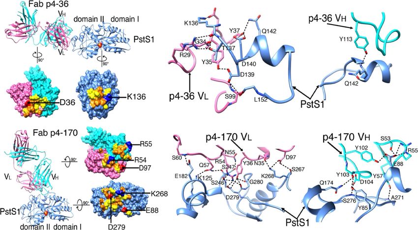
ARTICLE                                                                 NATURE COMMUNICATIONS | https://doi.org/10.1038/s41467-021-20930-0

E
      xposure to Mycobacterium tuberculosis (Mtb) results in a               light chains were amplified by single-cell Ig PCR;20 of these, 85
      spectrum of outcomes, one of which results in active                   constituted natural heavy and light pairs (Supplementary Fig. 2).
      tuberculosis (ATB) disease1. Both innate and adaptive arms             Consistent with previous reports of anti-Mtb B cell responses in
of the immune response have been implicated in immunity to                   humans7, the majority of the antibody sequences were not clonal.
Mtb2,3, but the role of humoral immunity and more specifically,               Based on VH DH JH, VL, and JL and >75% identity in CDRH321, a
antibodies, remains controversial4,5. Several studies have sug-              total of five clonal families were identified in 16 sequences
gested that antibodies may play a protective role in at least a              (Fig. 2c). As previously described21, the VH sequences of the
proportion of otherwise healthy individuals who have a history of            clonal heavy chains were more mutated when compared to the
exposure to Mtb4,6–8, and antibody responses have been corre-                VH sequences that were not part of a clone (Fig. 2d), but both
lated with the protective efficacy of an experimental TB vaccine9.            groups had a similar VH gene usage distribution (Supplementary
During active disease, higher antibody titers to the mycobacterial           Fig. 2b). We produced nine antibodies derived from four clones
glycolipid lipoarabinomannan (LAM) have been correlated with                 expressed in an IgG1 expression vector20 (Fig. 2e, f, expressed
decreased severity of infection10, and there is evidence of B cell           antibodies are in red). We were unable to amplify the light chains
dysfunction during active disease that resolves following treat-             from Clone 5 variants and therefore antibodies from this clone
ment11. In addition, during an active infection, serum antibodies            were not produced. All tested mAbs, except for p4-31, reacted
against the Mtb phosphate transporter PstS1 are detected12,13.               strongly both with PstS1 (with dissociation constants within the
However, functional characterization of monoclonal antibodies                nanomolar range, Supplementary Fig. 3) and with Mtb lysates by
isolated from patients with ATB has not been carried out, and it is          ELISA (Fig. 2g–i). Among the nine antibodies we produced,
not clear whether such antibodies exhibit any antibacterial effects.         mAbs p4-36 and p4-163 exhibited the strongest binding to
   In the present study, we explore B cell responses during ATB,             bacterial lysates (Fig. 2h, i). These mAbs also showed binding to
while focusing on the immune-dominant antigen PstS1. We                      whole-bacteria H37Ra-mCherry compared with isotype control
isolate and analyze a total of 85 monoclonal antibodies (mAbs)               (Supplementary Fig. 4). We therefore decided to focus on these
from one patient with elevated anti-PstS1 responses and find two              antibodies for the rest of the study.
antibodies, p4-36 and p4-163, from distinct clones that demon-                  First, since the anti-PstS1 mAbs were generated during natural
strate inhibitory activity against both BCG and Mtb. Structural              infection occurring in patient P.4, we sought to investigate the
analysis reveals that the two antibodies are directed against two            impact of affinity maturation on their activity. Both p4-36 and
different epitopes on PstS1 and do not compete with one another.             p4-163 showed relatively low levels of somatic hypermutations,
When administered prior to infection of Balb/c mice with Mtb,                with p4-36 exhibiting 6 and 12 amino acid changes in heavy and
both antibodies cause a 50% reduction in lung bacterial burden.              light chains, respectively, and p4-163 exhibiting 10 and 8 amino
Our data show that both p4-36 and p4-163 exhibit antibacterial               acid changes in heavy and light chains, respectively. Nevertheless,
activity and provide the first proof of concept that protective               we hypothesized that these mutations provide the mAbs with
antibody responses can be generated during the course of active              their ability to bind PstS1 and Mtb lysates. To test our hypothesis,
tuberculosis disease, a finding that may inform both therapeutic              we reverted the amino acid sequences of p4-36 and p4-163 back
and preventive vaccine design.                                               to their germline sequences (Supplementary Fig. 5a, b), similarly
                                                                             to what was previously carried out for other naturally elicited
                                                                             mAbs22, thus producing the unmutated predicted germline (gl)
Results                                                                      versions of each antibody: p4-36gl and p4-163gl. The binding of
Isolation of PstS1-specific mAbs. We recruited 26 in-patients                 the germline variants was compared to the binding of the mature
with ATB (Supplementary Table 1). All patients received stan-                antibodies to PstS1, as well as H37Rv and CDC1551 Mtb lysates.
dard treatment, recovered, and were eventually discharged. We                Both mAbs showed a dramatic reduction in their binding when
screened patient sera for anti-Mtb antibody responses prior to               somatic hypermutations were reverted, with the exception of
initiation of antibiotics using lysates from two pathogenic Mtb              mAb p4-36gl that exhibited similar binding to PstS1 as the
strains, H37Rv and CDC1551. Unlike healthy community con-                    mature mAb (p4-36mt) by ELISA. However, SPR measurements
trols, the majority of patients (23/26) showed serum reactivity by           showed a 4.7-fold reduction in affinity (Fig. 2j, Supplementary
ELISA to at least one lysate (Fig. 1a, b). Testing of antibody titers        Fig. 5c–g). To further investigate whether mutations occurring in
against soluble lysate versus cell-wall components of non-                   the heavy or the light chain contribute to antibody activity, we
pathogenic Mtb-H37Ra14 revealed responses against both com-                  produced chimeric antibodies where the heavy chain is mature
ponents, but with higher reactivity against the Mtb surface                  while the light chain is germline (HCmtLCgl), and vice versa
(Fig. 1c). To identify antibody reactivity against specific Mtb               (HCglLCmt). Testing those for binding showed that for p4-36,
proteins, we tested sera against five selected surface-exposed                mutations in the light chain were more significant for binding
proteins (produced in-house, Supplementary Fig. 1) that had been             than those in the heavy chain, as opposed to p4-163 where
described previously to elicit antibody responses in human                   mutations in the heavy chain were more essential for binding
infection (Fig. 1d–h)15,16. The strongest response was directed              compared to mutations in the light chain (Supplementary
against PstS112 (Fig. 1d, i), which has been implicated in Mtb               Fig. 5c–g). Overall, we conclude that both mAbs p4-36 and p4-
virulence13 and is one of three genes previously identified to                163 are specific for PstS1 and acquired somatic hypermutations
contain amino acid sequence variants undergoing diversifying                 during natural infection that resulted in their improved binding
selection in conserved epitopes17,18. Therefore, we decided to               to Mtb.
characterize antibody responses to PstS1.
   We focused on one patient, P.4, who had the strongest
individual response to PstS1 and retained high anti-PstS1                    Activity ex vivo. Next, we sought to test the effects of the mAbs
responses over his treatment course (Fig. 2a). Hence, we sought              p4-36 and p4-163 on Mtb infection in culture. First, we tested the
to isolate mAbs directed against PstS1 and test their effect on              effect of our mAbs on bacterial entry. We infected phorbol
experimental models of Mtb infection. We isolated peripheral                 12-myristate 13-acetate (PMA) differentiated THP-1 macro-
blood mononuclear cells (PBMCs) from whole blood and sorted                  phages23 with the attenuated H37Ra strain with and without
single IgG+, PstS1+ B cells, which comprised 0.5% of the total               PstS1-specific mAbs. The mAbs bound H37Ra within macro-
IgG+ B cell population (Fig. 2b)19. A total of 102 heavy and 90              phages and did not prevent, but rather slightly increased, bacterial

2                NATURE COMMUNICATIONS | (2021)12:602 | https://doi.org/10.1038/s41467-021-20930-0 | www.nature.com/naturecommunications
NATURE COMMUNICATIONS | https://doi.org/10.1038/s41467-021-20930-0 ARTICLE
P.1 P.8 P.15 P.25
a H37Rv lysate
b CDC1551 lysate
c H37Ra P.2 P.9 P.17 P.26
p = 0.0062 P.3 P.10 P.19 P.27
p < 0.0001 p < 0.0001 p < 0.0001 p < 0.0001
P.4 P.11 P.20 P.28
3 3 3 P.5 P.12 P.21 P.29
P.6 P.13 P.22
O.D 650
2
O.D650
O.D650
2 2 P.7 P.14 P.24
N1 N7 N13 N19
1 1 1 N2 N8 N14 N20
N3 N9 N15
0 0 0 N4 N10 N16
Active TB Healthy Active TB Healthy Active TB Healthy Active TB Healthy N5 N11 N17
Lysate Cell Wall
N6 N12 N18
d PstS1
e Ag85b
f CFP10+ESAT6
g Hbha
h Malate Synthase
p = 0.0226
3 3 ns 3 p = 0.0264 3 ns 3 ns
2 2 2 2 2
O.D 650
O.D 650
O.D 650
O.D 650
O.D 650
1 1 1 1 1
0 0 0 0 0
Active TB Healthy Active TB Healthy Active TB Healthy Active TB Healthy Active TB Healthy
i
P.10
P.12
P.13
P.14
P.15
P.17
P.19
P.20
P.21
P.22
P.24
P.25
P.26
P.27
P.28
P.29
P.11
P.1
P.2
P.3
P.4
P.5
P.6
P.7
P.8
P.9
H37Rv Lysate 3
CDC1551 Lysate
H37Ra Lysate
2
H37Ra Cell Wall
PstS1
Ag85b 1
Hbha
Malate Synthase 0
CFP10+ESAT6
Fig. 1 Serological profile of 26 Israeli actively infected tuberculosis patients. Serum responses by ELISA against a Mtb-H37Rv lysate, b Mtb-CDC1551
lysate, c Mtb-H37Ra lysate and cell-wall fraction, and recombinant proteins d PstS1, e antigen 85b (Ag85b), f CFP10 + ESAT6 complex, g Hbha, and
h Malate Synthase. Every symbol represents a single patient, with the legend given on the right side of the figure. ATB patients are in red symbols and
negative community controls are in green symbols. A heat map summarizing ELISA signals against the different antigens and lysates is shown in (i). Donor
P.4 is marked with a black arrow. All error bars are represented as mean ± SD. All statistics were derived for n = 26 ATB patients versus n = 20 negative
community controls, except for (a, b) n = 19 negative community controls. Significance was determined using a two-tailed unpaired Welch’s t test. ns no
significance. Data are representative of at least two independent experiments.
entry into the macrophages (Fig. 3a, upper panel, mAb binding is receptors (FcγRs)6,8,24. To test this, we compared the antibody
shown in the lower panel). We next asked whether the mAbs p4- activity in MGIA by comparing the wild-type IgG1 antibodies
36 and p4-163 could inhibit mycobacterial growth. For this, we with IgG1-N297A (NA), an aglycosylated Fc variant with no
used a whole-blood mycobacterial growth inhibition ex vivo assay binding to FcγRs27. Treating with the NA variants abolished
(MGIA) where human whole blood cells from healthy donors are protection compared with wild-type IgG1 mAbs (Fig. 3d). To
infected with pathogenic bacteria24. This system has been used evaluate the contribution of FcγRs+ effector cells to the inhibitory
previously to test polyclonal antibodies from healthy donors25 activity of the antibodies p4-36 and p4-163, we repeated the
and allows testing of antibody activity in a more physiologically MGIA experiments with selective FcγR blockers. Blocking the
relevant in vitro system. Here, p4-36 and p4-163 were able to main FcγRs expressed by human macrophages, CD16 (FcγRIIIA)
significantly restrict the growth of both Mycobacterium bovis- and CD32a, and CD32b (FcγRIIA and FcγRIIB) together
BCG (BCG) and pathogenic Mtb (Fig. 3b, c)24,26. This activity eliminated the inhibitory activity of both p4-36 and p4-163
was not dose-dependent and was unique to mAbs p4-36 and (Fig. 3e). By contrast, specific depletion of T cells or blocking
p4-163, and was not observed with other anti-PstS1 mAbs, such MHC class II molecules did not affect mAb-induced CFU
as p4-31 and p4-141, showing that ELISA binding to Mtb lysates reduction (Fig. 3e). Altogether, our data imply that the Fc domain
correlated well with their activity ex vivo. of the antibodies facilitates their antibacterial activity. We suggest
We next sought to investigate the mechanism of mAb-induced that FcγRs can mediate bacterial opsonization into the cells, thus
CFU reduction over time. MAbs p4-36 and p4-163 did not inhibit promoting the antibody inhibition of infection. Whether this Mtb
infection, and immediately after infection, more antibody-bound inhibition is due to an active restriction of intracellular bacterial
bacteria were found in THP-1 cells (Fig. 3a). This suggests replication or due to induction of bacterial killing is yet to be
antibody-dependent opsonization of bacteria via Fc gamma determined.
NATURE COMMUNICATIONS | (2021)12:602 | https://doi.org/10.1038/s41467-021-20930-0 | www.nature.com/naturecommunications 3
ARTICLE NATURE COMMUNICATIONS | https://doi.org/10.1038/s41467-021-20930-0
a b
3 P.4 sorted with PstS1
P.4 Sep-17 3.6% 0.5%
2 P.4 Nov-17
O.D 650
P.4 Dec-17
uninfected 3
PE Psts1
IgG-APC
uninfected 2 148 cells sorted
SSC
1
uninfected 1
PBS
0 45%
10 -3 10 -2
Serum dilution FSC VioBlue CD19 VioBlue CD19
e p4-31
f p4-141 Clone 2
Clone 4
c d p4-62 p4-31 Clone 4
p4-36
Clone 3 p4-36
p4-107
p = 0.0041 Clone 3
60 p4-13 p4-107
# of nucleotide mutations in Vh
102 Clone 5
p4-67
p4-48 p4-163
40
p4-85 Clone 2
p4-123
p4-141
p4-43
20 p4-110
p4-9 Clone 1
p4-66
16 p4-170 p4-66
0
Clonal Vh Not Clonal Vh p4-9 Clone 1
p4-110
p4-143
p4-170
p4-123
p4-163
0.05 0.05
g h i j
PstS1 CD1551 lysate H37Rv lysate p4-9
4
Binding relative to mature antibody
p4-110
p4-123 1
1 1
3 p4-163 p4-36mt
p4-170
O.D 650
O.D 650
O.D 650
p4-36gl
2 p4-141
p4-36 p4-163mt
0.5 0.5 0.5
p4-107 p4-163gl
1 p4-31
mGO53
PBSx1
0 0 0 0
H37Rv CDC1551 PstS1 H37Rv CDC1551 PstS1
01
01
01
01
1
1
01
10
01
1
1
1
1
1
10
0.
10
1
00
00
1
0.
0.
00
00
00
0.
00
0.
0.
0.
0.
0.
0.
0.
0.
mAb concentration (µg/ml) mAb concentration (µg/ml) mAb concentration (µg/ml) p4-163 p4-36
Fig. 2 Isolation of anti-PstS1 mAbs from memory B cells of P.4. a Donor P.4 serum responses taken at three different time points (indicated) to
recombinant PstS1 by ELISA. b Isolation of PstS1-specific B cells. Whole-blood-derived lymphocytes were stained for CD19, membrane IgG, and PstS1. A
total of 148 positive cells were single-cell sorted. c Pie charts representing the heavy chain sequences that were amplified from the CD19+/IgG+/PstS1+ B
cells from P.4. The number in the middle of the pies denotes the total number of sequences, and the colored slices indicate clonally related sequences.
Upper panel: all sequences. Lower panel: only the 16 clonally related sequences: clone 1—dark blue, clone 2—purple, clone 3—magenta, clone 4—teal,
clone 5—green. d Nucleotide mutations in VH of the clonal versus nonclonal sequences. Error bars are represented as mean ± SEM. n = 16 clonal Vh
sequences and n = 85 nonclonal Vh sequences. e, f are dendrograms (created using Geneious software) of the clonally related sequences, heavy and light
chains, respectively. The mAbs selected for expression are colored in red. g–i The binding of nine mAbs by ELISA to recombinant PstS1 (g), Mtb-CDC1551
lysate (h), and Mtb-H37Rv lysate (i). j Comparing binding by ELISA of the mature (mt) and the predicted germline (gl) versions of p4-36 (magenta) and
p4–163 (dark blue), to recombinant PstS1, Mtb-CDC1551 lysate, and Mtb-H37Rv lysate. AUC binding score of each antibody was determined as relative to
the AUC of the mature antibody, which was normalized to 1 (see also Supplementary Fig. 5). All data are representative of at least two independent
experiments (g–j).
Structure–function studies. To further understand the mechan- Table 3). Ten hydrogen bonds are observed at the interface, and
ism of the protective antibody binding to its target, we produced one salt bridge is formed between Asp36p4–36 CDRL1 and
antigen-binding fragments (Fabs) of p4-163 and p4-36 and pre- Lys136PstS1 (D36L[OD1]-K136PstS1[NZ]) (Fig.4b and Supple-
pared Fab–PstS1 complexes for crystallization. We determined the mentary Table 3).
structure of Fab p4-36 in complex with PstS1 at a resolution of No crystal could be obtained with the complex of Fab p4-163
2.1 Å (Fig. 4a and Supplementary Table 2). Two PstS1–Fab p4-36 and PstS1. Thus, we switched to p4-170, which is a closely related
heterodimers were in the asymmetric unit of the crystal and had variant of p4-163 and a member of the same B cell clone (Clone 1,
only minor differences in the constant domains of the bound Fig. 2e, f), and has 97.6% and 93.86% amino acid sequence
Fabs (Supplementary Fig. 6). P4-36 binds a contiguous epitope identity to p4-163 heavy and light chains, respectively (Supple-
located on an alpha-helix formed by residues 141–145 and pre- mentary Fig. 5a, b). In addition, p4-170 exhibited similar
ceding residues 136–137 and residues 139–140 (Fig. 4b and inhibitory activity in MGIA to p4-163 (Supplementary Fig. 9).
Supplementary Fig. 7a), within a small surface area of 583 Å 2. The Fab p4-170 and PstS1 complex could be crystallized, and the
The contacts between p4-36 and PstS1 are mostly Van der structure was determined at a resolution of 2.4 Å with one
Waals and hydrogen bonds and contributed largely by PstS1–Fab p4-170 heterodimer in the asymmetric unit (Fig. 4c
complementarity-determining region 1 (CDR1) and CDR3 of the and Supplementary Table 2). As revealed by the structure of the
light chain (CDRL1 and L3) and CDR3 of the heavy chain complexes, p4-170 binds to a different epitope from the one
(CDRH3) (Fig. 4b, Supplementary Fig. 8a, b and Supplementary recognized by p4-36.
4 NATURE COMMUNICATIONS | (2021)12:602 | https://doi.org/10.1038/s41467-021-20930-0 | www.nature.com/naturecommunicationsNATURE COMMUNICATIONS | https://doi.org/10.1038/s41467-021-20930-0 ARTICLE
p4-163 p4-170 p4-36 p4-141 mGO53 no antibody
a
6.5% 11.4% 8% 6.5% 2.9% 5.1%
FSC
mCherry
p4-163 p4-170 p4-36 p4-141 mGO53 no antibody
cell count
anti-human VioBlue
b c d
C
1.5x10⁶ 5 105
p = 0.0234
p = 0.0494
p = 0.0399
p = 0.0422
p = 0.0099
p = 0.0351
p = 0.0233
p = 0.0143
p = 0.0105
p = 0.0308
ns
4 105
p = 0.0156
p = 0.0254
ns ns ns ns
6 ns ns
1x10 1x10⁶
3 105
Mtb CFU
Mtb CFU
BCG CFU
2 105
0.5x10⁵
1 105
5
5x10
0 0 0
5 mg/ml
0.2 µg/ml
5 µg/ml
0.2 µg/ml
5 µg/ml
0.2 µg/ml
5 µg/ml
0.2 µg/ml
5 µg/ml
PBS
mGO53
PBS
mGO53
NA
NA
WT
WT
0.2 µg/ml
1 µg/ml
5 µg/ml
0.2 µg/ml
1 µg/ml
PBS
p4-36 p4-163
p4-36 p4-163 p4-36 p4-163 p4-31 p4-141
e
1.5
p < 0.0001
p = 0.0006
p < 0.0001
p < 0.0001
p = 0.0001
p < 0.0001
p < 0.0001
p < 0.0001
p < 0.0001
p = 0.0085
p < 0.0001
p = 0.0001
ns ns ns ns ns ns ns ns ns
Normalized infection
1.0
0.5
0.0
PBS
mGO53
p4-36
p4-163
mGO53
p4-36
p4-163
mGO53
p4-36
p4-163
mGO53
p4-36
p4-163
mGO53
p4-36
p4-163
mGO53
p4-36
p4-163
mGO53
p4-36
p4-163
blank CD3+ cell depletion CD8+ cell depletion CD4+ cell depletion anti-HLA antibody anti-CD16 antibody anti-CD16 antibody
anti-CD32 antibody
Fig. 3 Anti-PstS1 mAbs inhibit Mtb in culture. a Upper panel: gating strategy of H37Ra-infected macrophages, mCherry positive. PMA-differentiated THP-
1 cells preincubated with mAbs p4-163, p4-170, p4-36, p4-141, and the isotype control mAb mGO5320. For each treatment n = 90,000 cells were analyzed
by flow cytometry. Lower panel: histograms showing the frequencies of intracellular antibody-bound bacteria as detected by anti-human VioBlue antibody
staining. For each mAb, the binding histogram is shown in red and compared to the isotype control histogram, which is depicted in a gray overlay.
b, c Activity of anti-PstS1 mAbs at indicated concentrations in a human whole blood mycobacterial growth inhibition assay (MGIA) after 96 h of infection
with BCG or pathogenic Mtb, respectively. CFU was determined in n = 3 biological repetitions. d Activity of anti-PstS1 mAbs (5 µg/ml) used as IgG1
(named “WT”, full columns) or as N279A Fc variants (named “NA”, empty columns) in MGIA. Black and clear shapes correspond to two independent
experiments. CFU was determined in n = 5–6 biological repetitions. e Activity of anti-PstS1 mAbs (5 µg/ml) in MGIA following depletion of CD3+ T cells,
CD4+ T cells, CD8+ T cells, blockade of MHC II (anti-HLA), and CD16 or CD32 or both (anti-CD16, anti-CD32, marked with a red rectangle). Black,
cayenne, and clear shapes correspond to three representative independent experiments. In each experiment, the data points were compared to average
CFU infection in PBS, which was normalized to 1. CFU was determined in n = 4–10 biological repetitions. All error bars are represented as mean ± SD.
Significance was determined using a one-tailed unpaired t test (b), two-tailed unpaired t test (c, d) for black shapes (d), or one-way ANOVA with Tukey’s
multiple-comparison test (e). All statistical analyses are relative to PBS. ns no significance. Data are representative of at least two independent
experiments.
NATURE COMMUNICATIONS | (2021)12:602 | https://doi.org/10.1038/s41467-021-20930-0 | www.nature.com/naturecommunications 5ARTICLE NATURE COMMUNICATIONS | https://doi.org/10.1038/s41467-021-20930-0
a b
c d
p4-36 contact residues p4-170 contact residues
e
K136E D139A D140A K268E D279A S246G PstS1 WT
3 3 3 3 3 3 3
O.D 650
O.D 650
O.D 650
O.D 650
O.D 650
O.D 650
O.D 650
2 2 2 2 2 2 2
1 1 1 1 1 1 1
0 0 0 0 0 0 0
0.0001 0.001 0.01 0.1 1 0.0001 0.001 0.01 0.1 1 0.0001 0.001 0.01 0.1 1 0.0001 0.001 0.01 0.1 1 0.0001 0.001 0.01 0.1 1 0.0001 0.001 0.01 0.1 1
0.0001 0.001 0.01 0.1 1
mAb concentration (µg/ml)
p4-9 p4-123 p4-163 p4-170 p4-36 mGO53 PBSx1
Fig. 4 MAbs p4–170 and p4–36 recognize different epitopes on top of PstS1. a Top: Ribbon diagrams show the crystal structure of PstS1 in a complex
with Fab p4–36 (PDB ID 7DM1). The Fab p4-36 heavy and light chains are colored cornflower cyan and hot pink, respectively. The PstS1 structure is
colored blue. The bound phosphate (Pi) is represented as filled balls with oxygen and phosphorus atoms colored red and yellow, respectively. Bottom: an
open-up surface-shadowed representation showing the contact interface. Residues involved in hydrogen bonds and Van der Waals contacts are highlighted
in orange and yellow, respectively. Positively and negatively charged residues involved in the formation of the salt bridges are highlighted in blue and red,
respectively. b Close-up view of the interface between PstS1 and Fab p4-36. The dashed lines indicate hydrogen bonds and salt bridges. c Left: Ribbon
diagrams show the crystal structure of PstS1 in complex with Fab p4-170 (PDB ID 7DM2). Right: An open-up surface-shadowed representation showing
the contact interface. The color schemes used are the same as in (a). d Close-up view of the interface between PstS1 and Fab p4-170. The dashed lines
indicate hydrogen bonds and salt bridges. e Four Clone 1 variants (mAbs p4-9, p4-123, p4-163, and p4-170, dark blue), and mAb p4-36 (magenta), as well
as negative control mAb mGO5320 (gray), were tested for binding by ELISA to six-point mutant PstS1 proteins. The binding curves of PstS1 mutated
in p4-170 contact residues K268E, D279A, and S246G, as well as the PstS1-mutated p4-36 contact residues K136E, D139A, and D140A are shown.
The binding curves to wild-type PstS1 are shown in the right panel of the figure.
The epitope recognized by p4-170 is highly discontinuous and and Asp279PstS1[OD2]–Arg54L[NH2] (Fig. 4c, d and Supplemen-
conformationally proximate to the Pi binding site in the middle of tary Table 4).
the molecule, between the two domains I and II of PstS1 (Fig. 4c). To confirm the key antibody: antigen contact residues
The epitope is a large surface of 977 Å 2 and comprises three helices identified by the structures, we generated a panel of point
and four loops of PstS1 (Fig. 4d and Supplementary Fig. 7b). The mutations in PstS1. Mutating residues at the p4-170:PstS1
interactions between p4-170 and PstS1 are mediated through salt interface, S246GPstS1, K268EPstS1, and D279APstS1 reduced the
bridges, hydrogen bonds, and Van der Waals contacts (Fig. 4d binding of all tested Clone 1 mAb variants, but not the binding of
and Supplementary Table 4). Except for CDRH1, all the CDRs of p4-36 (Fig. 4e). Amino acid substitutions in the alpha-helix that
p4-170 are involved in the interactions with residues 85, 88, and 92 holds the epitope of p4-36 residues K136EPstS1, D139APstS1, and
of the PstS1 domain I and residues 125, 174, 177–178, 181–182, D140APstS1 reduced p4-36 binding. The binding of Clone 1 mAbs
190, 246–248, 267–268, 270–271, 275–276, and 279–281 of domain was also reduced, indicating that the alpha-helix bound by p4-36
II (Fig. 4d, Supplementary Fig. 8c, d and Supplementary Table 4). might be essential for the folding of PstS1.
The hydrogen bonds are formed with both the main-chain and We next asked whether the binding of p4-36 or p4-170 could
side-chain atoms of PstS1. Four salt bridges are formed at interfere with multimerization of the transporter. For this, we
the contact interface, including Glu88PstS1[OE1]–Arg55H[NE], performed modeling of the binding of PstS1 to the transporter
GLu88PstS1[OE1]–Arg55H[NH1], Lys268PstS1[NZ]–Asp97L[OD1], complex. According to our prediction, the bound antibodies show
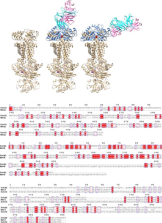
6 NATURE COMMUNICATIONS | (2021)12:602 | https://doi.org/10.1038/s41467-021-20930-0 | www.nature.com/naturecommunicationsNATURE COMMUNICATIONS | https://doi.org/10.1038/s41467-021-20930-0 ARTICLE
no blockage to the assembly of the PstA–B–C–S complex and do rather than IgG. However, in that study, ‘protection’ was defined
not block the binding of PstS1 to the PstA, B, C transporter as decreased viable Mtb within cells shortly after infection.
complex (Fig. 5). We conclude that p4-36 and p4-170 do not Unfortunately, the study evaluated neither in vivo activity nor the
function by inhibiting the transporter activities . precise molecular mode of antigen binding. While the Balb/c
mouse model we use in this study does not recapitulate lung
granulomas, which are the hallmark of active TB infection in
Activity in vivo. Finally, we tested the activity of the two mAbs
humans, passive transfer of mAbs before pathogenic Mtb infec-
p4-36 and p4-163 on Mtb infection in vivo. For this, we used
tion resulted in a significant reduction of bacterial lung loads in
wild-type Balb/c mice. We injected 0.5 or 1 mg mAb per mouse
mice indicating a preventive mAb activity.
intraperitoneally five hours prior to aerosol infection with
This is the first report of human anti-Mtb mAbs that have a
pathogenic Mtb. After 2 weeks, the mice were sacrificed and lung
modest, yet robust, activity in vivo, as well as resolving their
bacterial burden determined. Lung burdens were reduced in mice
structure along with their corresponding Mtb target. PstS1 DNA
pretreated with mAbs p4-36 and p4-163, or both by approxi-
was previously reported to elicit protective T-cell immunity
mately 0.5 log CFU (Fig. 6), which agrees with the MGIA results
against Mtb in mice37. The fact that the germline version of p4-
and verifying that the mAbs have anti-Mtb-inhibiting activity.
36, p4-36gl, binds recombinant PstS1 suggests that PstS1 could be
used as an immunogen in humans to elicit B cell responses and
Discussion p4-36-like antibodies in naive populations. Our study has
Here, we describe a patient, P.4, who had active tuberculosis and implications for the development of new anti-Mtb therapies and
was selected from a cohort of infected patients due to his strong prevention.
anti-Mtb serum responses to the known immunodominant
antigen PstS1. While the activity of the two anti-PstS1 mAbs we
isolated is modest (about 0.5 log), to our knowledge, this is the Methods
Study design. The objective of this study was to investigate whether antibodies
first description that naturally elicited PstS1 antibodies have anti- isolated during active tuberculosis (ATB) disease can inhibit Mtb in vivo and
Mtb activity in infection. elucidate their corresponding Mtb target and mechanism of action. Donor P.4 was
PstS1 is a 38-kDa phosphate-binding periplasmatic protein, one selected based on exceptional serum binding to Mtb lysates and to recombinant
PstS1 protein. Staining with PstS1 and single-cell sorting of antigen-positive
of three subunits of the Mtb phosphate-specific transporter (Pst) memory B cells from donor P.4 allowed isolation of four new clones of antibodies,
complex12. PstS1 is an immunodominant marker for ATB28–30. two of which were able to inhibit the growth of pathogenic Mtb ex vivo and in vivo.
PstS1 is also necessary for Mtb virulence; PstS1-deletion mutants We analyzed structurally the two mAbs and discovered that they are directed
were attenuated in a mouse-infection model13. Intriguingly, PstS1 against two nonoverlapping sites on PstS1. Their ability to inhibit Mtb growth was
was identified as one of only three Mtb genes that are subjected to tested by infecting healthy human whole blood cells in vivo, in Balb/c mice. The
epitopes were identified by X-ray crystallography.
evolutionary sequence diversification, suggesting that PstS1 var-
iation plays a role in Mtb immune evasion17,18.
In the present study, we characterize two different sites on Ethics statement. For the patient studies, the Tel Aviv University Institutional
PstS1 that can be targeted by antibodies capable of inhibiting Mtb Review Board (IRB) approved all studies involving patient enrollment, sample
collection, and clinical follow-up. Donor P.4 provided written informed consent
growth. P4-163 is a member of the largest B cell clone of nine prior to participating in this study, and The Tel Aviv University and Shmuel
mAbs that shared >93% sequence identity. The high-resolution Harofe Hospital Institutional Review Boards approved all studies involving patient
structure of a clonal variant of p4-163, p4-170, revealed a large, enrollment, sample collection, and clinical follow-up (protocol numbers 33.18 and
sparse, and highly conformational epitope that is adjacent to the 0058). Donor P.4 was selected from a group of 26 active pulmonary TB patients
that were followed by the Pulmonary and Tuberculosis Department of Shmuel
active site of PstS1. Intriguingly, seven of p4-170 contact residues Harofe hospital in Israel and is also referred to as subject ID 109004. Fresh human
(Lys268PstS1, Pro270PstS1, Ala271PstS1, Ile275PstS1, Ser276PstS1, whole blood for MGIA assays was obtained from volunteers under IRB-approved
Asp279PstS1, and GLy280PstS1) overlap with a highly conserved protocol (2014-2-25) of the Beijing Tumor and Thoracic Hospital. For the mouse
Mtb T-cell epitope 259-AAAGFASKTPANQAISMIDG-280, studies, this study was carried out in strict accordance with the guidelines of the
Chinese Association for Laboratory Animal Sciences and approved by the Animal
Immune Epitope Database number 3517,31. The mAb p4-36, a Ethics Committee of Beijing Chest Hospital, Capital Medical University.
member of a different and smaller B cell clone, binds an alpha-
helix structure within residues 136PstS1–145PstS1, with the epitope
being 30 Å distant from the epitope bound by p4-170. Study participants. Twenty-six active pulmonary TB patients were recruited from
the Pulmonary and Tuberculosis Department of Shmuel Harofe hospital in Israel.
Donor P.4 had active tuberculosis; therefore, by definition, the Patients were diagnosed with active pulmonary TB by sputum smear and positive
mAbs we isolated did not prevent or eliminate Mtb infection in bacterial culture and treated with antibiotics. Blood samples for screening for anti-
this patient. However, evidence from other infectious diseases Mtb serum antibodies were drawn before receiving antibiotic therapy. For patients
shows that B cell responses develop in parallel and subsequent to who exhibited high anti-Mtb serum activity, the blood draw was repeated two
high pathogen loads that stimulate B cell activation and additional times during hospitalization. Donor P.4, ID 109004 (Supplementary
Table 1), a 35-year-old male, was diagnosed with drug-sensitive TB, and a small
maturation, causing neutralizing/protective antibodies to arrive blood donation was first collected on September 25, 2017. This donor was asked for
‘too little, too late', without a significant benefit to the individual a large donation of whole blood, from which PBMCs were isolated by Ficoll gra-
that produced them22,32–34. Considering the low extent of affinity dient density centrifugation. Serum samples of healthy donors served as controls
maturation, as reflected by the relatively low number of somatic and were obtained from the Israeli blood bank. TB exposure history of these
subjects was not available.
hypermutations in p4-36 and p4-163, it is possible that these
B cell clones only started their evolution and did not reach their
full anti-Mtb potential. On the other hand, the fact that these Bacterial culture, lysate, and cell-wall fractions. Lysates of Mtb-H37Rv (Cat #
antibodies ultimately arise provides evidence that B cells play an NR-14822) and the Mtb-CDC1551 (Cat # NR-14823) were obtained from BEI
resources https://www.beiresources.org/. For the preparation of H37Ra14 lysate and
active role during ATB. cell-wall fractions, H37Ra bacterial culture was grown in Middlebrook 7H9 broth
Despite the long-standing controversy, recent studies have supplemented with 10% OADC, 0.05% Tween-80, and 0.5% glycerol, at 37 °C in a
demonstrated that in certain cases, anti-Mtb humoral responses shaking incubator to reach an optimal density of O.D600 = 0.4. The bacterial cul-
can be protective7,24,35 or correlate with lack of active disease or ture was centrifuged at 3180 g for 15 min at room temperature. The pellet was
resuspended in phosphate-buffered saline (PBS)×1 containing 1% phe-
even lack of infection4,6,35,36. Only one prior study profiled the nylmethylsulfonyl fluoride (Sigma) and was lysed by sonication. Following cen-
human B cell responses against Mtb antigens on a monoclonal trifugation at 29,800 g for 30 min at 4 °C, the lysate was clarified, and the cell-wall
level7. This study attributed bacterial inhibition to IgA isotypes fraction was resuspended in PBS×1.
NATURE COMMUNICATIONS | (2021)12:602 | https://doi.org/10.1038/s41467-021-20930-0 | www.nature.com/naturecommunications 7ARTICLE NATURE COMMUNICATIONS | https://doi.org/10.1038/s41467-021-20930-0 Fig. 5 Structure modeling of the binding of PstS1 to the PstA–B–C phosphate transporter complex. a Structural superimposition of the Fab–PstS1 complexes with MetQ of the MetNIQ ABC transporter complex (in gold) (PDB code: 6CVL) suggested a possible binding mode of PstS1 to the PstA–B–C phosphate transporter complex. The bound antibodies show no blockage to the assembly of the PstA–B–C–S complex. b Sequence alignments showing that the MetNIQ complex components share significant sequence similarities to these of the PstA–B–C phosphate transporter complex of Mtb. 8 NATURE COMMUNICATIONS | (2021)12:602 | https://doi.org/10.1038/s41467-021-20930-0 | www.nature.com/naturecommunications
NATURE COMMUNICATIONS | https://doi.org/10.1038/s41467-021-20930-0 ARTICLE
5 additional washing step, the plates were incubated with peroxidase-conjugated goat
p = 0.0036
p = 0.0051
p = 0.0024
p = 0.0238
p = 0.0026
5x10
anti-human IgG, developed, and read as indicated above.
ns For PstS1 mutant ELISA, plates were coated overnight at 4 °C with 5μg/ml of
5 recombinant PstS1 wild type and mutants. The next day, after washing and
4x10 blocking as indicated above, plates were incubated for 1 h at room temperature
with anti-PstS1 mAbs in eight consecutive fourfold dilutions, starting from
Lung TB CFU
5 concentration of 1.25 μg/ml. After an additional washing step, the plates were
3x10 incubated with peroxidase-conjugated goat anti-human IgG, developed, and read
as indicated above.
5
2x10
Single B cell sorting, sequencing, cloning, and expression of antibodies.
Patients’ purified PBMCs were isolated by ficoll (GE Healthcare) and frozen at
5 liquid nitrogen. PBMCs were thawed and enriched for B cells by CD20 magnetic
1x10
microbeads (MACS, Miltenyi Biotec). The CD20+ B cell fraction was stained for
15 min at 4 °C with anti-human antibodies: CD19-VioBlue (1:100, Miltenyi Biotec),
0 IgG-APC (1:20, Miltenyi Biotec), and with biotinylated PstS1 preincubated with
streptavidin-PE (1:10, Miltenyi Biotec). All the PstS1-PE-positive IgG+ CD19+
PBS
mGO53
0.5 mg/ml
1mg/ml
0.5 mg/ml
1mg/ml
0.5 mg/ml
cells were single-cell sorted into 4 μl of lysis buffer (PBS, RNAsin-Promega, and
0.1 M DTT). Rescue primers were used to amplify both heavy chains38 and Igλ
genes39, and regular primers were used for IgK chains40. All PCR products were
sequenced and analyzed for Ig gene usage, CDR3, and the number of VH/VL
p4-36 p4-163 p4-163 somatic hypermutations (IgBLAST, www.ncbi.nlm.nih.gov/igblast and IMGT,
+ www.imgt.org). Purified, digested PCR products were cloned into human Igγ1, Igk,
p4-36 or Igλ-expression vectors as previously described40 and produced by transient
transfection of IgH, IgK, and IgL expression plasmids into exponentially growing
Fig. 6 Anti-PstS1 mAbs inhibit Mtb in Balb/c mice. Activity of anti-PstS1 Expi293F cells, as previously described41.
mAbs: p4-36 (magenta bars), p4-163 (dark-blue bars), as well as the
negative control mAb mGO53 (gray bars) at indicated concentrations in Biacore (SPR). Biocare assay was performed on a Biacore T200 instrument at
Balb/c mice. Mice were injected once intraperitoneally with mAbs at 25 °C. In total, 5 μg/ml Capture SelectTM Biotin Anti-IgG-Fc (multispecies) Con-
indicated amounts 5 hours prior to aerosol infection with pathogenic Mtb. jugate (ThermoFisher Scientific) was immobilized to sensor chip SA, series S (GE
Healthcare) with a contact time of 600 s and flow rate of 10 μl/min. In total,
Mice were euthanized after 2 weeks, and lung bacterial burden measured.
0.25 μg/ml anti-PStS1 mAbs were injected with a contact time of 600 s and a flow
Error bars are represented as mean±SD. In each treatment n = 6 mice. rate of 10 μl/min. Following anti-PStS1 mAbs binding to the Capture Select, PstS1
Significance was determined using a two-tailed unpaired t test. ns no was injected in four concentrations: 7.81, 31.25, 125, and 500 nM. Following each
significance. The results are representative of three independent cycle, the regeneration step was done with 0.1 M glycine, pH 2 at a flow rate of
experiments. 30 μl/min for 1.5 min. Three replicates were performed for each mAb and all
samples were diluted in HBS-EP buffer (0.01 M HEPES, 0.15 M NaCl, 0.003 M
EDTA, and 0.05% Tween-20, pH 7.4). Sensograms were fitted to a 1:1 binding
Expression of recombinant Mtb antigens. Mtb antigens PtsS1 (Rv0934), Ag85b model using nonlinear regression in the BIA evaluation software. KD was calculated
(Rv1886c), Hbha (Rv0475), CFP10 (Rv3874), ESAT6 (Rv3875), and Malate Syn- using the ratio of the kinetic constants KD = Kd/Ka.
thase (Rv1837) were cloned into a pMALp vector as a fusion to maltose-binding
protein (MBP), with a TEV cleavage site separating the recombinant Mtb proteins Flow cytometry for direct and intracellular antibody binding. The binding of
and MBP (Supplementary Fig. 1). The recombinant Mtb proteins were C-terminally p4-mAbs to H37Ra-mCherry was determined as follows: bacteria were grown to
tagged with 6×His and a specific BirA biotinylation sequence and were expressed in OD600 = 0.4, centrifuged, and washed with PBS. A total of 108 colony-forming
Escherichia coli (E-coli, BL21 strain). Shortly, plasmid-transformed E. coli was grown units (CFU) were incubated with or without 50 μg of p4-mAbs for 1 h at room
to an optimal density of O.D600 = 0.6., at which protein expression was induced by temperature, washed with a fluorescence-activated cell sorting buffer (PBS, 1% FBS,
adding isopropyl-β-D-1-thiogalactopyranoside (IPTG) (0.5 mM), and the culture and 2 mM EDTA), and the bacteria were fixed and permeabilized (BioGems). The
was grown for 4 h at 30 °C in a shaking incubator of 225 rpm. Next, bacteria were fixated bacteria were incubated with anti-human IgG-VioBlue (1:50 dilution,
pelleted by centrifugation at 9700 g for 15 min at 4 °C, and the pellet was frozen and Miltenyi Biotec) for 20 min on ice, the unbound antibodies were washed, and the
kept at a temperature of −20 °C. Afterward, bacterial pellets were thawed and samples were analyzed by cytoflex. For staining of intracellular THP-1-infected
resuspended in 50 mM NaH2PO4, 300 mM NaCl, 10 mM imidazole, pH = 8, H37Ra-mCherry, human monocyte THP-1 cell line23 (ATCC) was cultured in
Triton-X 0.1% (Sigma), and protease inhibitor cocktail (Sigma) (“binding buffer”), RPMI medium (Biological Industries) and differentiated for 24 h by addition of
and lysed by sonication. Following additional centrifugation at 29,800 g for 30 min phorbol 12-myristate 13-acetate-(PMA) 160 ng/ml at 37 °C. On the day of infec-
at 4 °C, the protein was purified from the supernatant phase using Ni sepharose tion, bacteria were incubated with 50 μg/ml p4-mAbs and mGO5320, used as an
beads (GE Healthcare), TEV cleavage, and elution with 50 mM and 100 mM imi- isotype control for 30 min at room temperature. The p4-mAbs and bacterial mix,
dazole (Sigma). Enzymatic site-specific biotinylation was carried out by BirA bio- along with anti-human IgG-VioBlue (1:200 dilution, Miltenyi Biotec), were added
tinylation kit (Avidity). All recombinant Mtb proteins were stored in PBS×1. to 8 × 10⁵ differentiated THP-1 macrophages at MOI 1:10 for 3 h at 37 °C. Fol-
lowing 5 washes with PBS, the cells were incubated with 250 μg/ml Amikacin
sulfate salt (Sigma) for 1 h at 37 °C. Cells were washed and dislodged using cell
ELISAs. High-binding 96-well ELISA plates (Corning, 9018) were used for all dissociation solution (Biological Industries), and then fixed and permeabilized
experiments. For sera binding to Mtb strains, plates were coated overnight at 4 °C (BioGems).
with 1–2.5 μg/ml of pathogenic bacterial lysates, H37Rv, and CDC1551 (both
received from BEI resources), or H37Ra lysate or cell-wall fractions. For sera
binding to recombinant Mtb antigens, plates were coated overnight at 4 °C with Mycobacterial growth inhibition assay (MGIA). Blood from healthy volunteer
5 μg/ml of PstS1, Ag85b, Hbha, Malate Synthase, and ESAT6 + CFP10. ESAT6 + donors was drawn into a CPT vacutainer tube. Blood from the same donor was
CFP10 were premixed at a 1:1 ratio. The next day, after washing 3 times with used for any given set of experiments, to rule out donor-mediated variability. The
PBS + 0.05% Tween-20, the plates were blocked for 2 h at room temperature with blood was aspirated into a new 50-ml Falcon tube, and sodium citrate (3.2 g/L) was
3% bovine serum albumin, 20 mM EDTA, and 0.05% Tween-20 in PBS (“blocking added as an anticoagulant at a ratio of 1:9 (sodium citrate/blood). Blood was
buffer”), followed by a 1-hour incubation at room temperature with polyclonal diluted 1:1 with RPMI-1640 (Gibco). A local clinical isolate of pathogenic Mtb,
Mtb-infected sera or negative healthy controls at 1:100 and 1:300 dilutions in Beijing strain (strain 16542), was passed through a 5-µm filter to remove clumps.
blocking buffer. After an additional washing step, the plates were incubated for 1 h The OD was checked and diluted with the RPMI-1640 medium. Next, 0.1 ml of the
with peroxidase-conjugated goat anti-human IgG (Jackson ImmunoResearch) at a bacterium (105 CFU) was added to 0.9 ml of the diluted blood in 15-ml sterile
final concentration of 0.16 μg/ml at room temperature and developed by adding falcon tubes. A similar protocol was used with the MGIA and BCG infection. An
TMB substrate (Abcam). The plates were read in an ELISA plate reader after antibody (or PBS) was added to each tube in concentrations as indicated, in tri-
20 min at O.D650. plicates. The tubes were incubated for 96 h at 37 °C in a shaking incubator at
For anti-PstS1 and germline mAbs ELISA, plates were coated overnight at 4 °C 20 rpm. After incubation, the tubes were centrifuged for 10 min at 2000 g, and then
with 5–10 μg/ml of recombinant PstS1 antigen or pathogenic bacterial fractions as 8 ml of sterile water was added per tube and the tubes were incubated for 10 min at
described above. The next day, after washing and blocking as indicated above, plates room temperature. After blood lysis, tubes were spun at 2000 g for 10 min, the
were incubated for 1 h at room temperature with anti-PstS1 or germline mAbs in supernatant discarded, and the pellet resuspended in 1 ml of PBS. The samples
eight consecutive fourfold dilutions, starting from concentration of 5 μg/ml. After an were serially diluted and then plated onto OADC-supplemented 7H10 agar plates
NATURE COMMUNICATIONS | (2021)12:602 | https://doi.org/10.1038/s41467-021-20930-0 | www.nature.com/naturecommunications 9ARTICLE NATURE COMMUNICATIONS | https://doi.org/10.1038/s41467-021-20930-0
and incubated for 3 weeks at 37 °C. For the depletion and/or blocking experiments Data Bank (PDB), accession codes 7DM1 (a complex of PstS1 and Fab p4-36) and 7DM2
in the MGIA, the experiment was performed as above but with the following (a complex of PstS1 and Fab p4-170). The authors declare that all unique materials used
modifications24. For the T-cell depletion experiments, following a blood draw of are readily available from the authors upon MTA agreement. Source data are provided
50 µl of the Human CD3 MicroBeads, CD4 or CD8 MicroBeads (Miltenyi Biotec) with this paper.
were added separately into a 2-ml diluted blood sample each and incubated for
30 min at 4 °C. The LS columns (Miltenyi Biotec) were attached to the Midimacs
and primed with 3 ml of PBS and 3 ml of RPMI-1640 sequentially. Next, 6 ml of the
Received: 3 May 2020; Accepted: 4 January 2021;
diluted samples with the beads as above were added to the column. Control blood
samples without CD3, CD4, or CD8 microbead incubation were prepared under
the same conditions as above. For the receptor blocking experiments, the Fc
receptor antibodies anti-human CD32A (clone: 6C4, eBioscience) and anti-human
CD16 (clone: 3G8, BioLegend) were used as the blocking antibodies for this assay.
In the designated samples, 1 µg of CD32 or 2 µg of CD16 or both were used. For the References
MHC class II blocking experiments, 4 µg/ml mouse anti-human MHC class II 1. Pai, M. et al. Tuberculosis. Nat. Rev. Dis. Prim. 2, 16076 (2016).
antibody (clone Tu39, BD Biosciences catalog # 555557) was used in the assay. 2. Ardain, A. et al. Group 3 innate lymphoid cells mediate early protective
immunity against tuberculosis. Nature 570, 528–532 (2019).
Mouse-infection assay. Mice were caged in groups of six in the SPF animal 3. Scanga, C. A. et al. Depletion of CD4(+) T cells causes reactivation of murine
facility. Control mice were caged separately from experimental mice. Monoclonal persistent tuberculosis despite continued expression of interferon gamma and
antibodies (500 µg/mouse, unless indicated otherwise) were injected via the nitric oxide synthase 2. J. Exp. Med. 192, 347–358 (2000).
intraperitoneal (IP) route into 6–8-week-old, specific pathogen-free female Balb/c 4. Li, H. & Javid, B. Antibodies and tuberculosis: finally coming of age? Nat. Rev.
mice (Strain No. 211, purchased from Beijing Vital River Laboratory Animal Immunol. https://doi.org/10.1038/s41577-018-0028-0 (2018).
Technology Co. Ltd., China), 5 h prior to aerosol infection by the Glas-col inha- 5. Casadevall, A. Antibodies to Mycobacterium tuberculosis. N. Engl. J. Med. 376,
lation exposure system. PBS was injected as a control. Cultures of Mtb strain 165 283–285 (2017).
were diluted to the concentration of 1 × 10⁶ CFU/ml (10 ml) for the aerosol 6. Lu, L. L. et al. IFN-gamma-independent immune markers of Mycobacterium
infection. The mice were loaded into the basket of the inhalation exposure system tuberculosis exposure. Nat. Med. 25, 977–987 (2019).
with the lid secured, and the bacterial suspension was loaded into the nebulizer. 7. Zimmermann, N. et al. Human isotype-dependent inhibitory antibody responses
The set program of the inhalation exposure system was preheating for 15 min and against Mycobacterium tuberculosis. EMBO Mol. Med. 8, 1325–1339 (2016).
nebulizing for 30 min, cloud decay for 30 min, and decontamination for 15 min. 8. Chen, T. et al. Association of human antibodies to arabinomannan with
About 24 h following infection, 3 of the PBS control mice were euthanized by enhanced mycobacterial opsonophagocytosis and intracellular growth
cervical dislocation and the lungs were removed and homogenized by MP Fastprep. reduction. J. Infect. Dis. 214, 300–310 (2016).
The actual infection dose delivered to the lungs was verified as between 100 and 9. Fletcher, H. A. et al. T-cell activation is an immune correlate of risk in BCG
200 CFU/mouse for all experiments. The other mice were euthanized by cervical vaccinated infants. Nat. Commun. 7, 11290 (2016).
dislocation after 2 weeks, and the lungs were homogenized and plated for the CFU 10. Correia-Neves, M., Sundling, C., Cooper, A. & Kallenius, G.
load on OADC-supplemented 7H10 plates as above. The plates were incubated in a Lipoarabinomannan in active and passive protection against tuberculosis.
37 °C incubator for around 3 weeks before colonies were counted. Front. Immunol. 10, 1968 (2019).
11. Joosten, S. A. et al. Patients with tuberculosis have a dysfunctional circulating
Complex preparation and crystallization. The Fabs of p4-36 or p4-170 were B-cell compartment, which normalizes following successful treatment. PLoS
mixed with PstS1 at a molar ratio of 1:3 at 4 °C. The complex was purified by size- Pathog. 12, e1005687 (2016).
exclusion chromatography with a Superdex 200 Increase 10/300 column running in 12. Espitia, C., Cervera, I., Gonzalez, R. & Mancilla, R. A 38-kD Mycobacterium
150 mM NaCl and 20 mM Tris-HCl, pH 8.0. The peak fractions containing the tuberculosis antigen associated with infection. Its isolation and serologic
complex were collected and concentrated for crystallization. The PstS1–Fab p4-36 evaluation. Clin. Exp. Immunol. 77, 373–377 (1989).
crystals were grown at 18 °C by using the hanging-drop vapor diffusion method 13. Peirs, P. et al. Mycobacterium tuberculosis with disruption in genes encoding
with 1 μl protein (6.2 mg/ml) mixed with 1 μl of reservoir solution containing 0.2 M the phosphate binding proteins PstS1 and PstS2 is deficient in phosphate
sodium iodide and 22% (w/v) polyethylene glycol 3350. The PstS1–Fab p4-170 was uptake and demonstrates reduced in vivo virulence. Infect. Immun. 73,
concentrated to ~10 mg/ml, and the crystals were grown by using a similar method 1898–1902 (2005).
comprising 0.2 M lithium sulfate, 0.1 M Tris, pH 8.5, and 25% (w/v) polyethylene 14. Zheng, H. et al. Genetic basis of virulence attenuation revealed by comparative
glycol 3350 at 18 °C. Crystals were soaked in a reservoir solution supplemented genomic analysis of Mycobacterium tuberculosis strain H37Ra versus H37Rv.
with 15% glycerol and flash-frozen in liquid nitrogen for data collection. PLoS ONE 3, e2375 (2008).
15. Li, H. et al. Analysis of the antigenic properties of membrane proteins of
Data collection, structure determination, and refinement. The diffraction data Mycobacterium tuberculosis. Sci. Rep. 9, 3042 (2019).
were collected on the BL17U beamline at the Shanghai Synchrotron Research 16. Shete, P. B. et al. Evaluation of antibody responses to panels of M. tuberculosis
Facility. Indexing and integration were performed with the XDS software43, fol- antigens as a screening tool for active tuberculosis in Uganda. PLoS ONE 12,
lowed by scaling and merging with AIMLESS44. The structure was determined by e0180122 (2017).
molecular replacement using PHASER45. Manual building and adjustments of the 17. Comas, I. et al. Human T cell epitopes of Mycobacterium tuberculosis are
structures were performed in COOT46. The structures were refined by using evolutionarily hyperconserved. Nat. Genet. 42, 498–503 (2010).
PHENIX47. Data collection and refinement statistics are listed in Supplementary 18. Coscolla, M. et al. M. tuberculosis T cell epitope analysis reveals paucity of
Table 2. Structural analyses of antibody–antigen contacts were assessed through antigenic variation and identifies rare variable TB antigens. Cell Host Microbe
CCP4i48 (Supplementary Tables 3 and 4). All structural representations were 18, 538–548 (2015).
prepared through the use of the UCSF Chimera49. 19. Scheid, J. F. et al. A method for identification of HIV gp140 binding memory
B cells in human blood. J. Immunol. Methods 343, 65–67 (2009).
PstS1 mutation analysis. Site-directed mutagenesis by PCR was used to introduce 20. Wardemann, H. et al. Predominant autoantibody production by early human
point mutations into recombinant PstS1 in the pMALp vector. The PCR products B cell precursors. Science 301, 1374–1377 (2003).
were cleaned with KIT PCR purification (Life Technologies), and the methylated 21. Freund, N. T. et al. Coexistence of potent HIV-1 broadly neutralizing
parent DNA was digested using dpnI restriction enzyme (NEB). The restriction antibodies and antibody-sensitive viruses in a viremic controller. Sci. Transl.
products were cleaned and transformed into DH5αF- competent cells (Bio-Lab), Med. https://doi.org/10.1126/scitranslmed.aal2144 (2017).
after which the sequence was validated. The mutated PstS1 proteins were produced 22. Freund, N. T. et al. A new glycan-dependent CD4-binding site neutralizing
similarly to the procedure described above and tested for binding to p4-mAbs in antibody exerts pressure on HIV-1 in vivo. PLoS Pathog. 11, e1005238 (2015).
ELISA. Primers for PstS1 mutagenesis are listed in Supplementary Table 5. 23. Iona, E. et al. Infection of human THP-1 cells with dormant Mycobacterium
tuberculosis. Microbes Infect. 14, 959–967 (2012).
24. Li, H. et al. Latently and uninfected healthcare workers exposed to TB make
Reporting summary. Further information on research design is available in the Nature
protective antibodies against Mycobacterium tuberculosis. Proc. Natl Acad. Sci.
Research Reporting Summary linked to this article.
USA 114, 5023–5028 (2017).
25. Palivizumab, a humanized respiratory syncytial virus monoclonal antibody,
Data availability reduces hospitalization from respiratory syncytial virus infection in high-risk
All data are available in the main text, in the Supplementary Information, or in the infants. The IMpact-RSV Study Group. Pediatrics 102, 531–537 (1998).
Source Data file. Antibody sequence data are available in NCBI database (GenBank 26. Couvin, D. & Rastogi, N. Tuberculosis—a global emergency: tools and
accession numbers: p4-36_HC MW355506, p4-36_LC MW355507, p4-163_HC methods to monitor, understand, and control the epidemic with specific
MW355508, p4-163_LC MW355509, p4-170_HC MW355510, and p4-170_LC example of the Beijing lineage. Tuberculosis 95(Suppl 1), S177–S189
MW355511). Crystal structures presented in this work are available in RCSB Protein (2015).
10 NATURE COMMUNICATIONS | (2021)12:602 | https://doi.org/10.1038/s41467-021-20930-0 | www.nature.com/naturecommunicationsYou can also read