DOSE-DEPENDENT RESPONSE TO INFECTION WITH SARS-COV-2 IN THE FERRET MODEL AND EVIDENCE OF PROTECTIVE IMMUNITY - NATURE
←
→
Page content transcription
If your browser does not render page correctly, please read the page content below
ARTICLE
                  https://doi.org/10.1038/s41467-020-20439-y                  OPEN

                  Dose-dependent response to infection with
                  SARS-CoV-2 in the ferret model and evidence
                  of protective immunity
                  Kathryn A. Ryan 1, Kevin R. Bewley 1, Susan A. Fotheringham1, Gillian S. Slack1, Phillip Brown 1, Yper Hall1,
                  Nadina I. Wand 1, Anthony C. Marriott 1, Breeze E. Cavell 1, Julia A. Tree1, Lauren Allen1, Marilyn J. Aram1,
                  Thomas J. Bean1, Emily Brunt1, Karen R. Buttigieg1, Daniel P. Carter 1, Rebecca Cobb1, Naomi S. Coombes1,
1234567890():,;

                  Steve J. Findlay-Wilson1, Kerry J. Godwin1, Karen E. Gooch1, Jade Gouriet1, Rachel Halkerston1, Debbie J. Harris1,
                  Thomas H. Hender1, Holly E. Humphries 1, Laura Hunter1, Catherine M. K. Ho1, Chelsea L. Kennard1,
                  Stephanie Leung1, Stephanie Longet 1, Didier Ngabo 1, Karen L. Osman 1, Jemma Paterson1,
                  Elizabeth J. Penn1, Steven T. Pullan1, Emma Rayner1, Oliver Skinner1, Kimberley Steeds1, Irene Taylor1,
                  Tom Tipton 1, Stephen Thomas1, Carrie Turner1, Robert J. Watson1, Nathan R. Wiblin1, Sue Charlton1,
                  Bassam Hallis1, Julian A. Hiscox2, Simon Funnell 1, Mike J. Dennis1, Catherine J. Whittaker1,
                  Michael G. Catton3, Julian Druce3, Francisco J. Salguero1 & Miles W. Carroll1,4 ✉

                  There is a vital need for authentic COVID-19 animal models to enable the pre-clinical eva-
                  luation of candidate vaccines and therapeutics. Here we report a dose titration study of
                  SARS-CoV-2 in the ferret model. After a high (5 × 10⁶ pfu) and medium (5 × 10⁴ pfu) dose of
                  virus is delivered, intranasally, viral RNA shedding in the upper respiratory tract (URT) is
                  observed in 6/6 animals, however, only 1/6 ferrets show similar signs after low dose (5 ×
                  102 pfu) challenge. Following sequential culls pathological signs of mild multifocal bronch-
                  opneumonia in approximately 5–15% of the lung is seen on day 3, in high and medium dosed
                  groups. Ferrets re-challenged, after virus shedding ceased, are fully protected from acute lung
                  pathology. The endpoints of URT viral RNA replication & distinct lung pathology are observed
                  most consistently in the high dose group. This ferret model of SARS-CoV-2 infection presents
                  a mild clinical disease.

                  1 National Infection Service, Public Health England (PHE), Porton Down, Salisbury, Wiltshire SP4 0JG, UK. 2 Institute of Infection and Global Health, University

                  of Liverpool, Liverpool L69 2BE, UK. 3 Victorian Infectious Diseases Reference Laboratory, Peter Doherty Institute for Infection and Immunity, Melbourne,
                  Victoria 3000, Australia. 4 Nuffield Department of Medicine, Oxford University, Oxford OX1 3SY, UK. ✉email: miles.carroll@phe.gov.uk

                  NATURE COMMUNICATIONS | (2021)12:81 | https://doi.org/10.1038/s41467-020-20439-y | www.nature.com/naturecommunications                                        1
ARTICLE NATURE COMMUNICATIONS | https://doi.org/10.1038/s41467-020-20439-y
C
oronaviruses are positive sense, single stranded RNA URT viral shedding and lung pathology. This model aids our
viruses belonging to the family Coronaviridae1. These understanding of SARS-CoV-2 pathogenesis and naturally
viruses can infect a range of animals, including humans acquired immunity, additionally, it provides a pre-clinical
and usually cause a mild respiratory infection, much like the model for the evaluation of co-infections, vaccines and
common cold. Two highly pathogenic coronaviruses have therapeutics.
emerged in the human population in the last 20 years; severe
acute respiratory syndrome (SARS-CoV) and middle eastern
respiratory syndrome (MERS-CoV). SARS-CoV infected Results
approximately 8000 people worldwide with a case fatality rate Study design. Ferrets were challenged intranasally with Victoria/
(CFR) of 10%, while MERS-CoV has infected approximately 2500 1/202026 SARS-CoV-2, in 1 ml volume to increase the incidence
people with a CFR of 36%2. of virus reaching the lung9, at three different titres representing a
In December 2019 several pneumonia cases of unknown cause high, medium and low dose (Table 1). A high titre stock of
emerged. Deep sequencing analysis from lower respiratory tract challenge virus was prepared (passage 3), and quality control
samples from patients indicated the cause to be a novel cor- sequencing showed it was identical to the original stock received
onavirus3. The causative agent of this novel coronavirus disease from the Doherty Institute and did not contain a commonly
(COVID-19) was identified as SARS-CoV-2. Globally, as of 23 reported 8 amino acid deletion in the furin cleavage site27. Fol-
August 2020, there have been 23,057,288 confirmed cases of lowing the initial challenge, a re-challenge with the high dose (5 ×
COVID-19, including 800,906 deaths, reported to WHO4. The 106 PFU) was performed on day 28 post-challenge (pc). The four
global mortality rate is yet to be determined because there are (two per group) remaining ferrets in medium and low groups
concerns about the variability between countries of reporting were re-challenged via the same, intranasal, route using a 1 ml
rates, health care provision, socioeconomic and innate genetic volume alongside a control group of two naïve control ferrets
differences. Approximately 80% of patients display only mild (group 5).
symptoms, with approximately 14% displaying severe symptoms
such as dyspnoea and low blood oxygen saturation. Around 6% of Viral shedding following challenge. Viral RNA was detected in
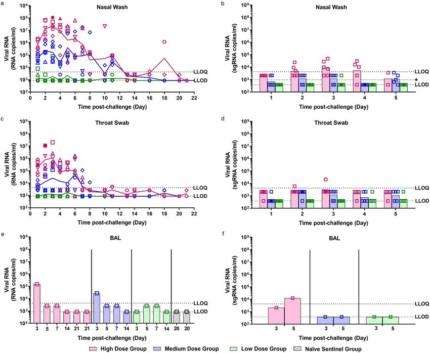
cases become critical, with respiratory failure, septic shock and/or the nasal wash of 6/6 ferrets in the high dose group from day 1 pc
multiple organ failure5. There is an urgent need to develop sui- and continued to be detected at varying levels until day 20 pc
table animal models to evaluate antivirals, therapeutics and vac- (Fig. 1a). The peak in viral RNA shedding was seen between days
cine candidates against SARS-CoV-2. 2 and 4 pc for all ferrets in the high dose group. Following a
Ferrets have been used extensively to model the disease caused decline in viral RNA (2/2 animals) to below the limit of quanti-
by influenza virus6–12 infection as well as human RSV13,14, fication of the assay at day 13 pc, an increase was seen at days 16
mumps virus15, Ebola virus16,17 and Nipah virus18,19. Due to the and 18 pc. Both Group 1 survivors were euthanised on day 21 pc
presence of a compatible form of ACE2, the virus receptor, on at which point no viral RNA was detected in their nasal washes.
cells of the ferret respiratory tract, these animals were developed Live virus was isolated in the nasal wash of 2/6 of the high dose
an as effective model for SARS-CoV20–23. The ferret has been ferrets at 3 and 4 dpc (Fig. 1a) at 30 and 110 pfu/ml, respectively.
shown to shed detectable virus from the URT as well as exhibiting This is in line with the viral subgenomic RNA (sgRNA) detected
comparable clinical symptoms associated with milder cases of the in the nasal wash (Fig. 1b), where these two samples, along with
infection21 and has shown similar pathology in the lung to that eight additional samples, are quantifiable for sgRNA.
observed in humans22. SARS-CoV-2 spike protein has been In the medium dose group 6/6 ferrets also had detectable viral
shown to exhibit many similarities in its amino acid sequence and RNA in nasal washes from day 1 pc. The peak of viral RNA
protein structure to the receptor binding domain of SARS-CoV24 shedding was more variable in the medium dose group, with
and also utilises ACE2 for cell entry25, suggesting ferrets would be some ferrets peaking at days 2–3 pc (4/6) and others peaking at
a suitable host for a model for COVID-19. days 5–6 pc (2/6). A decline was then seen until day 11 pc where
In this study, our aim is to understand if ferrets are a suitable viral RNA levels fell below the limit of quantification, but viral
species for a model of human SARS-CoV-2 infection. Animals RNA was still above the limit of detection of the assay. By day 16
are challenged intranasally with a range of titres of SARS-CoV- no more viral RNA was detected. Quantifiable viral RNA was
2 (5 × 10², 5 × 10⁴ and 5 × 10⁶ pfu) in 1 ml volume. We report only found in the nasal wash of 1/6 ferrets in the low challenge
that high and medium dose challenge induce URT RNA dose group. This ferret was euthanised on day 5 pc. No other
shedding, associated with LRT tissue viral RNA and lung ferrets in the low dose group were found to shed quantifiable viral
pathology. Re-challenge of recovered ferrets show a reduced RNA in their nasal wash. Additionally, live virus was not detected
Table 1 Experimental animal groups.
Group Number of Initial challenge virus Re-challenge virus Euthanasia/challenge days
animals titre (pfu/ml) titre (pfu/ml)
1 High 6 5 × 10⁶ 1 Ferret euthanised at day 3, 5, 7, 14 pc; 2 ferrets euthanised day at 21 pc
2 Medium 6 5 × 10⁴ 5 × 10⁶ 1 Ferret euthanised at days 3, 5, 7, 14 pc; 2 ferrets re-challenged at day
28 pc; 1 ferret euthanised at days 33 & 36 pc
3 Low 6 5 × 10² 5 × 10⁶ 1 Ferret euthanised at days 3, 5, 7, 14 pc; 2 ferrets re-challenged at day 28
pc; 1 ferret euthanised at days 33 & 36 pc
4 Naïve 2 PBS 2 Ferrets euthanised at day 20 pc
sentinel
5 Naïve 2 5 × 10⁶ 1 Ferret euthanised at days 33 & 36 pc
control
A total of 22 ferrets were distributed across 5 groups. All inoculations were performed intranasally with 1 ml of fluid. pc, post-challenge.
2 NATURE COMMUNICATIONS | (2021)12:81 | https://doi.org/10.1038/s41467-020-20439-y | www.nature.com/naturecommunications
NATURE COMMUNICATIONS | https://doi.org/10.1038/s41467-020-20439-y                                                                      ARTICLE

Fig. 1 Viral RNA Shedding. Nasal washes and swabs were collected at days 1 to 8, 10, 11, 13, 14, 16, 18 and 20 pc for all virus challenged groups. Viral
genomic RNA was quantified by RT-qPCR at all timepoints. No viral RNA was detected in any samples taken from the naïve sentinel ferrets. Samples that
were found to be positive by plaque assay are represented by a solid shape. (a) Nasal washes (c) Throat swabs (e) Bronchoalveolar lavage (BAL) collected
at necropsy (numbers indicate day post-challenge the ferret was euthanised). Symbols show values for individual animals, lines represented the calculated
group geometric means. The presence of viral subgenomic RNA was assessed in (b) nasal washes, (d) throat swabs and (f) BAL from 1 to 5 dpc. Bars show
geometric mean for each group and symbols show values for each individual animal. n = 6 ferrets per group with numbers decreasing by 1 at 3, 5, 7 and
14 dpc. The dashed horizontal lines show the lower limit of quantification (LLOQ) and the lower limit of detection (LLOD). [*LLOD range for nasal washes
represents 2 undetected samples at 5 dpc had
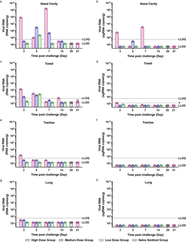
ARTICLE                                                                   NATURE COMMUNICATIONS | https://doi.org/10.1038/s41467-020-20439-y

Fig. 2 Viral RNA Shedding in Tissues. a, b Nasal cavity, c, d tonsil, e, f trachea and g, h lung were collected at euthanasia timepoints (days 3, 5, 7, 14, 20
and 21 pc) for all groups. n = 6 ferrets per group (n = 2 ferrets in the naive sentinel group), with one ferret from each group culled at each timepoint
(except day 20 and 21 pc where n = 2 ferrets from the naive sentinel and high dose groups were culled respectively). Viral genomic RNA (a, c, e and f) and
viral subgenomic RNA (b, d, f and h) was quantified by RT-qPCR. Bars show values for individual animals. The dashed horizontal lines show the lower limit
of quantification (LLOQ) and the lower limit of detection (LLOD). *LLOD range for tonsil represents undetected samples homogenised in smaller volume.

4                  NATURE COMMUNICATIONS | (2021)12:81 | https://doi.org/10.1038/s41467-020-20439-y | www.nature.com/naturecommunications
NATURE COMMUNICATIONS | https://doi.org/10.1038/s41467-020-20439-y                                                                  ARTICLE

genomic RNA findings, however at a much lower level (Fig. 2b).                   bronchopneumonia from days 3 to 14 pc was observed. Mild
Viral RNA was also detected in individual ferrets challenged with               necrosis of the bronchiolar epithelial cells was observed together
the medium dose culled at 3, 5 and 7 dpc. At 14 dpc viral RNA                   with inflammatory cell infiltration of neutrophils and mono-
was detected above the level of detection of the assay but below                nuclear cells within the bronchiolar luminae, mostly affecting
the level of quantitation in the high and medium dose ferrets. The              animals from the high dose group at days 3, 5 and 7 pc (Fig. 4c).
low dose ferret that was shown to shed viral RNA in its nasal                   This bronchopneumonia was characterised by the infiltration of
wash had a positive virus signal in nasal cavity and tonsil (Fig. 2c)           inflammatory cells, mostly neutrophils, but also macrophages and
tissue at 5 dpc when it was culled. Viral RNA was also quantified                lymphocytes, in approximately 10–15% of the lung section at day
in the tonsils (Fig. 2c) in high and medium dose ferrets at 3 and 5             3 pc decreasing to less than 5% at days 5 and 7 pc. The medium
dpc, with no detectable sgRNA found in the tonsils (Fig. 2d).                   dose group showed mild bronchopneumonia in less than 5% of
   Viral RNA was quantified in the trachea (Fig. 2e) of one of the               the lung sections at days 3 and 5 pc, while only occasional
ferrets euthanised at 3 dpc; and detected in high and medium                    infiltration was observed in animals from the low dose group.
dose ferrets across 3, 5 and 7 dpc. Viral RNA was detected above                Bronchiolar inflammatory infiltration was not observed in control
the level of detection of the assay but below the level of                      (PBS) animals (Fig. 4h). Few cells stained positive for viral RNA
quantitation in the lungs (Fig. 2g) of the high and medium dose                 using in situ hybridisation (RNAScope). Few type I and
ferrets culled at 3 dpc. Viral RNA was not detected in the lungs of             occasionally type II pneumocytes and alveolar macrophages were
any other ferrets euthanised at any other timepoints. Viral sgRNA               positive for viral RNA at days 3, 5 and 7 pc (Fig. 4d) in high and
was not detected in the trachea (Fig. 2f) or lungs (Fig. 2h) of any             middle dose animals. The presence of viral RNA was not
ferrets. No viral RNA was detected in the liver (Supplementary                  associated with histopathological lesions. Occasionally, mild
Fig. 1b), jejunum (Supplementary Fig. 1c) or colon (Supplemen-                  proliferation of bronchus-associated lymphoid tissue (BALT)
tary Fig. 1d) of any of the ferrets, from any of the groups.                    was observed surrounding damaged bronchi and bronchioles at
   Detection of viral RNA in the rectal swabs was found to be                   the early stages of the disease, with slightly more severity at days
variable across the different dose groups (Supplementary Fig. 1a).              14 and 21 pc (high dose) (Fig. 4e). Mild interstitial pneumonia
The highest viral RNA load was observed in a ferret in the high                 with an increase in the thickness of the interalveolar septa was
dose group but there was a less consistent pattern of RNA                       observed from day 3 pc towards the end of the experiment in high
detection which did not continue past day 7 pc. In the medium                   and medium dose groups (Fig. 4e). Ferrets from the high dose
dose group, 4/6 ferrets were found to have detectable viral RNA                 group showed mild proliferation of type II pneumocytes from day
in their rectal swabs between days 2 and 8 pc. No viral RNA was                 7 pc onwards (Fig. 4e).
detected in any of the rectal swabs collected from the low dose                    The liver showed multiple foci of inflammatory cell infiltration
group following challenge.                                                      in the portal areas, composed of mainly macrophages, lympho-
                                                                                cytes and occasional plasma cells (Fig. 4f). This multifocal
                                                                                infiltration was more severe in animals from the high and
Clinical signs. The normalised summed incidence of clinical
                                                                                medium dose groups from day 3 pc, compared to the low dose
scores for each group of ferrets is shown in Fig. 3a and total
                                                                                group or control (PBS) animals (Fig. 4i), which only showed
summed scores are shown in Table 2. At day 9 pc all 3/3 ferrets in
                                                                                minimal presence of portal inflammation. No other remarkable
the high dose group showed reduced activity, a similar observa-
                                                                                changes were observed in any other tissue. However, occasional
tion was made in the medium dose group but later, on day 10 pc.
                                                                                positive cells (absorbing epithelial enterocytes and goblet cells)
Reduced activity was accompanied by ruffled fur, a sign that the
                                                                                were also observed in the small and large intestine from high and
ferrets were not grooming regularly. By day 14 pc ferrets in the
                                                                                medium dose at days 3, 5 and 7 pc, not associated with
medium dose group stopped showing signs of reduced activity
                                                                                histopathological lesions (Fig. 4g).
and by day 15 pc the high dose groups stopped showing signs of
reduced activity. Ferrets in the high dose group had the highest
normalised cumulative clinical score (summed across all time
                                                                                Immune response to SARS-CoV-2 infection. Neutralising
points) (14.01), followed by the medium dose group (6.99) with
                                                                                antibody titres for ferrets infected in the high dose and medium
sporadic instances recorded in the low dose group. No fever
                                                                                dose groups generally increased longitudinally following chal-
(±1 °C from baseline at least two consecutive occasions) was
                                                                                lenge as illustrated in Supplementary Fig. 2a. The average fold
detected in any ferret, in any group (Fig. 3b); instead body
                                                                                increase of neutralising antibodies from days 8 to 14 pc was about
temperature remained within the normal range. No weight loss
                                                                                the same for both the high and medium dose groups. The low
was observed in any ferret in any group, below baseline; however,
                                                                                dose group had comparatively low neutralising antibodies
the SARS-CoV-2 infected ferrets failed to gain as much weight as
                                                                                throughout the time course. Further analysis of ferret sera showed
the ferrets in the control (phosphate buffered saline, PBS) group,
                                                                                that the presence of SARS-CoV-2 spike specific IgG (Supple-
although this difference was not statistically significant (Fig. 3c).
                                                                                mentary Fig. 2b) demonstrated a similar trend, with titres
                                                                                increasing over time in the high and medium dose groups. Upon
Histopathology. The nasal cavity from high dose ferrets showed a                euthanasia, lung mononucleocytes (MNCs) were isolated from
minimal to mild necrosis of epithelial cells and mild inflamma-                  the high dose ferrets at 14 and 21 dpc, the medium dose ferret at
tory cell infiltration (Fig. 4a) from days 3 to 7 pc. However,                   14 dpc, and the naïve sentinel ferrets culled at 20 dpc. Cellular
abundant epithelial cells from the nasal cavity were stained for                immune responses were assessed in lung MNCs using whole live
viral RNA at day 3 pc (Fig. 4b). Occasional scattered cells                     SARS-CoV-2 to stimulate the production of IFNγ (Supplemen-
expressing viral RNA were observed in high dose animals at days                 tary Fig. 3). SARS-CoV-2 specific immune responses were seen in
5 and 7 pc and medium dose animals at days 3, 5 and 7 pc.                       the high and medium dose ferrets euthanised at 14 and 21 dpc.
Similarly, very few scattered epithelial cells were stained for viral              One naïve sentinel group ferret was shown to have neutralising
RNA in the trachea and larynx from high and medium dose                         antibodies to SARS-CoV-2 as well as the presence of SARS-CoV-
animals at days 3, 5 and 7 pc.                                                  2 spike specific IgG upon euthanasia at day 20 pc. This ferret
   No remarkable gross lesions were observed in the infected                    showed no clinical signs of SARS-CoV-2 infection and samples
animals. Upon histological examination of the lungs of ferrets                  (nasal wash and swabs) taken at baseline, day 11 and day 20 pc
from the high and medium dose groups, a mild multifocal                         were shown to be PCR negative for SARS-CoV-2. Furthermore

NATURE COMMUNICATIONS | (2021)12:81 | https://doi.org/10.1038/s41467-020-20439-y | www.nature.com/naturecommunications                            5
ARTICLE NATURE COMMUNICATIONS | https://doi.org/10.1038/s41467-020-20439-y there was no evidence of pathology in any of the tissues taken immune response seen in the lung MNCs (Supplementary Fig. 3) from both naïve sentinel ferrets euthanised on day 20 pc. of the naïve sentinel ferret showed a high SARS-CoV-2 specific Following euthanasia, viral RNA was detected (but not quanti- immune response to whole live SARS-CoV-2, paralleling the fied) in tissue in the colon of this ferret. Viral RNA was not detection of neutralising antibodies and SARS-CoV-2 spike detected in any other tissues analysed (nasal cavity, tonsil, specific IgG. These results suggest that this ferret could have trachea, lung, liver and jejunum). Interestingly, the cellular inadvertently become infected with SARS-CoV-2. 6 NATURE COMMUNICATIONS | (2021)12:81 | https://doi.org/10.1038/s41467-020-20439-y | www.nature.com/naturecommunications
NATURE COMMUNICATIONS | https://doi.org/10.1038/s41467-020-20439-y                                                                            ARTICLE

Fig. 3 Clinical Observations. a Clinical observations were carried out four times daily (approximately 6 h apart) for the first 5 days and then twice daily
(approximately 8 h apart) for the remaining time. Observations were summed for each group of ferrets. b Temperatures were taken at the same time as
clinical observations, using the identifier chip, to ensure any peak of fever was recorded. Mean temperatures are displayed on the graph. c Weight was
recorded daily and percentage weight change from baseline was plotted. Points show values for individual animals, lines represented the calculated group
means. n = 6 ferrets per group (n = 2 ferrets in the naive sentinel group) with numbers decreasing by 1 at 3, 5, 7 and 14 dpc. The table illustrates the
summed scores for each clinical observation noted for each of the groups during specific days post-challenge and post re-challenge. Activity in ferrets was
scored as follows; 0 = alert and playful, 1 = alert, playful when stimulated, 2 = alert, not playful when stimulated, 3 = not alert or playful. Ruffled fur was
given a score of 1. Activity scores of 1 were given to ferrets during the initial challenge. Upon re-challenge ferret activity was recorded as 1 or 2 indicating
increased lethargy in ferrets following re-challenge.

 Table 2 Clinical scores.

 Group         Clinical scores (individual instances summed)

               Initial challenge                         Day 0–7 pc       Day 8–14 pc      Day 15–21 pc     Re-challenge                Day 0–8 post
                                                                                                                                        re-challenge
 1   High 1 Ferret euthanised at day 3, 5, 7,            Activity =1; 2 Activity =1; 19 Activity =1; 2
          14 pc; 2 ferrets euthanised at day                            Ruffled fur; 12
          21 pc
 2 Medium 1 Ferret euthanised at day 3, 5, 7,            Activity =1; 2 Activity =1; 12 0                   1 Ferret euthanised at day Activity =1; 4
          14 pc                                                         Ruffled fur; 8                       5 & 8 post re-challenge Activity =2; 2
                                                                                                                                       Ruffled fur; 1
 3 Low         1 Ferret euthanised at day 3, 5, 7,       Activity =1; 1   Activity =1; 1   0                1 Ferret euthanised at day Activity =1; 2
               14 pc                                     Sneeze; 1                                          5 & 8 post re-challenge Activity =2; 1
 4 Naïve       2 Ferrets euthanised at day 20 pc         0                0                0
   sentinel
 5 Naïve                                                                                                    1 Ferret euthanised at day Activity =1; 2
   control                                                                                                  5 & 8 post challenge

Re-challenge of ferrets with high dose SARS-Cov-2 results in                     observed at such an early stage in the initial challenge (Table 2).
absence of lung pathology. Four previously infected ferrets, two                 In contrast, the two naïve control animals did not experience
from the medium and low dose challenge groups, had neu-                          weight loss below baseline after infection and they did not suffer
tralising titres of 1:274, 1:250, 1:82 and 1:55 at day 26 pc,                    the same level of clinical observation as the re-challenged animals
respectively. At day 28 pc, these ferrets and two naïve control                  (Fig. 5c).
animals were challenged intranasally with the high dose of SARS-                    The cellular immune response in the lungs of a low dose
Cov-2 (5 × 10⁶ pfu). Though shedding of viral RNA from the                       (Group 3) re-challenge ferret and a naïve control (Group 5) ferret
nasal wash was similar in all groups on day 2 post re-challenge,                 at day 36 (8 days post re-challenge, respectively) were compared.
viral RNA levels subsequently decreased in the previously chal-                  Supplementary Fig. 6 shows SARS-CoV-2 specific cellular
lenged animals (n = 4), with the medium dose group showing                       immune responses, as determined by IFN-γ ELISpot. The
rapid decrease to below quantifiable levels by day 5 post re-                     number of secreting cells detected after re-stimulation of lung
challenge. Viral RNA levels in the nasal wash continued to stay                  MNCs with peptide pools spanning the spike protein varied
above quantifiable levels in the challenged naïve control group,                  between ferrets from each group. The strongest response is
although they began to fall at day 8 pc (Fig. 5a). Similar results               detected in the re-challenge ferret after ex vivo re-stimulation
were seen in the throat swab and rectal swabs (Supplementary                     with whole live virus. Upon histological examination the upper
Fig. 4a, b), with reduced viral shedding seen in the re-challenged               and lower respiratory tracts from animals in both re-challenged
animals. Viral RNA was only detected in the BAL of the ferret                    groups showed no remarkable lesions and an absence of
from the naïve control group euthanised at 5 dpc (Supplementary                  significant bronchopneumonia (Fig. 5d, e), that was observed in
Fig. 4c). Subgenomic RNA was detected but not quantified in                       ferrets initially challenged with 5 × 10⁶ pfu for the first time, i.e.
nasal wash (Supplementary Fig. 5a) and throat swabs (Supple-                     the original high dose ferret or the naïve control infected group
mentary Fig. 5b) for re challenged animals and animals chal-                     included for the ‘re-challenge’ (Fig. 5f). This parallels the absence
lenged for the first time.                                                        of pathology observed in the two naïve sentinel ferrets euthanised
   Analysis of viral RNA in the tissues clearly show reduced viral               at day 20 pc.
RNA present in the nasal cavity and tonsil in the medium dose
re-challenged group at 5 days post re-challenge compared to the
low dose re-challenged and naïve challenged control group                        Discussion
(Fig. 5b). Analysis of viral subgenomic RNA present in the nasal                 This study demonstrates ferrets are susceptible to experimental
cavity showed a reduction of the amount of RNA present in the                    intranasal infection with a low passage isolate of SARS-CoV-2
medium group compared to the low and naïve control groups at                     strain Victoria 128. A high dose (5 × 10⁶ pfu/ml in 1 ml volume)
day 5 post re-challenge (Supplementary Fig. 5c).                                 intranasal challenge in ferrets produced mild clinical signs, con-
   Animals in the re-challenged medium and low dose groups                       sistent lung pathology and a viral shedding pattern that aligns
exhibited weight loss from baseline that was not seen at initial                 with the mild to moderate disease seen in clinical cases of
challenge for any of the animals in any of the challenge groups                  COVID-1928.
(Fig. 5c). Re-challenged animals also experienced increased                         Previously published SARS-CoV-1 challenge studies conducted
clinical observations of lethargy and ruffled fur that was not                    in the ferret show that a lower dose of (103 TCID50) is sufficient

NATURE COMMUNICATIONS | (2021)12:81 | https://doi.org/10.1038/s41467-020-20439-y | www.nature.com/naturecommunications                                        7
ARTICLE NATURE COMMUNICATIONS | https://doi.org/10.1038/s41467-020-20439-y Fig. 4 Histopathological findings and presence of SARS-CoV-2 RNA in tissue sections from ferrets inoculated with SARS-CoV-2. Two sections from each tissue or organ were evaluated independently by two qualified pathologists and representative images are shown. a Nasal cavity, day 3 pc, Group 1, H&E staining. Mild epithelial cell necrosis (arrows) and minimal inflammatory cell infiltration within the epithelium; bar = 50 μm; inset = close up image of an inflammatory cell within the epithelial layer. b Nasal cavity, day 3 pc, Group 1, SARS-CoV-2 viral RNA detection (RNASCope staining). Presence of viral RNA in abundant epithelial and sustentacular cells from the nasal cavity mucosa; bar = 50 μm; inset = close up image showing abundant viral RNA within the olfactory epithelium. c Lung, day 5 pc, Group 1, H&E staining. Moderate bronchopneumonia with neutrophil and macrophage inflammatory infiltrate within the bronchiolar lumina (arrow). Mild peribronchiolar infiltration of mononuclear cells (*); bar = 50 μm; inset = clse up image of the bronchiolar inflammatory cell infiltration showing abundant neutrophils. d Lung, day 3 pc, Group 2, SARS-CoV-2 viral RNA detection (RNASCope staining). Presence of viral RNA in type II pneumocyte (arrow); bar = 50 μm. e Lung, day 21 pc, Group 1, H&E staining. A Bronchiole with mild inflammatory infiltration in the lumina (arrow) and attenuation of the epithelial cells. Moderate peribronchiolar infiltration of mononuclear cells (*) and mild interalveolar septal inflammatory cell infiltration with thickening of the wall (arrowheads); bar = 50 μm; inset = close up image of mils pneumocyte II proliferation in the lung at day 7 pc. f Liver, day 21 pc, Group 1, H&E staining. Moderate multifocal hepatitis with mononuclear cell infiltration in the portal areas (arrow); bar = 50 μm. g Colon, day 3 pc, Group 1, SARS-CoV-2 viral RNA detection (RNASCope staining). Presence of viral RNA in scattered cell within the absorptive epithelium (arrows); bar = 50μm. h Lung, naïve animal, H&E staining. Bronchiole showing no inflammatory reaction; bar = 50 μm. i Liver, naïve animal, H&E staining. Minimal hepatitis with mononuclear cell infiltration in the portal areas (arrow); bar = 50 μm. to cause a mild disease in the ferret20,21. We have shown that a release from isolation29. This report is in alignment with the high (5 × 10⁶ pfu) and medium (5 × 10⁴ pfu) dose intranasal observations in the two ferrets challenged with the high dose of challenge of SARS-CoV-2 results in an infection characterised by SARS-CoV-2 (euthanised at day 21 pc) which appeared to con- prolonged viral RNA shedding in all ferrets (days 1–11 pc), tinue to shed detectable viral RNA from the upper respiratory accompanied by observable clinical signs from day 8 pc for both tract up to day 18 pc even though these animals had developed high and medium dose groups. Onset of clinical symptoms were neutralising antibodies. delayed by approximately 24 h in the medium dose animals. Both The main histopathological finding in approximately 10% of doses also induced classical pathology of bronchial pneumonia the lung tissue sections in the high dose group consisted mainly of involving 10% and 3% of recipient lungs, respectively. A low dose a multifocal bronchiolitis, with inflammatory infiltrates within the intranasal challenge of the same SARS-CoV-2 virus (5 × 10² pfu) airways and some alveolar species. This finding is similar, but less appeared to result in infection of only one ferret which shed severe, to the findings in the published reports about SARS-CoV- detectable viral RNA in the upper respiratory tract (UTR) but 1 ferret challenge models22,30,31. In this study, mild alveolar failed to show any remarkable lesions in the respiratory tract. damage was observed in the acute phase. At later time points, In the high and medium dose groups, virus was readily detected mild proliferation of type II pneumocytes, with interstitial infil- using in situ hybridisation in the upper respiratory tract of ferrets, trates and peribronchiolar cuffing, was recoded, consistent with with a peak at 3 dpc. These findings aligned with the detected evolution from the acute phase. shedding of viral RNA and sgRNA from nasal washes and throat Mild to moderate multifocal hepatic inflammatory cell infil- swabs which also peaked at day 3–4 pc and detection of viral and tration has been widely reported in viral infections in animals, and sgRNA in the upper respiratory tract tissue This upper respiratory has been previously described in SARS-CoV-1 infected ferrets32. infection mirrors the clinical disease recently reported in mild However, the periportal infiltrates may not be associated with cases of humans infected with SARS-CoV-2 infection28. injury to the surrounding tissue and they are reported as a com- Recent reports indicate that COVID-19 patients appear to shed mon background finding in laboratory ferret species. The presence viral RNA intermittently after recovery from disease with some of infected enterocytes has been reported for SARS-CoV-1 and individuals being tested and found to be positive again after SARS-CoV-2 in humans33 and different ferret models34,35. 8 NATURE COMMUNICATIONS | (2021)12:81 | https://doi.org/10.1038/s41467-020-20439-y | www.nature.com/naturecommunications
NATURE COMMUNICATIONS | https://doi.org/10.1038/s41467-020-20439-y                                                                         ARTICLE

Fig. 5 Re-challenge of ferrets with SARS-CoV-2. a Nasal washes were collected at days 1–5 post re-challenge (days 29–33 post-original challenge). Viral
RNA was quantified by RT-qPCR. b Nasal cavity, tonsil, trachea and colon were collected at euthanasia timepoints (days 5 and 8 post re-challenge) for all
groups. Viral RNA was quantified by RT-qPCR. Bars show values for individual animals. The dashed horizontal lines show the lower limit of quantification
(LLOQ) and the lower limit of detection (LLOD). c Percentage weight change from baseline. Baseline was calculated as average of the two most recent
weights taken preceding re-challenge. n = 2 ferrets per group with numbers decreasing by 1 at 5 dpc. d Medium dose re-challenged ferret at day 5 post re-
challenge. No remarkable changes in alveoli or terminal bronchiole. e Low dose re-challenged ferret at day 5 post re-challenge. No remarkable changes in
alveoli or bronchiole. f Control group ferret challenged for the first time (day 28 pc). Inflammatory infiltration within bronchiolar lumen and mild infiltration
of alveolar septa; lesions comparable with those observed in the original high dose group. Bar = 50 μm.

   The upper respiratory virus replication, reported here, in the               BAL were pelleted prior to sampling for RNA extraction and live
high and medium dose groups of animals, support the observa-                    virus assays. It is feasible that virus recovered from animals
tions of Shi et al.34 and Kim et al.35 which found peak URT viral               remained strongly cell-associated and was depleted by cen-
RNA shedding between days 4 and 6 pc. Shi et al.34 also reported                trifugation. Alternatively, this result may have accurately reflected
mild lung pathology associated with SARS-CoV-2 infection                        low levels of viable virus presence which others have reported
similar to our medium dose animals, but this was not as extensive               even though viral RNA can be detected. For human swabs and
as that seen in our high dose challenge group ferrets.                          sputum samples, it has been noted that infectious virus was never
   Shi et al.34, Kim et al.35 and Richard et al.36 report live virus            recovered from samples with a viral RNA load of less than 106
isolation from RNA positive nasal wash samples. In this study,                  copies/ml28.
live virus was detected in a minority of nasal washes and throat                   Neutralising antibody levels developed in ferrets in the medium
swabs, even though high levels of viral RNA were detected.                      and high dose challenge groups within 14 days. While the levels
However, analysis of viral subgenomic RNA confirmed that a                       of serum neutralising antibodies did not increase in the low dose
minority of samples contained replicating virus, this aligned with              animals, mucosal, humoral and cellular immunity could have
samples in which live virus was detected. A possible reason for                 played a role during re-challenge. Accepting the small number of
this observation during this study could be how samples were                    animals in our re-challenge study, the finding that medium dosed
processed following collection. Nasal washes, throat swabs and                  animals displayed reduced viral RNA shedding in the URT, and

NATURE COMMUNICATIONS | (2021)12:81 | https://doi.org/10.1038/s41467-020-20439-y | www.nature.com/naturecommunications                                     9
ARTICLE                                                                         NATURE COMMUNICATIONS | https://doi.org/10.1038/s41467-020-20439-y

both low and medium dosed animals showed an absence of lung                           Animals. Twenty-two healthy, female ferrets (Mustela putorius furo) aged
pathology following re-challenge is encouraging; it suggests that                     7 months were obtained from a UK Home Office accredited supplier (Highgate
                                                                                      Farm, UK). The mean weight at the time of challenge was 1032 g/ferret (range
there may be potential benefits of naturally acquired immunity                         870–1239 g). Animals were housed in pairs at Advisory Committee on Dangerous
and is in line with the observation reported by Bao et al.37 in                       Pathogens (ACDP) containment level 3. Cages met with the UK Home Office
which previously infected rhesus macaques were protected                              ‘Code of Practice for the Housing and Care of Animals Bred, Supplied or Used for
against re-challenge with SARS-CoV-2.                                                 Scientific Procedures’ (December 2014). Access to food and water was ad libitum
   SARS-CoV-2 spike protein-specific immune responses seen in                          and environmental enrichment was provided. All experimental work was con-
                                                                                      ducted under the authority of a UK Home Office approved project licence that had
a low dose re-challenged ferret were compared to that of a pri-                       been subject to local ethical review at PHE Porton Down by the Animal Welfare
mary challenge ferret. This comparison showed that the response                       and Ethical Review Body (AWERB) as required by the ‘Home Office Animals
to the virus appears to be higher on re-challenge. However, ferrets                   (Scientific Procedures) Act 1986’.
challenged with our high dose of SARS-CoV-2 displayed
increased clinical observations and lost weight from baseline                         Experimental design. Before the start of the experiment animals were randomly
following re-challenge, hinting at enhanced disease but a larger                      assigned to challenge groups, to minimise bias. The weight distribution of the
study would be required to effectively assess this observation.                       animals was tested to ensure there was no statistically significant difference
                                                                                      between groups (one-way ANOVA, p > 0.05). An identifier chip (Bio-Thermo
Alternatively, these clinical signs may be a perfectly normal host                    Identichip, Animalcare Ltd, UK) was inserted subcutaneously into the dorsal
response to infection in a pre-immune individual whilst the                           cervical region of each animal. Prior to challenge animals were sedated by intra-
immune system is successfully clearing a large challenge dose.                        muscular injection of ketamine/xylazine (17.9 and 3.6 mg/kg bodyweight). Chal-
   In addition to the ferret, hamsters and non-human primates                         lenge virus was delivered by intranasal instillation (1.0 ml total, 0.5 ml per nostril)
                                                                                      diluted in phosphate buffered saline (PBS).
(NHPs) have also been developed as models of SARS-CoV-2. The                              Three different doses of virus were delivered to three groups (n = 6) of ferrets:
NHP model appears to exhibit a mild clinical disease much like                        high (5 × 10⁶ pfu/ml), medium (5 × 10⁴ pfu/ml) and a low (5 × 10² pfu/ml) dose.
the ferret but has an increased incidence of lung pathology.                          For the high, medium and low dose groups, individual ferrets were scheduled for
However, the Syrian hamster model has been found to exhibit                           euthanasia on day 3 (n = 1), day 5 (n = 1), day 7 (n = 1) and day 14 (n = 1). For
                                                                                      the high dose group, the remaining 2 ferrets were euthanised on day 21 (n = 2).
weight loss following SARS-CoV-2 infection and the virus                              The mock-infected animals (n = 2) received an intranasal instillation of sterile PBS
appears to replicate efficiently in the lungs causing severe                           and were euthanised on day 20.
pathological lesions commonly reported in COVID-19 patients                               On day 28 pc the remaining ferrets in the low (n = 2) and medium (n = 2)
with pneumonia38,39. The ferret has been well characterised for                       groups were re-challenged with 5 × 10⁶ pfu by the intranasal route. Additional
other respiratory viruses and provides a useful comparative model                     naïve control ferrets (n = 2) were also challenged on day 28, to provide a re-
                                                                                      challenge control. All 6 animals were monitored for clinical signs and one ferret
for the assessment of SARS-CoV-2 pathogenicity and transmis-                          from each group was euthanised on day 33 and the remaining animals were
sion, in addition to the evaluation of vaccines, drugs and ther-                      euthanised on day 36.
apeutics. Access to practical volumes of blood, in the ferret model,                      Nasal washes, throat and rectal swabs were taken at days −1, 1–8, 10, 11, 13, 14,
for sequential in-life samples and the availability of an immu-                       16, 18 and 20 pc. They were also taken at days 1–5 and 8 post re challenge (days
                                                                                      29–33 and 36 pc). Whole blood and serum were collected at 2, 5, 8, 11 and 14 dpc
nological tool kit are also advantages.                                               for all ferrets. Whole blood and serum were collected at days 2, 5 and 8 (days 30, 33
   This study demonstrates that ferrets challenged with 5 × 10⁶                       and 36 pc) post re-challenge for all remaining ferrets. The negative control ferrets
pfu or 5 × 10⁴ pfu displayed only mild clinical signs of SARS-                        (n = 2) had nasal washes, throat swabs, whole blood and serum taken at −1 and 11
CoV-2 infection. As seen in the clinical setting, pathological                        dpc. At necropsy nasal washes, throat and rectal swabs, whole blood and serum
                                                                                      were taken alongside tissue samples for histopathology. Nasal washes were
features appear to be less severe than those reported after ferrets                   obtained by flushing the nasal cavity with 2 ml PBS. For throat swabs, a flocked
were infected with SARS-CoV-121,31.                                                   swab (MWE Medical Wire, Corsham, UK) was gently stroked across the back of
   This ferret model of intranasal SARS-CoV-2 infection presents                      the pharynx in the tonsillar area. Throat and rectal swabs were processed, and
three key measurable endpoints: (a) consistent URT viral RNA                          aliquots stored in viral transport media (VTM) and AVL at −80 °C until assay.
shedding; (b) detectable lung pathology; and (c) post viral fatigue.
Reductions in URT RNA shedding during the first 14 days post                           Clinical and euthanasia observations. Animals were monitored for clinical signs
intranasal challenge could be an attractive indicator of the efficacy                  of disease four times daily (approximately 6 h apart) for the first 5 dpc and then
                                                                                      twice daily (approximately 8 h apart) for the remaining time. Clinical signs of
of candidate therapeutics and vaccines. It may be wise, however,                      disease were assigned a score based upon the following criteria. Activity was scored
to euthanise prior to 14 dpc to more accurately assess the impact                     as follows: 0 = alert and playful, 1 = alert, playful when stimulated, 2 = alert, not
on lung pathology especially when looking for signs of vaccine-                       playful when stimulated, 3 = not alert or playful. Ruffled fur was given a score of 1.
enhanced disease20,40,41. We believe the high dose intranasal                         No other clinical signs were noted. In order to meet the requirement of the project
                                                                                      license, immobility, neurological signs or a sudden drop in temperature were
challenge will provide the most distinct disease endpoints.                           automatic euthanasia criteria. Animals were also deemed to have reached a
However, with its reduced level of lung pathology, the medium                         humane endpoint if their body weight was at or below 30% baseline. If any ferret
dose challenge may provide a higher level of sensitivity to some                      reached any of these three euthanasia criteria, they were to be immediately
interventions, as was observed when assessing therapeutics to                         euthanised using a UK Home Office approved Schedule 1 procedure. However, no
influenza in the ferret model42.                                                       animals reached these end-points during this study.
                                                                                          Temperature was taken using a microchip reader and implanted temperature/
                                                                                      ID chip. Temperature was recorded at each clinical scoring point using the chip to
                                                                                      ensure any peak of fever was recorded. Animals were weighed at the same time
Methods                                                                               each day from the day before infection until euthanasia.
Viruses and cells. SARS-CoV-2 Victoria/01/202026 was generously provided by
The Doherty Institute, Melbourne, Australia, at P1 after primary growth in Vero/      Necropsy procedures. Ferrets were anaesthetised with ketamine/xylazine (17.9
hSLAM cells and subsequently passaged twice at PHE Porton in Vero/hSLAM cells         and 3.6 mg/kg bodyweight) and exsanguination was effected via cardiac puncture,
[ECACC 04091501]. Infection of cells was with ~0.0005 MOI of virus and har-           followed by injection of an anaesthetic overdose (sodium pentabarbitone Dole-
vested at day 4 by a single freeze thaw cycle and clarification by centrifugation at   lethal, Vetquinol UK Ltd, 140 mg/kg). A necropsy was performed immediately after
1000g for 10 min. Whole genome sequencing was performed, on the P3 challenge          confirmation of death. The BAL was collected at necropsy from the right lung. The
stock, using both Nanopore and Illumina as described previously43. Virus titre of     left lung was dissected prior to BAL collection and used for subsequent histo-
the challenge stocks was determined by plaque assay on Vero/E6 cells [ECACC           pathology and virology procedures.
85020206]. Cell lines were obtained from the European Collection of Authenticated
Cell Cultures (ECACC) PHE, Porton Down, UK. Cell cultures were maintained at
37 oC in MEM (Life Technologies, California, USA) supplemented with 10% foetal        RNA extraction. RNA was isolated from nasal wash, throat swabs, EDTA treated
bovine serum (Sigma, Dorset, UK) and 25 mM HEPES (Life Technologies). In              whole blood, BAL and tissue samples (nasal cavity, tonsil, trachea, lung liver,
addition, Vero/hSLAM cultures were supplemented with 0.4 mg/ml of geneticin           jejunum and colon). Weighed tissue samples were homogenised and inactivated in
(Invitrogen) to maintain the expression plasmid.                                      RLT (Qiagen) supplemented with 1%(v/v) Beta-mercaptoethanol. Tissue

10                  NATURE COMMUNICATIONS | (2021)12:81 | https://doi.org/10.1038/s41467-020-20439-y | www.nature.com/naturecommunications
NATURE COMMUNICATIONS | https://doi.org/10.1038/s41467-020-20439-y ARTICLE
homogenate was then centrifuged through a QIAshredder homogenizer (Qiagen) PBS/0.1% Tween 20, 50 µl/well of each dilution were added to the antigen coated
and supplemented with ethanol as per manufacturer’s instructions. Downstream plate and incubated for 2 h at room temperature. Following washing, anti-ferret
extraction was then performed using the BioSprint™96 One-For-All vet kit (Qiagen) IgG conjugated to HRP (Novus Biologics, NB7224) was diluted (1:5000) in 10%
and Kingfisher Flex platform as per manufacturer’s instructions. Non-tissue FBS in 1X PBS/0.1% Tween 20 and 100 µl/well were added to the plate, then
samples were inactivated in AVL (Qiagen) and ethanol, with final extraction using incubated for 1 h at room temperature. After washing, 1 mg/ml O-
the QIAamp Viral RNA Minikit (Qiagen) as per manufacturer’s instructions. Phenylenediamine dihydrochloride solution (Sigma P9187) was prepared and
100 µl/well were added. The development was stopped with 50 µl/well 1 M
Hydrochloric acid (Fisher Chemical, J/4320/15) and the absorbance at 490 nm was
Quantification of viral loads by RT-qPCR. Reverse transcription-quantitative read using Softmax 7.0. The cut-off was defined as the average Optical Density of
polymerase chain reaction (RT-qPCR) targeting a region of the SARS-CoV-2 naïve animals +2 standard deviation.
nucleocapsid (N) gene was used to determine viral loads and was performed using
TaqPath™ 1-Step RT-qPCR Master Mix, CG (Applied Biosystems™) and 2019-nCoV
CDC RUO Kit (Integrated DNA Technologies). Sequences of the N1 primers and Histopathological analysis. Samples from the left cranial and left caudal lung lobe
probe were: 2019-nCoV_N1-forward, 5′ GACCCCAAAATCAGCGAAAT 3′; together with spleen, kidney, liver, tracheobronchial and axillary lymph nodes, jejunum,
2019-nCoV_N1-reverse, 5′ TCTGGTTACTGCCAGTTGAATCTG 3′; 2019- colon, trachea, larynx and nasal cavity, were fixed by immersion in 10% neutral-
nCoV_N1-probe, 5′ FAM-ACCCCGCATTACGTTTGGTGGACC-BHQ1 3′. buffered formalin and processed routinely into paraffin wax. Nasal cavity samples were
Sequences can also be found in Supplementary Table 1. The cycling conditions decalcified using an EDTA-based solution prior to embedding. 4 µm sections were cut
were: 25 °C for 2 min, 50 °C for 15 min, 95 °C for 2 min, followed by 45 cycles of 95 and stained with haematoxylin and eosin (H&E) and examined microscopically. In
°C for 3 s, 55 °C for 30 s. Tissues samples were tested on the QuantStudio™ 7 Flex addition, samples were stained using the RNAscope technique to identify the SARS-
Real-Time PCR System with an in vitro transcribed RNA standard of the full CoV-2 virus RNA. Briefly, tissues were pre-treated with hydrogen peroxide for 10 min
length SARS-CoV-2 N ORF (accession number NC_045512.2) with quantification (room temperature), target retrieval for 15 min (98–101 °C) and protease plus for
between 1 × 10^1 and 1 × 10^6 copies/µl. Positive samples detected below the 30 min (40 °C) (Advanced Cell Diagnostics). A V-nCoV2019-S probe (Cat No. 848561,
lower limit of quantification (LLOQ) were assigned the value of 5 copies/µl, whilst Advanced Cell Diagnostics) was incubated on the tissues for 2 h at 40 °C. Amplification
undetected samples were assigned the value of ≤2.3 copies/µl, equivalent to the of the signal was carried out following the RNAscope protocol using the RNAscope 2.5
assays lower limit of detection (LLOD). Non-tissues samples were tested on the HD Detection kit – Red (Advanced Cell Diagnostics).
7500 Fast Real-Time PCR System (Applied Biosystems™) with a 100 bp Ultramer
RNA oligo standard (Integrated DNA Technologies) equivalent to 28274-28373 bp
of SARS-CoV-2 NC_045512.2, with quantification between 1 × 10^1 and 1 × 10^7 Isolation of lung mononuclear cells. Whole lungs were removed from each ferret.
copies/µl. Positive samples detected below the limit of quantification were assigned The lungs were dissected into small pieces and placed into a 12.5 ml solution of
the value of 6 copies/µl, whilst undetected samples were assigned the value of collagenase (715 collagenase units/ml) (Sigma-Aldrich) and DNase (350 DNase
≤2 copies/µl, equivalent to the assays LLOD. units/ml) (Sigma-Aldrich). Lungs were placed into gentleMACS C-tubes and agi-
tated whilst incubating, 37 oC for 1 h on an OctoMACS (Miltenyi Biotec, Surrey,
Subgenomic RT-qPCR. Subgenomic RT-qPCR was performed on the Quant- UK). Partially digested lung tissue was then dissociated using an OctoMACS. The
Studio™ 7 Flex Real-Time PCR System using TaqMan™ Fast Virus 1-Step Master tissue solution was passed through two cell sieves (100 µm then 70 µm) and then
Mix (Thermo Fisher Scientific) and oligonucleotides as specified by Wolfel et al., layered with Ficoll®- Paque Premium (GE Healthcare, Hatfield, UK). Density
with forward primer, probe and reverse primer at a final concentration of 250, 125 gradient centrifugation was carried out at 400g for 30 min. Buffy coats containing
and 500 nM, respectively. Sequences of the sgE primers and probe were: 2019- lymphocytes were collected and washed with medium by pelleting cells via cen-
nCoV_sgE-forward, 5′ CGATCTCTTGTAGATCTGTTCTC 3′; 2019-nCoV_sgE- trifugation at 400g for 10 min. The cells were counted using a Via1- cassette and a
reverse, 5′ ATATTGCAGCAGTACGCACACA 3′; 2019-nCoV_sgE-probe, 5′ Nucleocounter-200 before cryopreservation in 95% FCS/5% v/v DMSO. Cryopre-
FAM- ACACTAGCCATCCTTACTGCGCTTCG-BHQ1 3′. Sequences can also be served cells were then frozen at −80 °C in controlled rate freezer containers
found in Supplementary Table 1. Cycling conditions were 50 °C for 10 min, 95 °C overnight, before transfer to liquid nitrogen (vapour phase).
for 2 min, followed by 45 cycles of 95 °C for 10 s and 60 °C for 30 s. RT-qPCR
amplicons were quantified against an in vitro transcribed RNA standard of the full
length SARS-CoV-2 E ORF (accession number NC_045512.2) preceded by the Interferon-gamma (IFN-γ) ELISpot assay. An IFN-γ ELISpot assay was per-
UTR leader sequence and putative E gene transcription regulatory sequence formed to determine the production capacity of SARS-CoV-2-specific T cells in the
described by Wolfel et al. Positive samples detected below the LLOQ were assigned lung using a ferret IFN-γ kit (Mab-tech, Nacka. Sweden). Lung MNCs were
the value of 5 copies/µl, whilst undetected samples were assigned the value of ≤0.9 defrosted into pre-warmed medium (R10) consisting of RPMI 1640 medium
copies/µl, equivalent to the assays LLOD. (Sigma-Aldrich) supplemented with 2 mM L-glutamine (Sigma-Aldrich), 0.05 mM
2-mercaptoethanol (Invitrogen, Paisley, UK), 25 mM HEPES buffer (Sigma-
Aldrich, Dorset, UK), 100 U/ml Penicillin/100 µg/ml Streptomycin solution
SARS-CoV-2 virus plaque assay. Samples were diluted in serum-free MEM (Sigma-Aldrich), 10% heat inactivated foetal bovine serum (Sigma-Aldrich), and
containing antibiotic/antimycotic (Life Technologies) and incubated in 24-well benzonase (Novogen, Merck, Darmstadt, Germany). Cells were rested for 2 h prior
plates (Nunc, ThermoFisher Scientific, Loughborough, UK) with Vero E6 cell to use. Lung MNCs were assessed for responses to whole SARS-CoV-2 virus and a
monolayers. Virus was allowed to adsorb at 37 oC for 1 h, then overlaid with MEM COVID-19 Spike Protein (GenBank: QHQ82464.1) peptide panel. The peptide
containing 1.5% carboxymethylcellulose (Sigma), 4% (v/v) foetal bovine serum panel consisted of 15 mer peptides overlapping by 11 mer. Individual peptides were
(Sigma) and 25 mM HEPES buffer (Life Technologies). After incubation at 37 °C reconstituted in 10% v/v DMSO. The 10 peptide pools, each containing 32 pep-
for 5 days, they were fixed overnight with 20% (w/v) formalin/PBS, washed with tides, were created by combining equimolar amounts of each peptide. Three mega
tap water and stained with methyl crystal violet solution (0.2% v/v) (Sigma). pools spanning the whole spike protein (approximately 100 peptides in each mega
pool) were also created. Each peptide pool and mega pool was diluted for use in the
Plaque Reduction Neutralisation Test. Neutralising virus titres were measured in ELISpot assay in supplemented RPMI to achieve a final concentration of 2.5 µg per
heat-inactivated (56 °C for 30 min) serum samples. SARS-CoV-2 was diluted to a peptide. SARS-CoV-2 whole virus was also used at an MOI of 0.09 to re-stimulate
concentration of 933 pfu/ml (70 pfu/75 µl) and mixed 50:50 in 1% FCS/MEM with the lung MNCs. The virus was cell culture grown and was a direct match to the
doubling serum dilutions from 1:10 to 1:320 in a 96-well V-bottomed plate. The isolate used for ferret challenge. R10 media was used as a negative control and for
plate was incubated at 37 °C in a humidified box for 1 h to allow the antibody in the preparation and dilution of cells, peptide, virus and stimulants. Cell stimulation
serum samples to neutralise the virus. The neutralised virus was transferred into cocktail (PMA/Ionomycin 500x concentrate, Sigma-Aldrich, Dorset, UK), was used
the wells of a washed plaque assay 24-well plate (see plaque assay method), allowed as a positive control to prove cells were capable of a stimulation response. Pre-
to adsorb at 37°C for a further hour, and overlaid with plaque assay overlay media. coated ferret anti- IFN-γ ELISpot plates (mAb MTF14, Mab-tech, Nacka. Sweden)
After 5 days incubation at 37 °C in a humified box, the plates were fixed, stained were used and 500,000 lung MNCs were plated per well in 50 µl of R10, with or
and plaques counted. Median neutralising titres (ND50) were determined using the without antigen, in duplicate and incubated overnight (37 °C, 5% CO2). Following
Spearman-Karber44 formula relative to virus only control wells. cell stimulation, plates were washed 5x with 1x PBS (Gibco) and incubated at RT
for 2 h with biotinylated anti IFN-γ IgG. The plates were then washed 5x with 1x
PBS and incubated with streptavidin-ALP for 1 h, RT. The plates were washed
Spike-specific IgG ELISA. A full length trimeric and stabilised version of the again 5x with 1x PBS and spots were developed with 5-bromo-4-chloro-3-indoly
SARS-CoV-2 Spike protein developed by LakePharma (TP30943F, construct #6) phosphate (BCIP)-Nitro Blue tetrazolium (NBT) substrate. Plates were allowed to
was used as antigen. High-binding 96-well plates (Nunc Maxisorp, 442404) were dry overnight and decontaminated by formaldehyde fumigation before removal
coated with 50 µl/well of 2 µg/ml Spike trimer in 1X PBS (Gibco) and incubated from the CL3 facility. Plates were read, counted and quality control checked using
overnight at 4 °C. The ELISA plates were washed five times with wash buffer (1X the CTL ELISpot plate reader and ImmunoSpot 5.0 analyser software. Results from
PBS/0.05% Tween 20 (Sigma, P2287)) and blocked with 100 µl/well 5% Foetal duplicate test wells were averaged. Data were corrected for background by sub-
Bovine Serum (FBS, Sigma, F9665) in 1X PBS/0.1% Tween 20 for 1 h at room tracting the mean number of spots from the R10 media control wells from the
temperature. After washing, serum samples were serially diluted in 10% FBS in 1X mean counts of spots in the test wells.
NATURE COMMUNICATIONS | (2021)12:81 | https://doi.org/10.1038/s41467-020-20439-y | www.nature.com/naturecommunications 11ARTICLE NATURE COMMUNICATIONS | https://doi.org/10.1038/s41467-020-20439-y
Reporting summary. Further information on research design is available in the Nature 26. Clay, L. et al. Isolation and rapid sharing of the 2019 novel coronavirus
Research Reporting Summary linked to this article. (SARS-CoV-2) from the first patient diagnosed with COVID-19 in Australia.
Med. J. Aust. 212, 459–462 (2020).
27. Davidson, A. D. et al. Characterisation of the transcriptome and proteome
Data availability of SARS-CoV-2 reveals a cell passage induced in-frame deletion of the furin-
A data source file is provided alongside this manuscript. The authors declare that the data
like cleavage site from the spike glycoprotein. Genome. Medicine 12, 68
supporting the findings of this study are available within the paper and its supplementary
(2020).
information files. Source data are provided with this paper.
28. Wölfel, R. et al. Virological assessment of hospitalized patients with COVID-
2019. Nature 581, 465–469 https://doi.org/10.1038/s41586-020-2196-x (2020).
Received: 23 May 2020; Accepted: 30 November 2020; 29. Mahase, E. Covid-19: WHO and South Korea investigate reconfirmed cases.
BMJ 369, m1498 (2020).
30. Martina, B. E. E. et al. SARS virus infection of cats and ferrets. Nature 425,
915–915 (2003).
31. ter Meulen, J. et al. Human monoclonal antibody as prophylaxis for SARS
coronavirus infection in ferrets. Lancet 363, 2139–2141 (2004).
32. Weingartl, H. et al. Immunization with modified vaccinia virus Ankara-based
References recombinant vaccine against severe acute respiratory syndrome is associated
1. Cui, J., Li, F. & Shi, Z.-L. Origin and evolution of pathogenic coronaviruses.
with enhanced hepatitis in ferrets. J. Virol. 78, 12672–12676 (2004).
Nat. Rev. Microbiol. 17, 181–192 (2019).
33. Lamers, M. M. et al. SARS-CoV-2 productively infects human gut enterocytes.
2. Sutton, T. C. & Subbarao, K. Development of animal models against emerging
Science 369, 50–54 (2020).
coronaviruses: from SARS to MERS coronavirus. Virology 479–480, 247–258
34. Shi, J. et al. Susceptibility of ferrets, cats, dogs, and other domesticated animals
(2015).
to SARS–coronavirus 2. Science 368, 1016–1020 (2020).
3. Lu, R. et al. Genomic characterisation and epidemiology of 2019 novel
35. Kim, Y.-I. et al. Infection and rapid transmission of SARS-CoV-2 in ferrets.
coronavirus: implications for virus origins and receptor binding. Lancet 395,
Cell Host & Microbe 27, 704–709 (2020).
565–574 (2020).
36. Richard, M. et al. SARS-CoV-2 is transmitted via contact and via the air
4. WHO. WHO Coronavirus Disease (COVID-19) Dashboard. https://covid19.
between ferrets. Nature. Communications 11, 3496 (2020).
who.int/ (WHO, 2020).
37. Deng, W. et al. Primary exposure to SARS-CoV-2 protects against reinfection
5. WHO. Report of the WHO-China Joint Mission on Coronavirus Disease 2019
in rhesus macaques. Science 369, 818–823 https://doi.org/10.1126/science.
(COVID-19). https://www.who.int/docs/default-source/coronaviruse/who-
abc5343 (2020).
china-joint-mission-on-covid-19-final-report.pdf (WHO, 2020).
38. Imai, M. et al. Syrian hamsters as a small animal model for SARS-CoV-2
6. Maines, T. R. et al. Transmission and pathogenesis of swine-origin 2009 A
infection and countermeasure development. Proc. Natl. Acad. Sci. 117,
(H1N1) influenza viruses in ferrets and mice. Science 325, 484–487 (2009).
16587–16595 (2020).
7. Gustin, K. M. et al. Influenza virus aerosol exposure and analytical system for
39. Chan, J. F.-W. et al. Simulation of the clinical and pathological manifestations
ferrets. Proc. Natl. Acad. Sci. 108, 8432–8437 (2011).
of Coronavirus disease 2019 (COVID-19) in golden Syrian hamster model:
8. Chen, K. S., Bharaj, S. S. & King, E. C. Induction and relief of nasal
implications for disease pathogenesis and transmissibility.Clin. Infect. Dis. 71,
congestion in ferrets infected with influenza virus. Int. J. Exp. Pathol. 76,
2428–2446 (2020).
55–64 (1995).
40. Tseng, C.-T. et al. Immunization with SARS Coronavirus vaccines leads to
9. Belser, J. A., Eckert, A. M., Tumpey, T. M. & Maines, T. R. Complexities in
pulmonary immunopathology on challenge with the SARS virus. PLoS ONE 7,
ferret influenza virus pathogenesis and transmission models. Microbiol. Mol.
e35421 (2012).
Biol. Rev. 80, 733–744 (2016).
41. Bolles, M. et al. A double-inactivated severe acute respiratory syndrome
10. Ryan, K. A. et al. Cellular immune response to human influenza viruses differs
coronavirus vaccine provides incomplete protection in mice and induces
between H1N1 and H3N2 subtypes in the ferret lung. PLoS ONE 13,
increased eosinophilic proinflammatory pulmonary response upon challenge.
e0202675–e0202675 (2018).
J. Virol. 85, 12201–12215 (2011).
11. Gooch, K. E. et al. Heterosubtypic cross-protection correlates with cross-
42. Marriott, A. C. et al. Low dose influenza virus challenge in the ferret leads to
reactive interferon-gamma-secreting lymphocytes in the ferret model of
increased virus shedding and greater sensitivity to oseltamivir. PLoS ONE 9,
influenza. Sci. Rep. 9, 2617–2617 (2019).
e94090 (2014).
12. Thangavel, R. R. & Bouvier, N. M. Animal models for influenza virus
43. Lewandowski, K. et al. Metagenomic nanopore sequencing of influenza virus
pathogenesis, transmission, and immunology. J. Immunol. Methods 410,
direct from clinical respiratory samples. J. Clin. Microbiol. 58, e00963 (2019).
60–79 (2014).
44. Grist, N. R. Diagnostic Methods in Clinical Virology (Blackwell Scientific,
13. Stittelaar, K. J. et al. Ferrets as a novel animal model for studying human
Lincoln, UK, 1966).
respiratory syncytial virus infections in immunocompetent and
immunocompromised hosts. Viruses 8, 168 (2016).
14. Taylor, G. Animal models of respiratory syncytial virus infection. Vaccine 35,
469–480 (2017). Acknowledgements
15. Xu, P. et al. Infection of mice, ferrets, and rhesus macaques with a clinical The authors gratefully acknowledge the support from the Biological Investigations Group
mumps virus isolate. J. Virol. 87, 8158–8168 (2013). at the National Infection Service, PHE, Porton Down, UK. The views expressed in this
16. Kozak, R. et al. Ferrets infected with Bundibugyo virus or Ebola virus paper are those of the authors and not necessarily those of the funding body. This work
recapitulate important aspects of human Filovirus disease. J. Virol. 90, 9209 was funded by the US Food and Drug Administration [Contract number:
(2016). HHSF223201710194C].
17. de La Vega, M.-A. et al. Modeling Ebola virus transmission using ferrets.
mSphere 3, e00309–e00318 (2018).
18. Albrecht, R. A. et al. Moving forward: recent developments for the ferret Author contributions
biomedical research model. mBio 9, e01113–e01118 (2018). K.A.R., Y.H., S.G.F., C.J.W., J.A.H. and M.W.C. conceived the study. J.D. and M.C.
19. Clayton, B. A. et al. The nature of exposure drives transmission of Nipah provided virus strain. K.R.B. grew viral stock, optimised virology techniques and
viruses from Malaysia and Bangladesh in ferrets. PLoS Negl.Trop. Dis. 10, supervised virology experiments. S.A.F., D.J.H., I.T. and N.R.W. performed all animal
e0004775 (2016). procedures at containment level 3. K.A.R., P.B., B.E.C., K.G., C.M.K.H., T.B., O.S., D.N.,
20. Czub, M., Weingartl, H., Czub, S., He, R. & Cao, J. Evaluation of modified K.S. and S.T. processed all animal samples at containment level 3. L.H., C.L.K., E.R. and
vaccinia virus Ankara based recombinant SARS vaccine in ferrets. Vaccine 23, F.J.S. contributed to histology experiments and performed critical assessment of
2273–2279 (2005). pathology. L.A., E.B., K.R.B., N.S.C., K.J.G., H.E.H., S.L. and E.J.P. contributed wet vir-
21. Chu, Y.-K. et al. The SARS-CoV ferret model in an infection-challenge study. ology experiments and analysis of data. K.A.R., L.A., E.B., S F-W., C.T., T.H., O.S., J.G.,
Virology 374, 151–163 (2008). R.H., S.L., J.P., K.S. and N.I.W. contributed to inactivation, extraction and PCR of
22. van den Brand, J. M. A. et al. Pathology of experimental SARS Coronavirus samples. G.S. performed quality control and analytical assistance on PCR data D.P.C.,
infection in cats and ferrets. Vet. Pathol. 45, 551–562 (2008). S.T.P. and K.L.O. performed NGS and analysis data. K.A.R. performed analysis on data
23. Darnell, M. E. R. et al. Severe acute respiratory syndrome coronavirus generated. T.T., R.W. and M.J.D. provided technical assistance. K.A.R., J.A.T., F.J.S. and
infection in vaccinated ferrets. J. Infect. Dis. 196, 1329–1338 (2007). M.W.C. wrote the manuscript. A.C.M., C.J.W., K.S., B.E.C. and S.G.F. provided critical
24. Wu, F. et al. A new coronavirus associated with human respiratory disease in review.
China. Nature 579, 265–269 (2020).
25. Gralinski, L. E. & Menachery, V. D. J. V. Return of the Coronavirus: 2019-
nCoV.Viruses 12, 135 (2020).
Competing interests
The authors declare no competing interests.
12 NATURE COMMUNICATIONS | (2021)12:81 | https://doi.org/10.1038/s41467-020-20439-y | www.nature.com/naturecommunicationsYou can also read