SARS-COV-2 DETECTION BY A CLINICAL DIAGNOSTIC RT-LAMP ASSAY VERSION 1; PEER REVIEW: AWAITING PEER REVIEW
←
→
Page content transcription
If your browser does not render page correctly, please read the page content below
Wellcome Open Research 2021, 6:9 Last updated: 01 FEB 2021

METHOD ARTICLE

SARS-CoV-2 detection by a clinical diagnostic RT-LAMP assay
[version 1; peer review: awaiting peer review]
Michael D. Buck 1*, Enzo Z. Poirier 1*, Ana Cardoso1*, Bruno Frederico1*,
Johnathan Canton1, Sam Barrell1, Rupert Beale1, Richard Byrne1, Simon Caidan1,
Margaret Crawford1, Laura Cubitt 1, Sonia Gandhi1-3, Robert Goldstone1,
Paul R. Grant4, Kiran Gulati5, Steve Hindmarsh1, Michael Howell1,
Michael Hubank6,7, Rachael Instrell1, Ming Jiang1, George Kassiotis 1,
Wei-Ting Lu 1, James I. MacRae 1, Iana Martini8, Davin Miller5, David Moore2,3,
Eleni Nastouli1,3,9, Jerome Nicod 1, Luke Nightingale1, Jessica Olsen1,
Amin Oomatia8, Nicola O'Reilly1, Anett Rideg1, Ok-Ryul Song1, Amy Strange1,
Charles Swanton1-3, Samra Turajlic1,6, Mary Wu1, Caetano Reis e Sousa 1,
The Crick COVID-19 Consortium
1The Francis Crick Institute, London, NW1 1AT, UK
2University College London, London, WC1E 6BT, UK
3University College London Hospitals NHS Trust, London, NW1 2PG, UK
4Healh Service Laboratories, London, WC1H 9AX, UK
5New England Biolabs, Ipswich, MA, USA
6The Royal Marsden Hospital, Sutton, SM2 5NG, UK
7The Institute of Cancer Research, London, SW7 3RP, UK
8The Royal Free Hospital, London, NW3 2QG, UK
9University College London GOS Institute of Child Health, London, WC1N 1EH, UK

* Equal contributors

v1   First published: 21 Jan 2021, 6:9                                           Open Peer Review
     https://doi.org/10.12688/wellcomeopenres.16517.1
     Latest published: 21 Jan 2021, 6:9
     https://doi.org/10.12688/wellcomeopenres.16517.1                            Reviewer Status AWAITING PEER REVIEW

                                                                                 Any reports and responses or comments on the

Abstract                                                                         article can be found at the end of the article.
The ongoing pandemic of SARS-CoV-2 calls for rapid and cost-effective
methods to accurately identify infected individuals. The vast majority
of patient samples is assessed for viral RNA presence by RT-qPCR. Our
biomedical research institute, in collaboration between partner
hospitals and an accredited clinical diagnostic laboratory, established
a diagnostic testing pipeline that has reported on more than 252,000
RT-qPCR results since its commencement at the beginning of April
2020. However, due to ongoing demand and competition for critical
resources, alternative testing strategies were sought. In this work, we
present a clinically-validated procedure for high-throughput SARS-
CoV-2 detection by RT-LAMP in 25 minutes that is robust, reliable,
repeatable, sensitive, specific, and inexpensive.

                                                                                                                          Page 1 of 13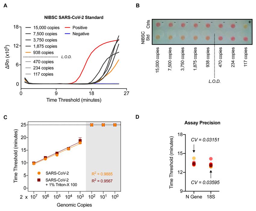
Wellcome Open Research 2021, 6:9 Last updated: 01 FEB 2021

Keywords
SARS-CoV-2, clinical diagnostic, RT-LAMP

               This article is included in the The Francis Crick
               Institute gateway.

                This article is included in the Coronavirus
                (COVID-19) collection.

 Corresponding authors: Michael D. Buck (michael.buck@crick.ac.uk), Enzo Z. Poirier (enzo.poirier@crick.ac.uk), Caetano Reis e Sousa (
 caetano@crick.ac.uk)
 Author roles: Buck MD: Conceptualization, Data Curation, Formal Analysis, Funding Acquisition, Investigation, Methodology, Project
 Administration, Supervision, Validation, Visualization, Writing – Original Draft Preparation, Writing – Review & Editing; Poirier EZ:
 Conceptualization, Data Curation, Formal Analysis, Funding Acquisition, Investigation, Methodology, Project Administration, Supervision,
 Validation, Visualization, Writing – Original Draft Preparation, Writing – Review & Editing; Cardoso A: Formal Analysis, Investigation;
 Frederico B: Formal Analysis, Investigation; Canton J: Formal Analysis, Investigation; Barrell S: Project Administration; Beale R:
 Conceptualization, Methodology, Project Administration; Byrne R: Project Administration; Caidan S: Project Administration; Crawford M:
 Project Administration; Cubitt L: Project Administration; Gandhi S: Conceptualization, Methodology, Project Administration; Goldstone
 R: Software; Grant PR: Methodology, Resources; Gulati K: Resources; Hindmarsh S: Software; Howell M: Conceptualization,
 Methodology, Project Administration, Resources; Hubank M: Methodology; Instrell R: Resources; Jiang M: Resources; Kassiotis G:
 Conceptualization, Methodology, Resources; Lu WT: Resources; MacRae JI: Conceptualization, Methodology, Project Administration,
 Resources; Martini I: Resources; Miller D: Resources; Moore D: Conceptualization, Methodology, Software; Nastouli E:
 Conceptualization, Methodology; Nicod J: Conceptualization, Methodology, Resources; Nightingale L: Software; Olsen J: Resources;
 Oomatia A: Resources; O'Reilly N: Resources; Rideg A: Resources; Song OR: Resources; Strange A: Resources; Swanton C:
 Conceptualization, Methodology, Project Administration; Turajlic S: Resources; Wu M: Resources; Reis e Sousa C: Conceptualization,
 Methodology, Project Administration;
 Competing interests: D. Miller and K. Gulati are employees of New England Biolabs, which provided the WarmStart Colorimetric LAMP
 2X Master Mix used in this work. C. Swanton receives or has received grant support from Pfizer, AstraZeneca, Bristol-Myers Squibb
 (BMS), Roche-Ventana, Boehringer-Ingelheim, and Ono Pharmaceutical and has consulted for or received an honorarium from Pfizer,
 Novartis, GlaxoSmithKline, Merck Sharp & Dohme, BMS, Celgene, AstraZeneca, Illumina, Genentech, Roche-Venatana, GRAIL, Medicxi,
 and the Sarah Cannon Research Institute. C. Swanton also is a shareholder of Apogen Biotechnologies, Epic Bioscience, and GRAIL and
 has stock options in and is a cofounder of Achilles Therapeutics.
 Grant information: This work was supported by The Rosetrees Trust and The John Black Charitable Foundation, as well as the Francis
 Crick Institute, which receives its funding from the UK Research Medical Council (FC001169, FC001078), Cancer Research UK (FC001169,
 FC001078) and the Wellcome Trust (FC001169, FC001078). This research was funded in part by the Wellcome Trust (FC001136). E.
 Nastouli is funded by MRC, NIHR, GSK and H2020. S. Gandhi has an MRC Senior Clinical Fellowship. E.Z. Poirier and M.D. Buck are
 supported by EMBO Long-Term Fellowships (ALTF 536-2018 and ALTF 1096-2018) and Marie Skłodowska-Curie Individual Fellowships
 (832511 and 837951).
 The funders had no role in study design, data collection and analysis, decision to publish, or preparation of the manuscript.
 Copyright: © 2021 Buck MD et al. This is an open access article distributed under the terms of the Creative Commons Attribution License
 , which permits unrestricted use, distribution, and reproduction in any medium, provided the original work is properly cited.
 How to cite this article: Buck MD, Poirier EZ, Cardoso A et al. SARS-CoV-2 detection by a clinical diagnostic RT-LAMP assay [version
 1; peer review: awaiting peer review] Wellcome Open Research 2021, 6:9 https://doi.org/10.12688/wellcomeopenres.16517.1
 First published: 21 Jan 2021, 6:9 https://doi.org/10.12688/wellcomeopenres.16517.1

                                                                                                                                Page 2 of 13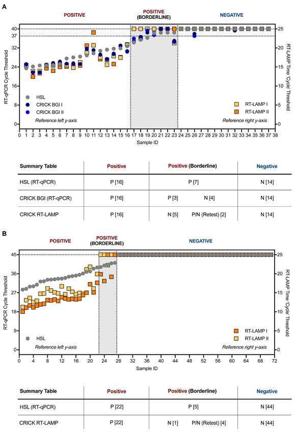
Wellcome Open Research 2021, 6:9 Last updated: 01 FEB 2021

Introduction                                                             Our results demonstrate that within the CCC pipeline,
The current pandemic caused by novel coronavirus SARS-CoV-2,             RT-LAMP can readily replace RT-qPCR as a means for
first detected in late 2019 in the province of Wuhan,                    detecting SARS-CoV-2 transcripts within RNA extracted from
China, has rapidly spread worldwide, infecting more than                 nose-throat swabs and endotracheal secretions/bronchoalveolar
68 million individuals as of 11 December 20201–3. Infection with         lavage fluid. RT-LAMP for the N gene shows absence of non-specific
SARS-CoV-2 can lead to development of COVID-19, a disease                amplification and cross-reactivity with other human coronaviruses
associated with severe acute respiratory syndrome, responsible           or respiratory viruses, and displays a sensitivity threshold
for hundreds of thousands of deaths globally. Controlling                almost equivalent to the gold standard RT-qPCR. Switching to
the spread of SARS-CoV-2 relies on the ability of healthcare             RT-LAMP translates into a ten-fold decrease in total reagent
systems to quickly identify infected individuals, which has              cost and a potential four-fold increase in pipeline output.
mainly relied on RT-qPCR for viral RNA detection4. International         Additionally, we provide preliminary data suggesting that
competition for commercial kits and reagents has negatively              RT-LAMP can be performed without prior RNA extraction,
impacted the ability of many countries to scale up testing capacity      allowing rapid and cost-effective testing that could potentially
to deal with the increased demand caused by rampant infection.           be extended to point-of-care. The entire workflow was validated
Implementing RT-qPCR testing programs requires specialised               under extended governance by public health authorities during
laboratory equipment and reagents, presenting additional                 the pandemic and inspected by a qualified UKAS assessor against
challenges.                                                              GenQA guidelines to verify compliance to ISO 15189:2012
                                                                         equivalent standard (US equiv. CAP/CLIA). As such, the pro-
In an effort to increase the diagnostic capacity for SARS-CoV-2          cedure developed here is ready to be deployed for diagnostic
infection in the UK, the Francis Crick Institute, a biomedi-             testing of SARS-CoV-2.
cal research institute based in London, rapidly repurposed its
staff and facilities in late March 2020 to serve as a clinical diag-     Methods
nostic testing facility through a partnership between a major            Patient samples
local healthcare provider (University College London Hospitals           In response to the pandemic, the Francis Crick Institute formed
National Health Services Trust) and an accredited clinical diag-         a partnership with University College London Hospitals (the
nostic laboratory (Health Services Laboratories, HSL), termed            Crick-COVID-Consortium) and set up a SARS-CoV-2 RT-qPCR
the CRICK COVID-19 Consortium (CCC)5. The pipeline uti-                  testing pipeline (CCC pipeline). Nasopharyngeal swabs tested
lises a series of in-house buffers to first inactivate patient sam-      in this study were obtained from the CCC pipeline5.
ples received from care homes and hospitals, and to then extract
RNA before using a CE marked commercial kit to detect                    CCC pipeline procedures and HSL RT-qPCR
SARS-CoV-2 by RT-qPCR. Patient results are reported through              All the samples processing through the CCC pipeline was done
a custom online web portal that interfaces with the reference            in accordance with Standard Operating Procedures (SOPs)
laboratory5. In order to avoid dependence on any singular testing        described recently5, which can be found at https://www.crick.
methodology, to continue increasing testing capacity, and to             ac.uk/research/covid-19/covid19-consortium. A redefined reaction,
provide a potential means to deliver diagnostics at the point-of-care,   running, and reporting SOP can be found in the Supplemental
the CCC was also tasked with developing and validating                   Methods (Extended data18).
alternative SARS-CoV-2 testing strategies.
                                                                         RT-LAMP assay
Herein, we describe the use of loop mediated isothermal                  RT-LAMP reaction was performed in a total volume of 15 µL,
amplification PCR coupled with reverse transcription (RT-                mixing 7.5 µL WarmStart Colorimetric LAMP 2X Master Mix
LAMP) as a robust method for SARS-CoV-2 detection in clinical            (New England Biolabs, M1800), 1.5 µL of 10X primer mix, 1.5 µL
specimens6. The entire strand displacement and amplification             SYTO 9 Green Fluorescent Nucleic Acid Stain (ThermoFisher
procedure is carried out at a single temperature in less than            Scientific, S34854), 1.5 µL of Nuclease-free water (Ambion,
30 minutes, alleviating the need for a thermocycler qPCR                 AM9932) and 3 µL of sample unless stated otherwise. 10X
system. Diagnostic tests utilising this technique have been              SYTO 9 solution at 5 µM in nuclease free water was prepared
developed for RNA viruses and other pathogens7–9, and there are          for a final concentration of 0.5 µM in the final RT-LAMP reac-
now several works focusing on the use of RT-LAMP to detect               tion. 10X primer mix was prepared from 100 µM desalted
SARS-CoV-210–17. We set up a SARS-CoV-2 RT-LAMP assay                    DNA primers obtained from Sigma Custom Oligos Service.
using the WarmStart Colorimetric LAMP 2X Mastermix                       10X primer mixes for N gene and 18S contained FIP and BIP
commercialised by New England Biolabs (NEB), which allows                primers at 16 µM, F3 and B3 at 2 µM, LF and LB at 4 µM.
for visual assessment of DNA amplification. Alternatively, DNA           RT-LAMP was ran following a SOP (cf. Extended data:
amplification can be quantified on a real-time PCR machine by            Supplementary Methods 1 and 218) on a 7500, 7500 Fast, Quant-
complexing the reaction with the DNA dye SYTO 910. We                    Studio 3, 5, or 7 Real-Time PCR System (Applied Biosystems).
made use of primers developed by Zhang et al. targeting the              A negative no template control (NTC) and a positive control (sup-
nucleocapsid phosphoprotein (N gene) of SARS-CoV-210 and,                plied by HSL, SARS-CoV-2 clinical sample) were included on
in a parallel reaction, primers that detect human 18S rRNA to            every run. Experiments utilising laboratory grown SARS-CoV-2
control for specimen quality9.                                           were performed in containment level 3 at the Francis Crick

                                                                                                                                 Page 3 of 13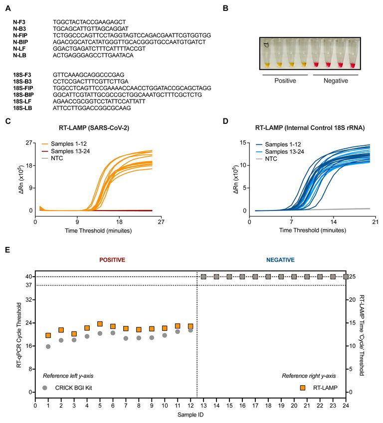
Wellcome Open Research 2021, 6:9 Last updated: 01 FEB 2021

Institute (FCI) by trained personnel according to health and          Results
safety guidelines.                                                    RT-LAMP test validity
                                                                      RT-LAMP was performed with 3 µL of RNA in a total reaction
We found that the assembled reaction mix was unstable when kept       volume of 15 µL unless stated otherwise. Each sample was
at 4°C for more than a few hours, and sensitive to freeze-thaw        tested in two separate reactions, one with primers targeting the
when kept at -20°C for more than a day. The RT-LAMP should            N gene of SARS-CoV-2, and the second targeting human 18S
be performed using freshly prepared reaction mix. The indi-           rRNA to control for specimen integrity and quality (Figure 1A).
vidual components should be stored at -20°C until use and avoid       The RT-LAMP mastermix contains a colorimetric pH indica-
repeated freeze/thaw cycles.                                          tor that turns from pink to yellow upon DNA amplification
                                                                      (Figure 1B). In addition, we benchmarked the RT-LAMP method
Accreditation and governance                                          by measuring DNA amplification using a SYBR based dye in
As outlined recently5, the CCC was formed in partnership with         a real-time PCR machine. When measuring fluorescence every
HSL, a UCLH UKAS accredited lab, who already had a COVID-19           minute (‘cycle’), double stranded DNA accumulation follows
RT-qPCR test in scope. All samples were received and com-             a characteristic exponential amplification phase that eventually
municated by HSL under their accreditation and the CCC                plateaus (Figure 1C and 1D).
RT-LAMP assay was validated against their existing RT-qPCR
test and the CCC’s validated RT-qPCR test, which uses a CE            An initial characterization of the technique was performed using
marked commercial kit (BGI). Given the urgent timeframe               24 RNA samples purified from patient nasopharyngeal swabs,
required to implement testing, it was not possible to secure offi-    12 of which were positive for SARS-CoV-2 as assessed by the
cial clinical laboratory accreditation for the FCI. However, full     CCC pipeline via RT-qPCR5. RT-LAMP could detect SARS-CoV-2
measures were taken to ensure that the CCC RT-LAMP test was           in the 12 positive samples with no amplification detected in
evaluated, verified and performed for diagnostic use in an envi-      all 12 negative samples, displaying 100% concordance to our
ronment that adhered to equivalent international standards (ISO       current clinical diagnostic platform (Figures 1C and Figures 1E).
15189:2012, US equiv. CAP/CLIA), overseen and audited by              Internal control signal was detected in all samples (Figure 1D).
HSL. These measures were implemented under the advice
and oversight of registered professionals from existing nearby        RT-LAMP background noise and specificity
ISO accredited medical laboratories, and included writing and         The background signal of the SARS-CoV-2 RT-LAMP assay
following clinical diagnostic SOPs for every stage of the pipe-       was assessed by running RNA elution buffer (7 samples) or
line from sample reception, processing to result reporting by         nuclease free water (7 samples) no template controls (NTCs)
qualified clinical scientists prior to results being communicated     performed 4 separate times by 4 different operators. Non-specific
to patients by HSL. Additional SOPs were followed for sample          signal could be detected in one well of one experiment after
storage, disposal of materials, batch certification of reagents and   25 minutes (Figure 2A). A similar experiment was performed
incident reporting. Appropriate risk assessments, training and        for the 18S rRNA internal control RT-LAMP assay and revealed
competency assessment procedures were established and docu-           signal in NTCs after 20 minutes (Figure 2B). Based on these
mented. Record sheets were created to document the receipt,           NTC data, a detection threshold was set for each RT-LAMP
batch acceptance testing, and start/end of use dates for key rea-     assay which eliminated all false positives in the N gene
gents and consumables. An inventory of all key equipment was          RT-LAMP assay and 21 of 23 false positives in the 18S rRNA
compiled which, where appropriate, included details of service        RT-LAMP assay (Figure 2C). This translated to a maximum run
and calibration records. Systems were also established for the con-   time for each reaction: 25 minutes for N gene and 20 minutes
trol of all key documents (version implementation, distribution       for 18S (Figure 1C). Implementing these criteria in a sepa-
and acknowledgement), audit trailing (what samples were tested        rate experiment yielded no false positives from 47 NTCs in the
when, by whom, with what equipment and using which                    SARS-CoV-2 RT-LAMP protocol and 4.5% false positives in
consumable/reagent batches), and the recording of all unto-           the 18S rRNA RT-LAMP assay (all with a time threshold > 19)
ward incidents/issues (thus facilitating appropriate investigation,   (Figure 2D–2E).
rectification and recurrence prevention). Samples were barcoded
and tracked using the Crick Clarity library information               We then asked if melting curves obtained from thermocycler
management system (LIMS). All key documents are available at          runs could be used to help discriminate true positives from false
https://www.crick.ac.uk/research/covid-19/covid19-consortium.         positives. N gene and 18S rRNA RT-LAMP reactions each gave
NHS governance was extended by a specific memorandum of               single peaks in melt curve plots (Extended data: Figure S1A18)
understanding for diagnostic testing between UCLH and the FCI         and consistent melting temperature values (Extended data:
enabled by NHS England. Assurance of the pipeline was per-            Figure S1B18) over multiple experiments performed on positive
formed in collaboration with GenQA, following their checklist         and negative patient samples. Moreover, positive samples that
for non-accredited laboratories and the lab and CCC workflow          amplified near the assay endpoint (i.e., high time threshold)
were inspected by a qualified UKAS assessor against the GenQA         still displayed a clear melting curve peak consistent with being
guidelines to verify compliance to IS015189:2012 equivalent           true positives (Extended data: Figure S2A18) and were distin-
standard.                                                             guishable from false positives (Extended data: Figure S2B18).

                                                                                                                              Page 4 of 13
Wellcome Open Research 2021, 6:9 Last updated: 01 FEB 2021
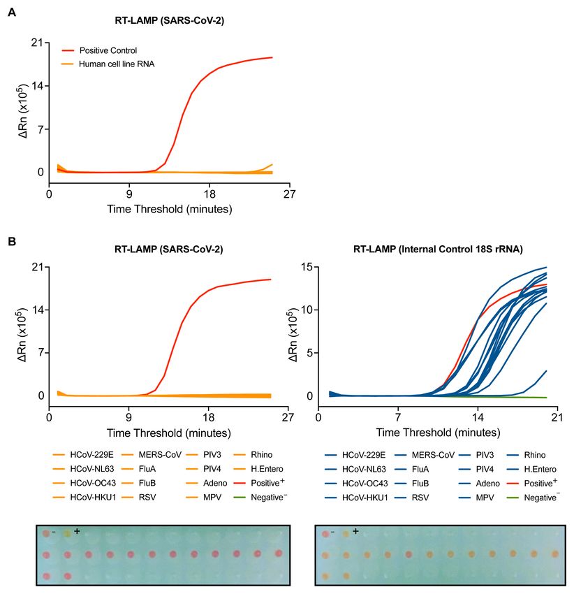
Figure 1. Validation of SARS-CoV-2 detection by RT-LAMP targeting the N gene. (A) The N gene of SARS-CoV-2 was targeted
using primers designed by Zhang et al.10, whilst the internal control implemented utilised primers for 18S rRNA initially published by
Lamb et al.9. (B) Upon DNA amplification, a colorimetric pH dye in the reaction mix will change from pink to yellow if the target is present.
Four SARS-CoV-2 positive and four SARS-CoV-2 negative patient samples are shown. (C–D) Amplification curves from 24 patient samples
assessed by SARS-CoV-2 (N gene) RT-LAMP (C) or 18S (D) in a Real-Time PCR machine. (E) Dot plot of the SARS-CoV-2 Ct values from
(C–D) compared to Ct values derived from the Crick’s RT-qPCR diagnostic pipeline using the CE marked BGI kit. Ct values of ‘undetermined’
are plotted as “Ct = 40” (left y-axis) or “Ct = 25” (right y-axis) for illustrative purposes. Data are normalised by assay run/cycles determined by
the two methods for comparative purposes. The clinical call from the reference laboratory is indicated above the graph.
Page 5 of 13Wellcome Open Research 2021, 6:9 Last updated: 01 FEB 2021
Figure 2. Optimisation of the RT-LAMP assay for accurate detection. (A–B) 7 samples of water and RNA elution buffer from the CCC
pipeline were run 4 independent times by 4 distinct operators by SARS-CoV-2. (A) or 18S (B) RT-LAMP. (C) Dot plot of ‘Ct’ values assessed in
(A–B). Assay endpoint detection threshold was set for 18S (dashed blue line) and SARS-CoV-2 (dashed orange line) based on these data.
(D–E) RT-LAMP amplification data from 47 samples of RNA elution buffer using the newly established assay endpoint of 25 min for SARS-
CoV-2 (D) and 20 minutes for 18S (E).
Page 6 of 13Wellcome Open Research 2021, 6:9 Last updated: 01 FEB 2021
Therefore, when implementing the assay using a real-time PCR To further assess the specificity of the N gene RT-LAMP assay,
machine (thermocycler) with SYBR-based detection, incorpo- we first determined possible cross-reactivity with human
ration of a traditional melting curve stage allows for elimina- RNA. 95 wells of RNA from a human cell line extracted by
tion of false positives and increases reporting confidence for the CCC pipeline gave no signal in the assay (Figure 3A). To
SARS-CoV-2 positive samples (see Extended data: Supplemental assess the assay’s specificity for SARS-CoV-2, the RT-LAMP
Methods21). assay was performed on RNA purified from clinical samples
Figure 3. SARS-CoV-2 RT-LAMP is highly specific. (A) SARS-CoV-2 RT-LAMP was performed on 95 wells of human cell line RNA extracted
by the CCC pipeline. (B–C) RT-LAMP targeting SARS-CoV-2 (B) or 18S (C) was performed on COVID-19 negative patient samples with
positively identified with other viral infections, including human coronaviruses (HCoV), influenzas (Flu), respiratory syncytial virus (RSV),
parainfluenzavirus (PIV), adenovirus (Adeno), metapneumovirus (MPV), rhinovirus (Rhino), and human enterovirus (H. Entero). The
colorimetric read-out from the actual run is depicted below. NTC (-) and positive control (+).
Page 7 of 13Wellcome Open Research 2021, 6:9 Last updated: 01 FEB 2021
from COVID-19 negative patients infected with a variety of RNA RT-LAMP sensitivity and precision
viruses, including seasonal coronaviruses strains 229E, NL63, In order to ascertain the sensitivity of the N gene RT-LAMP
OC43 and HKU1, MERS-CoV, Influenza A and B viruses, metap- assay, RNA from a SARS-CoV-2 quantified gene copy number
neumovirus (MPV), respiratory syncytial virus (RSV) or parain- standard obtained from the UK National Institute for Biologi-
fluenza viruses type 3 and 4 (PIV3, PIV4). All samples gave a cal Standards and Control (NIBSC) was extracted and assessed
signal in the 18S rRNA RT-LAMP, confirming the presence of by limiting dilution. The results indicate that the limit of reliable
human RNA (Figure 3B). With the N gene RT-LAMP, no detection of the N gene RT-LAMP assay is between 500–1000
signal was observed with any of the 14 distinct virus containing copies of N gene (Figure 4A and 4B) introduced in the assay. As
specimens tested, confirming that the assay is highly specific for the reaction is performed with 4.5 µL of sample, this assay
SARS-CoV-2 (Figure 3B). presents a limit of detection of 1.1 - 2.2 × 10⁵ N gene copies
Figure 4. SARS-CoV-2 RT-LAMP is highly sensitive, robust, and precise. (A–B) NIBSC SARS-CoV-2 standard was serially diluted and
the indicated number of copies in 4.5 µL was assessed by N gene RT-LAMP. Amplification curves shown with the limit of detection (L.O.D.)
determined by the presence or absence of amplification following the depicted dilution in (A) or via colorimetric read-out (B). (C) RNA
extracted from laboratory grown SARS-CoV-2 was serially diluted 10-fold and assessed by RT-LAMP in the presence or absence of 1%
Triton-X 100. (D) A COVID-19 positive patient sample was RNA extracted independently 5 times through the CCC pipeline and subjected to
5 independent N gene RT-LAMP reactions. Assay precision for N gene and 18S was determined by calculating the coefficient of variation
between Ct values observed (CV).
Page 8 of 13Wellcome Open Research 2021, 6:9 Last updated: 01 FEB 2021
per mL. This was confirmed using 10-fold serial dilutions of these validation experiments (3 µL versus 10 µL), partly due
RNA extracted from laboratory-grown SARS-CoV-2 quanti- to limitations in sample availability.
fied using the NIBSC standard (Figure 4C). Notably, a highly
linear response was observed, even in the presence of 1% Discussion
Triton-X 100, which has been proposed as a means of inactivating Rapid and reliable detection of SARS-CoV-2 is required to
SARS-CoV-219. efficiently diagnose infected individuals and to provide
governments and health systems with guidance for treatment and
The RT-LAMP assay reproducibility and precision were deter- quarantine strategies to reduce the risk of transmission. The CCC
mined by extracting RNA 5 times from a confirmed COVID-19 was formed to address a critical testing shortage in the London
positive patient sample through the CCC pipeline and assessing area, especially for frontline healthcare workers, with the addi-
by N gene and 18S RT-LAMP in 5 independent experiments, tional goal of rapidly validating and disseminating SOPs for
performed by two different operators (Figure 4D). The coeffi- others to scale up their own diagnostic programs. The major-
cient of variation was 0.03151 for the N gene RT-LAMP analy- ity of testing is currently performed by RT-qPCR amplifica-
sis and 0.03595 for the 18S rRNA internal control. We also tion of viral RNA obtained from nasopharyngeal samples. In the
verified that RT-LAMP can be performed in a 384-well plate face of continued global demand and competition for
format using a QuantStudio 5 real-time PCR machine with reagents and resources and to minimise reliance on any sin-
equivalent results (Extended data: Figure S318). gular testing strategy, we have outlined here procedures to
utilise RT-LAMP as a cost-effective and high throughput alter-
Clinical validation of RT-LAMP against two clinical native to RT-qPCR for detecting SARS-CoV-2 in clinical
diagnostic RT-qPCR assays specimens.
The RT-LAMP assay was benchmarked against the RT-qPCR
methods used by the reference clinical diagnostic laboratory, Implementing this testing method would reduce the CCC’s oper-
HSL, and that used by the CCC validated clinical diagnostic ational costs ten-fold when accounting for all consumables (data
platform by assessing 37 clinical samples processed in parallel not shown), provide the ability to scale up our output further
by both laboratories with duplicate RT-qPCR analyses. RNA (Extended data: Figure S318), and decrease reporting turnaround
extracted by the CCC pipeline was then tested by RT-LAMP time in comparison to our current method. We confirmed that
in duplicate runs. RT-LAMP detected 16 positives in both the assay is highly specific for SARS-CoV-2 and displays lack of
experiments, which was 100% concordant with results obtained cross-reactivity with other respiratory viruses, including sea-
by the CCC’s RT-qPCR assay using a CE marked kit from BGI sonal coronaviruses. Assay accuracy and robustness is denoted
and HSL’s N gene RT-qPCR assay. However, positives with by an absence of false positives and the further introduction
Ct > 35 near the limit of detection (Ct = 37) of the BGI RT-qPCR of a melt curve stage allows increased confidence in the iden-
assay, which were termed ‘borderline positives’, were not con- tification and reporting of genuine positives. When performed
sistently detected by the N gene RT-LAMP assay (Figure 5A). In with extracted RNA, the N gene RT-LAMP assay displays a
an additional set of experiments, 71 clinical specimens in sensitivity comparable to clinically approved RT-qPCR meth-
viral transport medium (VTM) stored at HSL were subjected ods. Additionally, preliminary experiments demonstrate that
to RNA extraction through the CCC pipeline and examined RT-LAMP can be performed directly without RNA extraction,
by HSL’s RT-qPCR and CCC’s RT-LAMP assay in duplicate by inactivating virus-containing dry swabs in a detergent-based
(Figure 5B). Again, samples with Ct < 39 in the HSL RT-qPCR solution. However, these experiments did not make use of true
analysis (which runs for 45 cycles as opposed to 40 in the BGI nasopharyngeal swabs that are likely to contain material that
assay) were confidently detected by RT-LAMP, whilst samples can cause RNA degradation or inhibit the assay (unpublished
with Ct values near the limit of detection (39 < Ct < 41), indicat- observations).
ing very low levels of SARS-CoV-2 RNA, displayed inconsist-
ent amplification by N gene RT-LAMP. Altogether, these data Our methodology makes use of a real-time qPCR machine that
demonstrate that RT-LAMP performed on extracted RNA accu- measures DNA amplification using a SYBR based dye, allow-
rately detects SARS-CoV-2 in clinical samples as verified by a ing for real-time detection and standardised reporting. We did
clinical diagnostic laboratory and a validated clinical diagnos- not test extensively if RT-LAMP could be assessed by the color-
tic pipeline. However, samples with Ct values within two cycles imetric indicator alone, although some of our data suggests
of the limit of detection of either RT-qPCR assays, were not that the colour change readout is concordant with the SYTO 9
consistently identified in duplicate RT-LAMP runs, with detec- dye results (Figure 1, Figure 3 and Figure 4). If RT-LAMP were
tion for some samples in one run, but not always in the other. coupled with a colorimetric read-out and the need for RNA
These results suggest that RT-LAMP is slightly less sensitive extraction could be obviated, the result would be a testing
than RT-qPCR when performed with extracted RNA. How- modality that tremendously reduces the cost and time for
ever, it is important to bear in mind that the RT-LAMP assay SARS-CoV-2 diagnostics and allow its application at point-of-care
was performed during these validation experiments with a third and in remote areas where sophisticated testing infrastructures
of the input RNA used by either RT-qPCR methods during currently do not exist.
Page 9 of 13Wellcome Open Research 2021, 6:9 Last updated: 01 FEB 2021
Figure 5. Clinical validation of SARS-CoV-2 RT-LAMP. (A) 37 patient samples were processed in parallel by HSL and the CCC pipeline
and interrogated by HSL’s RT-qPCR (N gene) and the CCC’s BGI RT-qPCR (ORF1a) in duplicate. RNA left from the CCC pipeline was assessed
in two separate experiments by N gene RT-LAMP. The graph indicates Ct values for HSL and CCC RT-qPCR runs by left y-axis and ‘Ct’ time
thresholds for RT-LAMP via the right y-axis. Data are normalised by assay run/cycles determined by the two methods for comparative
purposes. The clinical call from the reference laboratory is depicted above the summary table provided below. Positives (P) with low Ct values
were reliably detected by RT-LAMP, whilst ‘borderline positive’ (samples with Ct values near the limit of detection of each RT-qPCR assay)
were inconsistently detected, displaying no visible amplification at times – negative (N). (B) 71 VTM samples from HSL were RNA-extracted
through the CCC pipeline and interrogated by HSL RT-qPCR (45 cycles), or by two independent N gene RT-LAMP experiments (25 minutes
Page 10 of 13Wellcome Open Research 2021, 6:9 Last updated: 01 FEB 2021
Data availability Data are available under the terms of the Creative Commons
Underlying data Zero “No rights reserved” data waiver (CC0 1.0 Public domain
Open Science Framework: SARS-CoV-2 detection by a dedication).
clinical diagnostic RT-LAMP assay; raw data for figures, https://
doi.org/10.17605/OSF.IO/2XR8U20.
Acknowledgments
Open Science Framework: SARS-CoV-2 detection by a clini- The authors wish to thank all of the CCC members and CAP-
cal diagnostic RT-LAMP assay; raw data for supplementary TURE study members that have helped contribute reagents,
figures, https://doi.org/10.17605/OSF.IO/YFCSW21. advice, and time to this work.
Extended data The Crick COVID-19 Consortium (CCC): Jim Aitken1, Zoe
Open Science Framework: SARS-CoV-2 detection by a clini- Allen2, Rachel Ambler1, Karen Ambrose1, Emma Ashton2, Alida
cal diagnostic RT-LAMP assay, supplementary figures and Avola1, Samutheswari Balakrishnan1, Caitlin Barns-Jenkins1,
methods, https://doi.org/10.17605/OSF.IO/PAF3618. Genevieve Barr1, Sam Barrell1, Souradeep Basu1, Rupert
Beale1,3, Clare Beesley2, Nisha Bhardwaj1, Shahnaz Bibi2, Ganka
This project contains the following extended data: Bineva-Todd1, Dhruva Biswas3, Michael J. Blackman1,4, Domi-
- Figure S1. SARS-CoV-2 RT-LAMP and internal nique Bonnet1, Faye Bowker1, Malgorzata Broncel1, Claire
control 18S rRNA RT-LAMP display a consistent Brooks2, Michael D. Buck1, Andrew Buckton2, Timothy Budd1,
dissociation peak and melting temperature. (A) Alana Burrell1, Louise Busby2, Claudio Bussi1, Simon Butter-
Dissociation curves are shown for 11 SARS-CoV-2 worth1, Fiona Byrne1, Richard Byrne1, Simon Caidan1, Joanna
positive and 11 negative clinical samples along with 12 NTC Campbell5, Johnathan Canton1, Ana Cardoso1, Nick Carter1,
wells tested by RT-LAMP N gene assay (top) and 18S assay Luiz Carvalho1, Raffaella Carzaniga1, Natalie Chandler2, Qu
(bottom). (B) Dot plot of the melting temperatures deter- Chen1, Peter Cherepanov1, Laura Churchward6, Graham Clark1,
mined by 94 positive patient samples and 235 nega- Bobbi Clayton1, Clementina Cobolli Gigli1, Zena Collins1, Sally
tive patient samples tested by RT-LAMP assay. Data Cottrell2, Margaret Crawford1, Laura Cubitt1, Tom Cullup2, Heledd
are pooled from six separate experiments performed on Davies1, Patrick Davis1, Dara Davison1, Vicky Dearing1, Solene
3 separate RT-PCR machines. The average melting Debaisieux1, Monica Diaz-Romero1, Alison Dibbs1, Jessica
temperature (Tm) is depicted by a dotted line in (A) and Diring1, Paul C. Driscoll1, Annalisa D’Avola1, Christopher Earl1,
below the graph in (B) with the standard of deviation. Amelia Edwards1, Chris Ekin7, Dimitrios Evangelopoulos1,3,
Rupert Faraway1,3, Antony Fearns1, Aaron Ferron1, Efthymios
- Figure S2. Inclusion of a melt curve stage comple- Fidanis1 Dan Fitz1, James Fleming1, Bruno Frederico1, Alessan-
ments the ability of RT-LAMP to accurately distinguish dra Gaiba1, Anthony Gait2, Steve Gamblin1, Sonia Gandhi1,3,6,
true SARS-CoV-2 positive samples. (A) Amplification Liam Gaul1, Helen M. Golding1, Jacki Goldman1, Robert
curves (top) and dissociation curves (bottom) are Goldstone1, Belen Gomez Dominguez2, Hui Gong1, Paul R.
depicted from a range of positive samples for each assay Grant7, Maria Greco1, Mariana Grobler2, Anabel Guedan1,
along with a negative sample for reference. TP indicates Maximiliano G. Gutierrez1, Fiona Hackett1, Ross Hall1, Stei-
a true positive call based on amplification above back- nar Halldorsson1, Suzanne Harris1, Sugera Hashim2, Lyn Healy1,
ground plus consistent melting curve and Tm consist- Judith Heaney6, Susanne Herbst1, Graeme Hewitt1, Theresa
ent with genuine positives. (B) Amplification curve (left) Higgins1, Steve Hindmarsh1, Rajnika Hirani1, Joshua Hope1, Eliza-
and dissociation curve (right) are depicted of positive sam- beth Horton1, Beth Hoskins2, Catherine F. Houlihan6, Michael
ples and NTCs. TP indicates the same criteria outlined in Howell1, Louise Howitt1, Jacqueline Hoyle2, Mint R. Htun1,
(A), whereas FP denotes a false positive, which despite Michael Hubank8,9, Hector Huerga Encabo1, Deborah Hughes8, Jane
amplifying above background, does not have a melting Hughes1, Almaz Huseynova1, Ming-Shih Hwang1, Rachael Instrell1,
curve and temperature consistent with genuine positives. Deborah Jackson1, Mariam Jamal-Hanjani3,6, Lucy Jenkins2,
Ming Jiang1, Mark Johnson1, Leigh Jones1, Nnennaya Kanu3,
- Figure S3. RT-LAMP yields identical results across George Kassiotis1, Louise Kiely2, Anastacio King Spert Teixeira1,
different RT-PCR instruments and 96 versus 384-well Stuart Kirk7, Svend Kjaer1, Ellen Knuepfer1,12, Nikita Komarov1,3,
plate formats. Dot plot comparing results from RT-LAMP Paul Kotzampaltiris5, Konstantinos Kousis1, Tammy Krylova1,
assays (N gene, top; 18S, bottom) on the same clinical Ania Kucharska1, Robyn Labrum5, Catherine Lambe1, Michelle
positive and negative sample serially diluted 2-fold using Lappin1, Stacey-Ann Lee1, Andrew Levett7, Lisa Levett7,
an ABI 7500 Fast or QuantStudio 5 machine (96-well vs. Marcel Levi6, Hon Wing Liu1, Sam Loughlin2, Wei-Ting Lu1,
384-well plate format). James I. MacRae1, Akshay Madoo1, Julie A. Marczak1, Mimmi
Martensson1, Thomas Martinez1, Bishara Marzook1, John Mat-
- S
upplementary Method 1: RT-LAMP Mastermix
thews7, Joachim M. Matz1, Samuel McCall5, Laura E. McCoy3,
Preparation
Fiona McKay2, Edel C. McNamara1, Carlos M. Minutti1,
- S
upplementary Method 2: RT-LAMP RNA Loading and Gita Mistry1, Miriam Molina-Arcas1, Beatriz Montaner1,
Running RT-PCR Machine Kylie Montgomery2, Catherine Moore10, David Moore3,6,
Anastasia Moraiti1, Lucia Moreira-Teixeira1, Joyita Mukherjee1,
- Supplementary Method 3: RT-LAMP Results Reporting Cristina Naceur-Lombardelli3, Eleni Nastouli6,11, Aileen Nelson1,
Page 11 of 13Wellcome Open Research 2021, 6:9 Last updated: 01 FEB 2021
Jerome Nicod1, Luke Nightingale1, Stephanie Nofal1, Paul 3
University College London, London WC1E 6BT, UK
Nurse1, Savita Nutan2, Caroline Oedekoven1, Anne O’Garra1,
Jean D. O’Leary1, Jessica Olsen1, Olga O’Neill1, Nicola London School of Hygiene & Tropical Medicine, London
4
O’Reilly1, Paula Ordonez Suarez1, Neil Osborne1, Amar Pabari7, WC1E 7HT, UK
Aleksandra Pajak1, Venizelos Papayannopoulos1, Namita Patel1,
Yogen Patel5, Oana Paun1, Nigel Peat1, Laura Peces-Barba National Hospital for Neurology and Neurosurgery, University
5
Castano1, Ana Perez Caballero2, Jimena Perez-Lloret1, Magali S. College London Hospitals,
Perrault1, Abigail Perrin1, Roy Poh5, Enzo Z. Poirier1, James M.
Polke5, Marc Pollitt1, Lucia Prieto-Godino1, Alize Proust1, Clinda NHS Foundation Trust, London WC1N 3BG, UK
Puvirajasinghe2, Christophe Queval1, Vijaya Ramachandran2, Abhi-
nay Ramaprasad1, Peter Ratcliffe1, Laura Reed2, Caetano Reis e University College London Hospitals, NHS Foundation Trust,
6
Sousa1, Kayleigh Richardson1, Sophie Ridewood1, Fiona London NW1 2PG, UK
Roberts1, Rowenna Roberts2, Angela Rodgers1, Pablo Romero 7
Health Services Laboratories, London WC1H 9AX, UK
Clavijo1, Annachiara Rosa1, Alice Rossi1, Chloe Roustan1,
Andrew Rowan1, Erik Sahai1, Aaron Sait1, Katarzyna Sala1, Theo 8
The Institute of Cancer Research, London SW7 3RP, UK
Sanderson1, Pierre Santucci1, Fatima Sardar1, Adam Sateriale1, Jill A.
Saunders1, Chelsea Sawyer1, Anja Schlott1, Edina Schweighoffer1, 9
The Royal Marsden Hospital, Surrey SM2 5NG, UK
Sandra Segura-Bayona1, Rajvee Shah Punatar1, Joe Shaw2, Gee
Yen Shin6,7, Mariana Silva Dos Santos1, Margaux Silvestre1, 10
Public Health Wales, Heath Park, Cardiff CF14 4XW, UK
Matthew Singer1, Daniel M. Snell1, Ok-Ryul Song1, Moira J.
Spyer3, Louisa Steel2, Amy Strange1, Adrienne E. Sullivan1, University College London GOS Institute of Child Health,
11
Charles Swanton1,3,6, Michele S.Y. Tan1, Zoe H. Tautz-Davis1, Effie London WC1N 1EH, UK
Taylor1, Gunes Taylor1, Harriet B. Taylor1, Alison Taylor-Beadling2,
Fernanda Teixeira Subtil1, Berta Terré Torras1, Patrick Department of Pathobiology and Population Sciences, Royal
12
Toolan-Kerr1,3, Francesca Torelli1, Tea Toteva1, Moritz Treeck1, Veterinary College, University of London, London NW1 0TU, UK
Hadija Trojer13, Ming-Han C. Tsai1, James M.A.Turner1, Mela-
nie Turner7, Jernej Ule1,3, Rachel Ulferts1, Sharon P. Vanloo1,3, Royal Free London NHS Foundation Trust, London NW3 2QG,
13
Selvaraju Veeriah3, Subramanian Venkatesan1, Karen Vousden1, UK
Andreas Wack1, Claire Walder2, Philip A. Walker1, Yiran
Wang1, Sophia Ward1,3, Catharina Wenman2, Luke Williams1, CAPTURE study: Nicky Browne1, Kim Edmonds1, Yasir Khan1,
Matthew J. Williams1, Wai Keong Wong6, Joshua Wright1, Sarah Sarker1, Ben Shum1, Tim Slattery1, & Samra Turajlic1,2
Mary Wu1, Lauren Wynne1, Zheng Xiang1, Melvyn Yap1,
Julian A. Zagalak1,3, Davide Zecchin1,11 and Rachel Zillwood1
1
The Royal Marsden Hospital, Surrey, SM2 5NG, UK
1
The Francis Crick Institute, London NW1 1AT, UK
2
The Francis Crick Institute, London NW1 1AT, UK
2
Great Ormond Street Hospital for Sick Children NHS Foundation A previous version of this article is published on medRxiv:
Trust, London WC1N 3JH, UK. https://doi.org/10.1101/2020.06.29.20142430.
References
1. Zhu N, Zhang D, Wang W, et al.: A Novel Coronavirus from Patients with 7. Mori Y, Notomi T: Loop-mediated isothermal amplification (LAMP): a rapid,
Pneumonia in China, 2019. N Engl J Med. 2020; 382(8): 727–33. accurate, and cost-effective diagnostic method for infectious diseases.
PubMed Abstract | Publisher Full Text | Free Full Text J Infect Chemother. 2009; 15(2): 62–9.
PubMed Abstract | Publisher Full Text | Free Full Text
2. Wu F, Zhao S, Yu B, et al.: A new coronavirus associated with human
respiratory disease in China. Nature. 2020; 579(7798): 265–9. 8. Ahn SJ, Baek YH, Lloren KKS, et al.: Rapid and simple colorimetric detection
PubMed Abstract | Publisher Full Text | Free Full Text of multiple influenza viruses infecting humans using a reverse
transcriptional loop-mediated isothermal amplification (RT-LAMP)
3. Zhou P, Yang XL, Wang XG, et al.: A pneumonia outbreak associated with diagnostic platform. BMC Infect Dis. 2019; 19(1): 676.
a new coronavirus of probable bat origin. Nature. 2020; 579(7798): 270–3. PubMed Abstract | Publisher Full Text | Free Full Text
PubMed Abstract | Publisher Full Text | Free Full Text 9. Lamb LE, Bartolone SN, Tree MO, et al.: Rapid Detection of Zika Virus in Urine
4. Yan Y, Chang L, Wang L: Laboratory testing of SARS-CoV, MERS-CoV, and Samples and Infected Mosquitos by Reverse Transcription-Loop-Mediated
SARS-CoV-2 (2019-nCoV): Current status, challenges, and countermeasures. Isothermal Amplification. Sci Rep. 2018; 8(1): 3803.
Rev Med Virol. 2020; 30(3): e2106. PubMed Abstract | Publisher Full Text | Free Full Text
PubMed Abstract | Publisher Full Text | Free Full Text 10. Zhang Y, Odiwuor N, Xiong J, et al.: Rapid Molecular Detection of SARS-CoV-2
(COVID-19) Virus RNA Using Colorimetric LAMP. Infectious Diseases (except
5. Aitken J, Ambrose K, Barrell S, et al.: Scalable and Resilient SARS-CoV-2 testing
HIV/AIDS). 2020.
in an Academic Centre. Health Systems and Quality Improvement. 2020.
Publisher Full Text
Publisher Full Text
11. Rabe BA, Cepko C: SARS-CoV-2 detection using isothermal amplification and
6. Notomi T, Okayama H, Masubuchi H, et al.: Loop-mediated isothermal a rapid, inexpensive protocol for sample inactivation and purification. Proc
amplification of DNA. Nucleic Acids Res. 2000; 28(12): E63. Natl Acad Sci. 2020; 117(39): 24450–8.
PubMed Abstract | Publisher Full Text | Free Full Text PubMed Abstract | Publisher Full Text | Free Full Text
Page 12 of 13Wellcome Open Research 2021, 6:9 Last updated: 01 FEB 2021
12. Dao Thi VL, Herbst K, Boerner K, et al.: A colorimetric RT-LAMP assay and 16. Yoshikawa R, Abe H, Igasaki Y, et al.: Development and evaluation of a
LAMP-sequencing for detecting SARS-CoV-2 RNA in clinical samples. Sci rapid and simple diagnostic assay for COVID-19 based on loop-mediated
Transl Med. 2020; 12(556): eabc7075. isothermal amplification. PLoS Negl Trop Dis. 2020; 14(11): e0008855.
PubMed Abstract | Publisher Full Text | Free Full Text PubMed Abstract | Publisher Full Text | Free Full Text
13. Fowler VL, Armson B, Gonzales JL, et al.: A highly effective reverse- 17. Lalli MA, Langmade SJ, Chen X, et al.: Rapid and extraction-free detection
transcription loop-mediated isothermal amplification (RT-LAMP) assay for of SARS-CoV-2 from saliva by colorimetric reverse-transcription loop-
the rapid detection of SARS-CoV-2 infection. J Infect. 2020; mediated isothermal amplification. Clin Chem. 2020; hvaa267.
S0163-4453(20)30731-3. PubMed Abstract | Publisher Full Text | Free Full Text
PubMed Abstract | Publisher Full Text | Free Full Text
18. Poirier E: SARS-CoV-2 detection by a clinical diagnostic RT-LAMP assay. 2021.
14. Pang B, Xu J, Liu Y, et al.: Isothermal Amplification and Ambient http://www.doi.org/10.17605/OSF.IO/PAF36
Visualization in a Single Tube for the Detection of SARS-CoV-2 Using Loop-
19. Patterson EI, Prince T, Anderson ER, et al.: Methods of inactivation of SARS-
Mediated Amplification and CRISPR Technology. Anal Chem. 2020; 92(24):
CoV-2 for downstream biological assays. Microbiology. 2020.
16204–16212.
Publisher Full Text
PubMed Abstract | Publisher Full Text | Free Full Text
15. Taki K, Yokota I, Fukumoto T, et al.: SARS-CoV-2 detection by fluorescence 20. Poirier E: SARS-CoV-2 detection by a clinical diagnostic RT-LAMP assay. 2021.
loop-mediated isothermal amplification with and without RNA extraction. http://www.doi.org/10.17605/OSF.IO/2XR8U
J Infect Chemother. 2020; S1341-321X(20)30399-8. 21. Poirier E: SARS-CoV-2 detection by a clinical diagnostic RT-LAMP assay. 2021.
PubMed Abstract | Publisher Full Text | Free Full Text http://www.doi.org/10.17605/OSF.IO/YFCSW
Page 13 of 13You can also read