Susceptibility of Pimephales promelas and Carassius auratus to a strain of koi herpesvirus isolated from wild Cyprinus carpio in North America ...
←
→
Page content transcription
If your browser does not render page correctly, please read the page content below
www.nature.com/scientificreports
OPEN Susceptibility of Pimephales
promelas and Carassius auratus
to a strain of koi herpesvirus
isolated from wild Cyprinus carpio
in North America
Isaiah E. Tolo1,2, Soumesh K. Padhi1,2, Keiffer Williams2, Vikash Singh3, Sophie Halvorson1,
Sunil K. Mor1,3 & Nicholas B. D. Phelps1,2*
Cyprinid herpesvirus-3 (CyHV-3, syn. koi herpesvirus) is an important pathogen worldwide and a
common cause of mass mortality events of wild common carp (Cyprinus carpio) in North America,
however, reference strains and genomes obtained from wild carp are not available. Additionally, it is
unclear if fishes in North America are susceptible to CyHV-3 infection due to incomplete susceptibility
testing. Here we present the first North American type strain and whole-genome sequence of CyHV-3
isolated from wild carp collected from a lake with a history and recent incidence of carp mortality.
Additionally, the strain was used in an in-vivo infection model to test the susceptibility of a common
native minnow (Pimephales promelas) and goldfish (Carrasius auratus) which is invasive in North
America. Detection of CyHV-3 DNA was confirmed in the tissues of a single fathead minnow but the
same tissues were negative for CyHV-3 mRNA and samples from exposed fathead minnows were
negative on cell culture. There was no detection of CyHV-3 DNA or mRNA in goldfish throughout the
experiment. CyHV-3 DNA in carp tissues was reproducibly accompanied by the detection of CyHV-3
mRNA and isolation on cell culture. Additionally, environmental CyHV-3 DNA was detected on all tank
filters during the study. These findings suggest that fathead minnows and goldfish are not susceptible
to CyHV-3 infection and that detection of CyHV-3 DNA alone in host susceptibility trials should be
interpreted with caution.
Cyprinid herpesvirus 3, (CyHV-3, syn. koi herpesvirus), is a viral pathogen of common carp (Cyprinus carpio),
hereafter referred to as carp, and its ornamental variety, koi carp. CyHV-3 was first identified in the USA in
domestic ornamental koi1 and has since been detected with PCR-based methods in association with widespread
mass mortality events of wild carp in the U SA2,3, as well as in some clinically healthy p opulations4,5. However,
isolation of CyHV-3 endemic to carp in North America has been largely unsuccessful despite the use of known
permissible cell lines (koi fin, KF-1, and common carp brain, CCB) as well as other routinely used fish cell
lines3,6. Isolation of CyHV-3 has only been reported once in 2006, and on an atypical cell line, fathead minnow
cells (Pimpephales promelas, FHM)7. Since then, formation of cytopathic effect (CPE) on FHM using CyHV-3
positive tissue has not been replicated1,2,6,8,9. Isolation of CyHV-3 on cell lines is a critical step in establishing a
causal relationship between CyHV-3 detection and disease outbreaks. Furthermore, there is a lack of study iso-
lates for evaluating the genetic background, pathogenicity and host range of strains endemic to North America.
Though carp and its hybrids (carp x goldfish) are the only species known to be affected by koi herpesvirus
disease (KHVD), the range of species which may be susceptible to CyHV-3 infections remains unclear10. Gold-
fish (Carrasius auratus) have been shown to become infected and able to transmit CyHV-3 to naive carp11,12;
however, evidence of susceptibility of goldfish to infection with CyHV-3, and the ability of goldfish to act as a
1
Minnesota Aquatic Invasive Species Research Center and the Department of Fisheries Wildlife and Conservation
Biology, University of Minnesota, St. Paul, MN, USA. 2Department of Fisheries, Wildlife and Conservation
Biology, College of Food, Agriculture and Natural Resource Sciences, University of Minnesota, St. Paul, MN,
USA. 3Department of Veterinary Population Medicine and Veterinary Diagnostic Laboratory, College of Veterinary
Medicine, University of Minnesota, St. Paul, MN, USA. *email: phelp083@umn.edu
Scientific Reports | (2021) 11:1985 | https://doi.org/10.1038/s41598-021-81477-0 1
Vol.:(0123456789)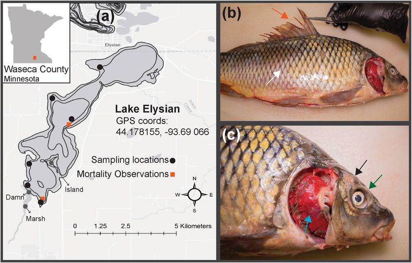
www.nature.com/scientificreports/
carrier for CyHV-3 has not been replicated in other studies13,14. Additionally, detection of CyHV-3 DNA and
transmission to carp by cohabitation experiments has been reported in a wide variety of non-carp species from
survey or disease trial studies without the presence of clinical signs15. In total, CyHV-3 DNA has been detected
in seven fish species native to North America: rainbow trout (Oncorhynchus mykiss)16, brown bullhead (Amei-
rus nebulosus) and three-spined stickleback (Gasterostues aculeatus)17, black bullhead (Ictaluris melas)18, silver
perch (Bairdiella chrysoura)19, Atlantic sturgeon (Acipenser oxyrinchus)20, and northern pike (Esox Lucius)21. The
absence of clinical signs or the employment of tools for differentiating sources of CyHV-3 DNA (e.g. subclini-
cal viral infection, environmental contamination, latent infection, or false positive) makes the interpretation of
these findings difficult.
A variety of diagnostic tools are available for determining the presence of CyHV-3 in suspected c ases10. Isola-
tion of CyHV-3 on permissive cell lines is a definitive method for determining the presence of infectious CyHV-3
in tissue and environmental samples but permissible cell lines, Koi fin (KF-1) and common carp brain (CCB),
are not highly susceptible to CyHV-321–23 and are not considered a sensitive diagnostic tool10. Recently, cell lines
with higher sensitivity to CyHV-3 have been reported24 but are not yet widely available.
PCR-based methods for detection of CyHV-3 DNA are the most sensitive diagnostic tools for CyHV-3. Of
these assays, the Gilad qPCR is the most sensitive method25. In-situ hybridization has been used as a confirma-
tory method for CyHV-3 DNA in t issues11 and the use of immunochemical assays such as ELISA have been
reported for determination of previous exposure of fish to CyHV-326–28. RNA and mRNA detection assays are
particularly useful in determining the host range of CyHV-3 since infectious hosts, in which viral replication
must occur, can be distinguished from vectors, which may transport CyHV-3 DNA and infectious particles
but without viral replication. Multiple PCR-based methods have been reported for the detection of CyHV-3
mRNA12,29; however, the mRNA detection test developed by Yuasa et al.29 does not detect CyHV-3 DNA when
present and is thus preferable to assays that rely on DNAse enzyme treatment which may not completely remove
DNA when concentrations are high.
Given the widespread detection of CyHV-3 in North A merica2 and the inevitable exposure of native fishes,
it is critical to understand potential impacts of local CyHV-3 strains on invasive carp populations and on native
fishes. Both native FHM and invasive goldfish are sympatric with carp and could occur in many of the same
habitats. While the host status of goldfish with regard to CyHV-3 has been partially described11,12, demonstration
of true susceptibility to CyHV-3 infection requires confirmation that indicates viral replication (i.e., presence of
mRNA)19,29,30. The susceptibility status of FHM has never been evaluated, despite the only previous isolation of
a North American CyHV-3 isolate on the FHM cell l ine7. Here, we further investigate the susceptibility of FHM
and goldfish to CyHV-3 in accordance with criteria for determination of host susceptibility defined by the OIE
(i.e., route of transmission is consistent with natural pathways for the infection, adequate identification of the
pathogen, and provision of evidence indicating that pathogen presence constitutes an infection)31 using a strain
of CyHV-3 that was associated with multiple, previously reported mass mortalities of wild carp in North America.
Materials and methods
Collection of wild carp from a CyHV‑3‑exposed population. This study was carried out in accord-
ance with the recommendations in the Guide for the Care and Use of Laboratory Animals of the National
Institutes of Health. All protocols for sampling, procedures and experimental endpoints involving live fish con-
ducted in this study were approved by the Institutional Animal Care & Use Committee (IACUC), University of
Minnesota (St. Paul, Minnesota, USA), under the approval numbers IACUC-1806-36036A and 1808-36276A.
Experiments were performed in compliance with the ARRIVE guidelines on animal r esearch32.
Wild carp were sampled from Lake Elysian (Waseca County, Minnesota, Coordinates: 44.178144, − 93.69066)
by boat electrofishing from September 3 to 9, 2019 (Fig. 1a). This lake was expected to have a CyHV-3-exposed
carp population following a confirmed outbreak in 20173. Captured wild adult carp (n = 116) were euthanized
by immersion in a solution of ~ 3 mL/L pure clove oil (90% Eugenol; Velona, Elk Grove Village, IL, USA) for
15 min and transported on ice to the University of Minnesota for necropsy. Brain, gill and kidney tissues from
up to three carp were pooled in a 1:5 (weight:volume) dilution of Hank’s Balanced Salt Solution (HBSS; Cellgro,
Lincoln, NE, USA) containing 100 IU/mL of Penicillin and Streptomycin and maintained at a pH of 7.4 at 4 °C
for 24 h prior to preparation for qPCR and cell culture screening for CyHV-3 (described below). Gill tissues
from ten freshly-dead carp obtained from a shallow bay in the Southern portion of the lake were also obtained
and pooled by five individuals for a total of two sample pools.
An additional 17 wild carp collected as part of the previously described sampling event were placed in an
aerated live well and transferred to the Minnesota Aquatic Invasive Species Research Center’s Containment
Laboratory (MCL). These carp were housed in a ~ 1400 L tank with flow through well water at 20 °C and treated
with 0.6% aquarium salt once per day. Carp were acclimated for 1 day and then anesthetized via immersion in
a solution of 100 µL/L of clove oil and uniquely marked using colored injectable elastomer (Northwest Marine
Technology, Anacortes, WA, USA). Additionally, a small portion (~ 0.2 cm2) of each carp’s gills were sampled
for qPCR screening for CyHV-3 and tested immediately. Carp determined to be CyHV-3 negative (n = 12) were
euthanized following testing. Carp determined to be CyHV-3-positive (n = 5) by specific qPCR were held for
a total of 5 days, during which, water temperature was gradually increased to 26 °C in order to increase viral
shedding. CyHV-3-positive carp gill biopsies were again sampled and screened on the fifth day to identify carp
with high qPCR copy numbers. All CyHV-3-positive carp were then euthanized, and the brain, gill and kidney
tissues were removed as previously described. Pooled tissues from two wild carp with clinical signs consistent
with KHVD (Fig. 1b,c) and with high qPCR copy numbers, were subjected to cell culture immediately following
necropsy. In addition, a 10 g portion of this pooled tissue was processed and used to challenge naive carp in the
in-vivo infection model. Tissues were homogenized in a 1:5 volume of HBSS containing 100 IU/mL Penicillin
Scientific Reports | (2021) 11:1985 | https://doi.org/10.1038/s41598-021-81477-0 2
Vol:.(1234567890)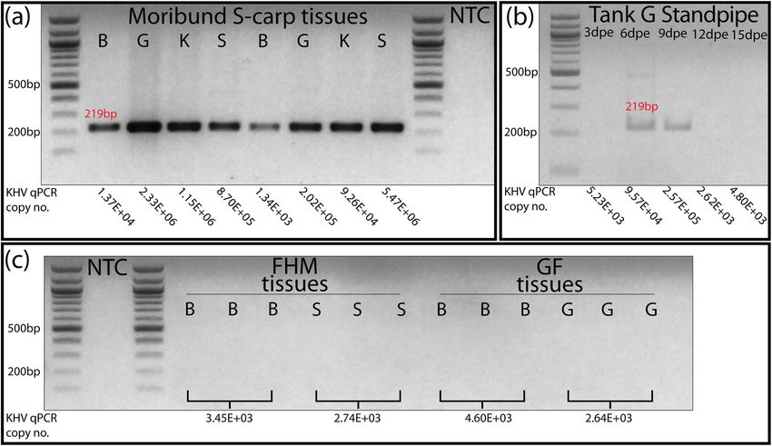
www.nature.com/scientificreports/
Figure 1. (a) Generated using ArcMap (v10.8.1, https://desktop.arcgis.com/en/arcmap/), shows the
approximate locations of sampling effort and mortality observations on Lake Elysian. Bathymetric contours
indicate depth in 5 ft increments. (b, c) Pathology of a representative individual wild carp sampled from Lake
Elysian. Arrows on (b, c) denote frayed fins (vermillion), loss of mucosal layer (white), loss of scales and
epidermis (black), enopthalmia (bluish green), gill necrosis (sky blue).
and Streptomycin (pH = 7.4). The sample was centrifuged at 2360 × g at 25 °C for 10 min, then the supernatant
was passed through a 0.45 µm syringe filter.
In‑vivo infection trial. To increase the potential of obtaining an isolate of CyHV-3, naïve carp previ-
ously determined to be CyHV-3 negative by qPCR, were challenged with CyHV-3-positive tissue homogenates
obtained from wild carp. Two naïve carp, purchased from Osage Catfisheries (Osage Beach, MO, USA), were
pair housed in a 60 L aquarium with flow through well water (flow rate = 3–4 tank volumes/h) at 21–22 °C.
Aquaria were set up with a standpipe drain covered by a cylindrical wire screen filter of approximately 15 cm
in length and 4.4 cm in diameter. Additionally, a PVC pipe section of 15 cm in length and 10 cm in diameter
was added to each tank for shelter. Each carp was exposed to 0.5 mL of CyHV-3-positive tissue homogenate by
IP-injection and monitored for signs of disease for 6 days and then euthanized. Pooled samples of brain, gill and
kidney tissue were subjected to qPCR and cell culture analysis. Following cell culture analysis (below) a second
infection trial was performed to satisfy River’s postulates (i.e. that CyHV-3 isolated from wild diseased carp
would cause similar disease in naïve carp)33. Two additional naïve carp purchased from Osage Catfisheries were
IP-injected with 0.5 mL of CyHV-3-positive (qPCR and cell culture positive) cell culture supernatant. Carp were
housed and observed for disease signs as previously described for 11 days and then sacrificed. Pooled samples of
brain, gill, and kidney then were tested by CyHV-3-specific qPCR to confirm the presence of CyHV-3.
Cell culture analysis. CCB cells were maintained in Eagle’s Minimum Essential Medium (EMEM) con-
taining Eagles’s salts (Sigma, St. Louis, MO, USA), 10% fetal bovine serum (FBS), 1% non-essential amino acids
(NEAA, Sigma), 2 mM l-glutamine and glucose (Sigma) up to 4.5 g/L. The KF-1 cells were cultured in EMEM
containing Eagles’s salts (Sigma), 10% FBS and 0.025 M HEPES. Penicillin 100 U/L and streptomycin 0.1 mg/L
(Sigma) were used as an anti-bacterial agent in both cell culture media and the cells were maintained at 25 °C.
Cell culture methods to isolate CyHV-3 were performed according to the US Fish and Wildlife Service and
American Fisheries Society-Fish Health Section Blue Book34. Briefly, pooled tissues were homogenized in Hank’s
Balanced Salt Solution (HBSS; Cellgro) and centrifuged at 2360 × g for 15 min. The inoculum was added to the
24-well plates with 80% confluent cell cultures in two dilutions, (1/10 and 1/100) and incubated at 25 °C for
14 days. A blind passage was performed for an additional 14 days if no cytopathic effects (CPE) were observed
on the first passage. If CPE was observed during the first passage, then the second passage was performed in a
25 cm2 flask. The virus was harvested when CPE reached 70–80% of the monolayer. The infected cultures were
exposed to two freeze/thaw cycles at − 80 °C, and then centrifuged at 3800 × g for 15 min at 4 °C. The clarified
supernatants and pellets were collected and stored at − 80 °C.
Whole‑genome sequencing and sequence analysis. Whole-genome sequencing was performed at
the University of Minnesota Veterinary Diagnostic Laboratory for genetic characterization of the CyHV-3 isolate
Scientific Reports | (2021) 11:1985 | https://doi.org/10.1038/s41598-021-81477-0 3
Vol.:(0123456789)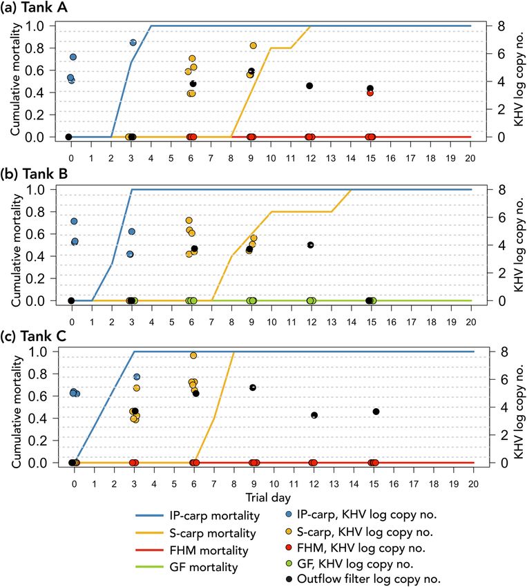
www.nature.com/scientificreports/

                                            Figure 2.  (a) Shows a schematic of the cohabitation disease trial. Vermillion arrows denote inoculation of
                                            IP-carp with CyHV-3 positive tissue homogenate, blue arrows denote introduction of IP carp for cohabitation
                                            with fishes in experimental tanks, and the reddish purple arrow indicates the tissue origin of CyHV-3-positive
                                            S-carp. (b) Shows a schematic of experimental flow through chambers with black arrows indicating the direction
                                            of water flow. (c) Shows a time-line of various samples.

                                            (KHV/Elysian/2019) obtained from wild carp. In brief, after CCB cells, infected with wild carp tissues, reached
                                            80% CPE, the supernatant was collected and stored at − 80 °C. The frozen supernatant was freeze-thawed three
                                            times, and centrifuged at 2896 × g for 25 min at 4 °C. Nucleic acid purification of CCB cell culture supernatant
                                            was done using a QIAamp MinElute Virus Spin Kit (Qiagen, Hilden, Germany) following manufacturer instruc-
                                            tions. The extracted nucleic acids were subjected to library preparation using Nextera Flex DNA library kit
                                            (Illumina, San Diego, CA, USA) following manufacturer instructions. The library was normalized according to
                                            the median fragment size measured by Tape Station 2.0 (Agilent, Santa Clara, CA, USA) and library concentra-
                                            tion measured by Qubit. The library was submitted to the University of Minnesota Genomic Center (UMGC) for
                                            sequencing via MiSeq V3 (2X75-bp) paired end chemistry.
                                                Raw fastq files were trimmed to remove Illumina adapters using Trimmomatic (v 0.39) with a minimum
                                            quality score of 20. Then, bowtie2 (v 2.3.5) was used to remove host contamination and unmapped reads were
                                            used for assembly with SPAdes (v3.13.0) with k-mer values of 29, 33 and 55 with the options “careful with a
                                            minimum coverage of 5 reads per contig”. Then contigs were searched into the RefSeq viral and non-redundant
                                            protein reference databases using Diamond BLASTx with an e-value of 1e − 5 for significant hits. Taxon assign-
                                            ments were made with MEGAN6 Community Edition according to the lowest-common-ancestor algorithm.
                                            ORFs prediction and genome annotation were done using Prokka (v1.14.5). The resulting alignment (GenBank
                                            accession no. MT914509) was aligned with 19 other CyHV-3 genomes available on NCBI using Mafft (v7)
                                            with the FFT-NS-2 alignment strategy and the following parameters: scoring matrix BLOUSUM62, gap open
                                            penalty 1.53, offset value 0. Model selection, maximum likelihood (ML) tree construction, and calculation of
                                            bootstrap values were done with R 4.0 (R Software) using phangorn (v2.5.5). ML trees were constructed using
                                            the top scoring model (GTR + G + I) and 100 bootstrap replicates were generated to assess the reliability of clades
                                            obtained in the tree. Additionally, this genome assembly was compared with the previously reported thymidine
                                            kinase gene sequence obtained from carp sampled during a large mortality event in Lake Elysian in 2017 (F36,
                                            GenBank accession no. MK987089).

                                            Investigation of species specificity.         To investigate the host range of KHV/Elysian/2019, six carp pur-
                                            chased from Osage Catfisheries, previously determined to be CyHV-3-negative by qPCR, were intraperitoneally
                                            (IP) injected with 0.5 mL of the filtered tissue homogenate material (Fig. 2a). The IP-injected carp (IP-carp)

          Scientific Reports |   (2021) 11:1985 |                https://doi.org/10.1038/s41598-021-81477-0                                                  4

Vol:.(1234567890)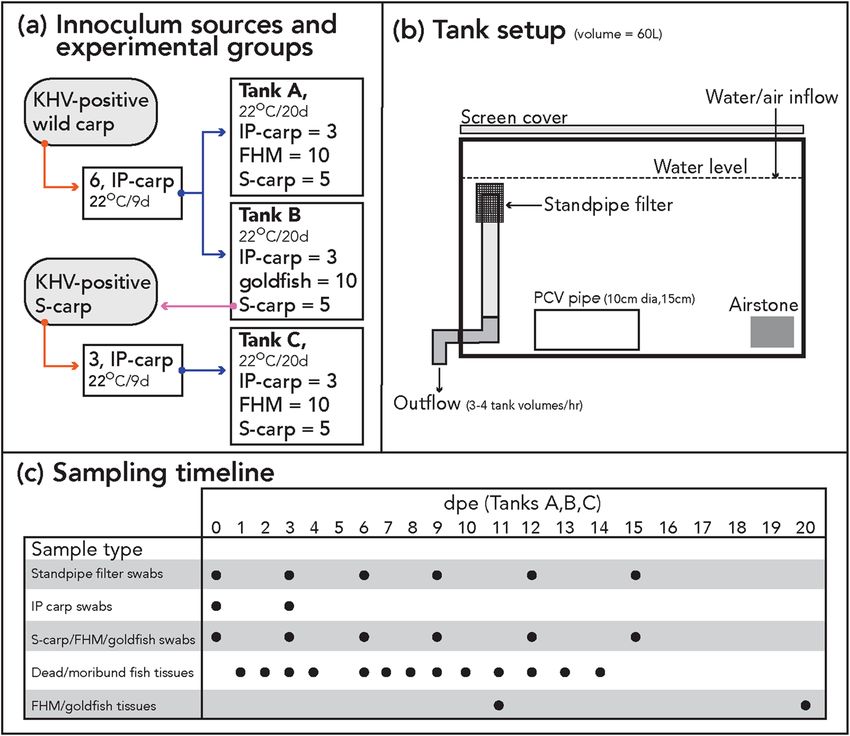
www.nature.com/scientificreports/
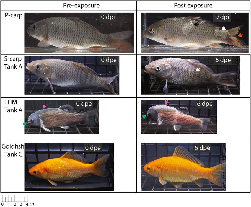
Figure 3. Representative fishes photographed before and after exposure to CyHV-3. Note, fishes photographed
at 0 dpe may not be the same individual as those at 6 dpe. dpe days post exposure via cohabitation, IP-carp
intraperitoneally injected carp, S-carp cohabitated sentinel carp, FHM fathead minnow. Arrows denote frayed
fins (vermillion), loss of mucosal layer (white), scale pocket edema (black). Additionally, normal morphological
features of mature male fathead minnows are indicated for nuptial tubercles (bluish green), and nape pads
varying in prominence (reddish purple).
were housed as previously described for 9 days prior to their use in the cohabitation trial (Fig. 2b). The IP-carp
were monitored twice daily for signs of disease. After 9 days the gills, skin and vent of each IP-carp was swabbed
aseptically with a single sterile cotton swab (Dynarex, Orangeburg, NY, USA) for determination of viral load by
qPCR. FHM and goldfish were challenged with CyHV-3 via cohabitation. One cohabitation tank (tank A) con-
tained ten naïve FHM, five naïve sentinel carp (S-carp) and three IP-carp (Fig. 2a). One cohabitation tank (tank
B) contained ten naïve goldfish, five naive S-carp and three of the IP-carp. S-carp were included in each tank
setup to act as a positive control for within-tank transmission of CyHV-3. Two additional negative control tanks
with the same stocking density and conditions contained ten naïve FHM (tank D) and ten naïve goldfish (tank
E), as well as eight naïve carp (confirmed to be CyHV-3-negative by specific qPCR). Average standard length
and weight for fishes used in these experiments was 13 cm and 64 g for carp, 7 cm and 13 g for FHM, and 10 cm
and 38 g for goldfish. All tanks consisted of ~ 60 L aquaria with flow-through well water as previously described.
Fishes were fed a commercial feed (Skretting classic trout, Skretting, Tooele, UT, USA) daily and monitored
twice daily to observe changes to fish health. IP-carp that died during the trial were allowed to remain in the
tank for 24 h prior to removal for necropsy, but any morbidity or mortality of other experimental groups were
immediately removed and necropsied.
At 0, 3, 6, 9, 12, and 15 days post exposure (dpe) by cohabitation, five FHM, five goldfish, and all IP-carp and
S-carp from each tank were anesthetized by immersion in a buffered solution of 100 mg/L of MS-222 and the
gills, skin and vent of each fish was swabbed with a sterile swab for determination of viral load by qPCR (Fig. 2c).
For FHM and goldfish, the five individuals were randomly sampled at each time-point. Additionally, the wire
screen filter of the outflow standpipe was swabbed at the same intervals during the course of the trial to evaluate
loading of CyHV-3 DNA in the environment. All swabs were stored at − 20 °C in individual plastic bags until
nucleic acid extraction could be performed. At 11 dpe, half of the FHM and goldfish from cohabitation tanks were
euthanized by immersion in a buffered solution of 3 g/L of MS-222 and necropsied (Fig. 2c). The remaining FHM
and goldfish were maintained until 20 dpe and then euthanized and necropsied. To visually record the presence
of gross pathology, representative IP carp, and fish from cohabitation groups (S-carp, FHM, and goldfish) were
randomly selected and photographed at 0 and 6 dpe in a small glass aquarium (Fig. 3).
For each necropsied fish, wet mounts of gill and skin scrapes were viewed at 40× magnification to identify
potential parasitic infections. Then the skin of each necropsied fish was rinsed briefly with 70% ETOH and clean
water. Brain, gill, kidney and skin tissue were collected individually for each fish and split into two duplicate
Scientific Reports | (2021) 11:1985 | https://doi.org/10.1038/s41598-021-81477-0 5
Vol.:(0123456789)
www.nature.com/scientificreports/
samples. The first sample duplicates were placed in Whirl–Pak sample bags (Nasco, Fort Atkinson, WI, USA) and
preserved at − 20 °C until nucleic acid extraction and screening for CyHV-3 DNA was performed. The second
sample duplicates were placed in 1 mL of RNAlater solution (Ambion) in 1.5 mL microcentrifuge tubes (Globe
Scientific, Mahwah, NJ, USA) and frozen at − 20 °C. An individual FHM and goldfish from each time-point (11-
and 20-dpe) was preserved in 10% NBF (TissuePro, Gainesville, FL, USA) for histological analysis. Individual
representatives of each species from control tanks and moribund S-carps from each experimental tank were also
preserved for histological analysis.
Due to the detection of CyHV-3 DNA in a single FHM in tank A, a second trial with FHM (tank C) was
performed as described previously (Fig. 2a). Brain, gill, kidney, and skin tissue from two S-carp exposed in the
first trial with disease signs and positive qPCR test for CyHV-3 (tank A) were pooled, homogenized and filtered
as previously described. Three new carp purchased from Osage Catfisheries were IP injected with 0.5 mL of this
tissue homogenate and maintained as previously described for 9 days prior to screening for CyHV-3 by qPCR
and used in the cohabitation trial. All other conditions and procedures were done as described for the first
cohabitation trial with the following exceptions. In the second trial, portions of brain, gill, kidney and skin tis-
sues obtained from a moribund S-carp at 5 dpe and four FHM at 11 dpe, respectively, were pooled as previously
described and subjected to cell culture. Additionally, duplicate swabs from the tank C outflow standpipe filter
were obtained and preserved in 1 mL of RNAlater solution (Sigma) as previously described for tissue samples.
Nucleic acid purification using chelex resin and detection of CyHV‑3 by qPCR. For nucleic acid
purification, chelex resin (Sigma) was used as described by Zida et al.35 and briefly summarized here. For pooled
tissue samples, approximately 100 mg of each tissue was homogenized in 1 mL of nuclease free water (NFW)
and then centrifuged, with 50 μL of the resulting supernatant later used as starting material. For swabs, the cot-
ton end was cut off and vortexed, then centrifuged and finally the cotton was removed leaving the supernatant.
For each sample type, 150 μL of chilled 80% ETOH was added, then centrifuged and the supernatant removed.
Samples were allowed to air dry for 10 min to remove residual ETOH. 150 μL of 20% Chelex was added to each
sample and vortexed. Samples were then incubated at 90 °C for 20 min and centrifuged and immediately used
for qPCR.
A Taqman probe-based qPCR was used for the detection of CyHV-3 DNA targeting the ORF89 gene36 using a
StepOnePlus thermocycler with default settings (Applied Biosystems). Nucleic acid purifications from all samples
were screened for CyHV-3 using a PrimeTime gene expression master mix kit (Integrated DNA Technologies,
Coralville, IA, USA), with each reaction containing 400 nM of primers (KHV-86f: GAC-GCC-GGA-GAC-CTT-
GTG, KHV-163r: CGG-GTT-GTT-ATT-TTT-GTC-CTT-GTT) and 250 nM of the probe (KHV-109p: [TAMRA]
CTT-CCT-CTG-CTC-GGC-GAG-CAC-G-[IBRQ]. The reaction mix was subjected to an initial denaturation
at 95 °C for 3 min, followed by 40 cycles of denaturation at 95 °C for five sec and annealing at 60 °C for 30 s. A
threshold cycle of 38 was used as a cut off. The standard curve for quantification of CyHV-3 genomes was per-
formed using a laboratory synthesized DNA fragment containing the ORF89 sequence as previously described by
Padhi et al.3. The results for virus load are presented as the number of viral copies per mL of tissue supernatant.
All samples obtained from FHM and goldfish were tested in triplicate with the exception of samples that had
positive qPCR Ct values, which were re-tested up to six times.
RNA purification and reverse transcription polymerase chain reaction (RT‑PCR). Individual tis-
sues of preserved brain, gill, kidney, and skin from one representative S-carp from each experimental tank (A, B
and C) were selected as positive controls for CyHV-3 mRNA detection (total of 12 tissue samples). All preserved
tissue samples from FHM or goldfish which had at least one positive qPCR test were also screened for CyHV-3
mRNA to determine if replicating virus was present (total of eight tissue samples). Additionally, preserved swabs
of the outflow standpipe filter were also screened. For RNA purification, RNA was extracted from tissues using
the RNeasy Mini Kit (Qiagen) according to the manufacturer instructions for animal tissues, using ~ 30 mg tis-
sue samples preserved in RNAlater. For swabs, cotton was cut from the end of the swab and used as the starting
material. CyHV-3 mRNA was detected using the RT-PCR developed by Yuasa et al.29 with the primers, (KHV
RT F3: GCC-ATC-GAC-ATC-ATG-GTG-CA, KHV RT R1: AAT-GCC-GCT-GGA-AGC-CAG-GT). The RT-
PCR was performed using a One-step RT-PCR kit (Qiagen) according to the manufacturer instructions. The
reaction mix was subjected to a single step of reverse transcription at 50 °C for 30 min and denaturation at 95 °C
for 15 min, followed by 40 cycles of: 94 °C for 30 s, 65 °C for 30 s, 72 °C for one minute and a final extension
step was 72 °C for 10 min. PCR products were separated and visualized on 2% agarose gels containing 0.75 μg/
mL ethidium bromide (Genesee Scientific, San Diego, CA, USA). PCR products for carp, FHM and goldfish
templates (clear band at the 219 bp location) were cut from gels and purified by precipitation with a 20% PEG,
2.5 M NaCl solution. Purified RT-PCR products were subjected to Sanger sequencing at the University of Min-
nesota Genomics Center (UMGC). Sequences were trimmed and analyzed using 4 peaks (v1.8) and consensus
sequences were generated using BioEdit (v7.2.1). Sequence identities were compared with available reference
sequences by BLASTn analysis of the National Center of Biotechnology sequence database.
Histology. Histology was used to demonstrate the presence or absence of lesions in cohabitation disease trial
specimens. Histological samples of gill tissue were prepared from formalin-fixed samples of representative fishes
of each species from trial and control tanks. Gill samples were dissected from formalin-fixed specimens and
decalcified in 10% ethylenediaminetetraacetic acid (EDTA) for 10 days. Following decalcification, samples were
dehydrated in an ethanol series to 100% ethanol, infiltrated with toluene, and subsequently embedded in paraf-
fin. Paraffin sections were cut at 6 µm thickness using a Leica Jung 820 Histocut Rotary Microtome and mounted
on slides. Sections were stained with Hematoxylin and Eosin using a protocol modified from H umasson37.
Scientific Reports | (2021) 11:1985 | https://doi.org/10.1038/s41598-021-81477-0 6
Vol:.(1234567890)www.nature.com/scientificreports/
Statistical analysis. R 4.0 (R Software) was used for statistical analysis and data presentation. CyHV-3
qPCR copy numbers are presented as averages of all positive tests for samples with duplicate tests and were Log
transformed prior to statistical testing. Significant differences (p < 0.05) in virus load of IP injected carp and
cohabitated fish were determined using a 1-way ANOVA with subsequent pairwise multiple comparisons using
the Holm-Sidak method and data were presented as box plots of 25–75% (+ minimum and maximum values)
with an indication of mean and median. Bivariate associations were measured with odds ratios and 95% confi-
dence intervals (Cis).
Results
Detection and isolation of CyHV‑3 from wild carp. Wild carp obtained from Lake Elysian (n = 133;
116 immediately euthanized and 17 transferred live to the MCL) ranged in standard length from 10 to 68 cm
(mean standard length = 49.16 cm, SD = 10.03 cm) and in weight from 0.95 to 6.29 kg (mean weight = 2.94 kg,
SD = 1.42 kg). Among this sample of wild carp 36 were male, 93 were female, and four were young of the year (sex
could not be determined). Water temperature at the time of sampling was between 20 and 22 °C at the surface.
Pooling of tissues from wild carp which were immediately euthanized for screening (n = 116) resulted in a
total of 50 tissue pools. A total of 10/50 tissue pools tested positive for CyHV-3 by qPCR with copy numbers
ranging between 2.81E + 03 and 1.63E + 06 (mean copy no. = 2.53E + 05). Gill necrosis was observed in 18 wild
carp (15.52%) and positively associated with the detection of CyHV-3 DNA by qPCR (p-value = 8.20E − 07,
OR = 89.37, 95% CI 8.60 4.78E + 03). However, the presence of gill necrosis was not observed in all fish with
positive qPCR tests. Two gill tissue pools collected from dead carp also tested positive for CyHV-3 by qPCR with
copy numbers of 6.03E + 05 and 2.36E + 06.
Five of the 17 live carp tested positive for CyHV-3 in gill samples with copy numbers ranging from 8.03E + 03
to 4.18E + 05. The two carp with the highest qPCR copy numbers of 4.18E + 05 and 2.04E + 04 were used for cell
culture inoculum and the in-vivo infection trial. Pooled tissue homogenate from these fish used in disease trials
and cell culture had a copy number of 2.00E + 04. The two live wild carp that had the highest qPCR copy numbers
for CyHV-3 also had signs of disease including necrotic gills, frayed fins, apparent loss of the mucosal layer (i.e.
rough sandpaper-textured scales), loss of scales and epidermis, and enopthalmia (Fig. 1b,c). Gyrodactylus sp.,
Trichodina sp. and Flavobacterium columnare were also observed in wet mounts of skin and gill scrapes from
several of the wild carp housed in the MCL. There were no clinical signs present in the other 15 carp held in the
MCL, including three additional CyHV-3 positive carp and 12 that were negative.
Tissue pools (n = 50) from 116 wild carp used for cell culture inoculum did not produce CPE on KF-1 or CCB
cell lines. However, CPE was observed on both the KF-1 and CCB cell lines when inoculated with tissue of the
carp held in the MCL. The CPE was characterized by the detachment of cells from the surface of the substrate
along with extensive vacuolation 9 days post-inoculation of the blind passage. The cultures were confirmed to
be CyHV-3-positive by qPCR with a viral copy number of 2.02E + 08.
In‑vivo infection trial. River’s postulates for associating CyHV-3 with disease of wild carp in Lake Elysian
were fulfilled in the initial in-vivo infection trial. Disease signs and subsequent morbidity at 6 dpe were observed
in naïve carp injected with CyHV-3-positive tissues obtained from the wild carp. Pooled samples of brain, gill,
and kidney obtained from experimentally infected individuals were positive for CyHV-3 with a qPCR copy num-
ber of 2.60E + 06 and produced CPE on CCB cells 14 days after inoculation. This CPE-positive CCB supernatant
had a qPCR copy number of 5.89E + 06. Additional naïve carp injected with this cell culture supernatant were
determined to be moribund at 11 dpe and were positive for CyHV-3 with a qPCR copy number of 9.74E + 05.
Whole‑genome of CyHV‑3 from wild carp. Sequencing and denovo assembly of the KHV/Elysian/2019
genome resulted in a 295,161 bp sequence. A total of 288 parsimoniously informative sites were identified
among the 20 strains included in the genome alignment. KHV/Elysian/2019 clustered with genomes in the
European clade, sharing between 98.74 and 99.16% sequence identity with genomes of the Asian lineage and
99.29–99.60% sequence identity with genomes of the European lineage (Fig. 4). KHV/Elysian/2019 was most
identical to KHV-E (ACC no. MG925489) which was isolated from England and first reported by Klafack et al.38.
The previously reported 1003 bp sequence containing the thymidine kinase gene obtained from dying carp dur-
ing a large mortality event in Lake Elysian in 2017 (CyHV-F36, ACC no. MK987089) was identical to that of
KHV/Elysian/2019.
Susceptibility of carp, fathead minnow and goldfish to a North American strain of
CyHV‑3. After 4–6 days post IP injection with CyHV-3-positive tissue obtained from wild carp IP-carp
began to show signs of disease. Clinical signs included a lack of response to feed, loss of the mucosal layer, frayed
fins, pale and discolored skin, enopthalmia and lethargy (Fig. 3). IP-carp in all experimental tanks died between
9 and 12 days post injection (within 4 days of initiation of the cohabitation trial) (Fig. 5). S-carp in tanks A, B
and C began showing similar signs of disease as IP-carp after 6 days (Fig. 3). Between 7 and 12 dpe, disease signs
in IP- and S-carp became more pronounced and included lethargy, loss of scales and epidermis, scale pocket
edema, enopthalmia and diffuse hemorrhages of the skin. Gill erosions were also observed in some IP-carp and
S-carp during necropsy. No external parasites were observed on wet mounts from any individuals from experi-
mental or control tanks.
The cumulative mortality of all IP and S-carp was 100%, occurring between 7 and 13 days after exposure to
CyHV-3 inoculum or infected carp (Fig. 5). Onset of mortality and complete mortality of naive carp occurred
earlier in tank C than in tank A. No lesions or change in behavior of FHM or goldfish were observed in any of
Scientific Reports | (2021) 11:1985 | https://doi.org/10.1038/s41598-021-81477-0 7
Vol.:(0123456789)www.nature.com/scientificreports/
Figure 4. Midpoint-rooted maximum likelihood phylogeny of publicly available CyHV-3 genomes and KHV/
Elysian/2019. The scale bar represents substitutions per site. Genotypes are indicated by vermillion (European)
and blue (Asian) boxes.
the experimental tanks (Fig. 3). However, a single mortality of FHM occurred in the negative control tank (tank
D); but no lesions were observed during necropsy.
The pooled tissue obtained from wild carp used for IP injection of IP-carp in tanks A and B had a copy
number of 1.13 E + 04. The pooled tissue obtained from tank A S-carp, used for IP injection of IP-carp in tank
C, had a copy number of 7.97 E + 05. All swabs and tissues obtained from IP-carp tested positive for CyHV-3
DNA at day 0 of the cohabitation trial (Fig. 5). Swabs from S-carp began to test positive for CyHV-3 at 3 dpe in
tank C and at day 6 in tanks A and B. Swabs from all S-carp continued to test positive until none remained in
cohabitation tanks. All tissues of IP- and S-carp tested positive for CyHV-3 DNA (Fig. 5). Copy numbers were
significantly higher in kidney tissue from IP-carp than tissues from cohabitated carp. On average, all tissues from
IP-carp had higher copy numbers than tissues from cohabitated fish (Fig. 6). Tissues from tank C S-carp were
also used to re-isolate CyHV-3 on CCB cells. CPE was observed in the first passage (14 dpe) after inoculation
with S-carp tissues collected from tank C.
Swabs from the outflow standpipe filter of all CyHV-3-exposed tanks were positive for CyHV-3 DNA with
copy numbers between 3.12 E + 03 and 5.50 E + 04, beginning at day 3 in tank C and at day 6 in tanks A and B
and continuing until day 12 in tank B and day 15 (final day of sampling from outflow standpipe filter) in tanks
A and C.
A skin swab from a single FHM at 15 dpe tested positive for CyHV-3 DNA (2/3 tests positive) with an average
copy number of 1.48 E + 03. All other swabs from FHM and goldfish were negative for CyHV-3 DNA. CyHV-3
DNA was detected (4/6 tests positive) in a single skin tissue sample from FHM collected on day 11 from tank
A with an average copy number of 2.74 E + 03, as well as in brain from the same individual (2/3 tests) with an
average copy number of 3.45 E + 03. No other tissues from FHM collected on day 11 from tank C were positive
for CyHV-3 DNA. One goldfish from tank B tested positive for CyHV-3 in brain tissue (1/6 tests positive) with
a copy number of 4.59 E + 03. A second goldfish in tank B tested positive for CyHV-3 in gill tissue (1/6 tests
positive) with a copy number of 2.64 E + 03. No other tissues from goldfish tested positive for CyHV-3 DNA on
day 11 dpe and no tissues from FHM or goldfish tested positive for CyHV-3 DNA on day 20 dpe. Tissues from
all fishes in negative control tanks (tanks d and e) were all negative for CyHV-3 at 20 dpe. Additionally, tissues
from FHM from tank C collected at 11 dpe and subjected to cell culture analysis did not produce CPE on CCB
cells during the first or second passages.
All tests of tissues from representative S-carp were positive for CyHV-3 mRNA with bands at the expected size
of 219 bp (12 tissue samples run in duplicate) (Fig. 7). None of the tissues from FHM or goldfish were positive
Scientific Reports | (2021) 11:1985 | https://doi.org/10.1038/s41598-021-81477-0 8
Vol:.(1234567890)www.nature.com/scientificreports/
Figure 5. (a–c) The cumulative mortality and CyHV-3 log copy number/mL of fishes in experimental
cohabitation tanks. IP-carp intraperitoneally injected carp, S-carp cohabitated sentinel carp, FHM fathead
minnow, goldfish. “Trial day” indicates days post introduction of IP carp into cohabitation tanks. Points
indicating log copy number are jittered to aid in visualization.
for CyHV-3 mRNA (eight tissue samples run in triplicate). Additionally, outflow standpipe filter swabs from tank
C at days 6 and 9 of the trial were also positive for CyHV-3 mRNA. CyHV-3 specific qPCR copy numbers for
corresponding tissues from S-carp testing positive for CyHV-3 mRNA ranged between 1.34E + 03 and 5.47E + 06.
CyHV-3 specific qPCR copy numbers for corresponding outflow standpipe filter swabs were 9.57E + 04 at 6 dpe
and 2.57E + 05 at 9 dpe (Fig. 7).
Sanger sequencing of 219 bp RT-PCR products from CyHV-3 infected carp confirmed the identity of these
fragments as the CyHV-3 DNA packaging terminase subunit gene sequence (ORF33 of the CyHV-3 E genome).
Sanger sequencing of purified gel extracts cut from the approximate location of potential low amplification RT-
PCR products from FHM and goldfish tissues resulted in no visible reaction.
Representative fish from each treatment group were examined by light microscopy. There were no lesions in
the FHM, goldfish or control carp. However, lesions consistent with KHVD were observed in the gill tissue of
CyHV-3-exposed carp, including fusion of the secondary lamellae to erosion of the primary lamellae, as well as
sporadic intranuclear inclusion bodies within epithelial cells.
Discussion
The primary goal of this study was to isolate a virulent strain of CyHV-3 present in wild carp in North America
and evaluate the susceptibility of two species, FHM and goldfish using a combination of methods chosen to
confirm the presence of viable CyHV-3 infection. Of these two species goldfish have been previously tested for
susceptibility to CyHV-3, but interpretation of results has been contradictory11,12,14. Under the experimental
conditions in the present trial, our results indicate that FHM and goldfish are unlikely to be natural hosts for
Scientific Reports | (2021) 11:1985 | https://doi.org/10.1038/s41598-021-81477-0 9
Vol.:(0123456789)www.nature.com/scientificreports/
Figure 6. Boxplots of CyHV-3 log copy numbers/mL of tissue supernatant of IP injected carp (IP-carp, light
grey boxes), cohabitated sentinel carp (S-carp, dark grey boxes), and in tissues of a single fathead minnow
(FHM), quantified using CyHV-3-specific qPCR. No boxes are displayed for FHM tissues since only a single
individual tested positive for CyHV-3.
Figure 7. (a–c) Show UV fluorescence of RT-PCR products on 2% agarose gels stained with ethidium bromide.
Bands for 500 and 200 bp on the DNA ladders are noted. The position of 219 bp is denoted in red. S-carp
cohabitated sentinel carp, FHM fathead minnow, goldfish, B brain, G gill, K kidney, S skin, NTC no template
control. In (b) “standpipe” indicates swabs of the tank outflow standpipe filer. CyHV-3 specific qPCR copy
numbers/mL of tissue supernatant from corresponding tissues are displayed below each lane. Samples in (a) are
representative tissues from S-carp from tank A (first 4 samples) and from tank C (last 4 samples). Samples in (c)
are triplicate tests of FHM and goldfish tissues which had positive qPCR tests.
Scientific Reports | (2021) 11:1985 | https://doi.org/10.1038/s41598-021-81477-0 10
Vol:.(1234567890)www.nature.com/scientificreports/
CyHV-3 and are not susceptible to KHVD. We recommend this diagnostic framework be adopted for future
CyHV-3 susceptibility studies.
This is the first reported isolation of CyHV-3 in North America that has been linked with a mortality event
of wild carp and fulfilled Koch’s postulates. Successful isolation of CyHV-3 had previously been documented by
Grimmet et al.7, however, this isolate could not be maintained in cell culture, was not re-exposed to carp and
only the molecular sequence of the TK gene is available. The difficulty of culturing CyHV-3 is most likely due
to relative fragility of virions, limited number and susceptibility of available cell lines8. In this study, none of the
samples of CyHV-3 positive carp tissues obtained from wild carp and maintained for 24–48 h at 4 °C induced
formation of CPE. However, CyHV-3 was successfully isolated when standard isolation protocols were used in
concert with live housing of infected carp and the in-vivo infection model to ensure that CyHV-3-positive tis-
sues were subjected to cell culture shortly (less than 2 h) after necropsy. This method was also useful since carp
could be screened non-lethally and euthanized during the peak of infection in order to avoid expending time
and resources on negative samples or samples with low viral loads. These results reinforce the necessity of using
either very fresh tissues from wild carp or tissues from carp with severe acute infections for cell culture isolation.
Inoculation of disease-free carp with CyHV-3 positive tissue homogenate may be a useful preliminary step in
propagating CyHV-3 in-vivo if cell culture is not immediately available.
Despite the importance of CyHV-3 worldwide, there are few publicly available genomes. Furthermore, most
CyHV-3 genomes available originated from cultured carp and koi and thus, CyHV-3 genomes originating from
wild carp are especially limited. The genome sequence of the isolate obtained in this study is of the same lineage
(European) as that of CyHV-3 detected in Elysian in 2 0173, as well as most strains of CyHV-3 originating prior
to 1999 when Asian lineage strains began to emerge in the USA39. Anthropogenic movement of wild carp in the
USA has been under strict regulation since the 1980s and it was well documented that carp was introduced to
North America from Europe, much earlier, in the late 1800s40. Based on the similarity of the genome presented
here to other European isolates it can be speculated that CyHV-3 in North America may have been circulat-
ing in wild populations prior to its initial report in 1 9981. This hypothesis however should be tested by further
molecular epidemiological studies.
CyHV-3 DNA was detected in at least one replicate qPCR test in brain and skin tissue of a single FHM and
brain and gill of a single goldfish but is unlikely to indicate the presence of replicating CyHV-3 as evidenced by
the lack CyHV-3 mRNA detection by RT-PCR. In contrast and as expected, all tissue samples from S-carp were
positive for CyHV-3 DNA and mRNA, which was confirmed by Sanger sequencing and histopathology. An
alternative interpretation of the DNA-positive and mRNA-negative results for FHM and goldfish may be due to
a higher sensitivity of the qPCR assay and the presence of very low-level infections that are not detectable by the
mRNA assay. However, this explanation is unlikely since S-carp tissues with lower CyHV-3 DNA concentrations
than those of positive FHM and goldfish samples had detectable levels of mRNA. Yuasa et al.29 also demonstrated
the sensitivity of the Yuasa RT-PCR assay to detect CyHV-3 mRNA in highly diluted positive control samples.
Additionally, re-isolation of CyHV-3 on CCB cell culture from S-carp tissues was successful though CyHV-3
could not be re-isolated in cell culture from tissues of cohabitated FHM and goldfish. Finally, replication of the
FHM disease trial (tank C) did not result in additional positive qPCR tests of any FHM swabs or tissues. Taken
together, these results indicate that detection of CyHV-3 DNA in FHM and goldfish tissues likely originated
from non-viable CyHV-3 virions or naked DNA.
The source of CyHV-3 DNA in external tissues of FHM (skin) and goldfish (skin and gill) was likely due
to contact contamination with feces, mucous and sloughing skin of infected cohabitated carp. Similar fomites
were also likely the origin of CyHV-3 DNA on the outflow standpipe filter, which was detectable by qPCR at
least 6 days after all S-carp had died and were removed from the tank. Interestingly, CyHV-3 mRNA was also
detectable on the outflow standpipe though it appeared to be partially degraded based on the appearance of
faint bands. The likely source of this mRNA was carp epidermal cells that became entrained in the filter mesh
of the outflow standpipe.
Given our inability to detect CyHV-3 in tissues of all but one FHM in tank A and none in the replicate
trial in tank C, the source of CyHV-3 DNA in an internal tissue of an FHM (brain) is most likely the result of
contamination of this sample with CyHV-3 virions or DNA on the skin or in oral cavity contents of the same
individual during necropsy. However, it is also possible that this DNA originated from CyHV-3 virions which
invaded or were taken up by FHM cells but without evidence of replication. CyHV-3 DNA has been previously
detected in internal tissues of various fish species without evidence of replication or d isease41. Several species
of fish have also been shown to transmit CyHV-3 to naive carp under experimental cohabitation conditions,
despite no detectable replication11,13,21. Thus, it has been speculated that many fish species may act as carriers of
CyHV-3, without the presence of disease signs or detectable mRNA16. The application of mRNA detection assays
could be further improved by development of a quantitative assay, which would be useful to verify the results of
species which are listed as having incomplete evidence for s usceptibility31, particularly if detection of CyHV-3
DNA represents sample contamination.
The difficulty in discriminating viable and non-viable aquatic pathogens (i.e. in non-viable virions or naked
DNA) in environmental sources and animal tissues is a well-known limitation of molecular t ests42–44. This is a
particular challenge for CyHV-3, where molecular tests are the only reliable methods of d etection10. The avail-
ability of a validated mRNA detection test is a convenient secondary test for confirmation of replicating CyHV-3
infections, though this test has only been employed in non-carp species susceptibility testing in two recent stud-
ies. For example, using the Gilad-qPCR and Yuasa-RT-PCR, McColl et al.19 evaluated the species-susceptibility
of a variety of species and found no detections of CyHV-3 mRNA despite an unexpectedly high detection rate
of CyHV-3 DNA. Though not conclusive, the authors interpreted this discrepancy as a high likelihood of con-
tamination occurring during sample collection and processing. In addition, Kim et al.30 used the Yuasa-RT-PCR
to confirm the resistance of Crucian carp (Carrasius auratus langsdorfii) to CyHV-3, but the authors did not
Scientific Reports | (2021) 11:1985 | https://doi.org/10.1038/s41598-021-81477-0 11
Vol.:(0123456789)www.nature.com/scientificreports/
accurately report the mRNA primers. No other studies acknowledge the potential for false-positive detections
of CyHV-3 DNA in susceptibility trials—an oversite considering the high sensitivity of PCR-based detection
methods and possibility for non-viable virions or sample contamination45.
Despite reduced sources of free CyHV-3 DNA contamination in this study (i.e. use of a flow through system,
few within-tank structures for fomite accumulation, and washing fish exteriors) it is likely that false-positive
detection of CyHV-3 still occurred in FHM and goldfish tissue samples. Unlike previous studies, we tested envi-
ronmental samples (tank outflow standpipe filter) to confirm this hypothesis. Indeed, CyHV-3 DNA concentra-
tions in the FHM and goldfish tissues samples were generally lower than those from the tank standpipe, even
6 days after all carp were removed. These findings highlight the fact that efforts should be made to minimize
contamination risk during the study design and sample collection. In addition, this suggests qPCR results should
be interpreted with caution, in particular when complimentary RT-PCR is not used for confirmation.
Data availability
The datasets generated during and/or analyzed during the current study are available in the Data Repository for
the University of Minnesota, (https://conser vancy.umn.edu/handle/11299/216462). Sequence data is available
at the NCBI database (ACC no. MT914509).
Received: 6 November 2020; Accepted: 4 January 2021
References
1. Hedrick, R. P. et al. A herpesvirus associated with mass mortality of juvenile and adult koi, a strain of common carp. J. Aquat.
Animal Health 12, 44–57 (2000).
2. Thresher, R. E., Allman, J. & Stremick-Thompson, L. Impacts of an invasive virus (CyHV-3) on established invasive populations
of common carp (Cyprinus carpio) in North America. Biol. Invasions 20, 1703–1718 (2018).
3. Padhi, S. K. et al. Koi herpesvirus and carp oedema virus: Infections and coinfections during mortality events of wild common
carp in the United States. J. Fish Dis. 42, 1609–1621 (2019).
4. Cornwell, E. R. et al. Low prevalence of cyprinid herpesvirus 3 found in common carp (Cyprinus carpio carpio) collected from
nine locations in the Great Lakes. J. Wildl. Dis. 48, 1092–1096 (2012).
5. Xu, J. R. et al. Analysis of koi herpesvirus latency in wild common carp and ornamental koi in Oregon, USA. J. Virol. Methods 187,
372–379 (2013).
6. Garver, K. A. et al. Mass mortality associated with koi herpesvirus in wild common carp in Canada. J. Wildl. Dis. 46, 1242–1251
(2010).
7. Grimmett, S. G., Warg, J. V., Getchell, R. G., Johnson, D. J. & Bowser, P. R. An unusual koi herpesvirus associated with a mortality
event of common carp (Cyprinus carpio) in New York State, USA. J. Wildl. Dis. 42, 658–662 (2006).
8. Davidovich, M., Dishon, A., Ilouze, M. & Kotler, M. Susceptibility of cyprinid cultured cells to cyprinid herpesvirus 3. Adv. Virol.
152, 1541–1546 (2007).
9. Neukirch, M., Böttcher, K. & Bunnajirakul, S. Isolation of a virus from koi with altered gills. Bull. Eur. Assoc. Fish Pathol. 19,
221–224 (1999).
10. OIE, World Organisation for Animal Health Infection with koi herpesvirus. In OIE, Manual of Diagnostic Tests for Aquatic Animals
(ed. Ingo, E.) (OIE, Paris, 2019).
11. Bergmann, S. M. et al. Goldfish (Carassius auratus auratus) is a susceptible species for koi herpesvirus (KHV) but not for KHV
disease (KHVD). Bull. Eur. Assoc. Fish Pathol. 30, 74–84 (2010).
12. El-Matbouli, M. & Soliman, H. Transmission of cyprinid herpesvirus-3 (CyHV-3) from goldfish to naïve common carp by cohabi-
tation. Res. Vet. Sci. 90, 536–539 (2011).
13. Hedrick, R. P., Waltzek, T. B. & McDowell, T. S. Susceptibility of koi carp, common carp, goldfish, and goldfish × common carp
hybrids to cyprinid herpesvirus-2 and herpesvirus-3. J. Aquat. Animal Health 18, 26–34 (2006).
14. Yuasa, K., Sano, M. & Oseko, N. Goldfish is not a susceptible host of koi herpesvirus (KHV) disease. Fish Pathol. 48, 52–55 (2013).
15. Bergmann, S. M. et al. Koi herpesvirus (KHV) and KHV disease (KHVD)—A recently updated overview. J. Appl. Microbiol. 129,
98–103 (2020).
16. Bergmann, S. M. et al. Is There any species specificity in infections with aquatic animal herpesviruses? The koi herpesvirus (KHV):
An Alloherpesvirus model. Fish. Aquacult. J. 7, 169 (2016).
17. Fabian, M., Baumer, A. & Steinhagen, D. Do wild fish species contribute to the transmission of koi herpesvirus to carp in hatchery
ponds?. J. Fish Dis. 36, 505–514 (2013).
18. Bergmann, S. M. et al. Similarities and heterogenicity of koi herpes virus (KHV) genome detected in ornamental fish without
clinical signs. Aquaculture 272, S245 (2007).
19. McColl, K. A. et al. Cyprinid herpesvirus 3 as a potential biological control agent for carp (Cyprinus carpio) in Australia: Suscep-
tibility of non-target species. J. Fish Dis. 40, 1141–1153 (2017).
20. Kempter, J. et al. Koi Herpes Virus: Do acipenserid restitution programs pose a threat to carp farms in the disease-free zones?.
Acta Ichthyol. Piscat. 39, 119–126 (2009).
21. Kempter, J. et al. Horizontal transmission of of koi herpesvirus (KHV) from potential vector species to common carp. Bull. Eur.
Assoc. Fish Pathol. 32, 212–219 (2012).
22. Bergmann, S. M., Kempter, J., Sadowski, J. & Fichtner, D. First detection, confirmation and isolation of koi herpesvirus (KHV) in
cultured common carp (Cyprinus carpio, L.) in Poland. Bull. Eur. Assoc. Fish Pathol. 26, 97–104 (2006).
23. Jordan, L.K. et al. Examination of infection parameters for replication of Israeli isolate of koi herpesvirus in common carp brain
cells. J. Virol. Antiviral Res. 06, (2017).
24. Eckart, V. et al. New cell lines for efficient propagation of koi herpesvirus and infectious salmon anaemia virus. J. Fish Dis. https
://doi.org/10.1111/jfd.12921(2018).
25. Bergmann, S. M., Riechardt, M., Fichtner, D., Lee, P. & Kempter, J. Investigation on the diagnostic sensitivity of molecular tools
used for detection of koi herpesvirus. J. Virol. Methods 163, 229–233 (2010).
26. Adkison, M. A., Gilad, O. & Hedrick, R. P. An enzyme linked immunosorbent assay (ELISA) for detection of antibodies to the koi
herpesvirus (KHV) in the serum of koi cyprinus carpio. Fish Pathol. 40, 53–62 (2005).
27. St-Hilaire, S. et al. Reactivation of koi herpesvirus infections in common carp Cyprinus carpio. Diseases Aquat. Organ. 67, 15–23
(2005).
28. Li, Y. et al. Preparation of monoclonal antibodies against KHV and establishment of an antigen sandwich ELISA for KHV detec-
tion. Microb. Pathog. 128, 36–40 (2019).
Scientific Reports | (2021) 11:1985 | https://doi.org/10.1038/s41598-021-81477-0 12
Vol:.(1234567890)www.nature.com/scientificreports/
29. Yuasa, K. et al. Development of mRNA-specific RT-PCR for the detection of koi herpesvirus (KHV) replication stage. Diseases
Aquat. Organ. 100, 11–18 (2012).
30. Kim, H. J., Kwon, S. R., Olesen, N. J. & Yuasa, K. The susceptibility of silver crucian carp (Carassius auratus langsdorfii ) to infection
with koi herpesvirus (KHV). J. Fish Dis. 42, 1333–1340 (2019).
31. OIE, World Organisation for Animal Health. Criteria for listing species as susceptible to infection with a specific pathogen. in
OIE—Aquatic Animal Health Code. 1.5 (OIE, 2019).
32. du Sert, N. P. et al. Reporting animal research: Explanation and elaboration for the ARRIVE guidelines 2.0. PLOS Biol. 18, e3000411.
https://doi.org/10.1371/journal.pbio.3000411 (2020).
33. Rivers, T. M. Viruses and Koch’s postulates. J. Bacteriol. 33, 1–12 (1937).
34. AFS-FHS (American Fisheries Society. Fish Health Section). FHS blue book: Suggested procedures for the detection and identifica-
tion of certain finfish and shellfish pathogens, 2014 edition. (AFS-FHS, 2014)
35. Zida, S. et al. Combined testing for herpes simplex virus and Mycobacterium tuberculosis DNA in cerebrospinal fluid of patients
with aseptic meningitis in Burkina Faso, West Africa. J. Clin. Lab. Anal. 33, e22719. https://doi.org/10.1002/jcla.22719 (2019).
36. Gilad, O. et al. Concentrations of a Koi herpesvirus (KHV) in tissues of experimentally-infected Cyprinus carpio koi as assessed
by real-time TaqMan PCR. Diseases Aquat. Organ. 60, 179–187 (2004).
37. Humason, G. L. Animal Tissue Techniques 3rd edn. (W.H. Freeman and Company, San Francisco, 1972).
38. Klafack, S. et al. Genetic variability of koi herpesvirus in vitro—A natural event? Front. Microbiol. 8, (2017).
39. Shahin, K., Soto, E., Martínez-López, B. & Barnum, S. Genetic diversity of cyprinid herpesvirus 3 from different geographical
locations during 1999–2019 in the United States of America. J. Aquat. Animal Health 32, 50–56. https: //doi.org/10.1002/aah.10098
(2020).
40. Cole, L. J. The status of the carp in America. Trans. Am. Fish. Soc. 34, 201–206 (1905).
41. Boutier, M. et al. Cyprinid herpesvirus 3. in Advances in Virus Research vol. 93, 161–256 (Elsevier, 2015).
42. Toze, S. PCR and the detection of microbial pathogens in water and wastewater. Water Res. 33, 3545–3556. https: //doi.org/10.1016/
S0043-1354(99)00071-8 (1999).
43. Laurin, E. et al. Design standards for experimental and field studies to evaluate diagnostic accuracy of tests for infectious diseases
in aquatic animals. J. Fish Dis. 41, 729–749 (2018).
44. Trujillo-González, A., Edmunds, R. C., Becker, J. A. & Hutson, K. S. Parasite detection in the ornamental fish trade using environ-
mental DNA. Sci. Rep. 9, (2019).
45. Schweiger, B., Pauli, G., Zeichhardt, H. & Kücherer, C. A multicentre quality assessment study to monitor the performance of
HIV-1 PCR. J. Virol. Methods 67, 45–55. https://doi.org/10.1016/S0166-0934(97)00075-X (1997).
Acknowledgements
Common carp brain cells (CCB) and Koi fin cells (KF-1) were kindly provided by Dr. Thomas B. Waltzek (Col-
lege of Veterinary Medicine, University of Florida). Valuable advice on best practices for wild carp field sampling
were provided by Dr. Peter Hundt and Dr. Przemeslaw Bajer and lab and fieldwork assistance were provided by
Morgan Hardy, Morgan Linn, and Nathan Swanson (College of Food Agriculture and Natural Resource Sciences,
University of Minnesota). Carp collection assistance was also kindly provided by local commercial fishing com-
pany, Mike’s Rough Fish. Funding for this study was provided by the Environment and Natural Resource Trust
Fund (L.A. 2017 (00080373)), as recommended by the Minnesota Aquatic Invasive Species Research Center,
and the State of Minnesota.
Author contributions
I.E.T. wrote the main manuscript text, performed field sampling, disease trials, molecular testing, statistical and
phylogenetic analysis, and produced all figures. S.K.P. performed cell culture and genome sequencing. K.W.
performed histology. V.S. performed genome assembly and annotation. S.H. assisted in field sampling, disease
trials and molecular testing. S.K.M. and N.B.D.P. provided oversight for all aspects of this work. Additionally,
N.B.D.P. also provided interpretation of histological samples and secured the funding. All authors contributed
to respective aspects of the text and reviewed the manuscript.
Competing interests
The authors declare no competing interests.
Additional information
Correspondence and requests for materials should be addressed to N.B.D.P.
Reprints and permissions information is available at www.nature.com/reprints.
Publisher’s note Springer Nature remains neutral with regard to jurisdictional claims in published maps and
institutional affiliations.
Open Access This article is licensed under a Creative Commons Attribution 4.0 International
License, which permits use, sharing, adaptation, distribution and reproduction in any medium or
format, as long as you give appropriate credit to the original author(s) and the source, provide a link to the
Creative Commons licence, and indicate if changes were made. The images or other third party material in this
article are included in the article’s Creative Commons licence, unless indicated otherwise in a credit line to the
material. If material is not included in the article’s Creative Commons licence and your intended use is not
permitted by statutory regulation or exceeds the permitted use, you will need to obtain permission directly from
the copyright holder. To view a copy of this licence, visit http://creativecommons.org/licenses/by/4.0/.
© The Author(s) 2021
Scientific Reports | (2021) 11:1985 | https://doi.org/10.1038/s41598-021-81477-0 13
Vol.:(0123456789)You can also read