DROMEDARY CAMELS AS A NATURAL SOURCE OF NEUTRALIZING NANOBODIES AGAINST SARS-COV-2
←
→
Page content transcription
If your browser does not render page correctly, please read the page content below
Dromedary camels as a natural source of neutralizing
nanobodies against SARS-CoV-2
Lotfi Chouchane, … , Murugan Subramanian, Jingxuan Shan
JCI Insight. 2021. https://doi.org/10.1172/jci.insight.145785.

 Research   In-Press Preview COVID-19

Graphical abstract

Find the latest version:
https://jci.me/145785/pdf
Dromedary camels as a natural source of neutralizing nanobodies against
                                  SARS-CoV-2

Lotfi Chouchane1,2,3*, Jean-Charles Grivel4, Elmoubasher Abu Baker Abd Farag5, Igor Pavlovski4,

Selma Maacha4, Abbirami Sathappan4, Hamad Eid Al-Romaihi5, Sirin W J Abuaqel           1,2,3
                                                                                               , Manar

Mahmoud Ahmad Ata6, Aouatef Ismail Chouchane6, Sami Remadi7, Najeeb Halabi 2,3, Arash Rafii 2,3,

Mohammed H Al-Thani8, Nico Marr6, Murugan Subramanian1,2, Jingxuan Shan2,3

1
    Department of Microbiology and Immunology, Weill Cornell Medicine, New York, USA.
2
    Genetic Intelligence Laboratory, Weill Cornell Medicine-Qatar, Qatar Foundation, Doha, Qatar.
3
    Department of Genetic Medicine, Weill Cornell Medicine, New York, USA.
4
    Deep Phenotyping Core, Research Branch, Sidra Medicine, Doha, Qatar.
5
    Department of Communicable Diseases Control, Ministry of Public Health, Doha, Qatar.
6
    Department of Immunology, Research Branch, Sidra Medicine, Doha, Qatar
7
    Laboratoire CYTOPATH, Sousse, Tunisia
8
    Ministry of Public Health, Doha, Qatar.

*
    Corresponding Αuthor: Dr. Lotfi Chouchane, Weill Cornell Medicine, 445 East 69th Street

Suite 432, New York, NY 10021; Phone: 646-962-4953. FAX: 646-962-4960, Email:

loc2008@med.cornell.edu

Conflict of interest: The authors have declared that no conflict of interest exists.

Keywords: SARS-CoV-2, Dromedary Camels, neutralizing antibody, nanobody, Virscan

                                                                                                     1
Abstract

The development of prophylactic and therapeutic agents for coronavirus disease 2019 (COVID-

19) is a current global health priority. Here, we investigated the presence of cross-neutralizing

antibodies against severe acute respiratory syndrome-coronavirus 2 (SARS-CoV-2) in

dromedary camels that were Middle East respiratory syndrome (MERS)-CoV-seropositive but

MERS-CoV-free. The tested 229 dromedaries had anti-MERS-CoV camel antibodies with

variable cross-reactivity patterns against SARS-CoV-2 proteins, including the S trimer, M, N,

and E proteins. Using SARS-CoV-2 competitive immunofluorescence immunoassays and

pseudovirus neutralization assays, we found medium-to-high titers of cross-neutralizing

antibodies against SARS-CoV-2 in these animals. Through linear B cell epitope mapping using

phage immunoprecipitation sequencing and a SARS-CoV-2 peptide/proteome microarray, we

identified a large repertoire of betacoronavirus cross-reactive antibody specificities in these

dromedaries and demonstrated that the SARS-CoV-2-specific VHH antibody repertoire is

qualitatively diverse. This analysis revealed not only several SARS-CoV-2 epitopes that are

highly immunogenic in humans, including a neutralizing epitope, but also epitopes exclusively

targeted by camel antibodies. The identified SARS-CoV-2 cross-neutralizing camel antibodies

are not proposed as a potential treatment for COVID-19. Rather, their presence in non-

immunized camels supports the development of SARS-CoV-2 hyperimmune camels, which

could be a prominent source of therapeutic agents for the prevention and treatment of COVID-

19.

                                                                                               2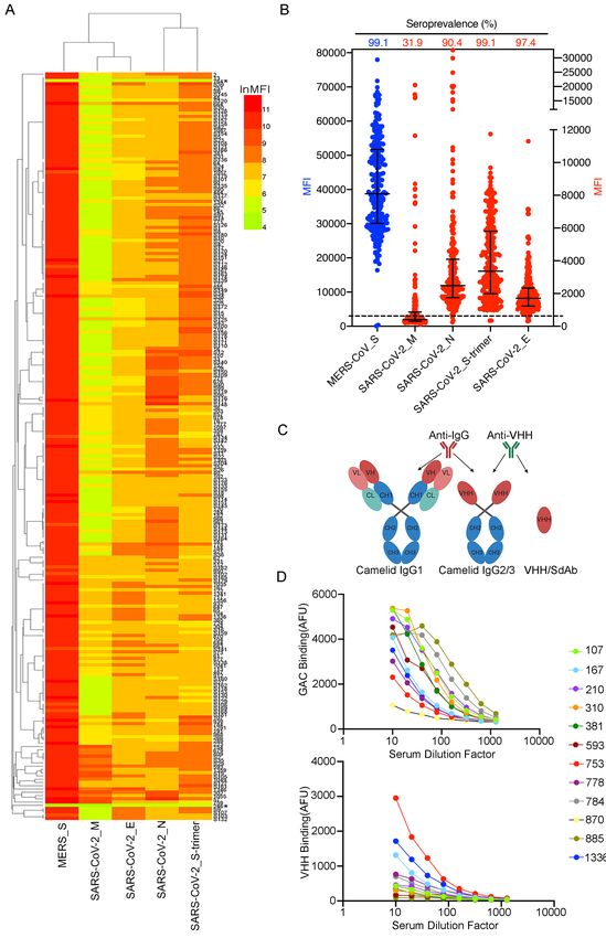
Introduction

Coronavirus (CoV) disease 2019 (COVID-19), caused by severe acute respiratory syndrome-

coronavirus 2 (SARS-CoV-2), is an increasing global threat to public health and economic

development. SARS-CoV-2 differs from SARS-CoV and Middle East respiratory syndrome

coronavirus (MERS-CoV) by its rapid spread and virulent human-to-human transmission (1).

Like its two predecessors, SARS-CoV-2 is a zoonotic virus, and it possibly has the same natural

reservoir (bats) (2) with an unknown intermediate host (3).

Although SARS-CoV-2 vaccine development is progressing at a rapid pace, widespread

vaccine availability must overcome various hurdles, including antigenic variation, low

efficacy, and short-term immune responses (4). Until herd immunity against SARS-CoV-2

develops within communities, preferably by means of effective vaccines, the global population

will remain at risk, and health care systems will continue to endure tremendous strain. Novel

therapeutic and preventive approaches are being designed and tested worldwide. Passive

antibody administration through the transfusion of plasma collected from donors who have

recovered from COVID-19, known as COVID-19 convalescent plasma (CCP), has emerged as

a promising therapy for the treatment of the disease (5). However, the potential benefits of CCP

therapy are hampered by the short-term efficacy of the human polyclonal antibodies, the

challenges of scaling up this intervention owing to the unavailability of large amounts of

convalescent plasma, the difficulty of mass production, and affordability (6). An attractive

alternative would be the use of animal-derived polyclonal antibody therapy, which has been

successfully and safely applied in several human conditions (7,8). An example of this approach

is the life-saving post-exposure prophylaxis against the rabies virus (9,10). Animal polyclonal

antibody products can be made cost-effectively in large quantities, which makes them suitable

                                                                                              3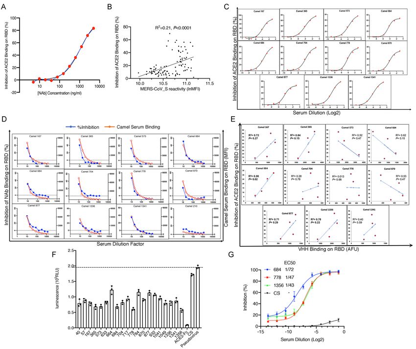
for responding to high endemic demand in low-income countries. Importantly, these products

may be valuable in addressing a pandemic situation such as the current COVID-19 outbreak.

Several studies have found that dromedary camels are the only intermediate host of MERS-

CoV, showing solely asymptomatic juvenile infection (11-15). MERS-CoV has been isolated

from the nasal swabs of young dromedaries (11-13), but despite extensive virus screening,

MERS-CoV has not been recovered from adult dromedaries (11,12). MERS-CoV

seropositivity rates increase with age to a seroprevalence of nearly 100% in adult dromedaries.

In contrast to humans, MERS-CoV-exposed dromedary camels develop no disease and show

only mild clinical respiratory signs (16-18). The absence of MERS-CoV in adult dromedary

camels is correlated with a dramatically potent virus-neutralizing antibody response (16).

These findings strongly suggest that dromedary immune system components have the ability

to efficiently limit MERS-CoV infection. The strong antibody response observed in

dromedaries could be caused by repeated exposure to the virus. However, some studies have

attributed this response to a characteristic of dromedaries (17-19), which produce relatively

unique heavy homodimeric chain-only antibodies, as well as conventional heterotetrameric

antibodies. The antigen-binding region of these homodimeric heavy chain-only antibodies

consists of one single domain, called the VHH. VHHs offer several advantages over common,

full-sized antibodies and currently used antibody-based fragments (Fabs, scFvs). These

advantages include high specificity, stability, and solubility, as well as a small size, which

allows them to recognize unusual antigenic sites and to deeply penetrate tissues. Since their

discovery, VHHs have been extensively used in diagnostics and therapies (20-23).

The highly proficient dromedary camel immune system against MERS-CoV and the close

structural and functional similarities between the different coronavirus species led us to

                                                                                             4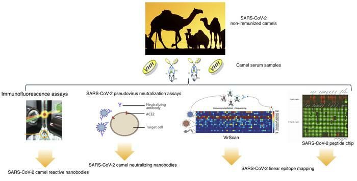
hypothesize that MERS-CoV-seropositive dromedary camels might have cross-reactive and

cross-neutralizing antibodies against SARS-CoV-2. We first assessed the dromedary

seroprevalence of antibodies against major SARS-CoV-2 proteins and evaluated their ACE2

binding, inhibitory, and pseudovirus-neutralizing effects. A systematic assessment of the

immunodominant B cell antigen determinants of betacoronaviruses among camels and the

degree to which these immunodominant B cell targets represent cross-reactive antigenic sites

is lacking. Therefore, using high throughput technologies, we extended our work to include a

comprehensive analysis of human and animal virus cross-reactive camel antibody specificities,

with an emphasis on the SARS-CoV-2 spike glycoprotein (S)-specific camel cross-reactive

VHH antibody repertoire.

                                                                                           5
Results
Identification of SARS-CoV-2 cross-reactive and cross-neutralizing antibodies in
dromedaries
Although the intermediate hosts of SARS-CoV-2, SARS-CoV, and MERS-CoV are different,
bats are the key natural reservoir of these three viruses, and humans are their final host (Figure
1A). Phylogenetic and structural studies have revealed that SARS-CoV-2, which belongs to
the genus Betacoronavirus, has a positive-sense, ~30 kilobase single-standard RNA genome
that has considerable similarity to the genomes of SARS-CoV (79%) and MERS-CoV (54%)
(Table 1). The striking homology of functional domains and epitope motifs between MERS-
CoV and SARS-CoV-2 viral structural proteins highlights the possibility that dromedary
MERS-CoV antibodies cross-react with SARS-CoV-2 proteins (Table 1, Figure 1B and C).
To gain deeper insight into dromedary camel SARS-CoV-2 cross-reactive antibodies, we
analyzed serum samples from 229 dromedaries of which 129 were collected prior to the
COVID-19 outbreak (to rule out any hypothetical SARS-CoV-2 interference); the remaining
100 samples were recent and traceable. No cases of dromedary camels carrying SARS-CoV-2
have been reported by the Ministry of Public Health of the State of Qatar despite extensive
screening for the presence of SARS-CoV-2 in domestic animals, including dromedary camels.
We used a highly sensitive fluorescent bead-based immunoassay that takes advantage of the
high dynamic range of fluorescent molecule detection by flow cytometry. In this assay,
biotinylated MERS-CoV S protein and SARS-CoV-2 proteins were immobilized on
polystyrene carboxylated beads to detect cross-reactive antibodies in the dromedary sera. In
agreement with previous reports, 227 of the 229 dromedary serum samples (99%) were found
to be highly seroreactive to MERS-CoV S protein, with a minimum value approximately 100
times greater than that of beads alone (Figure 2A). The fluorescence values of the two negative
6serum samples were less than two times that of beads alone (Figure 2B). Strikingly, all 227 of
the positive dromedary serum samples displayed variable but significant degrees of serological
reaction against the SARS-CoV-2 S trimer protein (Figure 2B). Over 90% of the serum
samples contained antibodies that bound to the SARS-CoV-2 N and E proteins, and
approximately 32% of the samples showed binding to the SARS-CoV-2 M protein (Figure
2B). Importantly, 15% to 30% of the animals exhibited relatively high binding activities to the
S trimer and N proteins (Figure 2B). High titers of SARS-CoV-2 S trimer-cross-reactive
antibodies were detected in several of the serum samples (Figure 2B). To investigate which
antibody subclass the cross-reactivity originated from, anti-camel antibodies that recognize
total IgG antibodies or that recognize only VHH antibodies were used to reveal the binding of
the camel serum to SARS-CoV-2 S protein (Figure 2C). Interestingly, in some of the serum
samples, the SARS-CoV-2 S trimer cross-reactivity predominantly resulted from VHH
antibody binding (Figure 2D).
To further evaluate the presence of SARS-CoV-2 cross-neutralizing antibodies in the camel
sera, we established a microsphere-based SARS-CoV-2 competition/inhibition assay to
monitor the binding of labeled ACE2 to beads conjugated with S protein or receptor binding
domain (RBD) in the presence or absence of camel sera. The assay was validated by
demonstrating an inhibitory effect of an anti-SARS-CoV-2 human neutralizing antibody (NAb)
isolated from a SARS-CoV-2-infected patient. This antibody targeted the RBD of the S protein
(Figure 3A), which indicated that this assay could detect antibodies that block SARS-CoV-2
S protein–ACE2 binding in vivo. We used this assay to test the inhibitory effect of 100 camel
serum samples. At a 10-fold dilution, nearly 70% of the samples showed obvious inhibitory
activity of S protein–ACE2 binding. Interestingly, the camel sera reactivity to MERS-CoV S
protein was significantly correlated with their SARS-CoV-2 cross-neutralizing potential
7(Figure 3B). We also performed a multi-dose inhibition assay with 11 serum samples which
showed a greater than 50% inhibition efficacy in a single-dose competition/inhibition assay. S-
RBD-specific cross-neutralizing antibodies were detectable in up to 50-fold serial dilutions of
these 11 serum samples, indicating high specificity and sensitivity (Figure 3C). Next, we
examined whether camel serum samples compete with the abovementioned patient-derived
human NAb for RBD binding. As shown in Figure 3D, the 11 camel RBD-specific cross-
neutralizing antibodies showed variable and partial binding inhibitory effects of the human
NAb on RBD protein (inhibition ranging between 20 and 50%). This result suggests that these
camel sera react with this particular conformational epitope revealed by the human NAb.
Moreover, this epitope could be also a neutralizing immunodominant epitope since it is reactive
with several neutralizing antibodies. Other camel sera, reacting with SARS-CoV-2 S protein
including serum 210, did not react with this conformational epitope (Figure 3D). To determine
whether VHH antibodies play a role in the cross-neutralizing activity, we simultaneously
detected VHH antibody–RBD binding and ACE2–RBD binding. VHH–S-RBD binding was
highly correlated with the inhibition of ACE2–RBD binding in serum from camels 167, 365,
684, 877, and 1336 (R2>0.7) (Figure 3E). In camel 684, the inhibitory activity was
significantly associated with VHH antibodies (R2=0.98, P=0.02).
To confirm the results of the camel SARS-CoV-2-neutralizing antibody screening obtained by
the microsphere-based SARS-CoV-2 competition/inhibition assay, we applied a cell-based
SARS-CoV-2 spike pseudovirus neutralization assay to assess the presence of antibodies
preventing pseudovirus entry into host cells in dromedaries showing high titers of SARS-CoV-
2 cross-reactive antibodies. A pseudoparticle-based model is a useful tool for evaluating the
efficacy of vaccine or antibody candidates against viruses (24-30). All tested camel sera
showed medium-to-high titers of SARS-CoV-2 cross-neutralizing antibodies by inhibiting
8pseudotyped luciferase SARS-CoV-2 spike entry into ACE2-expressing cells, while healthy
human serum did not (Figure 3F). The top three cross-neutralizing serum samples were further
analyzed in a multi-dose assay. The three samples showed high neutralizing potency against
the SARS-CoV-2 pseudovirus, with an EC50 range of 1:40 to 1:70 serum dilution (Figure
3G).
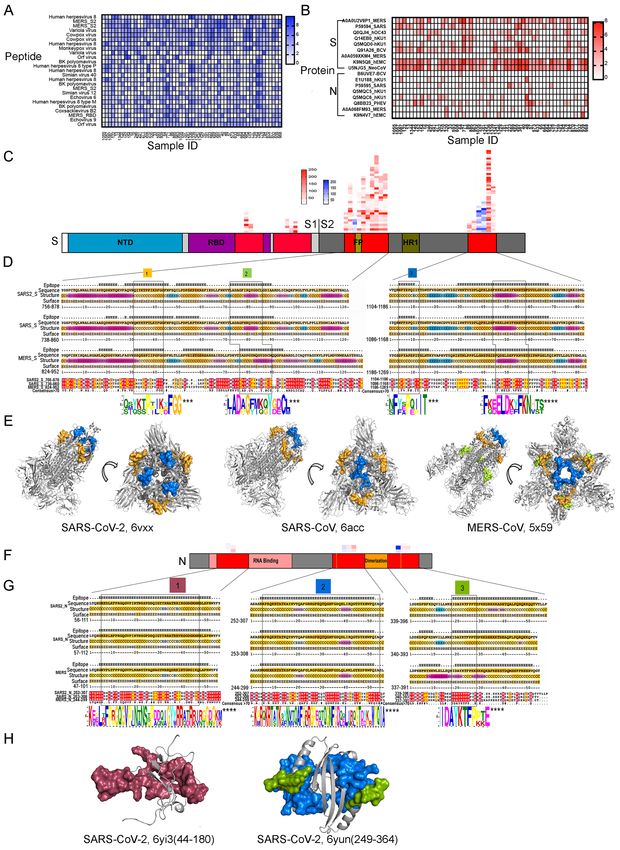
Epitope mapping of SARS-CoV cross-reactive dromedary camel antibodies using phage-
immunoprecipitation sequencing (VirScan)
The microsphere-based SARS-CoV-2 competition/inhibition assay and the cell-based SARS-
CoV-2 spike pseudovirus neutralization assay indicated the presence of SARS-CoV-2 spike
neutralizing antibodies induced by both conformational and linear epitopes. Although it is
known that neutralizing antibodies react more often with conformational epitopes, several
studies have revealed numerous linear epitopes of SARS-CoV-2 targeted by human
neutralizing antibodies (26,28,29,31). To reveal the large spectrum of linear epitopes targeted
by the camel cross-reactive antibody repertoire, we employed VirScan - a proteome-wide
programmable phage display and phage immunoprecipitation sequencing (PhIP-Seq) method
that comprehensively identifies epitope-specific antiviral antibody repertoires against MERS-
CoV and SARS-CoV (32,33). SARS-CoV-2 peptides are not included in the current version of
VirScan. We obtained a broadly diverse antibody repertoire targeting a myriad of camel
pathogen viral peptides in 56 of the serum samples. Several peptides corresponding to MERS-
CoV antigens and to many other animal viruses were enriched (Figure 4A). As expected, the
enriched epitopes were indeed located in the S and N proteins; these proteins are involved in
viral–host cell fusion and RNA replication, respectively, and are primary immunogenic targets
for viral neutralization in betacoronaviruses, including MERS, SARS-CoV, and SARS-CoV-2
(Figure 4B). Unexpectedly, most of the epitopes were concentrated in the S2 subunit of the S
9protein, while few were located in the S1 subunit (Figure 4C). Owing to the high degree of
glycosylation, S1 protein peptides present on phage display might not capture all potential
epitopes; however, five of the peptides from the S1 subunit were detected by VirScan (Figure
4C). To identify the conserved sequence motif, we performed a multiple sequence alignment
of highly enriched epitopes that shared linear sequence homology with the full-length proteins
of SARS-CoV and SARS-CoV-2. Accordingly, we identified four enriched epitopes shared by
the three coronaviruses. These epitopes were located in the S2 subunit, which is functionally
essential and a highly conserved region of the spike protein (Figure 4D). The S2 subunit
consists of a fusion peptide (FP), heptad repeat 1 and 2 (HR1 and HR2), a transmembrane
domain, and a cytoplasmic fusion domain. These domains interact with each other to form a
six-helix bundle fusion core and are responsible for viral entry. The potential antibody-binding
sites of both SARS-CoV and MERS-CoV were enriched in two regions, encompassing the FP
and overlapping HR1, which share high identities among these three viruses (Figure 4D).
These regions are positioned on the membrane fusion end of the S trimer structure (Figure
4E).
We also identified potential antibody-binding sites in the N protein, which comprises two
distinct RNA-binding domains: the N-terminal domain (NTD) and the C-terminal dimerization
domain (CTD) (Figure 4F and 4G). These domains are interconnected by a weakly structured
linkage region containing a serine-rich domain (Figure 4H).
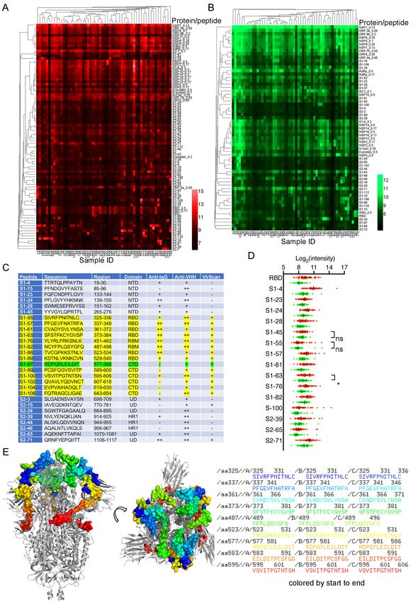
B cell epitope mapping of SARS-CoV-2 cross-reactive dromedary camel antibodies using
a SARS-CoV-2 peptide/proteome microarray
We used a recently validated SARS-CoV-2 peptide/proteome microarray to explore the
repertoire of SARS-CoV-2-specific cross-reactive camel VHH antibodies (31,34). We used
10different anti-camel Ig-isotype secondary antibodies specific for camel IgGs (all IgG isotypes)
and IgG2/3s (VHH, single-chain antibodies) to compare the repertoire of linear B cell epitopes
of the SARS-CoV-2 S1/S2 protein recognized by the different isotypes of camel MERS-CoV
antibodies.
Fifty-six serum samples from MERS-CoV-seropositive dromedaries and the appropriate
controls were probed in the SARS-CoV-2 peptide/proteome microarray. After data filtering
and normalization, we built a camel IgG and VHH profile for each serum sample and
performed cluster analysis to generate heatmaps of the enriched hits for visualization (Figure
5A and 5B). The MERS-CoV-seropositive serum samples and controls were perfectly
clustered for both the camel IgG and VHH antibodies, attesting to the specificity of the SARS-
CoV-2 peptide/proteome microarray (Figure 5A and 5B). In agreement with the VirScan
analysis, we found variable specificities among the 56 tested camel serum samples, but there
was significant cross-reactivity between the camel IgG and VHH antibodies and the full-length
SARS-CoV-2 proteins (Table 2). Several SARS-CoV-2 non-structural proteins (e.g., NSP1, -
2, -7, -8, and ORF6 and ORF7) elicited significant cross-reactivity with both camel Ig isotypes
(Table 2). NSP14, ORF6, ORF7b, and the S2 subunit of the S protein were predominantly
targeted by camel IgG1 rather than by VHH antibodies (Table 2). Strikingly, there was strong
IgG1 and VHH antibody reactivity against NSP7, NSP8, and the RNA-dependent RNA
polymerase (RdRp) of SARS-CoV-2 (nsp12) (Table 2). The RdRp of SARS-CoV-2, consisting
of the nsp12 catalytic subunit and the nsp7 and nsp8 cofactors, is a key component of the
replication/transcription machinery (35). In addition, variable specificities and significant
cross-reactivity against the S1 NTD, RBD, and CTD and the S2 HR1 and HR2 domains were
revealed (Table 2). A list of S1/S2 peptides targeted by VHH isotypes is presented in Figure
5C. Interestingly, 12 SARS-CoV-2 S1 peptides that reacted with camel antibodies were found
to be highly immunogenic in humans. Notably, eight of the 11 camel serum samples showing
11high SARS-CoV-2 cross-neutralizing antibody activity reacted with one or more linear RBD
peptides (Table 3); S1-82 reacted with five SARS-CoV-2-neutralizing camel serum samples;
S1-61 and S1-64 reacted with four samples; and S1-57, S1-63, and S1-76 reacted with three
samples. Although it is known that neutralizing antibodies react more often with
conformational epitopes, these RBD peptides, targeted by multiple cross-neutralizing serum
samples, could be SARS-CoV-2-neutralizing epitopes. Notably, the linear epitopes that we
identified (e.g., S1-76/97) are not only highly immunogenic in humans but are also
physiologically relevant because they have been identified in COVID-19 patients (31).
Therefore, these epitopes could serve as promising candidates for the development of broadly
neutralizing antibodies. Epitopes revealed exclusively by the camel antibodies could increase
the pool of neutralizing antibodies with potential therapeutic use.
By comparing the total IgG signal and VHH signal from the same antigens, we found that
antibodies recognizing the S1-45, S1-55, and S1-63 epitopes might all belong to the VHH
subclass (Figure 5D). We also located the identified epitopes on the S trimer structure (Figure
5E).
12Discussion
Our study showed the presence of SARS-CoV-2 cross-reactive and cross-neutralizing
antibodies in SARS-CoV-2 non-immunized dromedary camels and provides a comprehensive
structural analysis of the targeted SARS-CoV-2 proteins and linear epitopes. Because the titers
of these SARS-CoV-2 cross-neutralizing camel antibodies were not found consistently and
exceptionally high, they cannot be proposed as a potential treatment for COVID-19. Rather,
their presence in non-immunized camels suggests that these dromedaries might produce highly
efficient antibodies once they are actively immunized with SARS-CoV-2 antigens.
The camel cross-reactive antibody epitope mapping revealed not only several epitopes known
to be highly immunogenic in humans, including a neutralizing epitope (31), but also epitopes
exclusively targeted by camel antibodies. The identified highly immunogenic SARS-CoV-2
epitopes could be used as immunogens to develop SARS-CoV-2 hyperimmune camels.
One short-term implication of these findings is that after actively immunizing camels with
SARS-CoV-2 antigens, the SARS-CoV-2 hyperimmune dromedaries could generate novel
COVID-19 serotherapy tools to complement or replace the current CCP therapy. Given the
total blood volume of a camel and the large herd size of dromedary camels living in the Middle
East and North African regions, camel plasma would be available in quantities sufficient to
meet the needs of a large population. The proposed SARS-CoV-2 hyperimmune camel plasma-
based COVID-19 serotherapy could be used as a longer term treatment option, particularly in
low- and middle-income countries where resource constraints could bar access to novel
treatments (e.g., COVID-19 vaccines) even if they become widely available. Like any
immunoglobulin-based treatment, the proposed camel serum-based therapy for COVID-19
treatment should overcome potential pitfalls such as the exaggerated inflammatory response
seen in the antibody-dependent enhancement process (36). Additionally, after binding to the
13viral immune complexes, subneutralizing antibodies could bind to FcγR-bearing cells, leading
to increased viral uptake and replication (37).
Numerous human SARS-CoV-2-neutralizing antibodies have been recently reported; however,
their binding affinities and pseudoviral- and viral-neutralizing abilities have varied (25-30, 38-
40). Liu et al. highlighted the short-duration protective effect of human SARS-CoV-2-
neutralizing antibodies and raised concerns about the efficacy of future SARS-CoV-2 vaccines
(41). In contrast to humans, camel anti-MERS-CoV-neutralizing antibodies, which were shown
to cross-react with SARS-CoV-2, persist several years post infection. This could be because of
repeated exposures to the virus, but some studies indicate that these camel antibodies have
long-lasting efficacy (17-19). It remains unclear whether this durable effect arises from the
structural and/or functional features of camel antibodies or from some other component of the
camel plasma. Relatedly, adverse reactions in humans to animal-derived polyclonal antibodies
are usually due to the presence of highly immunogenic animal proteins. This is less likely to
occur with camel serum-based therapy because camel IgGs are less immunogenic than most
mammalian IgGs, and when administered intravenously, they are less likely to induce serum
sickness and anaphylactic adverse reactions (42,43). Growing research in the field of antibody
engineering has focused on enhancing the therapeutic efficacy of VHHs. Strategies that enable
VHHs to cross the blood–brain barrier have recently shown promise. These findings have
driven tremendous growth in the use of VHHs for treating central nervous system diseases (44-
46). A limiting factor for the clinical use of VHH domains is that the hydrodynamic radius of
VHHs falls below the kidney's glomerular filtration limit, which can contribute to rapid renal
clearance and a weak pharmacokinetic (PK) profile, considerably impacting their therapeutic
effectiveness. To address this issue, several antibody engineering techniques have been
employed to increase the VHH size and improve the PK profile. The simple formation of
14genetic fusions has frequently been used, for example with conventional Fc (47), VHH repeat
domains (mono- or polyspecific), serum proteins (e.g., HSA), and anti-serum albumin VHH
(48). As an alternative to genetic fusions and modularity, chemical methods such as
PEGylation and lipidation have been applied to increase the VHH half-life (49-51).
Dromedary camels may constitute a competitive source for the development of COVID-19
targeted antibody therapy. Interest has been growing in recent years in the generation and use
of camel single-chain antibodies (VHHs or nanobodies) and their derivatives for a wide
spectrum of applications (52,53). The therapeutic properties of camel VHHs can be enhanced
by protein engineering to improve their efficacy (54,55). The use of camel nanobodies is of
special interest for the recognition of epitopes that are usually not antigenic for conventional
antibodies. In the case of COVID-19, the unique feature of VHHs to easily penetrate tissues,
including the lungs (the main target of SARS-CoV-2), gives them additional potential curative
properties to treat SARS-CoV-2 infection. Camel nanoantibodies might be an appropriate
choice for generating a COVID-19 treatment because these single-chain antibodies are highly
soluble, small, stable proteins and can be produced in large quantities (52).
Recent studies have found that SARS-CoV-2 non-exposed individuals have cross-reactive
antibodies to a number of coronaviruses, including SARS-Co-V2 (56,57). Particularly, Ng et
al. found that in SARS-Co-V2 non-exposed individuals possess neutralizing antibodies
targeted to the S2 protein (58). Moreover, many of these cross-reactive antibodies found in
humans are unique to fusion peptide epitopes or adjacent S2 subunit epitopes which suggested
to neutralize the coronaviruses by blocking viral membrane fusion and host cell entry (59).
Importantly, recent studies have shown that antibodies induced by the active immunization of
llamas with MERS-CoV and SARS-CoV viral antigens (in particular, SARS VHH-72) cross-
15react with the spike protein of SARS-CoV-2 (60,61). Although dromedary camels and llamas
belong to the same Camelidae family and their antibodies share the single-chain antibody
feature, the lack of natural infection in llamas by MERS-CoV means they cannot replicate the
efficient response of dromedary camels to MERS-CoV infection. Dromedaries experience
rapid viral clearance without showing any disease symptoms. This could arise from innate
immunity, efficient neutralizing antibodies, or other antiviral immunity components, providing
support for the use of dromedaries in the development of COVID-19 targeted antibody
therapies. Indeed, in a recent study, serum from dromedary camels that was seropositive for
MERS-CoV was highly efficient when administered to mice infected with MERS-CoV (62).
Camel serum given both pre- and post-exposure protected the infected mice from weight loss,
diminished the histological changes in the lungs, and accelerated viral clearance (62).
Moreover, the low 12–30 kDa MW of nanobodies also offers new and noninvasive routes of
administration such as delivery by inhalation, which has been proven in clinical trials to be safe
and successful in preventing respiratory syncytial virus infection (63,64). Numerous
nanobodies are being investigated in clinical trials (65), and one nanobody (caplacizumab) has
already been approved by the US Food and Drug Administration for the treatment of
thrombotic thrombocytopenic purpura (66). Additionally, camel milk, with its unique
nutritional composition and abundance of secreted IgA and VHH nanobodies (67), has
potential implications for the induction of passive immunity to SARS-CoV-2. Immunizing
lactating camels with SARS-CoV-2 antigens might induce SARS-CoV-2-neutralizing
antibodies in camel serum and milk.
In summary, we identified the presence of SARS-CoV-2 cross-reactive neutralizing antibodies
in dromedary camels that were not previously immunized with SARS-CoV-2 antigens and have
revealed the structures of the corresponding major target linear B cell epitopes. Our findings
16advocate for the development of SARS-CoV-2 hyperimmune camels as a prominent source of
therapeutic agents for the prevention and treatment of COVID-19. With adequate testing and
clinical trials, the proposed SARS-CoV-2 camel serum-based serotherapy could have a major
impact as a preventive and curative intervention for COVID-19. By taking advantage of the
unique features of the camel immune system, the suggested intervention might provide
protective passive immunization in humans before and after exposure to SARS-CoV-2 and in
patients with established disease, thus helping alleviate the burden of the current pandemic.
17Methods
Dromedary camel sample collection
Serum from 229 dromedary camels was collected from two camel cohorts in Qatar. All camels
were females, with an age range of 4-15 years. A total of 129 serum samples were collected
from a camel slaughterhouse before September 2019; the other 100 samples were taken from
live camels by jugular puncture for routine infection screening in 2020. All samples were stored
at −80°C until testing. The Ministry of Public Health has extensively tested dromedaries in
Qatar, including the living camels enrolled in the present study, for SARS-CoV-2 infection
using nasal swab sampling, and no positive cases have been reported.
Seroconversion assay
The camel serum samples were tested for the presence of antibodies binding SARS-CoV-2 M,
N, S trimer, and E and MERS-CoV S proteins using a laboratory-made bead array. The
following Spherotech carboxyl microspheres were used: 107 microspheres of peaks 8, 6, 4, and
2 from the blue particle array kit (Spherotech #CPACK-5067) and peak 11 from the UV
particle array kit (Spherotech #UVCPACK-5042-1). The microspheres were washed once in
diH2O and activated in the presence of 80 mM monobasic sodium phosphate pH 6.2, 5 mg/ml
sulfo-NHS (Pierce #24520), and 5 mg/ml EDC (Pierce #77149) under shaking for 20 minutes
at room temperature. The activated microspheres were then washed three times with PBS pH
7.4. Blue peaks 8, 6, 4, and 2 and UV peak 11 were respectively incubated with 100 µg of
recombinant SARS-CoV-2 E, S trimer, N, M, and MERS-CoV S1 under rotation overnight at
room temperature (Table S1). Finally, the microspheres were washed twice with PBS-TBN
(0.2% Tween-20, 0.1% BSA, 0.05% sodium azide) and stored at 4°C in PBS-TBN until further
use. To assess the antibody titers, the serum samples were diluted 20-fold (initial dilution
followed by serial dilution) in assay buffer (10 mM Tris-HCl pH 7.5, 0.1% BSA, 0.01%
18Tween-20) and incubated with the SARS-CoV-2 M, N, S trimer, E, and MERS-CoV S1
microspheres (2500 microspheres for each peak) under shaking for 1 hour at room temperature
in a Multiscreen HV filter plate (Millipore #MSHVN4510). After three vacuum washes in
assay buffer, the microspheres were incubated in 1 µg/ml goat anti-IgG camel antibody (ADI
#30835-UL) conjugated in-house to Alexa Fluor 594 (AF594) or in 1 µg/ml anti-camelid VHH
antibodies conjugated to Phycoerythrin (PE) (GenScript #A02018). These incubations were
conducted in assay buffer, with shaking for 30 minutes at room temperature. The microspheres
were then vacuum-washed three times in wash buffer (10 mM Tris-HCl pH 7.5, 0.05% Tween-
20), resuspended in the same buffer, and analyzed in a BD FACS Symphony™ A5. The flow
cytometry experiment analyzed each bead region with the UV and blue laser, and the detection
antibody was analyzed with the yellow-green laser. To assess the participation of VHH
antibodies in the overall camel seroconversion, 1 µg/ml iFluor647-labeled anti-VHH cocktail
antibody (GenScript #A02019) was used to detect overall seroconversion. The data were
analyzed on FlowJo, gating on each bead region to measure antibody binding. A minimum of
100 beads per region was acquired. The median fluorescence intensity of each bead set was
used in the subsequent calculations.
In vitro competition/inhibition assay
The neutralization activity of the camel sera against SARS-CoV-2 was tested using an in vitro
competition/inhibition assay. A standard inhibition curve was first prepared from a standard
solution of a SARS-CoV-2 RBD human neutralizing antibody isolated from a SARS-CoV-2-
infected patient (Acro Biosystems #SAD-S35). The curve started at 5 µg/ml and proceeded in
a 12-step two-fold dilution series in assay buffer. The camel sera were serially diluted (1:2,
1:6, 1:18, 1:54, 1:162, and 1:486) in assay buffer. Next, 0.5 µg/ml biotinylated human ACE2
(Acro Biosystems #AC2-H82F9) was added to the neutralizing antibody standards and to the
19camel sera dilutions. Mixed samples with ACE2 were then added to the SARS-CoV-2-S1 and
SARS-CoV-2-RBD microspheres (2500 microspheres for each peak) (Table S1) and incubated
with shaking for 45 minutes at room temperature. The microspheres were then washed three
times with assay buffer and incubated in 4 µg/ml streptavidin-PE with shaking for 20 minutes
at room temperature. After three washes in wash buffer, the microspheres were resuspended in
the same buffer and detected in a BD FACS Symphony A5. The competition for binding to
RBD between the RBD-specific human IgG1 monoclonal antibody AS35 (Acro Biosystems
#SAD-S35) and camel serum samples was assayed using RBD-coupled microspheres. 2500
RBD-coupled microspheres in 50 µl were incubated for 45 minutes with a 50 µl mixture of
AS35 at 0.15 µg/ml and 2-fold serially diluted camel serum samples from 1:10 to 1:1280 in a
MultiScreen filter plate (Millipore #MSHVN4510). After 3 vacuum washes, camel antibody
binding on RBD was revealed by goat anti-camel IgG antibody (ADI #30835-UL) conjugated
with AF594 and the binding of the Human Nab was revealed with AF488-labelled-goat anti-
human anti-IgG1 antibodies (SouthernBiotech #9052-30). The maximum binding (MFImax)
was determined as the MFI read on beads incubated only with the human Nab. Inhibition of
binding at a given dilution of camel serum was calculated by using the median fluorescent
intensity (MFIdil) in the following formula: % Inhibition = (1-(MFIdil/MFImax) ×100. The
inhibition of NAb binding on RBD and camel serum binding on RBD were calculated at
different serum dilution factorTo investigate the participation of VHH antibodies in
neutralizing S trimer binding to ACE2, the competition/inhibition assay was performed as
described above until the detection step, which was accomplished with a mix of iFluor647-
labeled anti-VHH cocktail antibody (GenScript #A02019) and streptavidin-PE to
concomitantly reveal the binding of VHH antibodies and hACE2 to RBD or SARS-CoV-2 S1
proteins. The data were analyzed on FlowJo. A minimum of 100 beads per region was acquired.
The median fluorescence intensity of each bead set was used in the subsequent calculations.
20Pseudovirus neutralization assay
A SARS-CoV-2 pseudovirus neutralization assay kit (GenScript #SC2087A) was used to
evaluate the ability of the camel sera to block cell entry of the pseudotyped lentiviral particle
of SARS-CoV-2 spike. HEK293 cells overexpressing ACE2 (HEK293-ACE2) were seeded
into 96-well plates and infected with 50 μL of the pseudotyped luciferase SARS-CoV-2 spike
with or without diluted camel serum. ACE2-Fc was used as a positive control, and serum from
a healthy person was used as a negative control. After 6 hours of incubation at 37°C, the
pseudovirus-containing medium was replaced with fresh cell culture medium and the plate was
incubated for another 48 hours. After removing the culture media, Bio-Glo™ luciferase
substrate working solution (Promega #G7940) was added to the HEK293-ACE2 cells.
Luciferase activity, expressed in relative luminescence units, was measured with an EnVision
plate reader.
VirScan analysis
Serological profiling of the camel serum antiviral IgG repertoire was performed using PhIP-
Seq as described by Xu et al. (32). Briefly, the VirScan 2.0 library was programmed into an
Agilent microarray and was then amplified and ligated into bacteriophage T7 DNA, packaged
into phage particles, and amplified in E. coli. The amplified libraries were incubated with 2 μL
of camel serum at 4°C overnight with protein A and G magnetic beads (Invitrogen). Antibody-
bound phage were immunoprecipitated, and balanced amplicon libraries were pooled and
sequenced. The read counts per peptide were converted to a relative antibody epitope binding
signal, and the magnitude was reported as an epitope-specific Z-score.
21Protein/peptide microarray
Microarray-based serum analysis of anti-SARS-CoV-2 antibodies in the camel serum was
performed as previously described (31,34). The arrays were detected by incubating with APC-
conjugated goat anti-camel IgG antibodies (Alpha Diagnostic#30385) and iFluor555-
conjugated anti-camelid VHH antibodies (GenScript #A01863). The fluorescence signals were
scanned by a LuxScan 10K-A (CapitalBio Corporation), and the fluorescence intensity data
were extracted by GenePix Pro 6.0 software (Molecular Devices).
Bioinformatics analysis
Multiple sequences were aligned by Clustal Omega on an EBI server or by T-Coffee (68) and
illustrated using ESPript 3.0 (69). Linear B cell epitopes with a threshold greater than 0.5 were
predicted by BepiPred-2.0 and are displayed with an orange gradient (70). The common motif
was calculated and illustrated by MEME (71). The secondary structures of helix, sheet, and
coil were predicted using NetsurfP (72). Structure visualization was conducted using pdb files
downloaded from the Protein Data Bank (PDB) in Europe (https://www.ebi.ac.uk/pdbe) and
PyMOL 2.4 (https://pymol.org/2/).
Statistics
All statistical analyses were performed using GraphPad Prism 7. Statistical parameters
are reported in the figure legends. An unpaired or paired two-tailed Student’s t-test was used
for two-group comparisons for normally distributed data. An asymmetric sigmoidal five-
parameter logistic model was used to generate standard curve for in-vitro
competition/inhibition assay. A typical four-parameter logistic model was used to fit dose
(dilution) vs. response (inhibition percentage) curve to the data of camel serum samples. The
22correlation between inhibition percentage and fluorescence signal was analyzed by linear
regression.
Study approval
The study was approved by the Institutional Animal Care and Use Committee of Weill Cornell
Medicine-Qatar.
Author Contributions
L.C. designed the study. L.C., J.S., J.C.G., M.S., S.W.J.A., E.A.B.A.F., and M.H.A.T.
performed experiments and collected the data. J.C.G., I.P., S.M., and A.S. performed in-vitro
binding and neutralization assay. N.M., M.M.A.A., H.E.A.R., and A.I.C. generated and
interpreted data. L.C., J.S., M.S., J.C.G. S.R. and A.R. analyzed and interpreted the data. N.H.
generated the epitope 3-dimentional structure confirmation data. L.C., M.S., and J.S. wrote the
manuscript, did the literature search, analyzed and interpreted data. L.C. supervised and
coordinated the study. All authors contributed to reviewing and approved the final version
Acknowledgments
This work was supported by the BMRP Funding of Weill Cornell Medicine-Qatar and by Qatar
National Research Fund (NPRP12S-0317-190379). We thank Dr. Johan Hoebeke for the
intellectual discussion, Ms. Sinéad O’Rourke, Content Development Specialist, Distributed
eLibrary, Weill Cornell Medicine-Qatar and Katherine Thieltges, from Edanz Group for
manuscript editing.
23References
1. Zhu N, et al. A Novel Coronavirus from Patients with Pneumonia in China, 2019. N Engl
J Med. 2020;382(8):727-733.
2. Zhou P, et al. A pneumonia outbreak associated with a new coronavirus of probable bat
origin. Nature. 2020;579(7798):270-273.
3. Huang C, et al. Clinical features of patients infected with 2019 novel coronavirus in
Wuhan, China. The lancet. 2020;395(10223):497-506.
4. P Padron-Regalado E. Vaccines for SARS-CoV-2: Lessons from Other Coronavirus
Strains. Infect Dis Ther. 2020;9(2):1-20.
5. Bloch EM, et al. Deployment of convalescent plasma for the prevention and treatment of
COVID-19. J Clin Invest. 2020;130(6):2757-2765.
6. Bloch EM, et al. Guidance for the Procurement of COVID‐19 Convalescent Plasma:
Differences between High and Low‐Middle Income Countries [published online ahead of
print, 2020 Jun 13]. Vox Sang. 2020;10.1111/vox.12970.
7. Baud FJ, et al. Treatment of severe colchicine overdose with colchicine-specific Fab
fragments. N Eng J Med. 1995;332(10):642-645.
8. Antman EM, et al. Treatment of 150 cases of life-threatening digitalis intoxication with
digoxin-specific Fab antibody fragments. Final report of a multicenter study. Circulation.
1990;81(6):1744-1752.
9. Dixit R, et al. Benefits of using heterologous polyclonal antibodies and potential
applications to new and undertreated infectious pathogens. Vaccine. 2016;34(9):1152-
1161.
10. Sparrow E, et al. Therapeutic antibodies for infectious diseases. Bull World Health
Organ. 2017;95(3):235-237.
2411. Sabir JS, et al. Co-circulation of three camel coronavirus species and recombination of
MERS-CoVs in Saudi Arabia. Science. 2016;351(6268):81-84.
12. Alagaili AN, et al. Middle East respiratory syndrome coronavirus infection in dromedary
camels in Saudi Arabia. MBio. 2014;5(2):e00884-14.
13. Wernery U, et al. Acute middle East respiratory syndrome coronavirus infection in
livestock Dromedaries, Dubai, 2014. Emerg Infect Dis. 2015;21(6):1019-1022.
14. Chu DK, et al. MERS coronaviruses in dromedary camels, Egypt. Emerg Infect Dis.
2014;20(6):1049-1053.
15. Reusken CB, et al. Middle East respiratory syndrome coronavirus neutralising serum
antibodies in dromedary camels: a comparative serological study. Lancet Infect Dis.
2013;13(10):859-866.
16. Wernery U, et al. Middle East respiratory syndrome (MERS) coronavirus and
dromedaries. Vet J. 2017;220:75-79.
17. Adney DR, et al. Replication and shedding of MERS-CoV in upper respiratory tract of
inoculated dromedary camels. Emerg Infect Dis. 2014;20(12):1999-2005.
18. Haagmans BL, et al. An orthopoxvirus-based vaccine reduces virus excretion after
MERS-CoV infection in dromedary camels. Science. 2016;351(6268):77-81.
19. Saad M, et al. Clinical aspects and outcomes of 70 patients with Middle East respiratory
syndrome coronavirus infection: a single-center experience in Saudi Arabia. Int J Infect
Dis. 2014;29:301-306.
20. Rothbauer U, et al. Targeting and tracing antigens in live cells with fluorescent
nanobodies. Nat Methods. 2006;3(11):887-889.
21. Stalin Raj V, et al. Chimeric camel/human heavy-chain antibodies protect against
MERS-CoV infection. Sci Adv. 2018;4(8):eaas9667.
2522. Muyldermans S. Nanobodies: natural single-domain antibodies. Annu Rev Biochem.
2013;82:775-797.
23. Danquah W, et al. Nanobodies that block gating of the P2X7 ion channel ameliorate
inflammation. Sci Transl Med. 2016;8(366):366ra162.
24. Li Q, et al. An LASV GPC pseudotyped virus based reporter system enables evaluation
of vaccines in mice under non-BSL-4 conditions. Vaccine. 2017;35(38):5172-5178.
25. Pinto D, et al. Cross-neutralization of SARS-CoV-2 by a human monoclonal SARS-CoV
antibody. Nature. 2020;583(7815):290-295.
26. Liu L, et al. Potent neutralizing antibodies against multiple epitopes on SARS-CoV-2
spike. Nature. 2020;584(7821):450-456.
27. Cao Y, et al. Potent Neutralizing Antibodies against SARS-CoV-2 Identified by High-
Throughput Single-Cell Sequencing of Convalescent Patients' B Cells. Cell.
2020;182(1):73-84.e16.
28. Chen X, et al. Human monoclonal antibodies block the binding of SARS-CoV-2 spike
protein to angiotensin converting enzyme 2 receptor. Cell Mol Immunol. 2020;17(6):647-
649.
29. Chi X, et al. A neutralizing human antibody binds to the N-terminal domain of the Spike
protein of SARS-CoV-2. Science. 2020;369(6504):650-655.
30. Wang C, et al. A human monoclonal antibody blocking SARS-CoV-2 infection. Nat
Commun. 2020;11(1):2251.
31. Li Y, et al. Linear epitope landscape of SARS-CoV-2 Spike protein constructed from
1,051 COVID-19 patients [preprint]. https://doi.org/10.1101/2020.07.13.20152587i.
Posted on medRxiv on July 14, 2020.
32. Xu GJ, et al. Viral immunology. Comprehensive serological profiling of human
populations using a synthetic human virome. Science. 2015;348(6239):aaa0698.
2633. Mohan D, et al. PhIP-Seq characterization of serum antibodies using oligonucleotide-
encoded peptidomes. Nat Protoc. 2018;13(9):1958-1978.
34. Jiang HW, et al. SARS-CoV-2 proteome microarray for global profiling of COVID-19
specific IgG and IgM responses. Nat Commun. 2020;11(1):3581.
35. Hillen HS, et al. Structure of replicating SARS-CoV-2 polymerase. Nature.
2020;584(7819):154-156.
36. Nguyen AA, et al. Immunoglobulins in the treatment of COVID-19 infection: Proceed
with caution!. Clin Immunol. 2020;216:108459.
37. Murrell S, et al. Review of dengue virus and the development of a vaccine. Biotechnol
Adv. 2011;29(2):239-247.
38. Wu Y, et al. A noncompeting pair of human neutralizing antibodies block COVID-19
virus binding to its receptor ACE2. Science. 2020;368(6496):1274-1278.
39. Wan J, et al. Human-IgG-Neutralizing Monoclonal Antibodies Block the SARS-CoV-2
Infection. Cell Rep. 2020;32(3):107918.
40. Rogers TF, et al. Isolation of potent SARS-CoV-2 neutralizing antibodies and protection
from disease in a small animal model. Science. 2020;369(6506):956-963.
41. Liu T, et al. Prevalence of IgG antibodies to SARS-CoV-2 in Wuhan - implications for
the ability to produce long-lasting protective antibodies against SARS-CoV-2 [preprint].
https://doi.org/10.1101/2020.06.13.20130252. Posted on medRxiv on June 16, 2020.
42. Herrera M, et al. Factors associated with adverse reactions induced by caprylic acid-
fractionated whole IgG preparations: comparison between horse, sheep and camel
IgGs. Toxicon. 2005;46(7):775-781.
43. Mahase E. Covid-19: what treatments are being investigated?. BMJ. 2020;368:m1252.
2744. Kunz P, Flock T, Soler N, et al. Exploiting sequence and stability information for
directing nanobody stability engineering. Biochim Biophys Acta Gen Subj.
2017;1861(9):2196-2205.
45. Stanimirovic DB, Sandhu JK, Costain WJ. Emerging Technologies for Delivery of
Biotherapeutics and Gene Therapy Across the Blood-Brain Barrier. BioDrugs.
2018;32(6):547-559.
46. Li T, Bourgeois JP, Celli S, et al. Cell-penetrating anti-GFAP VHH and corresponding
fluorescent fusion protein VHH-GFP spontaneously cross the blood-brain barrier and
specifically recognize astrocytes: application to brain imaging. FASEB J.
2012;26(10):3969-3979.
47. Harmsen MM, Van Solt CB, Fijten HP, Van Setten MC. Prolonged in vivo residence
times of llama single-domain antibody fragments in pigs by binding to porcine
immunoglobulins. Vaccine. 2005;23(41):4926-4934.
48. Beirnaert E, Desmyter A, Spinelli S, et al. Bivalent Llama Single-Domain Antibody
Fragments against Tumor Necrosis Factor Have Picomolar Potencies due to
Intramolecular Interactions. Front Immunol. 2017;8:867.
49. van Witteloostuijn SB, Pedersen SL, Jensen KJ. Half-Life Extension of
Biopharmaceuticals using Chemical Methods: Alternatives to
PEGylation. ChemMedChem. 2016;11(22):2474-2495.
50. Ding L, Tian C, Feng S, et al. Small sized EGFR1 and HER2 specific bifunctional
antibody for targeted cancer therapy. Theranostics. 2015;5(4):378-398.
51. Hoefman S, Ottevaere I, Baumeister J, Sargentini-Maier ML. Pre-clinical intravenous
serum pharmacokinetics of albumin binding and non-half-life extended Nanobodies®.
Antibodies. 2015;4(3):141-156.
2852. De Meyer T, et al. Nanobody-based products as research and diagnostic tools. Trends
Biotechnol. 2014;32(5):263-270.
53. Rothbauer U, et al. Targeting and tracing antigens in live cells with fluorescent
nanobodies. Nat Methods. 2006;3(11):887-9.
54. Jovčevska I, Muyldermans S. The Therapeutic Potential of Nanobodies. BioDrugs.
2020;34(1):11-26.
55. Zhao G, et al. A Novel Nanobody Targeting Middle East Respiratory Syndrome
Coronavirus (MERS-CoV) Receptor-Binding Domain Has Potent Cross-Neutralizing
Activity and Protective Efficacy against MERS-CoV. J Virol. 2018;92(18):e00837-18.
56. Shrock E, et al. Viral epitope profiling of COVID-19 patients reveals cross-reactivity and
correlates of severity. Science. 2020;370(6520):eabd4250.
57. Grifoni A, et al. Targets of T Cell Responses to SARS-CoV-2 Coronavirus in Humans
with COVID-19 Disease and Unexposed Individuals. Cell. 2020;181(7):1489-1501.e15.
58. Ng KW, et al. Preexisting and de novo humoral immunity to SARS-CoV-2 in humans.
Science. 2020;370(6522):1339-1343.
59. Poh CM, et al. Two linear epitopes on the SARS-CoV-2 spike protein that elicit
neutralising antibodies in COVID-19 patients. Nat Commun. 2020;11(1):2806.
60. Wrapp D, et al. Structural Basis for Potent Neutralization of Betacoronaviruses by
Single-Domain Camelid Antibodies. Cell. 2020;181(6):1436-1441.
61. Huo J, et al. Neutralizing nanobodies bind SARS-CoV-2 spike RBD and block
interaction with ACE2. Nat Struct Mol Biol. 2020;27(9):846-854.
62. Zhao J, et al. Passive immunotherapy with dromedary immune serum in an experimental
animal model for Middle East respiratory syndrome coronavirus infection. J Virol.
2015;89(11):6117-6120.
2963. Broadbent L, et al. Comparative Therapeutic Potential of ALX-0171 and Palivizumab
against Respiratory Syncytial Virus Clinical Isolate Infection of Well-Differentiated
Primary Pediatric Bronchial Epithelial Cell Cultures. Antimicrob Agents Chemother.
2020;64(2):e02034-19.
64. Larios Mora A, et al. Delivery of ALX-0171 by inhalation greatly reduces respiratory
syncytial virus disease in newborn lambs. MAbs. 2018;10(5):778-795.
65. Morrison C. Nanobody approval gives domain antibodies a boost. Nat Rev Drug Discov.
2019;18(7):485-487.
66. Scully M, et al. Caplacizumab Treatment for Acquired Thrombotic Thrombocytopenic
Purpura. N Engl J Med. 2019;380(4):335-346.
67. El Agamy EI, et al. Antibacterial and antiviral activity of camel milk protective
proteins. J Dairy Res. 1992;59(2):169-175.
68. Notredame C, et al. T-Coffee: A novel method for fast and accurate multiple sequence
alignment. J Mol Biol. 2000;302(1):205-217.
69. Robert X, Gouet P. Deciphering key features in protein structures with the new
ENDscript server. Nucleic Acids Res. 2014;42(Web Server issue):W320-W324.
70. Jespersen MC, et al. BepiPred-2.0: improving sequence-based B-cell epitope prediction
using conformational epitopes. Nucleic Acids Res. 2017;45(W1):W24-W29.
71. Bailey TL, et al. MEME SUITE: tools for motif discovery and searching. Nucleic Acids
Res. 2009;37(Web Server issue):W202-W208.
72. Klausen MS, et al. NetSurfP-2.0: Improved prediction of protein structural features by
integrated deep learning. Proteins. 2019;87(6):520-527.
30Figure 1. Transmission, structural and functional homologies of 3 Betacoronaviruses (SARS- CoV, MERS-CoV, SARS-CoV-2). (A) Emergence of coronaviruses pathogenic for humans from ancestral bat viruses. (B) Schematic representation of the genome organization and functional domains of Spike glycoprotein and Nucleoprotein proteins for SARS-CoV, MERS- CoV and SARS-CoV-2. The single-stranded RNA genomes of SARS-CoV, MERS-CoV and SARS- CoV-2 include two large genes, the ORF1a and ORF1b genes, which encode 16 non-structural proteins (nsp1–nsp16) that are highly conserved throughout coronaviruses. The structural
genes encode the structural proteins, spike (S), envelope (E), membrane (M), and
nucleocapsid (N), which are common features to all coronaviruses. Other accessory genes are
unique to different coronaviruses in terms of number, genomic organization, sequence, and
function. The structure of each S and N protein is shown beneath the genome organization.
The S protein mainly contains the S1 and S2 subunits. The residue numbers in each region
represent their positions in the S or N protein, respectively. CP, cytoplasm domain; IDR,
intrinsically disordered region; FP, fusion peptide; HR, heptad repeat; NTD, N-terminal
domain; RBD, receptor-binding domain; RBM, receptor-binding motif; SP, signal peptide; SR-
rich, Serine and Arginine rich; TM, transmembrane domain. (C) The linear epitope B prediction
of Spike glycoprotein (S) NTD and RBD, and Nucleoprotein protein (N) of SARS-CoV, MERS-
CoV and SARS-CoV-2. The peak highlighted in yellow represents for predicted linear epitope
by BLEP 2.0 software. The motifs of RBD and N are highly similar among the three viruses.
Figure 2. Seroreactivity of dromedary camels to MERS-CoV S protein and SARS-CoV-2
proteins. (A) Antibody-binding activities of 229 dromedary sera diluted at 1:20. * stands for
serological negative samples which are below two times value of beads only. Each rectangle
indicates the camel serum (row) reactivity to MERS-CoV S protein and to SARS-CoV-2 proteins
(column). Fluorescence intensity (MFI) is shown by a color gradient scale. (B) The
seroprevalence and distribution of antibody-binding activities (mean with interquartile range)
of 229 serum samples to each protein. (---): baseline, which is two times value of beads only.
(C) Schematic structure of IgG1, IgG2/3 and VHH/SdAb (Single domain antibody, also known
as nanobody). Anti-IgG antibody can recognize total camel IgG antibodies while anti-VHH
antibody can only recognize heavy-chain alone antibody, IgG2/3 and SdAb. (D) SARS-CoV-2 S-
trimer binding curves for 12 camel sera revealed by anti-IgG camel antibodies (upper plot)
and anti-VHH antibodies (lower plot), both of which are conjugated with fluorochrome. The
sera were diluted ten times and then subjected to 7-step two-fold series dilutions. Each serum
was Each datum point is the median of up to 500 individual beads. GAC: Goat Anti-Camel IgG.
AFU: arbitrary fluorescence units.
Figure 3. The analysis of the virus-neutralizing potential of SARS-CoV-2 cross-reactive camel
sera. (A) SARS-CoV-2 RBD competition immunofluorescence assay using human neutralizing
antibody against SARS-CoV-2. The curve started with an [NAb] concentration at 5 µg/ml and
proceeded in a 12-step two-fold dilution series. The [NAb] concentration v. inhibition curve
was fit by a four-parameter logistic model. (B) The correlation between RBD binding inhibitory
effects and seroreactivity with MERS-CoV S protein in camel sera. MERS-CoV S seroreactive
positive sera from living camels were included. (C) Inhibition of ACE2 binding to SARS-CoV-2
RBD by 11 camel sera. Eleven camel sera with over 50% inhibition efficacy in single dose
inhibition (1:10) assay was serially diluted to 1:2, 1:6, 1:18, 1:54, 1:162, and 1:486 to test the
ability to competitively bind SARS-CoV-2 RBD. Each datum point is the median of up to 500
individual beads. The Log2(dilution) vs. inhibition curves were fit by a four-parameter logistic
model. (D) (D) Competition for RBD binding between the RBD-specific human IgG1
monoclonal Antibody AS35 and camel serum. RBD-coupled microspheres were incubated
with a mixture of AS35 at 0.15µg/ml and 2-fold serially diluted sera from 1:10 to 1:1280.
Camel antibody binding on RBD was revealed by AF594-labeled goat anti-camel antibodies,
and human NAb binding was revealed with AF488-labelled-goat anti-human anti-IgG1
You can also read