Neutralizing Antibodies Targeting HIV-1 gp41 - MDPI
←
→
Page content transcription
If your browser does not render page correctly, please read the page content below
viruses
Review
Neutralizing Antibodies Targeting HIV-1 gp41
Christophe Caillat , Delphine Guilligay, Guidenn Sulbaran and Winfried Weissenhorn *
 Institut de Biologie Structurale (IBS), University Grenoble Alpes, Commissariat à L’énergie Atomique et aux
 Énergies Alternatives (CEA), Centre National de la Recherche Scientifique (CNRS), 38000 Grenoble, France;
 caillatchristophe@gmail.com (C.C.); Delphine.guilligay@ibs.fr (D.G.); Guidenn.Sulbaran-Machado@ibs.fr (G.S.)
 * Correspondence: winfried.weissenhorn@ibs.fr

 Received: 1 October 2020; Accepted: 20 October 2020; Published: 23 October 2020

 Abstract: HIV-1 vaccine research has obtained an enormous boost since the discovery of many broadly
 neutralizing antibodies (bnAbs) targeting all accessible sites on the HIV-1 envelope glycoprotein (Env).
 This in turn facilitated high-resolution structures of the Env glycoprotein in complex with bnAbs.
 Here we focus on gp41, its highly conserved heptad repeat region 1 (HR1), the fusion peptide (FP) and
 the membrane-proximal external region (MPER). Notably, the broadest neutralizing antibodies target
 MPER. Both gp41 HR1 and MPER are only fully accessible once receptor-induced conformational
 changes have taken place, although some studies suggest access to MPER in the close to native
 Env conformation. We summarize the data on the structure and function of neutralizing antibodies
 targeting gp41 HR1, FP and MPER and we review their access to Env and their complex formation
 with gp41 HR1, MPER peptides and FP within native Env. We further discuss MPER bnAb binding to
 lipids and the role of somatic mutations in recognizing a bipartite epitope composed of the conserved
 MPER sequence and membrane components. The problematic of gp41 HR1 access and MPER bnAb
 auto- and polyreactivity is developed in the light of inducing such antibodies by vaccination.

 Keywords: HIV-1; Env; gp41; MPER; 4E10; 10E8; DH511; VRC42; LN01; PGZL1; HK20;
 PGT151; VRC34

1. Introduction
     The HIV-1 envelope glycoprotein (Env) is essential for virus entry into target cells to establish
infection. Env forms a trimer of heterodimers composed of the receptor-binding subunit gp120 and
the fusion protein gp41. The latter anchors the Env trimer to the membrane and catalyzes membrane
fusion. The Env trimer is metastable and gp120 binding to the cellular receptor CD4 induces first
conformational changes in Env that subsequently allow interaction with chemokine receptors CXCR4
or CCR5 [1], which leads to the activation of gp41-mediated membrane fusion [1–4]. Ample structural
data suggest that the current structures of the Env ectodomain based on the SOSIP design [5,6]
represent the native Env prefusion conformation [7–9]. The conformation observed in Env SOSIP
structures is in agreement with medium resolution structures of membrane-bound Env [10] and
membrane-anchored Env solubilized in detergent or nanodiscs [11–13]. In addition, an alternative
conformation that precedes the SOSIP conformation has been proposed to explain some discrepancies
on antibody binding to membrane-anchored versus soluble Env [14,15].
     Although no vaccine has yet been developed, that is capable of inducing a broadly neutralizing
antibody (bnAb) response much progress has been made to understand the complex antibody
response during infection. Notably, the development of new technologies that allow the identification
and isolation of bnAbs from donors whose serum has been identified to have potent and broadly
neutralizing activity have revolutionized the field [16]. This led to the discovery of a plethora of
antibodies targeting many exposed regions of the prefusion Env trimer and in turn accelerated the

Viruses 2020, 12, 1210; doi:10.3390/v12111210                                      www.mdpi.com/journal/viruses
Viruses 2020, 12, 1210 2 of 19
structural characterization and optimization of Env-based immunogens [17,18]. Key structures of
bnAbs target V1/V2 [19–21], glycan-V3 [22–24], the CD4-binding site [25–29], the fusion peptide [11,30],
the gp120/gp41 interface [31,32], the silent face of gp120 [33], the N-terminal region of the gp41
membrane-proximal external region (MPER) [34] as well as C-terminal MPER [35–41]. Structural
Viruses 2020, 12, x FOR PEER REVIEW 2 of 19
biology together with longitudinal studies on B cell linage development has played a central role in
understanding accelerated
the role theof structural
somatic characterization
hypermutation and optimization of Env-based
in the generation ofimmunogens [17,18]. Key antibodies.
broadly neutralizing
structures of bnAbs target V1/V2 [19–21], glycan-V3 [22–24], the CD4-binding site [25–29], the fusion
Despite the peptide
advances, [11,30],vaccine development
the gp120/gp41 poses
interface [31,32], still face
the silent enormous challenges
of gp120 [33], dueregion
the N-terminal to the antigenic
diversity, theofhigh Envmembrane-proximal
the gp41 glycosylation content, the length
external region (MPER) variability
[34] as well of Env variable
as C-terminal MPERloops,
[35–41].long HCDR 3
Structural
regions of bnAbs, whichbiologyoccur together
withwith
onlylongitudinal studies in
low frequency onnaïve
B cell linage
B cells,development
high levels hasofplayed
somatic a mutations,
central role in understanding the role of somatic hypermutation in the generation of broadly
bnAb glycanneutralizing
recognition or accommodation
antibodies. andvaccine
Despite the advances, polyreactivity
development [42–44].
poses stillHere we challenges
enormous review the structural
principles ofdue
bnAbsto thetargeting gp41, with
antigenic diversity, a specific
the high focus oncontent,
Env glycosylation MPER-specific antibodies,
the length variability of Env their bipartite
variable loops,
epitope composed of the long HCDRMPER
linear 3 regionsepitope
of bnAbs, and
whichspecific
occur withand
only non-specific
low frequency inmembrane
naïve B cells, interaction.
high levels of somatic mutations, bnAb glycan recognition or accommodation and polyreactivity [42–
Understanding these
44]. Here weprinciples have important
review the structural implications
principles of bnAbs for MPER-based
targeting gp41, vaccine
with a specific focus on MPER- development.
specific antibodies, their bipartite epitope composed of the linear MPER epitope and specific and
2. Antibodies Targeting
non-specific gp41 interaction. Understanding these principles have important implications for
membrane
MPER-based vaccine development.
GP41 is a target for vaccine development because of the high sequence conservation of FP, MPER
2. Antibodies
and the heptad repeat regionTargeting gp41 (Figure 1A). These gp41 regions are highly conserved (Figure 1B,C)
1 (HR1)
GP41 is a target for
as they play critical roles in membrane vaccine development because of the
fusion including thehigh sequence conservation
conformational changesof FP, that
MPERtransform the
and the heptad repeat region 1 (HR1) (Figure 1A). These gp41 regions are highly conserved (Figure
native prefusion gp41 structure to the gp41 post-fusion conformation [1,3,45]. Notably, MPER contains
1B,C) as they play critical roles in membrane fusion including the conformational changes that
a tryptophan-rich
transformregion that
the native has been
prefusion gp41 implicated
structure to thein membrane
gp41 fusion and virusNotably,
post-fusion conformation[1,3,45]. infectivity [46,47].
However, mostMPER contains a tryptophan-rich
antibodies induced against region gp41
that hasduring
been implicated
naturalininfection
membrane fusion and virus
are non-neutralizing and
infectivity [46,47]. However, most antibodies induced against gp41 during natural infection are non-
target immunodominant regions within the C-C loop [48]. In addition, early
neutralizing and target immunodominant regions within the C-C loop [48]. In addition, early anti-
anti-gp41 responses
are polyreactive revealing
gp41 responses arecross-reactivity
polyreactive revealingwith commensal
cross-reactivity withbacteria
commensal ofbacteria
the gut, with
of the gut, cellular
with proteins
cellular
and with lipids proteins Thus,
[49–53]. and withthe
lipids [49–53]. Thus,
detection the detection oftargeting
of antibodies antibodies targeting the linear
the linear MPER MPERepitope varies
epitope varies substantially within the different patient cohorts analyzed and neutralizing MPER
substantiallyantibodies
within the different patient cohorts analyzed and neutralizing MPER antibodies are less
are less prevalent than other broadly neutralizing antibodies targeting the native Env
prevalent than other
trimer broadly neutralizing antibodies targeting the native Env trimer [36,54–57].
[36,54–57].
Figure 1. Gp41 sequence conservation and antibody epitopes. Comparison of 5447 sequences of
HIV-1; M group (A–K plus recombinants) gp41 sequences are from the sequence database website
http://www.hiv.lanl.gov/. (A) Organization of gp41 with the different domains: FP, fusion peptide;
FPPR, fusion peptide proximal region; HR1, heptad repeat 1; CC, cysteine-linked loop; HR2, heptad
repeat 2; MPER, membrane-proximal external region; TM, transmembrane region; Cyt, cytoplasmic
domain. Logo showing the amino acid sequence conservation of HR1 (B), MPER (C) and FP (D) [58].
Below the consensus sequence, the epitope of each antibody is shown.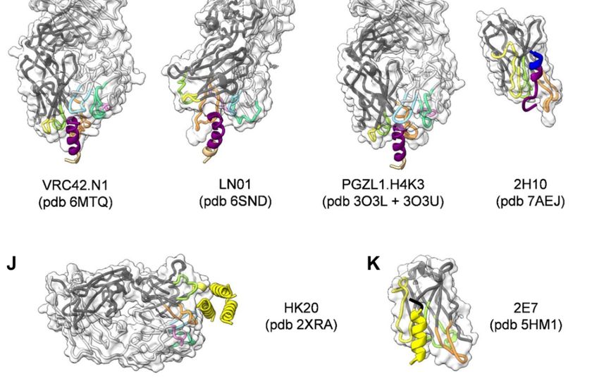
Viruses 2020, 12, 1210                                                                                3 of 19

3. Structure of MPER
     Several different MPER conformations have been described based on MPER peptide studies.
NMR analyses revealed a kinked MPER structure [59], a continuous helical conformation of MPER
fused to the N-terminal part of the transmembrane region (TM) [60] and the complete TM [61].
The physiological role of the continuous helical MPER-TM conformation was confirmed by the
structure of the bnAb LN01 in complex with MPER-TM [40]. Furthermore, trimeric models of MPER,
TM and MPER-TM have been proposed [62–64] and MPER was shown to form a continuous helix
with HR2 in the gp41 post-fusion conformation [65,66]. Together, the structural studies indicate the
conformational flexibility of MPER with potential hinges within MPER and between MPER and TM.
No high-resolution structure of MPER within native Env is yet available, but a recent medium resolution
structure suggests that MPER spans approximately 12 Å between the C-terminal gp41 HR2 residue
D664 and the membrane boundary at R/K683, thereby forming a “twisting tripod” configuration [10].
As this region contains 19 MPER residues, it remains yet unclear which conformation they might adopt
to span this short distance.

4. Neutralizing Antibodies Targeting gp41 MPER
      To date, a number of different broadly neutralizing antibodies or antibody lineages thereof
have been isolated from patients targeting linear epitopes of MPER (Figure 1C). They are among the
broadest neutralizing antibodies in cell-free infection models, but they are less efficient in blocking
cell-to-cell transfer [67–69]. 2F5 recognizes an extended epitope within N-terminal MPER [34,70]
(Figure 2A,B). Additional 2F5-like antibodies m66 and m66.6 have been isolated as well though with a
lower extent of somatic mutations and reduced neutralization breadth and potency [71]. Z13E binds to
an S-shaped epitope that overlaps with that of 2F5 and 4E10 [37,72]. Abs 4E10 [35,72,73] (Figure 2C),
10E8 [36] (Figure 2D), CAP206-CH12 [74], DH511 [38] (Figure 2E), VRC42 [39] (Figure 2F), LN01 [40]
(Figure 2G), and PGZL1 [41] (Figure 2H) recognize linear sequences forming helical epitopes adjacent
to the transmembrane region (TM). Recent studies suggest that the MPER epitope extends into the
TM since LN01 requires the first helical turn of the TM for interaction [40] and the binding affinity of
10E8 increases in the presence of TM [75], consistent with increased 4E10 binding in the presence of
TM [76]. Indeed, the structure of LN01 in complex with MPER fused to the complete TM reveals a
continuous helix of the MPER epitope and the TM. Molecular dynamics simulation of the complex
within a bilayer resembling the lipid composition of the HIV-1 envelope [77] revealed that MPER-TM
is inserted into the membrane with an ~18◦ tilt, that allows the HCDR3 to dip into the bilayer as
well as additional interactions of the Fab surface with the bilayer (Figure 3A,B) [40]. It remains to
be determined whether the N-terminal region of the TM is a general feature of the MPER epitope of
all anti-MPER antibodies recognizing the helical epitope adjacent to TM. In addition to the isolation
of human bnAbs, llama immunization with a gp41 MPER-TM mimetic led to the isolation of the
2H10 nanobody. 2H10 recognizes a 2F5-overlapping epitope (Figures 1C and 2I) and has modest
neutralization breadth and potency as a bi-head [78].
      MPER is likely sterically occluded within native Env and only accessible upon initial receptor-induced
conformational changes [79,80]. Nevertheless, a first encounter complex can be formed with native Env,
which however induces conformational changes to facilitate access to the epitope [11,12]. MPER bnAb-Env
structures [11,12] suggest further that trimeric MPER [64] needs to “open” up to accommodate bnAb
binding. In addition, the different approach angles of MPER bnAbs indicates that MPER is likely in a
“monomeric” conformation upon bnAb binding [40]. Furthermore, an induced interaction of Env trimers
was likely observed by super-resolution microscopy of MPER bnAb recognition on native virions [81].
This initial interaction with native Env, however, may depend on the neutralization sensitivity of the
virus clade. Other studies reveal high-affinity binding to a fusion intermediate gp41 conformation that
bridges the viral and cellular membranes [82,83]. Thus, the first encounter with native Env, interaction
with early Env conformations and with gp41 intermediates along the fusion pathway suggest that
MPER antibodies have a long window of action to inhibit the molecular transitions of gp41 required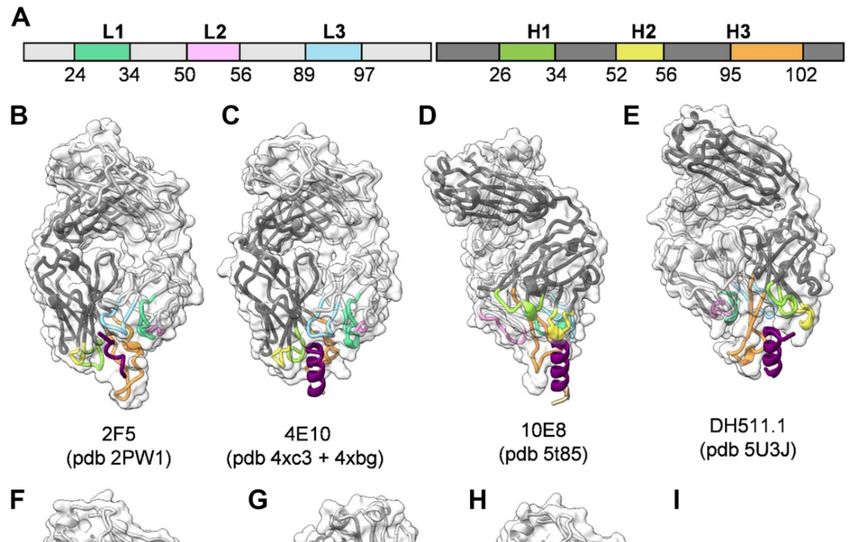
Viruses 2020, 12, x FOR PEER REVIEW                                                                       4 of 19

       induced interaction of Env trimers was likely observed by super-resolution microscopy of MPER
       bnAb recognition on native virions [81]. This initial interaction with native Env, however, may
       depend on the neutralization sensitivity of the virus clade. Other studies reveal high-affinity binding
Virusesto2020,
           a fusion   intermediate gp41 conformation that bridges the viral and cellular membranes [82,83].4 of 19
               12, 1210
       Thus, the first encounter with native Env, interaction with early Env conformations and with gp41
       intermediates along the fusion pathway suggest that MPER antibodies have a long window of action
for entry    by membrane
       to inhibit               fusion.
                     the molecular        This is inofagreement
                                       transitions                 with for
                                                       gp41 required     their relatively
                                                                             entry        long half-life
                                                                                    by membrane          of This
                                                                                                    fusion. neutralization.
                                                                                                                  is in
       agreement
Notably,     MPERwith bnAbstheirblock
                                  relatively long when
                                       infection  half-lifeadded
                                                            of neutralization.
                                                                   up to almostNotably,
                                                                                  30 minMPER   bnAbs block
                                                                                          post-exposure    of infection
                                                                                                              target cells to
HIV-1when      added
          [38,84].      up to possible
                    Another     almost 30mechanism
                                            min post-exposure     of target to
                                                         that contributes   cells to HIV-1 [38,84].
                                                                               neutralization        Another
                                                                                               is their        possible
                                                                                                        capacity   to induce
gp120 shedding [80,85]. Moreover, IgG avidity is not required for neutralization, since LN01[80,85].
       mechanism       that contributes    to neutralization   is their capacity  to induce gp120    shedding    Fabs show
       Moreover, IgG avidity is not required for neutralization, since LN01 Fabs show the same breadth and
the same breadth and potency as LN01 IgG1 [40].
       potency as LN01 IgG1 [40].

      FigureFigure  2. Structures
              2. Structures       of gp41-specific
                             of gp41-specific      antibodies
                                              antibodies       in complex
                                                         in complex        with epitope.
                                                                     with their  their epitope. Theare
                                                                                         The Fabs    Fabs are
                                                                                                       represented
            represented  in ribbon colored according  to the light and heavy  chain  scheme, highlighting
      in ribbon colored according to the light and heavy chain scheme, highlighting the position          theof the
      different CDRs (A). The surface of the Fabs is represented by a semi-transparent white surface. The gp41
      epitope is represented as cartoon colored according to the gp41 scheme shown in Figure 1A. The PBD
      coordinate files used for generating the figure for each antibody are indicated. (B) 2F5; (C) 4E10;
      (D) 10E8; (E) DH511.1; (F) VRC42.N1; (G) LN01; (H) PGZL1.H4K3; (I) llama nanobody 2H10; (J) HK20
      targeting HR1; (K) llama nanobody 2E7 targeting HR1-CC. Images shown in Figures 2–4 were rendered
      by ChimeraX [86].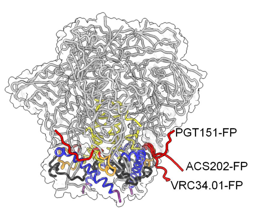
Viruses 2020, 12, x FOR PEER REVIEW 5 of 19
position of the different CDRs (A). The surface of the Fabs is represented by a semi-transparent white
surface. The gp41 epitope is represented as cartoon colored according to the gp41 scheme shown in
Figure 1A. The PBD coordinate files used for generating the figure for each antibody are indicated.
(B) 2F5; (C) 4E10; (D) 10E8; (E) DH511.1; (F) VRC42.N1; (G) LN01; (H) PGZL1.H4K3; (I) llama
Viruses 2020, 12, 1210 5 of 19
nanobody 2H10; (J) HK20 targeting HR1; (K) llama nanobody 2E7 targeting HR1-CC. Images shown
in Figures 2 to 4 were rendered by ChimeraX [86].
Figure 3. The orientation
Figure of the MPER-TM
3. The orientation epitope
of the MPER-TM bound
epitope to LN01
bound to LN01in the lipid
in the bilayer.
lipid bilayer.(A,B)
(A) andRepresentation
(B)
Representation ofcomplex
of the LN01-MPER-TM the LN01-MPER-TM
placed into complex
a bilayerplaced intoa alipid
with bilayer with a lipid resembling
composition composition the viral
resembling the viral envelope obtained by molecular dynamics (MD) simulations. The LN01-MPER-
envelope obtained by molecular dynamics (MD) simulations. The LN01-MPER-TM complex and lipid
TM complex and lipid analogues are represented as shown in figures 1A and 2A. Only the phosphate
analoguesgroup
are represented
of the lipids ofastheshown in Figures
bilayer are displayed1A andand 2A. Only
represented the phosphate
as spheres. group
(A) The tilted of the lipids of
orientation
the bilayerofare
thedisplayed
TM segmentand withrepresented
respect to thatas
of spheres.
the bilayer(A) The
allows tiltedK683
residue orientation
to interactof thelipid
with TMheadsegment with
groups
respect to that of of
theone leaflet and
bilayer allowsR696residue
to contact headto
K683 groups of the
interact opposite
with lipidleaflet;
head residues
groups R707
of one andleaflet
R709 and R696
terminating the TM helix interact as well with lipid head groups in the simulation. (B) Close up of the
to contact head groups of the opposite leaflet; residues R707 and R709 terminating the TM helix interact as
interaction between LN01, gp41 and the lipids. The gp41 residues located on the same side of the
well with lipid head
MPER-TM helixgroups in the
as K683 are simulation.
more accessible(B)toClose up of the
the interaction withinteraction between
the antibody. LN01, gp41
This orientation of and the
lipids. Thethegp41 residues located on the same side of the MPER-TM helix as K683
TM in the lipid bilayer could explain why only one side of the MPER-TM helical epitope (residues are more accessible
673–686) with
to the interaction is targeted by all knownThis
the antibody. MPER bnAbs. Theof
orientation MD simulation
the TM in the supports
lipidthe interaction
bilayer could of the
explain why
Fab surface with the bilayer and underlines together with specific lipid-binding shown in later
only one side of the MPER-TM helical epitope (residues 673–686) is targeted by all known MPER bnAbs.
Figures, the recognition of a bipartite epitope composed of MPER and the membrane by MPER bnAbs.
The MD simulation supports the interaction of the Fab surface with the bilayer and underlines together
with specific lipid-binding
5. Neutralizing Antibodiesshown in latergp41
Targeting Figures,
HR1 the recognition of a bipartite epitope composed of
MPER andSeveral
the membrane by MPER bnAbs.
antibodies targeting the heptad repeat region 1 (HR1) (Figure 1A,B) with modest breadth
have been isolated as well. These antibodies target a gp41 pre-hairpin conformation that exposes HR1
5. Neutralizing Antibodies Targeting gp41 HR1
and bridges the viral and cellular membranes before refolding into the six-helical bundle
conformation required for membrane fusion [87]. The inhibitory action of HR1 targeting Abs is thus
Several antibodies targeting the heptad repeat region 1 (HR1) (Figure 1A,B) with modest breadth
comparable to the function of peptide fusion inhibitors [88].
have been isolated as well. These
The HR1-specific antibodies
Fab 3674 target
was isolated a gp41
from pre-hairpin
a human non-immune conformation
phage library that exposes HR1
[89] and
and bridges the viral and cellular membranes before refolding into the six-helical bundle
further affinity matured (Fab8066) [90]. D5 was selected by vaccination with HR1 mimetics [91] and conformation
HK20
required for was isolated
membrane from memory
fusion [87]. TheB cells from an infected
inhibitory action individual [92]. D5, HK20,
of HR1 targeting Abs and 8066comparable
is thus bind to
into a conserved hydrophobic pocket of the HR1 triple-stranded coil [93–95] (Figure 1A,B and 2J) that
the function of peptide fusion inhibitors [88].
is occupied by HR2 in the post-fusion conformation [87,96]. Although D5 and HK20 have modest
The HR1-specific Fab 3674
neutralizing breadth and was isolated
potency from aIgGs,
as complete human non-immune
the HK20 breadth andphage library
potency [89] and further
are greatly
affinity matured (Fab8066) [90]. D5 was selected by vaccination with HR1 mimetics [91] and HK20
was isolated from memory B cells from an infected individual [92]. D5, HK20, and 8066 bind into a
conserved hydrophobic pocket of the HR1 triple-stranded coil [93–95] (Figure 1A,B and Figure 2J) that
is occupied by HR2 in the post-fusion conformation [87,96]. Although D5 and HK20 have modest
neutralizing breadth and potency as complete IgGs, the HK20 breadth and potency are greatly increased
when used as Fab or scFv, the latter neutralizing 100% out of a 45 pseudovirus panel compared to
15% neutralization by the HK20 IgG [94]. This thus indicates that the size of the complete IgG limits
access to this epitope in most clades during the fusion process. Nevertheless, antibodies recognizing
the HK20 epitope footprint are present in a significant fraction of HIV-1 infected individuals [94].
Furthermore, it has been shown that the presence of the Fc gamma receptor I (FcγRI) on the target cells
substantially increases breadth and potency of mAb D5 and sera from HR1 mimetic immunized guinea
pigs neutralizing not only Tier 1 but also Tier-2 viruses from multiple clades in a FcγRI-dependent
manner. Whether this is also the case for HK20 needs to be tested. Notably, FcγRI is expressed on
macrophages and dendritic cells, which are present at mucosal surfaces and are implicated in the early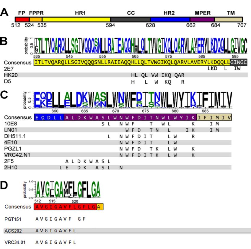
Viruses 2020, 12, 1210                                                                                                     6 of 19

establishment of HIV-1 infection following sexual transmission, which may reestablish HR1 antibodies
as an interesting prevention strategy [97]. An additive effect of FcγRI on neutralization has been also
reported for MPER antibodies 2F5 and 4E10 [98,99] in line with the importance of the Fc function of
2F5 for dose-dependent protection of rhesus macaques upon vaginal challenge with SHIV-BaL [100].
     Llama immunization with an “open” conformation of HIV-1 Env gp140 CN54 and UG37 led to the
isolation of the HR1-targeting nanobody 2E7 [101] that recognizes a C-terminal HR1 epitope extending
into the C-C loop of gp41 [102]. In contrast to D5 and HK20, which require coiled-coil interaction
for HR1 interaction, 2E7 binds to a linear sequence forming a helical epitope (Figures 1B and 2K).
2E7 neutralizes 80% out of a 26 pseudovirus panel [101] but revealed highly increased breadth and
potency when linked to nanobodies recognizing the CD4 binding site [102]. Thus the small size of
nanobodies is not only advantageous to penetrate the Env glycan shield [103] but permits also easier
access to the HR1 intermediate conformation present in the gp41 pre-hairpin conformation thereby
preventing membrane fusion [104].

6. Neutralizing Antibodies Targeting gp41 FP
     Recent work has identified the fusion peptide as a promising target for vaccine development.
A number of broadly neutralizing antibodies have been identified that target the conserved FP
sequence (Figure 1A,D) at the interface of gp41 and gp120 in the native Env trimer conformation.
These include human bnAb PGT151 [105], bnAb VRC34 targeting the N-terminus of FP [30,106]
and bnAb ACS202 (Figure 4) [107,108]. In addition to FP, these bnAbs contact other components of
the native trimer as well as complex glycans at the gp120-gp41 interface. Notably, immunization
schemes employing fusion peptide-coupled carriers combined with Env trimers induced an impressive
neutralization of 31% of a cross-clade panel of 208 HIV-1 strains. Isolation of two vaccine-induced
murine bnAbs, v1602 and vFP20.01, confirmed the recognition of FP [109] and escape mutants of
VRC34 and vaccine-induced bnAbs were mapped to mutations in FP and distal interacting sites of the
Env trimer [110]. Together, the structural analyses of Env trimers in complex with FP interacting bnAbs
and gp120-gp41 interface bnAbs highlights the conformational plasticity of FP within proteolytically
processed native Env trimers [109]. However, FP is not exposed in all native Env trimers and was
found to be sequestered in the hydrophobic core of an HIV-1 transmitted founder Env trimer [111].
Nevertheless, immunogens targeting FP have yet produced the broadest cross-clade neutralization
by immunization.
          Viruses 2020, 12, x FOR PEER REVIEW                                             7 of 19

      Figure 4. Figure   4. Conformational
                 Conformational             plasticity
                                       plasticity   of of
                                                       FP.FP.Color-coding
                                                              Color-coding ofof
                                                                              gp41 is asisshown
                                                                                gp41            in Figure
                                                                                           as shown    in 1A. The Cα
                                                                                                          Figure  1A. The Cα
                 trace of gp120 is shown in grey and the molecular envelope of Env is shown transparently. The three
      trace of gp120 is shown in grey and the molecular envelope of Env is shown transparently. The three
                 different conformations of FP in complex with the indicated bnAbs are shown in red.
      different conformations of FP in complex with the indicated bnAbs are shown in red.
             7. Membrane Interaction and Polyreactivity
                  The hallmark of neutralizing MPER bnAbs is a long HCDR3 region exposing hydrophobic
             residues at its tip that can insert into the bilayer upon gp41 interaction, which is essential for
             neutralization [112–116]. In addition, non-specific lipid and membrane interaction in vitro has been
             observed for 4E10, 2F5 and VRC42.01 [39,52,117]. In contrast, 10E8, DH511 lineage bnAbs and LN01
             do not show significant membrane interaction in vitro or with cellular membranes tested by indirect
Viruses 2020, 12, 1210 7 of 19
7. Membrane Interaction and Polyreactivity
The hallmark of neutralizing MPER bnAbs is a long HCDR3 region exposing hydrophobic
residues at its tip that can insert into the bilayer upon gp41 interaction, which is essential for
neutralization [112–116]. In addition, non-specific lipid and membrane interaction in vitro has been
observed for 4E10, 2F5 and VRC42.01 [39,52,117]. In contrast, 10E8, DH511 lineage bnAbs and LN01
do not show significant membrane interaction in vitro or with cellular membranes tested by indirect
immunofluorescence binding to HEp-2 cells [36,38,40] although some membrane binding using different
assays has been reported as well [118]. However, structures of 4E10, 10E8, LN01 and PGZL1 provide
evidence for specific lipid binding. 4E10 can bind to lipid components such as glycerol-1-phosphate,
glycerol-3-phosphate, phosphatidic acid and phosphatidylglycerol (Figure 5A) [119], 10E8 interacts
with glycerol, phosphatidylglycerol and phosphatidic acid (Figure 5B) [120] and LN01 with
phosphatidylserine and the phosphocholine group of Fos-choline-12 mimicking a phospholipid
interaction (Figure 5C) [40]. Furthermore, PGZL1 and its variant H4K3 coordinate two phosphatidic
acid molecules [41] (Figure 5D). To further increase membrane interaction with negatively charged
phospholipids basic patches are present in the 10E8 light chain that are positioned at the putative
Fab-membrane interface, which are either germline encoded or generated by somatic mutations.
Viruses 2020, 12, x FOR PEER REVIEW 8 of 19
Mutations of these basic residues did not affect epitope binding but influenced neutralization.
Importantly, mutation
Immunization ofKI
of 2F5 themice
lipid-binding
with MPER site abrogated
peptide neutralization
coated liposomes [120].
rescued, Similarly,
however, LN01
anergic variants
B-cells
that for
lackneutralizing
the lipid-binding
antibody production, in agreement with some autoreactive B-cells being able several
site deploy poor neutralization [40]. Like 10E8, LN01 has to
basicescape
residuesclonal atselection
the Fab-membrane interface
[130]. In addition, 2F5 germthat
line can contact
KI mice, whencharged
immunized lipid
withhead groups
different Env [40].
Thus,immunogens,
increasing membraneinduced theinteraction
deletion ofby2F5 precursors, additional
engineering although anergic B cells specific
hydrophobic for MPER
and basic residues at
could be still activated
the Fab-membrane interfacewith Env immunogens
augments the potency [131]. This neutralization
of 10E8 indicates that mechanisms
[118,121–123].controlling
immunological tolerance may limit the generation of 2F5 and 4E10-like bnAbs.
Figure
Figure 5. Close-ups
5. Close-ups of lipid
of lipid oror lipidfragment
lipid fragment binding
binding of
ofMPER
MPERbnAbs.
bnAbs. (A)(A)
4E10; (B) 10E8;
4E10; (C) LN01
(B) 10E8; (C) LN01
and (D) PGZL1. The gp41 epitope and Fabs are represented as in Figure 2. The lipid or lipid fragment
and (D) PGZL1. The gp41 epitope and Fabs are represented as in Figure 2. The lipid or lipid fragment
visible in the structures are shown as spheres. The residues coordinating the binding of the lipids are
visible in the structures are shown as spheres. The residues coordinating the binding of the lipids are
shown as stick and balls, the identity and number of these residues are indicated below each structure,
shown as stick and balls, the identity and number of these residues are indicated below each structure,
an asterisk indicates which residues are present in the UCA.
an asterisk indicates which residues are present in the UCA.
8. V-Gene Usage and Somatic Mutations
MPER-specific bnAbs isolated from different patients use members of 6 different VH-genes and
3 different Vκ or Vλ genes. Notably, none of the VH gene usages is rare (Table 1). Most bnAbs
targeting Env undergo a large set of somatic mutations to reach breadth and potency. To follow the
track of somatic mutations from the unmutated common ancestor (UCA) to the mature bnAb requires
samples from different time points of infection, which are often not available complicating the
identification of early lineage members. Identification of the UCA and early lineage members would,Viruses 2020, 12, 1210 8 of 19
Besides membrane interaction, MPER bnAbs 2F5, 4E10, 10E8 and DH511 lineage bnAbs have been
shown to cross-react with cellular proteins [38,124,125]. It was thus suggested that polyreactivity of
MPER bnAbs may be the limiting factor to induce such antibodies by vaccination because auto-reactive
MPER and membrane-specific B-cells may be eliminated from the B cell repertoire during clonal
selection [126,127]. This was strengthened by studying B-cell development in 2F5 and 4E10 knock-in (KI)
mice, which showed defects in the transition of pre-B to immature B cells [128,129]. Immunization of 2F5
KI mice with MPER peptide coated liposomes rescued, however, anergic B-cells for neutralizing antibody
production, in agreement with some autoreactive B-cells being able to escape clonal selection [130].
In addition, 2F5 germ line KI mice, when immunized with different Env immunogens, induced the
deletion of 2F5 precursors, although anergic B cells specific for MPER could be still activated with Env
immunogens [131]. This indicates that mechanisms controlling immunological tolerance may limit the
generation of 2F5 and 4E10-like bnAbs.
8. V-Gene Usage and Somatic Mutations
MPER-specific bnAbs isolated from different patients use members of 6 different VH-genes and 3
different Vκ or Vλ genes. Notably, none of the VH gene usages is rare (Table 1). Most bnAbs targeting
Env undergo a large set of somatic mutations to reach breadth and potency. To follow the track of
somatic mutations from the unmutated common ancestor (UCA) to the mature bnAb requires samples
from different time points of infection, which are often not available complicating the identification of
early lineage members. Identification of the UCA and early lineage members would, however, help to
design immunogens that can specifically target naïve B-cells expressing UCA receptors.
In case of 10E8, next-generation sequencing (NGS) was used to identify potential early lineage
members [132]. Combination with structure-function studies permitted to reconstruct a germline
version and early Ab intermediates in the maturation process. This indicated that 10E8 develops from
a UCA with no significant MPER binding and substantial differences in HCDR 2 and 3 compared to the
mature 10E8. Indeed, structural comparison of unliganded 10E8 with its proposed UCA version and
10E8 with a mature light chain paired with the germline VH revealed significant structural changes of
the Cα positions of HCDR 2 and 3 residues. This thus suggests that Ab maturation affects the structure
of the HCDRs. Early intermediates showed some weak binding to MPER and modest neutralization,
whereas extensive hypermutation is required for broad and potent neutralization [133].
Table 1. VH and VL gene usage by MPER bnAbs.
bNAb VH-Gene VL-Gene D-Segment VH Gene Usage (%) *
4E10 IGVH1-69 IGVκ3-20*01 D3-10 ~2
PGZL1 IGVH1-69 IGVκ3-20 D3-10*01 ~2
VRC42 IGVH1-69 IGVκ3-20 D3-10 ~2
CAP206-CH12 IGVH1-69 IGVκ3-20*01 n.i. ~2
2F5 IGVH2-05 IGVκ1-13*02 D3-03*01 ~1.9
10E8 IGVH3-15 IGVλ3-19 D3-03*01 ~2.8
DH511 IGVH3-15 IGVκ1-39 n.i. ~2.8
DH517 IGVH4-34 IGVλ3-19 n.i. ~2.8
M46 IGVH4-34*01 IGVκ1-9*01 D5-12*01 ~2.8
LN01 IGVH4-39 IGVκ1-39 D3-3*01 ~2.3
Z13e1 IGVH4-59*03 IGVκ3-11*01 n.i. ~8
M44 IGVH4-61*01 IGVκ3-20*01 D3-10*02 ~8
* VH gene usage in humans [134]; n.i.: not identified.Viruses 2020, 12, 1210 9 of 19
DH511 UCA did not bind to MPER peptides and only the late inferred maturational intermediate,
DH511-I6 VH has a detectable affinity for the MPER peptide. Accumulation of neutralization breadth
correlates with increased somatic mutations and affinity. Furthermore, in Luminex assay and ELISA,
the DH511 UCA reacted with U1 small nuclear RNP (U1-snRNP), and in protein microarray assay,
the DH511 UCA was both polyreactive and autoreactive with a number of proteins [38].
A longitudinal study on VRC42 using samples from days 85 to 646 post-infection allowed an
accurate reconstruction of the maturation process. This suggests that the Ab becomes broad with
only a few somatic mutations starting from day 154. Although the VRC42 UCA (inferred from an
early transcript with only 5 nucleotide changes) did not bind to MPER peptides nor does it neutralize,
the inferred later intermediate VRC42.I3 neutralized 51% of heterologous viruses with only 13 amino
acids different from the inferred VRC42 UCA [39]. Furthermore, VRC42 UCA bound to MPER presented
as a multimer on KLH. Although VRC42 showed some degree of polyreactivity and cardiolipin binding,
the VRC42 UCA did not interact with HEp-2 cells nor with cardiolipin [39].
No longitudinal information is available on LN01. The inferred LN01 UCA does not interact
with MPER peptides. Mixed hc and lc variants having only mature CDRs of LN01 showed that
neutralization correlated with binding affinity as expected. Notably, only a few somatic mutations
within the CDRs are required for MPER interaction and lipid recognition in the final mature bnAb.
Phosphatidylserine (PS) binding is acquired and stabilized by 2 somatic mutations in HCDR1 and
while 3 other polar PS interactions from HCDR3 are germline-encoded. Furthermore, LN01 exhibits a
low degree of autoreactivity comparable to the one reported for 10E8 [40].
PGZL1 shares germline V/D-region genes with 4E10, but has a shorter CDRH3 and low
polyreactivity comparable to 10E8. Notably, a germline revertant of PGZL1 with mature CDR3s
still neutralizes 12% of a 130-isolate panel. As an exception, the complete germline reversion of PGZL1
still binds MPER [41] providing a starting point to specifically activate naïve B cells by immunization.
In summary, quite a remarkable number of germline residues are implicated in the binding to
the MPER epitope. This implies that only a limited number of somatic mutations are theoretically
required to achieve a large breadth and potency. However, long-range structural effects are likely also
important for nAb breadth and potency [135]. Of further note, specific lipid binding is not completely
germline-encoded and requires somatic mutations for optimal coordination [40,41].
9. Gp41 MPER-Based Vaccine Approaches
Although a number of different approaches with different prime-boost strategies have been
employed to generate Abs that target MPER (Table 2), none of them showed significant success beyond
the induction of some modest neutralization of tier 1 and 2 pseudoviruses. This may be due to multiple
reasons. First, polyreactivity as described above may be a major hindrance to generate MPER bnAbs.
Thus a hypothesis was put forward suggesting that the clonal lineage UCA of bnAb DH511 may have
been initiated by self-antigens while gp41 MPER antigens engaged later to further mature the initial
cross-reactive Ab into an anti-MPER bnAb [49–51]. Secondly, the immunization strategies followed
so far did not take into account an efficient targeting of the naïve B cell receptors. Thirdly, the aspect
of the membrane and specific lipids as part of the bipartite epitope has not yet been fully explored,
although some approaches presented MPER in a lipid environment. Fourth, the orientation of MPER
within a membrane environment needs to be taken into account. This may be a particular problem as
the MPER sequence has a high tendency to fold back and interact itself with the membrane thereby
occluding its access. Fifth, the lipid composition including cholesterol seems to be important as it may
constrain the antigenic conformation of the MPER epitope [136].Viruses 2020, 12, 1210 10 of 19
Table 2. MPER immunogens that elicit modest neutralizing antibody responses in animal models.
Immunogen Reference
MPER-TM proteoliposomes [136]
His-tagged MPER bound to liposomes [137]
Trimeric MPER fused to the diphtheria toxin domainA [138]
A peptide containing 4 copies of the 10E8 epitope [139]
Three different 6-helical bundle gp41 constructs containing different bundle
[140]
destabilizing mutations
MPER peptide associated with liposomes [141]
Chimeric human Rhinoviruses expressing MPER [142]
The MPER and gp41 ectodomain was expressed separately as N-terminal
fusions to the E2 protein of Geobacillus stearothermophilus; Immunization in [143]
conjunction with DNA encoding full-length SF162 gp160
Hybrid antigens containing the MPER and the FPPR of gp41 of HIV-1 and
[144]
sequences of the TM protein p15E of PERV
Tri-repeat of the MPER epitope of gp41 plus defending containing
[145]
proteoliposomes
Fusion intermediate conformation of gp41 covalently linked to liposomes [83]
Gp41 HR2-MPER-TM proteoliposomes [78]
Bovine papillomavirus VLPs with extended 2F5 or 4E10 epitopes or the MPER
[146]
domain grafted into the D-E loop of BPV L1
A HEK293 cell line expressing membrane-anchored gp41 [147]
Gp41-subunit antigens grafted onto liposomes/ virosomes [148]
Gp140 oligomer prime followed by MPER peptide-liposome boost [149]
Trimeric MPER fusion proteins [150]
Gp41 ectodomain fused to an influenza HA2 region [151]
10. Conclusions
Much progress in understanding the molecular details of bnAbs targeting HR1, FP and MPER in
conjunction with components of the bilayer have been made. Notably, FP-specific bnAb generation by
immunization is currently the most promising approach. However, regarding MPER as an immunogen,
a number of questions are still open. First, which is the gp41 conformation that is finally locked by
MPER bnAbs: an early fusion-activated conformation, close to that of native Env, a fusion-intermediate
extended conformation or others or all together? Second, can broad and potent MPER-specific bnAbs be
induced by vaccination by overcoming high somatic hypermutation and autoreactivity? The findings
that bnAbs VRC42, LN01 and especially PGZL1 may require relatively few somatic mutations to
acquire high neutralizing activity provides some clues for feasibility. Furthermore, direct lipid binding
is not germline-encoded but partly acquired by somatic mutations, suggesting that MPER recognizing
germline B-cell receptors may not be clonally deleted because of membrane autoreactivity. Finally,
among the first MPER bnAbs, the UCA of PGLZ1 recognizes MPER providing a blueprint to develop
vaccination protocols that employ a series of potent immunogens that can first activate naïve B cells
and subsequently mature antibodies into high-affinity bnAbs recognizing the bipartite MPER peptide
and lipid bilayer epitopes.
Author Contributions: Writing—original draft preparation, W.W.; writing—review and editing, C.C., D.G., G.S.
and W.W.; funding acquisition, W.W. All authors have read and agreed to the published version of the manuscript.
Funding: This work was supported by the Institute Universitaire de France (IUF)(W.W.), the European Union’s
Horizon 2020 research and innovation program under grant agreement No. 681137, H2020 EAVI, by FRISBIViruses 2020, 12, 1210 11 of 19
(ANR-10-INBS-05-02) and GRAL, a project of the University Grenoble Alpes graduate school (Ecoles Universitaires
de Recherche) CBH-EUR-GS (ANR-17-EURE-0003)(W.W.).
Acknowledgments: We thank the two anonymous reviewers for providing insightful comments.
Conflicts of Interest: The authors declare no conflict of interest.
References
1. Chen, B. Molecular Mechanism of HIV-1 Entry. Trends Microbiol. 2019, 27, 878–891. [CrossRef] [PubMed]
2. Schibli, D.J.; Weissenhorn, W. Class I and class II viral fusion protein structures reveal similar principles in
membrane fusion. Mol. Membr. Biol. 2006, 21, 361–371. [CrossRef] [PubMed]
3. Harrison, S.C. Viral membrane fusion. Nat. Struct. Mol. Biol. 2008, 15, 690–698. [CrossRef] [PubMed]
4. Blumenthal, R.; Durell, S.; Viard, M. HIV entry and envelope glycoprotein-mediated fusion. J. Biol. Chem.
2012, 287, 40841–40849. [CrossRef] [PubMed]
5. Sanders, R.W.; Vesanen, M.; Schuelke, N.; Master, A.; Schiffner, L.; Kalyanaraman, R.; Paluch, M.; Berkhout, B.;
Maddon, P.J.; Olson, W.C.; et al. Stabilization of the soluble, cleaved, trimeric form of the envelope
glycoprotein complex of human immunodeficiency virus type 1. J. Virol. 2002, 76, 8875–8889. [CrossRef]
6. Klasse, P.J.; Depetris, R.S.; Pejchal, R.; Julien, J.P.; Khayat, R.; Lee, J.H.; Marozsan, A.J.; Cupo, A.; Cocco, N.;
Korzun, J.; et al. Influences on trimerization and aggregation of soluble, cleaved HIV-1 SOSIP envelope
glycoprotein. J. Virol. 2013, 87, 9873–9885. [CrossRef]
7. Lyumkis, D.; Julien, J.P.; de Val, N.; Cupo, A.; Potter, C.S.; Klasse, P.J.; Burton, D.R.; Sanders, R.W.; Moore, J.P.;
Carragher, B.; et al. Cryo-EM Structure of a Fully Glycosylated Soluble Cleaved HIV-1 Envelope Trimer.
Science 2013, 342, 1484–1490. [CrossRef]
8. Julien, J.P.; Cupo, A.; Sok, D.; Stanfield, R.L.; Lyumkis, D.; Deller, M.C.; Klasse, P.J.; Burton, D.R.; Sanders, R.W.;
Moore, J.P.; et al. Crystal Structure of a Soluble Cleaved HIV-1 Envelope Trimer. Science 2013, 342, 1477–1483.
[CrossRef]
9. Pancera, M.; Zhou, T.; Druz, A.; Georgiev, I.S.; Soto, C.; Gorman, J.; Huang, J.; Acharya, P.; Chuang, G.Y.;
Ofek, G.; et al. Structure and immune recognition of trimeric pre-fusion HIV-1 Env. Nature 2014, 514, 455–461.
[CrossRef]
10. Li, Z.; Li, W.; Lu, M.; Bess, J., Jr.; Chao, C.W.; Gorman, J.; Terry, D.S.; Zhang, B.; Zhou, T.; Blanchard, S.C.; et al.
Subnanometer structures of HIV-1 envelope trimers on aldrithiol-2-inactivated virus particles. Nat. Struct.
Mol. Biol. 2020, 27, 726–734. [CrossRef]
11. Lee, J.H.; Ozorowski, G.; Ward, A.B. Cryo-EM structure of a native, fully glycosylated, cleaved HIV-1
envelope trimer. Science 2016, 351, 1043–1048. [CrossRef]
12. Rantalainen, K.; Berndsen, Z.T.; Antanasijevic, A.; Schiffner, T.; Zhang, X.; Lee, W.H.; Torres, J.L.; Zhang, L.;
Irimia, A.; Copps, J.; et al. HIV-1 Envelope and MPER Antibody Structures in Lipid Assemblies. Cell Rep.
2020, 31, 107583. [CrossRef] [PubMed]
13. Pan, J.; Peng, H.; Chen, B.; Harrison, S.C. Cryo-EM Structure of Full-length HIV-1 Env Bound With the Fab of
Antibody PG16. J. Mol. Biol. 2020, 432, 1158–1168. [CrossRef] [PubMed]
14. Lu, M.; Ma, X.; Castillo-Menendez, L.R.; Gorman, J.; Alsahafi, N.; Ermel, U.; Terry, D.S.; Chambers, M.;
Peng, D.; Zhang, B.; et al. Associating HIV-1 envelope glycoprotein structures with states on the virus
observed by smFRET. Nature 2019, 568, 415–419. [CrossRef]
15. Wang, Q.; Finzi, A.; Sodroski, J. The Conformational States of the HIV-1 Envelope Glycoproteins. Trends
Microbiol. 2020, 28, 655–667. [CrossRef] [PubMed]
16. Kwong, P.D.; Mascola, J.R. HIV-1 Vaccines Based on Antibody Identification, B Cell Ontogeny, and Epitope
Structure. Immunity 2018, 48, 855–871. [CrossRef]
17. Kwong, P.D. What Are the Most Powerful Immunogen Design Vaccine Strategies? A Structural Biologist’s
Perspective. Cold Spring Harb. Perspect. Biol. 2017, 9, 1–12. [CrossRef]
18. Ward, A.B.; Wilson, I.A. Innovations in structure-based antigen design and immune monitoring for next
generation vaccines. Curr. Opin. Immunol. 2020, 65, 50–56. [CrossRef]
19. Lee, J.H.; Andrabi, R.; Su, C.Y.; Yasmeen, A.; Julien, J.P.; Kong, L.; Wu, N.C.; McBride, R.; Sok, D.;
Pauthner, M.; et al. A Broadly Neutralizing Antibody Targets the Dynamic HIV Envelope Trimer Apex via a
Long, Rigidified, and Anionic beta-Hairpin Structure. Immunity 2017, 46, 690–702. [CrossRef]Viruses 2020, 12, 1210 12 of 19
20. McLellan, J.S.; Pancera, M.; Carrico, C.; Gorman, J.; Julien, J.P.; Khayat, R.; Louder, R.; Pejchal, R.; Sastry, M.;
Dai, K.; et al. Structure of HIV-1 gp120 V1/V2 domain with broadly neutralizing antibody PG9. Nature 2011,
480, 336–343. [CrossRef]
21. Cale, E.M.; Gorman, J.; Radakovich, N.A.; Crooks, E.T.; Osawa, K.; Tong, T.; Li, J.; Nagarajan, R.; Ozorowski, G.;
Ambrozak, D.R.; et al. Virus-like Particles Identify an HIV V1V2 Apex-Binding Neutralizing Antibody that
Lacks a Protruding Loop. Immunity 2017, 46, 777–791.e710. [CrossRef] [PubMed]
22. Pejchal, R.; Doores, K.J.; Walker, L.M.; Khayat, R.; Huang, P.S.; Wang, S.K.; Stanfield, R.L.; Julien, J.P.;
Ramos, A.; Crispin, M.; et al. A potent and broad neutralizing antibody recognizes and penetrates the HIV
glycan shield. Science 2011, 334, 1097–1103. [CrossRef]
23. Julien, J.P.; Sok, D.; Khayat, R.; Lee, J.H.; Doores, K.J.; Walker, L.M.; Ramos, A.; Diwanji, D.C.; Pejchal, R.;
Cupo, A.; et al. Broadly neutralizing antibody PGT121 allosterically modulates CD4 binding via recognition
of the HIV-1 gp120 V3 base and multiple surrounding glycans. PLoS Pathog. 2013, 9, e1003342. [CrossRef]
[PubMed]
24. Kong, L.; Lee, J.H.; Doores, K.J.; Murin, C.D.; Julien, J.P.; McBride, R.; Liu, Y.; Marozsan, A.; Cupo, A.;
Klasse, P.J.; et al. Supersite of immune vulnerability on the glycosylated face of HIV-1 envelope glycoprotein
gp120. Nat. Struct. Mol. Biol. 2013, 20, 796–803. [CrossRef] [PubMed]
25. Liao, H.X.; Lynch, R.; Zhou, T.; Gao, F.; Alam, S.M.; Boyd, S.D.; Fire, A.Z.; Roskin, K.M.; Schramm, C.A.;
Zhang, Z.; et al. Co-evolution of a broadly neutralizing HIV-1 antibody and founder virus. Nature 2013,
496, 469–476. [CrossRef] [PubMed]
26. Zhou, T.; Xu, L.; Dey, B.; Hessell, A.J.; Van Ryk, D.; Xiang, S.H.; Yang, X.; Zhang, M.Y.; Zwick, M.B.; Arthos, J.; et al.
Structural definition of a conserved neutralization epitope on HIV-1 gp120. Nature 2007, 445, 732–737. [CrossRef]
[PubMed]
27. Zhou, T.; Lynch, R.M.; Chen, L.; Acharya, P.; Wu, X.; Doria-Rose, N.A.; Joyce, M.G.; Lingwood, D.; Soto, C.;
Bailer, R.T.; et al. Structural Repertoire of HIV-1-Neutralizing Antibodies Targeting the CD4 Supersite in 14
Donors. Cell 2015, 161, 1280–1292. [CrossRef]
28. Zhou, T.; Georgiev, I.; Wu, X.; Yang, Z.Y.; Dai, K.; Finzi, A.; Kwon, Y.D.; Scheid, J.F.; Shi, W.; Xu, L.; et al.
Structural basis for broad and potent neutralization of HIV-1 by antibody VRC01. Science 2010, 329, 811–817.
[CrossRef]
29. Wu, X.; Zhou, T.; Zhu, J.; Zhang, B.; Georgiev, I.; Wang, C.; Chen, X.; Longo, N.S.; Louder, M.; McKee, K.; et al.
Focused evolution of HIV-1 neutralizing antibodies revealed by structures and deep sequencing. Science
2011, 333, 1593–1602. [CrossRef]
30. Kong, R.; Xu, K.; Zhou, T.; Acharya, P.; Lemmin, T.; Liu, K.; Ozorowski, G.; Soto, C.; Taft, J.D.; Bailer, R.T.;
et al. Fusion peptide of HIV-1 as a site of vulnerability to neutralizing antibody. Science 2016, 352, 828–833.
[CrossRef]
31. Scharf, L.; Scheid, J.F.; Lee, J.H.; West, A.P., Jr.; Chen, C.; Gao, H.; Gnanapragasam, P.N.; Mares, R.;
Seaman, M.S.; Ward, A.B.; et al. Antibody 8ANC195 reveals a site of broad vulnerability on the HIV-1
envelope spike. Cell Rep. 2014, 7, 785–795. [CrossRef]
32. Huang, J.; Kang, B.H.; Pancera, M.; Lee, J.H.; Tong, T.; Feng, Y.; Imamichi, H.; Georgiev, I.S.; Chuang, G.Y.;
Druz, A.; et al. Broad and potent HIV-1 neutralization by a human antibody that binds the gp41-gp120
interface. Nature 2014, 515, 138–142. [CrossRef]
33. Zhou, T.; Zheng, A.; Baxa, U.; Chuang, G.Y.; Georgiev, I.S.; Kong, R.; O’Dell, S.; Shahzad-Ul-Hussan, S.;
Shen, C.H.; Tsybovsky, Y.; et al. A Neutralizing Antibody Recognizing Primarily N-Linked Glycan Targets
the Silent Face of the HIV Envelope. Immunity 2018, 48, 500–513.e506. [CrossRef]
34. Ofek, G.; Tang, M.; Sambor, A.; Katinger, H.; Mascola, J.R.; Wyatt, R.; Kwong, P.D. Structure and mechanistic
analysis of the anti-human immunodeficiency virus type 1 antibody 2F5 in complex with its gp41 epitope.
J. Virol. 2004, 78, 10724–10737. [CrossRef] [PubMed]
35. Cardoso, R.M.; Zwick, M.B.; Stanfield, R.L.; Kunert, R.; Binley, J.M.; Katinger, H.; Burton, D.R.; Wilson, I.A.
Broadly neutralizing anti-HIV antibody 4E10 recognizes a helical conformation of a highly conserved
fusion-associated motif in gp41. Immunity 2005, 22, 163–173. [CrossRef]
36. Huang, J.; Ofek, G.; Laub, L.; Louder, M.K.; Doria-Rose, N.A.; Longo, N.S.; Imamichi, H.; Bailer, R.T.;
Chakrabarti, B.; Sharma, S.K.; et al. Broad and potent neutralization of HIV-1 by a gp41-specific human
antibody. Nature 2012, 491, 406–412. [CrossRef]Viruses 2020, 12, 1210 13 of 19
37. Pejchal, R.; Gach, J.S.; Brunel, F.M.; Cardoso, R.M.; Stanfield, R.L.; Dawson, P.E.; Burton, D.R.; Zwick, M.B.;
Wilson, I.A. A Conformational Switch in Human Immunodeficiency Virus gp41 Revealed by the Structures
of Overlapping Epitopes Recognized by Neutralizing Antibodies. J. Virol. 2009, 83, 8451–8462. [CrossRef]
[PubMed]
38. Williams, L.D.; Ofek, G.; Schatzle, S.; McDaniel, J.R.; Lu, X.; Nicely, N.I.; Wu, L.; Lougheed, C.S.; Bradley, T.;
Louder, M.K.; et al. Potent and broad HIV-neutralizing antibodies in memory B cells and plasma. Sci. Immunol.
2017, 2, eaal2200. [CrossRef]
39. Krebs, S.J.; Kwon, Y.D.; Schramm, C.A.; Law, W.H.; Donofrio, G.; Zhou, K.H.; Gift, S.; Dussupt, V.;
Georgiev, I.S.; Schatzle, S.; et al. Longitudinal Analysis Reveals Early Development of Three MPER-Directed
Neutralizing Antibody Lineages from an HIV-1-Infected Individual. Immunity 2019, 50, 677–691. [CrossRef]
40. Pinto, D.; Fenwick, C.; Caillat, C.; Silacci, C.; Guseva, S.; Dehez, F.; Chipot, C.; Barbieri, S.; Minola, A.;
Jarrossay, D.; et al. Structural Basis for Broad HIV-1 Neutralization by the MPER-Specific Human Broadly
Neutralizing Antibody LN01. Cell Host Microbe 2019, 26, 623–637.e628. [CrossRef]
41. Zhang, L.; Irimia, A.; He, L.; Landais, E.; Rantalainen, K.; Leaman, D.P.; Vollbrecht, T.; Stano, A.; Sands, D.I.;
Kim, A.S.; et al. An MPER antibody neutralizes HIV-1 using germline features shared among donors.
Nat. Commun. 2019, 10, 5389. [CrossRef] [PubMed]
42. Sok, D.; Burton, D.R. Recent progress in broadly neutralizing antibodies to HIV. Nat. Immunol. 2018,
19, 1179–1188. [CrossRef]
43. Haynes, B.F.; Burton, D.R.; Mascola, J.R. Multiple roles for HIV broadly neutralizing antibodies.
Sci. Transl. Med. 2019, 11, eaaz2686. [CrossRef] [PubMed]
44. Stephenson, K.E.; Wagh, K.; Korber, B.; Barouch, D.H. Vaccines and Broadly Neutralizing Antibodies for
HIV-1 Prevention. Annu. Rev. Immunol. 2020, 38, 673–703. [CrossRef] [PubMed]
45. Weissenhorn, W.; Hinz, A.; Gaudin, Y. Virus membrane fusion. FEBS Lett. 2007, 581, 2150–2155. [CrossRef]
46. Munoz-Barroso, I.; Salzwedel, K.; Hunter, E.; Blumenthal, R. Role of the membrane-proximal domain in the
initial stages of human immunodeficiency virus type 1 envelope glycoprotein-mediated membrane fusion.
J. Virol. 1999, 73, 6089–6092. [CrossRef]
47. Salzwedel, K.; West, J.T.; Hunter, E. A conserved tryptophan-rich motif in the membrane-proximal region of
the human immunodeficiency virus type 1 gp41 ectodomain is important for Env-mediated fusion and virus
infectivity. J. Virol. 1999, 73, 2469–2480. [CrossRef]
48. Tomaras, G.D.; Yates, N.L.; Liu, P.; Qin, L.; Fouda, G.G.; Chavez, L.L.; Decamp, A.C.; Parks, R.J.; Ashley, V.C.;
Lucas, J.T.; et al. Initial B-cell responses to transmitted human immunodeficiency virus type 1: Virion-binding
immunoglobulin M (IgM) and IgG antibodies followed by plasma anti-gp41 antibodies with ineffective
control of initial viremia. J. Virol. 2008, 82, 12449–12463. [CrossRef]
49. Liao, H.X.; Chen, X.; Munshaw, S.; Zhang, R.; Marshall, D.J.; Vandergrift, N.; Whitesides, J.F.; Lu, X.; Yu, J.S.;
Hwang, K.K.; et al. Initial antibodies binding to HIV-1 gp41 in acutely infected subjects are polyreactive and
highly mutated. J. Exp. Med. 2011, 208, 2237–2249. [CrossRef]
50. Trama, A.M.; Moody, M.A.; Alam, S.M.; Jaeger, F.H.; Lockwood, B.; Parks, R.; Lloyd, K.E.; Stolarchuk, C.;
Scearce, R.; Foulger, A.; et al. HIV-1 envelope gp41 antibodies can originate from terminal ileum B cells that
share cross-reactivity with commensal bacteria. Cell Host Microbe 2014, 16, 215–226. [CrossRef]
51. Williams, W.B.; Liao, H.X.; Moody, M.A.; Kepler, T.B.; Alam, S.M.; Gao, F.; Wiehe, K.; Trama, A.M.; Jones, K.;
Zhang, R.; et al. Diversion of HIV-1 vaccine-induced immunity by gp41-microbiota cross-reactive antibodies.
Science 2015, 349, aab1253. [CrossRef]
52. Haynes, B.F.; Fleming, J.; St Clair, E.W.; Katinger, H.; Stiegler, G.; Kunert, R.; Robinson, J.; Scearce, R.M.;
Plonk, K.; Staats, H.F.; et al. Cardiolipin polyspecific autoreactivity in two broadly neutralizing HIV-1
antibodies. Science 2005, 308, 1906–1908. [CrossRef] [PubMed]
53. Haynes, B.F.; Moody, M.A.; Verkoczy, L.; Kelsoe, G.; Alam, S.M. Antibody polyspecificity and neutralization
of HIV-1: A hypothesis. Hum. Antib. 2005, 4, 59–67. [CrossRef]
54. Braibant, M.; Brunet, S.; Costagliola, D.; Rouzioux, C.; Agut, H.; Katinger, H.; Autran, B.; Barin, F. Antibodies
to conserved epitopes of the HIV-1 envelope in sera from long-term non-progressors: Prevalence and
association with neutralizing activity. AIDS 2006, 20, 1923–1930. [CrossRef] [PubMed]
55. Gray, E.S.; Madiga, M.C.; Moore, P.L.; Mlisana, K.; Abdool Karim, S.S.; Binley, J.M.; Shaw, G.M.; Mascola, J.R.;
Morris, L. Broad neutralization of human immunodeficiency virus type 1 mediated by plasma antibodies
against the gp41 membrane proximal external region. J. Virol. 2009, 83, 11265–11274. [CrossRef]Viruses 2020, 12, 1210 14 of 19
56. Landais, E.; Huang, X.; Havenar-Daughton, C.; Murrell, B.; Price, M.A.; Wickramasinghe, L.; Ramos, A.;
Bian, C.B.; Simek, M.; Allen, S.; et al. Broadly Neutralizing Antibody Responses in a Large Longitudinal
Sub-Saharan HIV Primary Infection Cohort. PLoS Pathog. 2016, 12, e1005369. [CrossRef]
57. Gonzalez, N.; McKee, K.; Lynch, R.M.; Georgiev, I.S.; Jimenez, L.; Grau, E.; Yuste, E.; Kwong, P.D.; Mascola, J.R.;
Alcami, J. Characterization of broadly neutralizing antibody responses to HIV-1 in a cohort of long term
non-progressors. PLoS ONE 2018, 13, e0193773. [CrossRef]
58. Crooks, G.E.; Hon, G.; Chandonia, J.M.; Brenner, S.E. WebLogo: A sequence logo generator. Genome Res.
2004, 14, 1188–1190. [CrossRef]
59. Sun, Z.Y.; Oh, K.J.; Kim, M.; Yu, J.; Brusic, V.; Song, L.; Qiao, Z.; Wang, J.H.; Wagner, G.; Reinherz, E.L.
HIV-1 broadly neutralizing antibody extracts its epitope from a kinked gp41 ectodomain region on the viral
membrane. Immunity 2008, 28, 52–63. [CrossRef]
60. Apellaniz, B.; Rujas, E.; Serrano, S.; Morante, K.; Tsumoto, K.; Caaveiro, J.M.; Jimenez, M.A.; Nieva, J.L.
The Atomic Structure of the HIV-1 gp41 Transmembrane Domain and Its Connection to the Immunogenic
Membrane-proximal External Region. J. Biol. Chem. 2015, 290, 12999–13015. [CrossRef]
61. Chiliveri, S.C.; Louis, J.M.; Ghirlando, R.; Baber, J.L.; Bax, A. Tilted, Uninterrupted, Monomeric HIV-1 gp41
Transmembrane Helix from Residual Dipolar Couplings. J. Am. Chem. Soc. 2018, 140, 34–37. [CrossRef]
[PubMed]
62. Reardon, P.N.; Sage, H.; Dennison, S.M.; Martin, J.W.; Donald, B.R.; Alam, S.M.; Haynes, B.F.; Spicer, L.D.
Structure of an HIV-1-neutralizing antibody target, the lipid-bound gp41 envelope membrane proximal
region trimer. Proc. Natl. Acad. Sci. USA 2014, 111, 1391–1396. [CrossRef] [PubMed]
63. Dev, J.; Park, D.; Fu, Q.; Chen, J.; Ha, H.J.; Ghantous, F.; Herrmann, T.; Chang, W.; Liu, Z.; Frey, G.; et al.
Structural basis for membrane anchoring of HIV-1 envelope spike. Science 2016, 353, 172–175. [CrossRef]
[PubMed]
64. Fu, Q.; Shaik, M.M.; Cai, Y.; Ghantous, F.; Piai, A.; Peng, H.; Rits-Volloch, S.; Liu, Z.; Harrison, S.C.;
Seaman, M.S.; et al. Structure of the membrane proximal external region of HIV-1 envelope glycoprotein.
Proc. Natl. Acad. Sci. USA 2018, 115, E8892–E8899. [CrossRef]
65. Shi, W.; Bohon, J.; Han, D.P.; Habte, H.; Qin, Y.; Cho, M.W.; Chance, M.R. Structural characterization of HIV
gp41 with the membrane-proximal external region. J. Biol. Chem. 2010, 285, 24290–24298. [CrossRef]
66. Buzon, V.; Natrajan, G.; Schibli, D.; Campelo, F.; Kozlov, M.M.; Weissenhorn, W. Crystal structure of HIV-1
gp41 including both fusion peptide and membrane proximal external regions. PLoS Pathog. 2010, 6, e1000880.
[CrossRef]
67. Dufloo, J.; Bruel, T.; Schwartz, O. HIV-1 cell-to-cell transmission and broadly neutralizing antibodies.
Retrovirology 2018, 15, 51. [CrossRef]
68. Schiffner, T.; Sattentau, Q.J.; Duncan, C.J. Cell-to-cell spread of HIV-1 and evasion of neutralizing antibodies.
Vaccine 2013, 31, 5789–5797. [CrossRef]
69. Malbec, M.; Porrot, F.; Rua, R.; Horwitz, J.; Klein, F.; Halper-Stromberg, A.; Scheid, J.F.; Eden, C.; Mouquet, H.;
Nussenzweig, M.C.; et al. Broadly neutralizing antibodies that inhibit HIV-1 cell to cell transmission.
J. Exp. Med. 2013, 210, 2813–2821. [CrossRef]
70. Muster, T.; Guinea, R.; Trkola, A.; Purtscher, M.; Klima, A.; Steindl, F.; Palese, P.; Katinger, H. Cross-neutralizing
activity against divergent human immunodeficiency virus type 1 isolates induced by the gp41 sequence
ELDKWAS. J. Virol. 1994, 68, 4031–4034. [CrossRef]
71. Ofek, G.; Zirkle, B.; Yang, Y.; Zhu, Z.; McKee, K.; Zhang, B.; Chuang, G.Y.; Georgiev, I.S.; O’Dell, S.;
Doria-Rose, N.; et al. Structural basis for HIV-1 neutralization by 2F5-like antibodies m66 and m66.6. J. Virol.
2014, 88, 2426–2441. [CrossRef] [PubMed]
72. Zwick, M.B.; Labrijn, A.F.; Wang, M.; Spenlehauer, C.; Saphire, E.O.; Binley, J.M.; Moore, J.P.; Stiegler, G.;
Katinger, H.; Burton, D.R.; et al. Broadly neutralizing antibodies targeted to the membrane-proximal external
region of human immunodeficiency virus type 1 glycoprotein gp41. J. Virol. 2001, 75, 10892–10905. [CrossRef]
[PubMed]
73. Stiegler, G.; Kunert, R.; Purtscher, M.; Wolbank, S.; Voglauer, R.; Steindl, F.; Katinger, H. A potent cross-clade
neutralizing human monoclonal antibody against a novel epitope on gp41 of human immunodeficiency
virus type 1. AIDS Res. Hum. Retrovir. 2001, 17, 1757–1765. [CrossRef] [PubMed]You can also read