CLEC-2 and Syk in the megakaryocytic/platelet lineage are essential for development
←
→
Page content transcription
If your browser does not render page correctly, please read the page content below

From www.bloodjournal.org by guest on October 6, 2015. For personal use only.

PLATELETS AND THROMBOPOIESIS

CLEC-2 and Syk in the megakaryocytic/platelet lineage are essential for
development
*Brenda A. Finney,1 *Edina Schweighoffer,2 Leyre Navarro-Núñez,1 Cecile Bénézech,3 Francesca Barone,3
Craig E. Hughes,1 Stacey A. Langan,1 Kate L. Lowe,1 Alice Y. Pollitt,1 Diego Mourao-Sa,4 Steve Sheardown,5
Gerard B. Nash,1 Nicholas Smithers,5 Caetano Reis e Sousa,4 Victor L. J. Tybulewicz,2 and Steve P. Watson1
1Centre for Cardiovascular Sciences, Institute for Biomedical Research, College of Medical and Dental Sciences, University of Birmingham, Birmingham,

United Kingdom; 2Medical Research Council National Institute for Medical Research, London, United Kingdom; 3School of Immunity and Infection, Institute for
Biomedical Research, College of Medical and Dental Sciences, University of Birmingham, Birmingham, United Kingdom; 4Immunobiology Laboratory, Cancer
Research UK, London Research Institute, London, United Kingdom; and 5Epinova DPU, Immuno-Inflammation Centre of Excellence for Drug Discovery,
GlaxoSmithKline, Medicines Research Centre, Stevenage, United Kingdom

The C-type lectin receptor CLEC-2 sig-                 karyocyte/platelet lineage, also causes             through a pathway that depends on
nals through a pathway that is critically              defects in brain vascular and lymphatic             CLEC-2 and Syk. These studies found
dependent on the tyrosine kinase Syk.                  development, although the mice are vi-              that megakaryocyte/platelet expression
We show that homozygous loss of either                 able. In contrast, conditional deletion of          of CLEC-2 and Syk is required for nor-
protein results in defects in brain vascu-             Syk in other hematopoietic lineages had             mal brain vasculature and lymphatic
lar and lymphatic development, lung infla-             no effect on viability or brain vasculature         development and that platelet CLEC-2
tion, and perinatal lethality. Furthermore,            and lymphatic development. We show                  and Syk directly modulate lymphatic
we find that conditional deletion of Syk in            that platelets, but not platelet releasate,         endothelial cell behavior in vitro. (Blood.
the hematopoietic lineage, or conditional              modulate the migration and intercellular            2012;119(7):1747-1756)
deletion of CLEC-2 or Syk in the mega-                 adhesion of lymphatic endothelial cells

Introduction
Recently, several mutant mouse models have shown a defect in the                phatic endothelial cells (LECs), lung type-1 alveolar cells, and
separation of the lymphatic vasculature from the blood vasculature              kidney podocytes but not in blood endothelial cells (BECs).
typically resulting in the appearance of blood-filled lymphatic                 Podoplanin-deficient mice die shortly after birth because of an
vessels in the skin at embryonic day (E) 14.5 (review in Tammela                inability to inflate their lungs and, like Syk-deficient mice, show
and Alitalo1). Mice deficient in the tyrosine kinase Syk show this              dilated, tortuous blood-filled lymphatics in mid-gestation.19,20 A
phenotype during gestation and die around the time of birth.2-4 A               similar phenotype is seen in mice lacking megakaryocytes/
similar defect is found in mice deficient in the adapter protein                platelets.21 A series of recent studies has shown that deletion of
SLP76 (Lcp2)4 or in PLC␥2,5 which play vital roles downstream of                CLEC-2 resulted in blood-filled lymphatics and vascular defects in
Syk in immunoreceptor tyrosine-based activation motif (ITAM)                    mid-gestation and in perinatal lethality in most offspring.15,16,22,23
and integrin signaling cascades, providing circumstantial evidence              Furthermore, the same phenotype was observed after the targeted
that the Syk-SLP76-PLC␥2 pathway is required for normal lym-                    disruption of SLP76 in the megakaryocyte/platelet lineage by
phatic development.                                                             crossing SLP76fl/fl mice to PF4-Cre transgenic mice,22 although this
    The C-type lectin-like protein type 2 (CLEC-2, encoded by the               strategy also induces a limited excision in a subpopulation of other
Clec1b gene) is highly expressed on platelets and at lower levels on            hematopoietic cells.24 Furthermore, it is notable that CLEC-2
other hematopoietic cells6-9 and signals through a cytosolic YxxL               signaling partially depends on this adaptor protein, and constitutive
sequence known as a hemITAM.10,11 These receptors signal through                SLP76-deficient mice are viable in contrast to CLEC-2–deficient
a similar pathway used by ITAM receptors which have a dual                      mice.4,6 Together, these studies indicated that separation of the
YxxL/I sequence. HemITAM receptors activate Syk, initiating a                   lymphatics from blood vessels requires podoplanin and CLEC-2
signaling cascade partially dependent on SLP76 that leads to                    signaling.
activation of PLC␥2.6,12,13 The role of CLEC-2 in hemostasis and                    These data support a model in which Syk and SLP76-dependent
thrombosis is debatable because some lines of evidence suggest                  platelet activation through engagement of CLEC-2 by podoplanin
that it is required14,15and others show that it has no significant              (presumably on LECs) is essential for separation of LECs from the
involvement in these processes.16                                               cardinal vein in mid-gestation. However, these studies do not address
    CLEC-2 has been recognized as a receptor for the transmem-                  whether other cells contribute to this phenotype, because both
brane protein podoplanin.17,18 Podoplanin is expressed on lym-                  CLEC-2 and Syk are expressed elsewhere in the hematopoietic

Submitted September 19, 2011; accepted December 12, 2011. Prepublished          The publication costs of this article were defrayed in part by page charge
online as Blood First Edition paper, December 20, 2011; DOI 10.1182/blood-      payment. Therefore, and solely to indicate this fact, this article is hereby
2011-09-380709.                                                                 marked ‘‘advertisement’’ in accordance with 18 USC section 1734.

*B.A.F. and E.S. contributed equally to this study.

The online version of this article contains a data supplement.                  © 2012 by The American Society of Hematology

BLOOD, 16 FEBRUARY 2012 䡠 VOLUME 119, NUMBER 7                                                                                                          1747
From www.bloodjournal.org by guest on October 6, 2015. For personal use only.
1748 FINNEY et al BLOOD, 16 FEBRUARY 2012 䡠 VOLUME 119, NUMBER 7
system.9,25 Moreover, it has been suggested that defective lymphat- Table 1. Offspring resulting from Clec1bⴙ/ⴙ matings
ics in Syk- and SLP76-deficient mice may be caused by loss of key Number of Clec1bⴚ/ⴚ mice
functions for both proteins in endothelial precursors.26 Another Stage Total number of mice Expected Found
proposal is that the lymphatic defect in Syk-deficient mice may be E10.5 70 17.5 14
because of loss of the kinase in macrophages.27 None of these E12.5 79 19.75 19
studies, however, provide an insight into the underlying mechanism E14.5 45 11.25 11
by which platelets contribute to the normal development of E16.5 44 11 7
lymphatics and do not explain the high level of perinatal morbidity E18.5 16 4 5
of CLEC-2– and Syk-deficient mice. P0 40 10 7
To investigate the role of CLEC-2 and Syk in the development Up to P30 205 38.75 3*
of the embryo, we analyzed mice constitutively deficient in Time-mated Clec1b⫹/⫺ females were killed, and the resultant offspring were
CLEC-2 and Syk throughout gestation and compared these with genotyped. Shown are the expected and actual numbers of Clec1b⫺/⫺ offspring.
*Significant reduction of mice found by 2 test (P ⬍ .005).
mice with a selective deletion of CLEC-2 and Syk in several
lineages. We have also investigated the effects of platelets on LEC
behavior in vitro. Flow cytometry
The following Abs were used for flow cytometry: CD31-FITC clone 390,
gp38/podoplanin-PE clone eBio8.1.1, Ter-119–peridinin chlorophyll pro-
tein complex–cyanine 5.5 clone Ter-119, and CD45 APC clone 30-F11
Methods (eBioscience). Four-color flow cytometric analysis was performed with the
use of FACSCalibur (BD Biosciences). Data were analyzed with FlowJo
Mouse strains
Version 8.8.6 software (TreeStar) and presented as the ratio of the
All animal experimentation was performed under an approved license from percentage of LECs (podoplanin⫹, CD31⫹) counted per the percentage of
the UK Home Office. CLEC-2–deficient mice (Clec1b⫺/⫺) and radiation BECs (podoplanin⫺, CD31⫹) counted in the stromal fraction (CD45⫺,
chimeras reconstituted with Clec1b⫺/⫺ fetal liver cells have been previously Ter119⫺) of each cell preparation.
described.16 Conditional deletion of CLEC-2 was achieved by insertion of
loxP sites flanking exons 3 and 4 of the Clec1b gene (Clec1bfl/fl), using FITC-dextran injection
standard methods. Cre-mediated recombination of the Clec1bfl allele results For all studies FITC-dextran (150 kDa; Sigma-Aldrich) was used at a
in deletion of exons 3 and 4, and a frameshift in exons 5 and 6. Syk-deficient concentration of 25 mg/mL in PBS. For kinetic studies on Clec1bfl/fl
mice (Syktm1Tyb/tm1Tyb, Syk⫺/⫺) on a C57BL/6J background were described animals, mice were anesthetized, and mesenteric circulation was visual-
earlier.2 Mice carrying conditional alleles of Syk (Sykfl/fl) had loxP sites ized with bright field microscopy (Olympus BX-061WI). Recording at
introduced flanking exon 11 (N.S. and S.S., manuscript in preparation) of 50-millisecond intervals with the use of Slidebook 5 (Intelligent
the Syk gene. PF4Cre, Vav-iCre, hCD2-iCre, LysMCre, and Tie1Cre Imaging Innovations) was started ⬃ 20-30 seconds before FITC-dextran
transgenic mice have been described previously.28-32 Sykfl/fl-CD11c mice infusion through a carotid cannula. Flow through the mesenteric vessels
were generated by breeding C57BL/6-Sykfl/fl33 (gift from Alexander Tarak- was recorded for ⱖ 3 minutes. Studies on Sykfl/fl animals were per-
hovsky, Rockefeller University) and CD11c-Cre (B6.Cg-Tg [Itgax-cre] formed by injecting FITC-dextran (150 L) into the left ventricle of the
1-1Reiz) mice.32 Genotyping was performed by PCR with the use of heart immediately after cervical dislocation. After 60 seconds the
genomic DNA isolated from tail/ear tissue with primers listed in supplemen- mesentery was visualized with Zeiss Stemi SV11 microscope equipped
tal Table 1 (available on the Blood Web site; see the Supplemental Materials with a Hamamatsu C4742 camera on Openlab Version 4 software
link at the top of the online article). (Perkin-Elmer).
Lymphatic endothelial cell transmigration and network
Histologic analyses
formation assays
Time-mated females (day of plug ⫽ E0.5), and embryos were culled by
The effect of platelet-expressed CLEC-2 on LEC migration was assessed
Schedule 1 procedures. P0 lungs were inflated with 50L of PBS and fixed
with the transfilter assay. Cell culture polyethylene terephthalate inserts
in 10% neutral buffered formalin (NBF) overnight, as were E12.5 heads. All
(BD Biosciences) with 8-m pores were placed in 24-well plates and
tissues were processed to 5-m paraffin sections stained with H&E. P0 lung
complete growth medium MV2 (Promocell) containing 350 ng/mL VEGF-C
sections were stained for podoplanin (Angiobio) localization with visualiza-
(R&D Systems) in the lower wells. Human LECs (HLECs; Promocell)
tion with the use of the Vectastain ABC kit (Vector Laboratories) and
were nonenzymatically detached, resuspended in MV2, and plated on top at
counterstained with Harris Hematoxylin (Sigma-Aldrich), dehydrated, and
3 ⫻ 104 cells/insert.
mounted with Vectamount (Vector Laboratories). Sections were photo-
Whole mouse blood was drawn into acid citrate dextrose (1/9 vol) from
graphed by Axiocam (Zeiss) in brightfield illumination at 10⫻ and 63⫻ on
CO2-asphyxiated mice after isofluorane anesthesia. Washed platelet suspen-
an Axiovert 200M (Zeiss).
sions were obtained by centrifugation and resuspended as previously
described.13,16 Platelets (108), Tyrode buffer, or platelet releasate from
Isolation of cells rhodocytin-stimulated (300nM) 108 platelets was added 1 hour after
seeding and incubated for 18 hours at 37°C, 5% CO2. Podoplanin cross-
Mesenteric vessels and intestine were disaggregated by incubation in linking was achieved by treating LECs with 2 g/mL of the rat-anti–human
2.5 mg/mL collagenase/dispase (Roche), 100 g/mL DNase I (Sigma- podoplanin Ab NZ-1.3 (eBioscience), plus a cross-linking anti–rat IgG2a
Aldrich) in RF10 media at 37°C for 30 minutes. Lungs were disaggregated Ab (Biolegend) at a 1:15 ratio. Negative controls contained 2 g/mL rat
mechanically and digested in sequential incubations (45 minutes and IgG with and without anti–rat IgG2a. Cells attached to the insert membrane
20 minutes) with 2.5 mg/mL collagenase D, 0.2 mg/mL DNase I and were washed with PBS, fixed with 2% formaldehyde, and stained with
2.5 mg/mL collagenase dispase, 100 g/mL DNase I in 2% FCS RPMI 2 g/mL bisbenzimide (Sigma-Aldrich). The stained nuclei were visualized
media at 37°C. Single-cell suspensions were obtained by pipetting. Final with an AxioVert 200M inverted fluorescent microscope (Zeiss), and the
incubation was in 5mM EDTA for 5 minutes at 37°C before the suspension numbers of cells above and below the filter were counted in 20 fields/insert.
was filtered through 40-m cell strainers. Cells were then washed and Percentage of transmigration was calculated as the number of migrated
resuspended in MACS buffer for staining. cells/total number of cells.
From www.bloodjournal.org by guest on October 6, 2015. For personal use only.
BLOOD, 16 FEBRUARY 2012 䡠 VOLUME 119, NUMBER 7 CLEC-2/Syk ARE ESSENTIAL FOR LYMPHATICS 1749
Figure 1. Clec1bⴚ/ⴚ newborn pups are nonviable and visually distinct
from Clec1bⴙ/ⴙ littermates at birth. (A) Blood-filled lymphatic vessels
(black arrowhead) in the subcutaneous region of the skin persist at P0
(scale bar ⫽ 500 m). (B) Clec1b⫺/⫺ newborn pups that feed develop
chylous ascites (black arrow) in the abdominal cavity (L indicates lobes of
the liver; I, intestine; scale bar ⫽ 1 mm). (C) Example of flow cytometric
analysis identifying LECs (podoplanin⫹CD31⫹) and BECs (podoplan-
in⫺CD31⫹) in the stromal fraction (CD45⫺Ter119⫺) of mesenteric vessel
preparations from Clec1b⫹/⫹ (left) and Clec1b⫺/⫺ (right) offspring. (D) Ra-
tio of LECs to BECs in preparations of isolated mesenteric vessels (left;
**P ⫽ .002 by unpaired t test; n ⫽ 3 Clec1b⫹/⫹; n ⫽ 5 Clec1b⫺/⫺) and
intestine (right; **P ⫽ .012 by unpaired t test; n ⫽ 3 for each genotype)
show significant decreases in P0 Clec1b⫺/⫺ offspring.
The network formation assay was performed on 12-well plates coated ⫾ SD. All data were tested with 2-tailed unpaired t tests in GraphPad Prism
with 100 L Matrigel (BD Biosciences) diluted at 6 mg/mL in culture Version 4 software, and differences were considered significant when
medium. After polymerization, HLECs (2 ⫻ 105) resuspended in 2 mL of P ⱕ .05.
MV2 were added to each well and incubated at 37°C, 5% CO2 for 2 hours.
The medium was changed, and 200 L of Tyrode buffer, washed platelet
suspension (2.5 ⫻ 108 platelets/mL), or platelet releasate from rhodocytin-
stimulated (300nM) 108 platelets was added to the wells. Results
The effect of platelets on network formation was evaluated 3 hours after
CLEC-2–deficient mice show hallmarks of defective lymphatic
application of platelets (5 hours after endothelial cell seeding) with the use
development and function
of an inverted microscope (Olympus) at 4⫻ magnification. Total tube
length in the resulting images (5 fields/well) was blindly quantified with Up to P0, the number of Clec1b⫺/⫺ offspring from Clec1b⫹/⫺ timed
ImageJ Version 1.42q software (National Institutes of Health).34
matings was at Mendelian frequency (Table 1). In contrast, only
3 Clec1b⫺/⫺ offspring survived beyond P10 from a total of
Statistical analyses 205 offspring. These 3 survived ⬍ 30 days postpartum and had to
Numbers of mice obtained from transgenic lines were subjected to be humanely killed because of their deteriorating condition. These
chi-square test. Ratio of LECs to BECs is presented as mean ⫾ SEM. results are similar to those described for other constitutive Clec1b⫺/⫺
Transmigration and tube-forming assay numbers are presented as mean models.15,22,23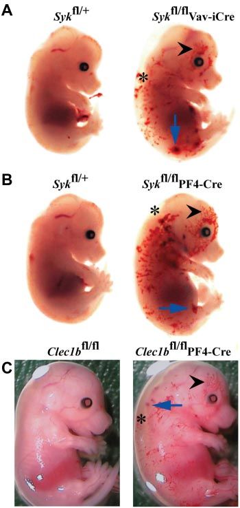
From www.bloodjournal.org by guest on October 6, 2015. For personal use only.
1750 FINNEY et al BLOOD, 16 FEBRUARY 2012 䡠 VOLUME 119, NUMBER 7
Figure 2. Lungs of P0 Clec1bⴚ/ⴚ and Sykⴚ/ⴚ mice
have a similar pathology. (A) Representative low magni-
fication (10⫻) photomicrographs of H&E-stained paraffin
sections from P0 lung showed detail of the lung’s contribu-
tion to the lethal phenotype of the Clec1b⫺/⫺ and Syk⫺/⫺
mice (n ⱖ 3 for each genotype). Lungs from P0 wild-type
(⫹/⫹) mice showed large open bronchi and expanded
terminal air sacs with thin septae between airspaces. In
contrast, lungs from Clec1b⫺/⫺ and Syk⫺/⫺ mice showed
fluid accumulation in the bronchi (black arrows) as well
as incompletely expanded terminal airways. Scale
bar ⫽ 100 m. (B) Lymphatic vessels (LVs) localized
next to blood vessels (BVs) were identified with an Ab to
podoplanin (brown staining). These vessels were clear in
Clec1b⫹/⫹ and Syk⫹/⫹ (⫹/⫹) mice but contained red
blood cells in Clec1b⫺/⫺ and Syk⫺/⫺ mice, indicating a
connection between the lymphatic and circulatory sys-
tems. Scale bar ⫽ 20 m.
Clec1b⫺/⫺ embryos showed numerous, blood-filled vessels in found alive after birth always presented with dyspnea. Histologic
the skin, confirmed to be lymphatic in origin by expression of analysis found a similar morphology in the lungs from Clec1b⫺/⫺
LYVE-1, from E14.5 to E18.5 with no evidence of this before and Syk⫺/⫺ mice (Figure 2A). The amount of airspace in both sets
E14.5. Clec1b⫺/⫺ embryos presented with edematous swelling of lungs was markedly reduced. Furthermore, in both Clec1b⫺/⫺
throughout their back and limbs from E14.5 onward. At birth, and Syk⫺/⫺ mice, the terminal air sacs did not appear to be properly
Clec1b⫺/⫺ offspring are readily distinguishable from their wild- inflated, and the larger airways contained fluid droplets. Immuno-
type littermates because of their edematous swelling and signs of histochemistry of LECs in sections of P0 lung with the use of an Ab
severe respiratory distress with ⬎ 90% of the animals surviving to podoplanin showed that LECs detected in Clec1b⫺/⫺ and Syk⫺/⫺
⬍ 24 hours. In addition, occasional discreet hemorrhages were lungs formed a vessel that contained red blood cells (Figure 2B). As
seen in the skin (supplemental Figure 1). All Clec1b⫺/⫺ offspring with the mesentery and intestine, there is a significant decrease in
had blood-filled vessels in their skin (Figure 1A). Most Clec1b⫺/⫺ the ratio of LEC/BEC of ⬎ 50% established in Clec1b⫺/⫺ lungs
offspring did not survive long enough to feed, but those that did during development (supplemental Figure 2).
developed chylous ascites in the abdomen (Figure 1B), a further
indication of defective lymphatic function. Loss of CLEC-2 and Syk results in persistent CNS hemorrhage
To determine the nature of the lymphatic defect causing the
developmental and perinatal phenotype, flow cytometric analysis An additional phenotype of hemorrhages within the developing
was performed on isolated mesenteric vessels and intestine of P0 brain and spinal cord at E12.5 was noted in most Clec1b⫺/⫺ (18 of
Clec1b⫺/⫺ mice to identify LECs (CD31⫹podoplanin⫹) and BECs 19; Figure 3A) and Syk⫺/⫺ embryos (13 of 14; Figure 3B) persisting
(CD31⫹podoplanin⫺; Figure 1C). The LEC/BEC ratio was consis- through E18.5 (not shown). In Clec1b⫺/⫺ mice hemorrhages were
tently and significantly reduced in the Clec1b⫺/⫺ offspring localized to at least 1 ventricle and the brain parenchyma, whereas
(0.35 ⫾ 0.07) in comparison to Clec1b⫹/⫹ littermates (0.70 ⫾ 0.07) in Syk⫺/⫺ mice they were restricted to the parenchyma (Figure
in the mesenteric vessels surrounding the intestine (Figure 1D). A 3C-D). These hemorrhages were never seen in wild-type embryos
significant decrease was also seen in the intestine proper of (n ⱖ 15 for each colony; Figure 3A-B). Thus, both CLEC-2 and
Clec1b⫺/⫺ mice (0.06 ⫾ 0.02) in comparison to Clec1b⫹/⫹ litter- Syk are required for the integrity of the brain vasculature.
mates (1.60 ⫾ 0.35). The decreased ratio at P0 may be the result of
earlier changes during gestation, because the LEC/BEC ratio is CLEC-2 is required in the hematopoietic lineage for normal
significantly reduced in the mesentery and intestine of Clec1b⫺/⫺ lymphatic integrity
embryos at both E16.5 and E18.5 (supplemental Figure 2).
Syk plays a critical role in signaling by CLEC-2.6,13 Signifi- To investigate whether CLEC-2 was required in adult mice for
cantly, many aspects of the Clec1b⫺/⫺ phenotype, including normal lymphatic vessel integrity, we reconstituted the hematopoi-
blood-filled lymphatic vessels in the skin beginning at E14.5, etic system of irradiated mice with wild-type or Clec1b⫺/⫺ fetal
perinatal lethality, and chylous ascites after feeding, have been liver cells as previously published.16 Seven weeks after reconstitu-
previously reported for Syk-deficient mice.2-4 Comparison of the tion, radiation chimeras reconstituted with wild-type cells showed
blood-filled vessels in Clec1b⫺/⫺ and Syk⫺/⫺ embryos showed no abnormalities in the abdominal cavity, whereas chimeras
similar appearance at all times (not shown). reconstituted with CLEC-2–deficient cells showed blood in the
mesenteric lymphatic vessels and in Peyer patches (supplemental
Lung histopathology of P0 Clec1bⴚ/ⴚ and Sykⴚ/ⴚ Figure 3), suggesting that, as with Syk and SLP76,4 CLEC-2 was
required in hematopoietic cells for integrity of lymphatic vessels.
Possibly contributing to the early postnatal lethality of both the Interestingly, 12 weeks after reconstitution with Clec1b⫺/⫺ fetal
Clec1b⫺/⫺ and Syk⫺/⫺ pups is the effect of disruption of CLEC-2/ liver cells, 2 of 6 chimeras died, and the remaining 4 were killed
Syk signaling on the physiology of the lungs. Clec1b⫺/⫺ pups because of deteriorating health. Bloody fluid was found in the chestFrom www.bloodjournal.org by guest on October 6, 2015. For personal use only.
BLOOD, 16 FEBRUARY 2012 䡠 VOLUME 119, NUMBER 7 CLEC-2/Syk ARE ESSENTIAL FOR LYMPHATICS 1751
Figure 3. Abnormal hemorrhage within the develop-
ing central nervous system of Clec1bⴚ/ⴚ and Sykⴚ/ⴚ
mice from E12.5. (A) Both constitutive (Clec1b⫺/⫺; middle;
n ⫽ 18 of 19) and platelet/megakaryocyte-specific
(Clec1bfl/flPF4-Cre; right; n ⫽ 3 of 5) ablation of Clec1b
results in hemorrhaging within the brain and developing
spinal column (blue arrows). (B) Constitutive (Syk⫺/⫺; middle;
n ⫽ 13 of 14) and platelet/megakaryocyte-specific (Sykfl/flPF4-
Cre; n ⫽ 10 of 13) ablation of Syk presented with a similar
hemorrhaging phenotype in similar regions as the Clec1b⫺/⫺
embryos (blue arrows). None of the littermate wild-type
controls (⫹/⫹; left; n ⱖ 5) showed this phenotype. Scale
bars ⫽ 1 mm. (C) H&E-stained sections from E12.5 brains
show that hemorrhages in Clec1b⫺/⫺ embryos were localized
to at least 1 ventricle (middle; red arrowhead) and within the
parenchyma (right; yellow arrowhead). (D) Sections from
E12.5 Syk⫺/⫺ embryos showed no hemorrhaging in the
ventricles, but consistent hemorrhaging in the parenchyma
(middle; yellow arrowhead). None of the wild-type controls
showed hemorrhaging in any section (left). Scale
Bars ⫽ 100 m.
cavity of these mice (4 of 6) on autopsy. In contrast, all chimeras tion (not shown) and no lethality (Table 2). To examine whether the
reconstituted with wild-type fetal liver cells were healthy 12 weeks reduced severity of the phenotype in Sykfl/flVav-iCre mice com-
after reconstitution (n ⫽ 8). Because radiation is known to cause pared with Syk⫺/⫺ mice was because of a role for Syk in endothelial
gastrointestinal syndrome,35 it is not readily possible to compare cells secondary to that in hematopoietic cells, we deleted Syk in
the severity of this phenotype with the phenotypes seen in adult both lineages by crossing Sykfl/flVav-iCre mice with Tie1-Cre
mice produced by genetic approaches. transgenic mice. In the resultant Sykfl/flVav-iCre Tie1-Cre mice the
phenotype of blood-filled lymphatics at E14.5 was no more severe
Megakaryocyte/platelet-specific ablation of CLEC-2 and Syk
than that seen in Sykfl/flVav-iCre mice, and, mice survived to
To identify the cells in which loss of Syk resulted in the lymphatic adulthood (Table 2). Taken together, these results show that normal
phenotype, mice with a conditional loxP-flanked allele of Syk lymphatic development requires Syk expression in hematopoietic
(Sykfl/fl) were crossed with mice expressing Cre recombinase under cells but not in BECs.
the control of tissue-specific promoters. Deletion of Syk in the To determine which hematopoietic lineage played a critical role
whole hematopoietic system with the use of Vav-iCre resulted in in normal lymphatic–blood vessel separation, we crossed Sykfl/fl
the characteristic appearance of edema and blood-filled vessels in mice to hCD2-iCre, LysMCre, CD11c-Cre, and PF4-Cre transgen-
the skin of E14.5 embryos (Figure 4A). This phenotype appeared ics, resulting in loss of Syk in B and T lymphocytes, in macro-
similar to that seen in Syk⫺/⫺ embryos. However, unlike Syk⫺/⫺ phages and neutrophils, in dendritic cells, or in megakaryocytes
mice, there was little perinatal lethality, with most Sykfl/flVav-iCre and platelets, respectively. No edema, blood-filled lymphatics, or
mice surviving to adulthood (Table 2). Thus, Syk is required within perinatal lethality (Table 2) was seen in any of these crosses
the hematopoietic system for normal lymphatic development. In except in Sykfl/flPF4-Cre mice that had lost Syk expression in
contrast, deletion of Syk in endothelial cells with the use of megakaryocytes and platelets (Figure 4B). Notably, Sykfl/flPF4-
Tie1-Cre caused no detectable lymphatic phenotype during gesta- Cre mice showed a phenotype at E14.5 indistinguishable fromFrom www.bloodjournal.org by guest on October 6, 2015. For personal use only.
1752 FINNEY et al BLOOD, 16 FEBRUARY 2012 䡠 VOLUME 119, NUMBER 7
ics and, in some cases, chylous fluid around the intestine (supple-
mental Figure 4; data not shown). Injection of FITC-dextran into
the carotid artery of 8-week-old Clec1bfl/fl control mice led to
sequential labeling of the mesenteric arteries and veins but not the
lymphatic vessels, which run in parallel (Figure 5A; supplemental
Video 1). In contrast, rapid labeling of the arteriolar, venous, and
lymphatic systems could be readily seen in the Clec1bfl/flPF4-Cre
mice, suggesting that there are direct interconnections between the
blood and lymphatic vessels (Figure 5A). Labeling of the mesen-
teric lymphatics occurred ⬃ 15 seconds after fluorescence was
detected in the artery and several seconds after labeling of the veins
(supplemental Video 2). The sudden appearance of the fluorescent
dextran in the lymphatic vessels argues against a slow, remote
leakage site. Similarly to Clec1bfl/fl mice, Sykfl/fl and Sykfl/flLysMCre
mice show circulation of injected FITC-dextran only through the
systemic blood vasculature (Figure 5B). In contrast, injection of
FITC-dextran into the left ventricle of Sykfl/flVav-iCre and Sykfl/flPF4-
Cre mice resulted in rapid labeling of the mesenteric lymphatic
vessels (Figure 5B). Together, these results show that the loss of
either CLEC-2 or Syk in the megakaryocyte/platelet lineage results
in persistent inappropriate connections between the blood and
lymphatic vasculatures of adult mice.
In contrast to the lymphatic defect, Clec1bfl/flPF4-Cre P0 pups
and adults (6-8 weeks old) or Sykfl/flPF4-Cre P0 mice showed no
visible abnormalities in the chest cavity. Lungs were fully ex-
panded with no sign of hemorrhage or fluid collection. Histologic
analyses showed patent conducting airways with normal alveoli
(not shown). Because this phenotype was different from that
observed in the constitutive deletion models, the efficiency of
CLEC-2 and Syk ablation in adult platelets was analyzed by FACS
and immunoblotting. CLEC-2 and Syk proteins were undetectable
in platelets from adult Clec1bfl/flPF4-Cre and Sykfl/flPF4-Cre mice,
respectively (supplemental Figure 5).
Figure 4. Hematopoetic and platelet-specific deficiency of Syk and Clec1b
result in defective lymphatic development. The phenotypes of (A) Sykfl/flVav-iCre,
Platelet effects on lymphatic endothelial cell behavior are
(B) Sykfl/flPF4-Cre, and (C) Clec1bfl/flPF4-Cre embryos at E14.5 are indistinguishable
and include edematous swelling (asterisk), hemorrhages (blue arrow) and blood- modulated by CLEC-2 and Syk
filled lymphatics in the skin (black arrowhead). This phenotype is not seen in control
littermates. The figures shown are representative of ⬎ 5 embryos of each genotype. HLECs have been previously shown to induce aggregation of
human platelets.17 Similarly, HLECs also cause aggregation of
wild-type mouse platelets. This response was abrogated in Clec1b⫺/⫺
that seen in Sykfl/flVav-iCre mice strongly suggesting that Syk
mouse platelets, indicating that aggregation requires binding of
was required in megakaryocytes and/or platelets for normal
podoplanin on the HLECs to CLEC-2 on platelets (supplemental
lymphatic development.
Figure 6A). In contrast, the effect of platelets on LEC behavior is
Extending this analysis to the lineage-specific requirement for
CLEC-2, we generated a conditional loxP-flanked allele of Clec1b
and crossed it to the PF4-Cre transgenic mouse. The resultant Table 2. Offspring resulting from Cre-transgenic matings
Clec1bfl/flPF4-Cre E14.5 embryos showed edema and blood-filled Number of Sykfl/flCreⴙ or
Clec1bfl/flCreⴙ
vessels in the skin, closely resembling the phenotype seen in
Sykfl/flPF4-Cre embryos (Figure 4C). In addition, at E12.5, Clec1bfl/ Cre Total number of mice Expected Found
flPF4-Cre (3 of 5; Figure 3A) and Sykfl/flPF4-Cre (10 of 13; Figure Syk
3B) embryos had hemorrhages within the developing brain similar LysM 139 34.8 51
to the constitutive loss of CLEC-2 and Syk, although generally they hCD2 93 18.9 18
were fewer and smaller. Together these studies suggest that Vav 207 54.5 35*
Tie1 155 37.4 28
CLEC-2 and Syk are required in megakaryocytes and/or platelets
CD11c 62 37.8 36
during development by both the lymphatic and blood brain
PF4 108 32.5 17†
vasculatures. Tie1⫹Vav 50 12.5 4
Clec1b
Conditional deletion of Clec1b and Syk in megakaryocytes and PF4 81 29.8 26
platelets results in interconnected veins and lymphatics but
does not affect perinatal lung function Lineage-specific deletion of Syk and Clec1b with the use of the Cre-recombinase
lines indicated. The number of mice with homozygous deletion of Syk or Clec1b
expected versus the actual number genotyped is shown.
Next, we examined the phenotype of newborn and adult Clec1bfl/
*Significant reduction in the number of mice as indicated by the 2 test (P ⬍ .05).
flPF4-Cre and Sykfl/flPF4-Cre mice. Both strains showed signs of †Significant reduction in the number of mice as indicated by the 2 test
defective lymphatics with blood visible in the mesenteric lymphat- (P ⬍ .005).From www.bloodjournal.org by guest on October 6, 2015. For personal use only.
BLOOD, 16 FEBRUARY 2012 䡠 VOLUME 119, NUMBER 7 CLEC-2/Syk ARE ESSENTIAL FOR LYMPHATICS 1753
Figure 5. Disruption of Clec1b or Syk in the megakaryocyte/platelet
lineage results in interconnection of the blood and lymphatic vascu-
latures. (A) FITC-dextran was injected into an anesthetized mouse via a
carotid cannula after exteriorization of the abdominal mesentery. In both
Clec1bfl/fl (top) and Clec1bfl/flPF4-Cre (bottom) FITC-dextran was initially
visualized in the artery (1) followed shortly by the vein (2). In the
Clec1bfl/flPF4-Cre mice FITC-dextran was visualized in the mesenteric
lymphatic vasculature (3) shortly after the flowing through the vein. This
phenomenon was never seen in the Clec1bfl/fl mice. Scale bar ⫽ 0.5 mm.
This figure is representative of 3 experiments. (B) FITC-dextran was
injected into the left ventricle of the heart of mice of the indicated
genotypes, and vessels in the gut were visualized for FITC fluorescence
60 seconds later. FITC-dextran was only detected in the systemic blood
circulation (1 and 2) of Sykfl/fl (top left) and Sykfl/flLysM-Cre mice (top right).
In contrast, leakage of the FITC-dextran into the gut mesenteric lymphatic
vasculature (3) was detected in Sykfl/flVav-iCre (bottom left) and Sykfl/flPF4-
Cre (bottom right).
unknown. LEC migration plays a critical role in lymphangiogen- of platelets from Clec1bfl/flPF4-Cre or Sykfl/flPF4-Cre mice (Figure
esis. Therefore, to identify a potential mechanism by which 6B,D,G). Application of platelets to HUVEC cultures did not affect
platelets modulate lymphatic vasculature formation, we assessed network formation (not shown).
the effect of platelets on LEC migration with the use of a transfilter These data show that direct contact of platelets affects the
assay.36 The presence of wild-type, Clec1bfl/flPF4-Cre, or Sykfl/flPF4- ability of HLECs to migrate, stabilize cell–cell interactions, and
Cre platelets did not modify the total number of cells attached to the form networks in vitro in a CLEC-2/Syk–dependent manner.
filters as measured at 24 hours (not shown), suggesting the
presence of platelets did not affect cell survival or adhesion. The
presence of platelets from Clec1bfl/fl or Sykfl/fl mice inhibited Discussion
transmigration through the filter by ⬎ 60% relative to transmigra-
tion of HLECs not treated with platelets (Figure 6A,C). Platelets This study shows that loss of CLEC-2 and Syk in megakaryocytes
from Clec1bfl/flPF4-Cre and Sykfl/flPF4-Cre mice also decreased and platelets results in similar defects in fetal lymphatic develop-
transmigration in comparison to untreated HLECs, but this effect ment, as does the loss of Syk within the whole hematopoietic
was significantly weaker than that seen in the presence of platelets lineage. In contrast, lineage-specific deletions of Syk from B and
from Clec1bfl/fl or Sykfl/fl mice (Figure 6A,C). Application of T lymphocytes, endothelial cells, macrophages and neutrophils, or
platelet releasate did not affect LEC transmigration (supplemental dendritic cells do not result in defective lymphatics. These results
Figure 6B). This suggests that platelets inhibit LEC migration in suggest that CLEC-2 signaling via Syk in megakaryocytes/platelets
vitro by contact-dependent and CLEC-2/Syk–dependent and inde- is required for normal lymphangiogenesis which correspond to
pendent mechanisms. previous studies that used both Clec1b and SLP76 deletion
The effects of Ab-mediated podoplanin cross-linking in HLECs models.15,22 The present study further shows that defects in
were tested to determine whether platelet regulation of HLEC lymphatic function in CLEC-2–deficient mice are associated with a
behavior relies on direct podoplanin cross-linking by CLEC-2. decrease in the number of LECs relative to BECs and that platelets
Treatment with anti–human podoplanin plus a secondary cross- regulate LEC behavior via a contact-dependent mechanism that
linking Ab decreased VEGF-C–induced HLEC migration, whereas involves CLEC-2 and Syk. The altered migration and network
an irrelevant rat IgG or the primary Ab did not have any significant formation capacity of LECs in the absence of platelet CLEC-2 or
effect (Figure 6E). These data suggest that podoplanin cross- Syk may give rise to blood-filled lymphatics, possibly as a
linking leads to the inhibition of HLEC migration, possibly because consequence of impaired separation of LECs from blood vessels or
of altered constitutive podoplanin signaling. stabilization of mis-connections between the two vessels.
To further characterize the modulatory role of platelets on The lethality from constitutive deletion of CLEC-2 and Syk
HLEC behavior, we assessed the effect of platelets on HLEC could be because of the functions of the two proteins in nonhemato-
network formation in vitro ⱖ 3 hours after seeding the HLECs on poietic lineages, although this seems unlikely, given that CLEC-2
Matrigel. The addition of Clec1bfl/fl or Sykfl/fl platelets significantly has not been detected outside the hematopoietic system. More
disrupted network formation by almost 70% (Figure 6B,D), probable, the milder phenotype, as evidenced by the viability of
whereas addition of Clec1bfl/fl platelet releasate had no effect mice with PF4-Cre–mediated or Vav-Cre–mediated deletion, could
(Figure 6F). This inhibitory effect was reduced ⬃ 40% with the use be because of incomplete deletion of the genes. Although we wereFrom www.bloodjournal.org by guest on October 6, 2015. For personal use only.
1754 FINNEY et al BLOOD, 16 FEBRUARY 2012 䡠 VOLUME 119, NUMBER 7
Figure 6. Platelets can modulate lymphatic endothe-
lial cell behavior. VEGF-C–driven migration of LECs in
the absence or presence of washed platelets from Clec1bfl/
fl or Clec1bfl/flPF4-Cre (A; n ⫽ 4) and Sykfl/fl or Sykfl/flPF4-
Cre (C; n ⫽ 3) mice was assessed with the transfilter
assay. The percentage of LECs that migrated through
the filter was reduced significantly when platelets from
Clec1bfl/fl or Sykfl/fl mice were applied to the LECs.
Application of platelets from Clec1bfl/flPF4-Cre or
Sykfl/flPF4-Cre mice significantly increased the amount of
migration in comparison to the migration seen in the
presence of platelets from Clec1bfl/fl or Sykfl/fl mice.
Cross-linking of podoplanin (E) with the use of the Ab
NZ-1.3 with a cross-linking secondary IgG2a resulted in a
significant decrease in VEGF-C–mediated migration,
whereas application of irrelevant IgG or the Ab without the
cross-linking secondary showed no effect. Data repre-
sent mean values from ⱖ 3 independent experiments
performed in duplicate (mean ⫾ SD; **P ⬍ .006,
*P ⬍ .02). Network formation by LECs on Matrigel in the
absence or presence of washed platelets from Clec1bfl/fl
or Clec1bfl/flPF4-Cre (B) Sykfl/fl or Sykfl/flPF4-Cre (D) and
Clec1bfl/fl platelet releasate (F) was assessed by seeding
LECs in Matrigel-coated 12-well plates. The complexity of
networks formed by LECs were reduced significantly
when platelets from Clec1bfl/fl or Sykfl/fl mice were applied,
whereas application of platelets from Clec1bfl/flPF4-Cre
or Sykfl/flPF4-Cre mice only partially reduced network
complexity, and platelet releasate showed no significant
effects on network formation. Data represent mean val-
ues from 3 independent experiments performed in dupli-
cate (mean ⫾ SD; **P ⬍ .005, *P ⬍ .05). (G) Represen-
tative pictures of networks analyzed for the network-
forming assay after application and incubation with either
buffer (left) or platelets from Clec1bfl/fl (middle) or Clec1bfl/
flPF4-Cre (right) mice. Scale bars ⫽ 100 m.
unable to detect expression of CLEC-2 or Syk in platelets from of birth and have lungs lacking inflated airspaces.40 Because it
adult Clec1bfl/flPF4-Cre or Sykfl/flPF4-Cre, respectively, it is pos- appears that alveolar formation is disrupted in the constitutive
sible that a small fraction of cells still expressed protein which fell CLEC-2 and Syk-deficient mice, podoplanin-expressing alveolar
below the level of detection. Another explanation for the milder type-1 cells could be particularly affected by the absence of these
phenotype in the conditional mutants is that Clec1b and Syk proteins.
undergo partial deletion in platelets formed by primitive rather than The disruption of lymphatic function is a potential cause of the
definitive hematopoiesis. Primitive hematopoiesis originates in the fluid in the large airways of the Clec1b⫺/⫺ and Syk⫺/⫺ lungs and
yolk sac at E6.5 with megakaryocyte/erythroid progenitors de- ultimately of their failure to inflate. During development, the
tected at E7.5 and yolk-sac–derived platelets appearing at E9.5.37-39 airway epithelium secretes fluid into the lumen of the lung which
Megakaryocytes derived through primitive hematopoiesis have a influences its branching dynamics and structure.41 This liquid must
lower ploidy level than those in fetal liver or BM, whereas their be cleared at birth, and a large portion (⬃ 40%) of the clearance is
platelets are larger. Further, under culture conditions, platelets are because of flow through lymphatics.42 The viability and apparently
generated more rapidly from primitive hematopoiesis relative to normal patency of the lungs in Clec1bfl/flPF4-Cre and Sykfl/flPF4-
definitive hematopoiesis. Thus, although PF4 has been shown to be Cre mice could be because of a reduced severity of the lymphatic
expressed during primitive hematopoiesis,37 it is possible that the phenotype, which could allow lung drainage and therefore expan-
more rapid formation of platelets enables residual levels of sion to occur. However, it is also possible that megakaryocyte/
CLEC-2 and Syk to be expressed. The relative contribution of platelet-expressed CLEC-2 and Syk are only partially involved or
primitive and definitive hematopoiesis to lymphatic development is not involved in the lung defect and that, unlike the lymphatic
an important area for further investigation. defect, the lung disorder is the result of loss of these proteins in
The present study reports that most of the constitutive CLEC-2 another cell type.
and Syk-deficient mice die shortly after birth. The lungs in these Preceding the appearance of blood-filled lymphatics CLEC-2–
mice fail to inflate normally, and fluid is present in the larger and Syk-deficient embryos showed hemorrhaging in the mid- and
airways. The few mice that survive for at least a few hours have hind-brain of Clec1b⫺/⫺ and Syk⫺/⫺ embryos at E12.5. In Clec1b⫺/⫺
marked difficulties in breathing. A similar pathology is seen in embryos, blood was detected in at least 1 ventricle, whereas in
podoplanin-deficient mice in which most mice die within minutes Syk⫺/⫺ embryos it was restricted to the parenchyma. TheseFrom www.bloodjournal.org by guest on October 6, 2015. For personal use only.
BLOOD, 16 FEBRUARY 2012 䡠 VOLUME 119, NUMBER 7 CLEC-2/Syk ARE ESSENTIAL FOR LYMPHATICS 1755
hemorrhagic foci persist throughout gestation. Tang et al have also network formation depends on the direct contact between platelets
reported hemorrhaging in the hind brain in a constitutive CLEC-2– and LECs. This could be mediated through regulation of one or
deficient mouse.23 This phenotype is present in Clec1bfl/flPF4-Cre more platelet surface receptors, although this would appear to
and Sykfl/flPF4-Cre mice, although it is less marked. This may exclude platelet integrins because the phenotype has not been
reflect a role for CLEC-2 and Syk in other lineages or residual described in mice deficient in 1- or 3-integrins50,51 or in mice
protein in a subset of platelets during early development as deficient in the global regulator of integrin function, Talin.
discussed earlier. Clec1b⫺/⫺ mice have a reduced LEC/BEC ratio, implying a
The hemorrhaging in the brain cannot be because of defective reduction in LEC number because BECs have not been reported to
lymphatic function because this system is absent from the CNS. be affected by the deletion of CLEC-2, Syk, or SLP76. This
However, the choroid plexus, which is responsible for secretion of reduction opens up several questions about the nature of the
cerebrospinal fluid (CSF),43 expresses podoplanin during develop- lymphatic defect in these mice, because platelets have not been
ment.44 The podoplanin-expressing epithelial cells of the choroid shown to alter proliferation, survival, or differentiation of LECs, at
plexus form a barrier between the blood and CSF which is distinct least in vitro.22 These results, in conjunction with the results of the
from the endothelial structure of the blood-brain barrier. Choroid migration and network assays, suggest a critical role for CLEC-2 in
plexus can be readily identified in histologic sections of embryos at the establishment of functional lymphatic vessels.
day 12.5-13 in the fourth and lateral ventricles,45 which correspond In summary, with the use of several unique lineage-specific
with the sites of bleeding into the brain and shortly after the deletion mouse models, this study shows the critical role of platelet
appearance of platelets. We, therefore, propose that platelet interac- CLEC-2 and Syk in lymphangiogenesis and in the development of
tion with podoplanin-expressing cells of the choroid plexus may be the brain vasculature and found that platelets directly influence
important for the correct formation of the blood–CSF barrier. LEC migration and formation of junctions through a CLEC-2– and
There is considerable evidence of a role for podoplanin in Syk-dependent process.
regulating cell migration. Podoplanin up-regulation in cancer cells
is associated with altered actin cytoskeleton reorganization and
increased tumor cell migration and invasiveness.46 Similarly, in Acknowledgments
lung microvascular LECs, small interfering RNA–mediated podo-
planin knockdown causes a dramatic reduction in directional The authors thank Milan Fernando, Beata Grygielska, Phil Stone,
migration47 and abrogated formation of capillary tubes on Matri- and Hannah Jeffery for excellent technical assistance.
gel.48 These effects appear to be independent of ligand engagement This work was supported by the Wellcome Trust (ref: 088410).
and may reflect constitutive signaling from podoplanin. Thus, L.N.N. holds a postdoctoral fellowship from the Spanish Ministry
changes in podoplanin-regulated migration of LECs as a conse- of Education (EX2009-0242). E.S. and V.L.J.T. are supported by
quence of interaction with CLEC-2– and Syk-dependent platelet the Medical Research Council UK (Program no. U117527252).
activation could lead to altered lymphangiogenesis. S.P.W. holds a British Heart Foundation Chair (CH/03/003).
We show that platelets decrease VEGF-C–stimulated HLEC
migration through a pathway that partially depends on CLEC-2 and
Syk, raising the possibility that binding of CLEC-2 to podoplanin Authorship
may inhibit migration. Moreover, the observed effects of platelets
on formation of LEC networks on Matrigel suggest that activation Contribution: B.A.F., E.S., L.N.-N., C.B., F.B., C.E.H., S.A.L., and
of Syk after the binding of CLEC-2 to podoplanin destabilizes K.L.L. performed experiments; A.Y.P., D.M.-S., S.S., G.B.N., N.S.,
LEC–LEC interactions. These results are consistent with the and C.R.e.S. provided reagents and mouse models; B.A.F., V.L.J.T.,
observation that podoplanin-Fc, which interferes with endogenous and S.P.W. wrote the manuscript with critical editing provided by
ligand binding to podoplanin, also inhibits transmigration and all of the authors; and V.L.J.T. and S.P.W. designed experiments
network-forming ability of LECs.49 The partial effect of CLEC-2 and oversaw the research program.
and Syk deletion in these 2 assays suggests that additional platelet Conflict-of-interest disclosure: The authors declare no compet-
receptors or alternative mechanisms may also influence LEC ing financial interests.
function. An inhibitory action of CLEC-2 on podoplanin signaling The current affiliation for S.S. is Takeda Cambridge, Cam-
however does not explain the similar in vivo phenotypes of bridge, United Kingdom.
CLEC-2 and Syk deficiency. It is therefore possible that activation Correspondence: Victor L. J. Tybulewicz, MRC National Insti-
of Syk by CLEC-2 is required in vivo to maintain binding of tute for Medical Research, London NW7 1AA, United Kingdom;
platelets to LECs and possibly the degree of clustering of podopla- e-mail: vtybule@nimr.mrc.ac.uk; and Steve P. Watson, Centre for
nin thereby influencing their modulatory effect. This hypothesis is Cardiovascular Sciences, Institute for Biomedical Research, College of
supported by our data showing that cross-linking of podoplanin Medical and Dental Sciences, University of Birmingham, Birmingham
interferes with the migration of LECs and that disruption of B15 2TT, United Kingdom; e-mail: s.p.watson@bham.ac.uk.
References
1. Tammela T, Alitalo K. Lymphangiogenesis: mo- viability and B-cell development. Nature. 1995; 6. Suzuki-Inoue K, Fuller GLJ, Garcia A, et al. A
lecular mechanisms and future promise. Cell. 378(6554):303-306. novel Syk-dependent mechanism of platelet acti-
2010;140(4):460-476. 4. Abtahian F, Guerriero A, Sebzda E, et al. Regula- vation by the C-type lectin receptor CLEC-2.
tion of blood and lymphatic vascular separation Blood. 2006;107(2):542-549.
2. Turner M, Joseph Mee P, Costello PS, et al. Peri-
by signaling proteins SLP-76 and Syk. Science. 7. Colonna M, Samaridis J, Angman L. Molecular
natal lethality and blocked B-cell development in
2003;299(5604):247-251. characterization of two novel C-type lectin-like
mice lacking the tyrosine kinase Syk. Nature.
5. Ichise H, Ichise T, Ohtani O, Yoshida N. Phospho- receptors, one of which is selectively expressed
1995;378(6554):298-302.
lipase C gamma 2 is necessary for separation of in human dendritic cells. Eur J Immunol. 2000;
3. Cheng AM, Rowley B, Pao W, Hayday A, Bolen JB, blood and lymphatic vasculature in mice. Devel- 30(2):697-704.
Pawson T. Syk tyrosine kinase required for mouse opment. 2009;136(2):191-195. 8. Kerrigan AM, Dennehy KM, Mourao-Sa D, et al.From www.bloodjournal.org by guest on October 6, 2015. For personal use only.
1756 FINNEY et al BLOOD, 16 FEBRUARY 2012 䡠 VOLUME 119, NUMBER 7
CLEC-2 is a phagocytic activation receptor ex- brary for secreted and transmembrane proteins. 38. Xie X, Chan RJ, Johnson SA, et al. Thrombopoi-
pressed on murine peripheral blood neutrophils. Nat Biotechnol. 2010;28(7):749-755. etin promotes mixed lineage and megakaryocytic
J Immunol. 2009;182(7):4150-4157. 24. Chagraoui H, Kassouf M, Banerjee S, et al. SCL- colony-forming cell growth but inhibits primitive
9. Mourao-Sa D, Robinson MJ, Zelenay S, et al. mediated regulation of the cell-cycle regulator and definitive erythropoiesis in cells isolated from
CLEC-2 signaling via Syk in myeloid cells can p21 is critical for murine megakaryopoiesis. early murine yolk sacs. Blood. 2003;101(4):1329-
regulate inflammatory responses. Eur J Immunol. Blood. 2011;118(3):723-735. 1335.
2011;41(10):3040-3053. 25. Mocsai A, Ruland J, Tybulewicz VLJ. The SYK 39. Tober J, Koniski A, McGrath KE, et al. The mega-
10. Watson SP, Herbert JMJ, Pollitt AY. GPVI and tyrosine kinase: a crucial player in diverse biologi- karyocyte lineage originates from hemangioblast
CLEC-2 in hemostasis and vascular integrity. cal functions. Nat Rev Immunol. 2010;10(6):387- precursors and is an integral component both of
J Thromb Haemost. 2010;8(7):1456-1467. 402. primitive and of definitive hematopoiesis. Blood.
2007;109(4):1433-1441.
11. Suzuki-Inoue K, Inoue O, Ozaki Y. Novel platelet 26. Sebzda E, Hibbard C, Sweeney S, et al. Syk and
activation receptor CLEC-2: from discovery to Slp-76 mutant mice reveal a cell-autonomous he- 40. Ramirez MI, Millien G, Hinds A, Cao YX, Seldin DC,
prospects. J Thromb Haemost. 2011;9(Suppl 1): matopoietic cell contribution to vascular develop- Williams MC. T1 alpha, a lung type I cell differentia-
44-55. ment. Dev Cell. 2006;11(3):349-361. tion gene, is required for normal lung cell proliferation
and alveolus formation at birth. Dev Biol. 2003;
12. Spalton JC, Mori J, Pollitt AY, Hughes CE, Eble JA, 27. Bohmer R, Neuhaus B, Buhren S, et al. Regula-
256(1):61-72.
Watson SP. The novel Syk inhibitor R406 reveals tion of developmental lymphangiogenesis by
mechanistic differences in the initiation of GPVI Syk(⫹) leukocytes. Dev Cell. 2010;18(3):437- 41. Warburton D, El-Hashash A, Carraro G, et al.
and CLEC-2 signaling in platelets. J Thromb Hae- 449. Lung organogenesis. Curr Top Dev Biol. 2010;90:
most. 2009;7(7):1192-1199. 73-158.
28. Tiedt R, Schomber T, Hao-Shen H, Skoda RC.
13. Hughes CE, Pollitt AY, Mori J, et al. CLEC-2 acti- Pf4-Cre transgenic mice allow the generation of 42. Humphreys PW, Normand ICS, Reynolds EOR,
vates Syk through dimerization. Blood. 2010; lineage-restricted gene knockouts for studying Strang LB. Pulmonary lymph flow and the uptake
115(14):2947-2955. megakaryocyte and platelet function in vivo. of liquid from the lungs of the lamb at the start of
14. May F, Hagedorn I, Pleines I, et al. CLEC-2 is an Blood. 2007;109:1503-1506. breathing. J Physiol-London. 1967;193(1):1-29.
essential platelet-activating receptor in hemosta- 29. de Boer J, Williams A, Skavdis G, et al. Trans- 43. Pardridge WM. Drug transport in brain via the ce-
sis and thrombosis. Blood. 2009;114(16):3464- genic mice with hematopoietic and lymphoid spe- rebrospinal fluid. Fluids Barriers CNS. 2011;
3472. cific expression of Cre. Eur J Immunol. 2003; 8(1):7.
15. Suzuki-Inoue K, Inoue O, Ding G, et al. Essential 33(2):314-325. 44. Williams MC, Cao Y, Hinds A, Rishi AK, Wetterwald A.
in vivo roles of the C-type lectin receptor CLEC-2. 30. Clausen BE, Burkhardt C, Reith W, Renkawitz R, T1 alpha protein is developmentally regulated and
J Biol Chem. 2010;285(32):24494-24507. Förster I. Conditional gene targeting in macro- expressed by alveolar type I cells, choroid plexus,
16. Hughes CE, Navarro-Núñez L, Finney BA, phages and granulocytes using LysMcre mice. and ciliary epithelia of adult rats. Am J Respir Cell
Mourão-Sá D, Pollitt AY, Watson SP. CLEC-2 is Transgenic Res. 1999;8(4):265-277. Mol Biol. 1996;14(6):577-585.
not required for platelet aggregation at arteriolar 31. Gustafsson E, Brakebusch C, Hietanen K, 45. Kaufmann MH. The Atlas of Mouse Development.
shear. J Thromb Haemost. 2010;8(10):2328- Fassler R. Tie-1-directed expression of Cre re- London, United Kingdom: Elsevier; 1992.
2332. combinase in endothelial cells of embryoid bodies 46. Martin-Villar E, Megias D, Castel S, Yurrita MM,
17. Suzuki-Inoue K, Kato Y, Inoue O, et al. Involve- and transgenic mice. J Cell Sci. 2001;114(Pt 4): Vilaro S, Quintanilla M. Podoplanin binds ERM
ment of the snake toxin receptor CLEC-2, in 671-676. proteins to activate RhoA and promote epithelial-
podoplanin-mediated platelet activation, by can- 32. Caton ML, Smith-Raska MR, Reizis B. Notch- mesenchymal transition. J Cell Sci. 2006;119(Pt
cer cells. J Biol Chem. 2007;282(36):25993- RBP-J signaling controls the homeostasis of 21):4541-4553.
26001. CD8- dendritic cells in the spleen. J Exp Med. 47. Navarro A, Perez RE, Rezaiekhaligh MH, Mabry SM,
18. Christou CM, Pearce AC, Watson AA, et al. Renal 2007;204(7):1653-1664. Ekekezie II. Polarized migration of lymphatic en-
cells activate the platelet receptor CLEC-2 33. Saijo K, Schmedt C, Su IH, et al. Essential role of dothelial cells is critically dependent on podopla-
through podoplanin. Biochem J. 2008;411(1):133- Src-family protein tyrosine kinases in NF-kappaB nin regulation of Cdc42. Am J Physiol Lung Cell
140. activation during B cell development. Nat Immu- Mol Physiol. 2011;300(1):L32-L42.
19. Schacht V, Ramirez MI, Hong YK, et al. T1 alpha/ nol. 2003;4(3):274-279. 48. Navarro A, Perez RE, Rezaiekhaligh M, Mabry SM,
podoplanin deficiency disrupts normal lymphatic 34. Abramoff MD, Magelhaes PJ, Ram SJ. Image Ekekezie II. T1 alpha/podoplanin is essential for cap-
vasculature formation and causes lymphedema. processing with ImageJ. Biophot Int. 2004;11(7): illary morphogenesis in lymphatic endothelial cells.
EMBO J. 2003;22(14):3546-3556. 36-42. Am J Physiol Lung Cell Mol Physiol. 2008;295(4):
20. Uhrin P, Zaujec J, Breuss JM, et al. Novel func- 35. Aparicio-Vergara M, Shiri-Sverdlov R, de Haan G, L543-L551.
tion for blood platelets and podoplanin in devel- Hofker MH. Bone marrow transplantation in mice 49. Cueni LN, Chen L, Zhang H, et al. Podoplanin-Fc
opmental separation of blood and lymphatic cir- as a tool for studying the role of hematopoietic reduces lymphatic vessel formation in vitro and in
culation. Blood. 2010;115(19):3997-4005. cells in metabolic and cardiovascular diseases. vivo and causes disseminated intravascular co-
21. Carramolino L, Fuentes J, Garcia-Andres C, Atherosclerosis. 2010;213(2):335-344. agulation when transgenically expressed in the
Azcoitia V, Riethmacher D, Torres M. Platelets 36. Makinen T, Veikkola T, Mustjoki S, et al. Isolated skin. Blood. 2010;116(20):4376-4384.
play an essential role in separating the blood and lymphatic endothelial cells transduce growth, sur- 50. Hodivala-Dilke KM, McHugh KP, Tsakiris DA, et al.
lymphatic vasculatures during embryonic angio- vival and migratory signals via the VEGF-C/D re- Beta3-integrin-deficient mice are a model for Glanz-
genesis. Circ Res. 2010;106(7):1197-1201. ceptor VEGFR-3. EMBO J. 2001;20(17):4762- mann thrombasthenia showing placental defects and
22. Bertozzi CC, Schmaier AA, Mericko P, et al. 4773. reduced survival. J Clin Invest. 1999;103(2):229-238.
Platelets regulate lymphatic vascular develop- 37. Xu M-j Matsuoka S, Yang F-C, et al. Evidence for 51. Nieswandt B, Brakebusch C, Bergmeier W, et al.
ment through CLEC-2-SLP-76 signaling. Blood. the presence of murine primitive megakarycyto- Glycoprotein VI but not alpha 2 beta 1 integrin is
2010;116(4):661-670. poiesis in the early yolk sac. Blood. 2001;97(7): essential for platelet interaction with collagen.
23. Tang T, Li L, Tang J, et al. A mouse knockout li- 2016-2022. EMBO J. 2001;20(9):2120-2130.From www.bloodjournal.org by guest on October 6, 2015. For personal use only.
2012 119: 1747-1756
doi:10.1182/blood-2011-09-380709 originally published
online December 20, 2011
CLEC-2 and Syk in the megakaryocytic/platelet lineage are essential for
development
Brenda A. Finney, Edina Schweighoffer, Leyre Navarro-Núñez, Cecile Bénézech, Francesca Barone,
Craig E. Hughes, Stacey A. Langan, Kate L. Lowe, Alice Y. Pollitt, Diego Mourao-Sa, Steve
Sheardown, Gerard B. Nash, Nicholas Smithers, Caetano Reis e Sousa, Victor L. J. Tybulewicz and
Steve P. Watson
Updated information and services can be found at:
http://www.bloodjournal.org/content/119/7/1747.full.html
Articles on similar topics can be found in the following Blood collections
Platelets and Thrombopoiesis (597 articles)
Information about reproducing this article in parts or in its entirety may be found online at:
http://www.bloodjournal.org/site/misc/rights.xhtml#repub_requests
Information about ordering reprints may be found online at:
http://www.bloodjournal.org/site/misc/rights.xhtml#reprints
Information about subscriptions and ASH membership may be found online at:
http://www.bloodjournal.org/site/subscriptions/index.xhtml
Blood (print ISSN 0006-4971, online ISSN 1528-0020), is published weekly by the American Society
of Hematology, 2021 L St, NW, Suite 900, Washington DC 20036.
Copyright 2011 by The American Society of Hematology; all rights reserved.You can also read