Carbon Nanotube Reinforced Supramolecular Hydrogels for Bioapplications
←
→
Page content transcription
If your browser does not render page correctly, please read the page content below
Full Paper
Nanocomposite Hydrogels www.mbs-journal.de
Carbon Nanotube Reinforced Supramolecular Hydrogels
for Bioapplications
Marko Mihajlovic, Milos Mihajlovic, Patricia Y. W. Dankers, Rosalinde Masereeuw,
and Rint P. Sijbesma*
1. Introduction
Nanocomposite hydrogels based on carbon nanotubes (CNTs) are known to
possess remarkable stiffness, electrical, and thermal conductivity. However, Hydrogels are materials composed of
they often make use of CNTs as fillers in covalently cross-linked hydrogel hydrophilic polymers crosslinked into a 3D
networks or involve direct cross-linking between CNTs and polymer chains, network.[1] They represent a very attractive
class of materials due to their soft nature
limiting processability properties. Herein, nanocomposite hydrogels are
and ability to absorb water. Hydrogels
developed, in which CNTs are fillers in a physically cross-linked hydrogel. mimic biological environments and, as
Supramolecular nanocomposites are prepared at various CNT concentra- such, they are promising materials for
tions, ranging from 0.5 to 6 wt%. Incorporation of 3 wt% of CNTs leads to an constructing scaffolds for biomedical
increase of the material’s toughness by over 80%, and it enhances electrical applications.[2–5] To this end, it is required
conductivity by 358%, compared to CNT-free hydrogel. Meanwhile, the to develop hydrogels that are both biocom-
patible and mechanically tough.
nanocomposite hydrogels maintain thixotropy and processability, typical of
There is a large demand for hydrogels
the parent hydrogel. The study also demonstrates that these materials display which are easy to prepare, and which have
remarkable cytocompatibility and support cell growth and proliferation, while multiple functionalities, and tunable prop-
preserving their functional activities. These supramolecular nanocomposite erties. In the past decades, a new class
hydrogels are therefore promising candidates for biomedical applications, in of hydrogels, known as nanocomposite
hydrogels,[6] has been designed to improve
which both toughness and electrical conductivity are important parameters.
mechanical performance. These gels,
next to the polymeric network, contain
inorganic particles, such as clay, graphene, carbon nanotubes
(CNTs), or silica.[7] Besides improving mechanical properties,
M. Mihajlovic, Prof. R. P. Sijbesma new optical, electrical, and thermal properties are imparted to the
Laboratory of Macromolecular and Organic Chemistry nanocomposite material due to the inorganic component.[8–10]
Department of Chemical Engineering and Chemistry CNTs are widely used to make nanocomposite hydrogels
Eindhoven University of Technology because of their unique properties, such as electrical and
P.O. Box 513, 5600 MB Eindhoven, The Netherlands
E-mail: r.p.sijbesma@tue.nl
thermal conductivity, high mechanical strength, high spe-
M. Mihajlovic, Prof. P. Y. W. Dankers, Prof. R. P. Sijbesma
cific area, and low mass density.[11] CNTs are often used as
Institute for Complex Molecular Systems reinforcing agents to enhance the mechanical properties of
Eindhoven University of Technology hydrogels. By incorporating CNTs in hydrogel formulations, it
P.O. Box 513, 5600 MB Eindhoven, The Netherlands is possible to obtain very tough[12,13] and electrically conductive
M. Mihajlovic, Prof. R. Masereeuw hydrogels.[14–17] CNT-based nanocomposites represent a versa-
Division of Pharmacology tile platform for developing hydrogels with multiple responsive
Utrecht Institute for Pharmaceutical Sciences
Utrecht University properties and remarkable mechanical performance. However,
3584 CG Utrecht, The Netherlands there is a concern about the toxic effects of CNTs and, therefore,
Prof. P. Y. W. Dankers hydrogel biocompatibility. Studies reported CNT toxicity that
Department of Biomedical Engineering seemed to be dose-dependent, but which could be reduced when
Laboratory of Chemical Biology CNTs are functionalized and incorporated in networks.[15,18]
Eindhoven University of Technology
P.O. Box 513, 5600 MB Eindhoven, The Netherlands
Most of the reported CNT-based nanocomposite hydrogels
contain covalent cross-links between CNTs and polymer chains,
The ORCID identification number(s) for the author(s) of this article
can be found under https://doi.org/10.1002/mabi.201800173. or between polymer chains, with CNTs being only physically
embedded in the network.[13,14,19–22] In addition, some of the prepa-
© 2018 The Authors. Published by WILEY-VCH Verlag GmbH & Co. KGaA,
Weinheim. This is an open access article under the terms of the Creative ration procedures are quite complicated and laborious due to the
Commons Attribution-Non Commercial License, which permits use, challenges in dispersing CNTs efficiently in the matrix. In addition
distribution and reproduction in any medium, provided that the original to improved mechanical behaviour, various responsive properties,
work is properly cited and is not used for commercial purposes. and biocompatibility, it would be an asset to have completely physi-
DOI: 10.1002/mabi.201800173 cally crosslinked networks, which in contrast to covalent systems,
Macromol. Biosci. 2019, 19, 1800173 1800173 (1 of 12) © 2018 The Authors, Published by WILEY-VCH Verlag GmbH & Co. KGaA, Weinheim
www.advancedsciencenews.com                                                                                                   www.mbs-journal.de

would allow for easy processability, manipulation, and reshaping          dispersed in the mixture of sulfuric and nitric acid (3:1, v/v)
features of the hydrogels. To the best of our knowledge, there are        and sonicated for 24 h at room temperature (RT). Deionized
limited studies on physical nanocomposite gels containing CNTs.           water was then slowly added (at 0 °C) to dilute the mixture
Some of the described systems are based on PVA[23] or alginate.[24]       and ox-MWNT were filtered (Millipore, JHWP 0.45 μm filter),
However, these gels are either not tested for biocompatibility or         resuspended in water and washed until the pH of the filtrate
lack significant improvement in physical or mechanical properties         was neutral. The black powder was dried under vacuum over-
of the polymer counterpart, with processability not being discussed.      night to yield 304 mg of ox-MWNT.
   In the present work, these properties were targeted in a single,
fully physically cross-linked nanocomposite hydrogel, whose fab-
rication is simple and easy. The aim was to develop a CNT-based           2.3. Fabrication of Nanocomposite Hydrogel
hydrogel that is characterized by high toughness, electrical con-
ductivity, processability, and biocompatibility, as all of these fea-     PE PEG2000 was synthesized by a polycondensation reaction
tures are essential for bioapplications. We chose to work with a          in the melt, under vacuum, between PEG2000 and DFA. Seg-
hydrophobically associating physical hydrogel, which we devel-            mented copolymer and oxidized MWNT were used to prepare
oped previously.[25] The polymer forming the gel is a segmented           the nanocomposite hydrogels at four different CNT contents
copolyester of polyethylene glycol (PEG, Mw 2000) and dimer               (0.5, 1.5, 3, and 6 wt% relative to the weight of PE PEG2000).
fatty acid (DFA). The segmented copolymer gives rise to a stable          In order to embed ox-MWNT homogeneously within the
and free-standing hydrogel due to strong hydrophobic associa-             polymer network, we made use of the following procedure:
tions between DFA segments. It was estimated that each DFA                typically, 0.5 wt% MWNT sample was prepared by dispersing
nanodomain is composed of approximately 200 DFA units. It                 1.5 mg of ox-MWNTs in 0.8 mL water and sonication at RT for
was shown that this hydrogel is processable and displays high             2 h. PE PEG2000 copolymer (300 mg) was dissolved in 2 mL
toughness, therefore it was combined with CNTs in order to                of acetone. Next, the aqueous solution of ox-MWNT was added
improve the toughness and to achieve new functionalities and              to the acetone solution of PE PEG2000 and the resulting blend
properties. In particular, short multi-walled CNTs (MWNTs)                was mixed thoroughly. Additional 0.5 mL of water was added,
were employed, so as to achieve easier functionalization and              until a semisolid gel was formed. At this point, ox-MWNT were
dispersion in the hydrogel. We anticipated that this PEG/DFA/             completely absent from the liquid and incorporated in the gel
MWNT nanocomposite hydrogel would result in a material with               phase. After blending, the gel was kept in a large amount of
tunable physical, mechanical, and biochemical properties.                 deionized water for a day, with water changed several times in
   Herein, the development and easy fabrication of purely                 order to remove the remaining acetone. The hydrogel was then
physically assembled nanocomposite hydrogels, at different                dried in an oven, under reduced pressure, at 40 °C for 24 h.
MWNT content and without covalent cross-links, is described.              The obtained dry material was then used for making homo-
In addition, detailed physical and mechanical characterization,           geneous samples by compression molding. The material was
hydrogel’s biocompatibility and interaction with conditionally            press-melted at 95 °C and at 100 bar for 10 min, using a stain-
immortalized renal proximal tubule epithelial cells (ciPTEC) and          less steel mold. Teflon sheets were used to prevent the mate-
cervical adenocarcinoma epithelial cells (HeLa) were addressed.           rial from sticking. After cooling down to ambient temperature,
                                                                          polymer nanocomposite disks were removed from the mold,
                                                                          weighed, and placed in water for at least 24 h to yield 0.5 wt%
2. Experimental Section                                                   MWNT nanocomposite hydrogel. The prepared polymer disks
                                                                          were 0.5 mm thick, with 25 mm diameter. Following the same
2.1. Materials                                                            procedure, hydrogels at 1.5, 3, and 6 wt% MWNT content
                                                                          were prepared, using the appropriate amount of ox-MWNT
Multi-walled nanotubes (MWNT) were purchased from                         and adjusting the volume of water. The control sample was
Nanoamor Inc. (stock# 1237YJS, 95%, OD 20–30 nm, length                   fabricated in the same way, with the exception that pure water
0.5-2 μm). The segmented copolymer PE PEG2000 was syn-                    instead of CNT solution was used.
thesized as described previously,[25] yielding the polymer with
Mn of 42 kg mol−1 and PDI of 2.07. Poly(ethylene glycol) 2000
(PEG 2000) was purchased from Merck. Tin (II) chloride anhy-              2.4. Equilibrium Water Content and Stability of Hydrogels
drous (SnCl2) (99%) was obtained from Alfa Aesar. Dimer fatty
acid (DFA) was purchased from Sigma-Aldrich. Concentrated                 Equilibrium water contents (EWCs) of the hydrogels was deter-
sulfuric acid (H2SO4, 98%), nitric acid, (HNO3, 65%), and bulk            mined by the gravimetric method. Dry polymer disks were
solvents were obtained from Biosolve BV Chemicals. PEG was                weighed and placed in a large amount of water for at least 24 h
dried by azeotropic distillation with toluene before use, all other       until they reached the equilibrium swelling state. The amount
reagents were used without further purification.                          of water absorbed by materials was determined by taking the
                                                                          disks from the water bath and gently blotting with paper to
                                                                          remove the surface water. Then, the weight was recorded and
2.2. Oxidation of MWNT                                                    the amount of water determined with the following equation:

Oxidized MWNT (ox-MWNT) were prepared according to the                               m − md
                                                                          EWC(%) =          ⋅ 100
reported procedure.[26] Briefly, 350 mg of pristine MWNT were                          m          (1)

Macromol. Biosci. 2019, 19, 1800173    1800173 (2 of 12)                © 2018 The Authors, Published by WILEY-VCH Verlag GmbH & Co. KGaA, Weinheim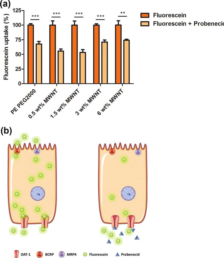
www.advancedsciencenews.com                                                                                               www.mbs-journal.de

where md and m are the weights of the dry sample and hydrogel        inner electrodes was recorded by a Keithely 6517A electrom-
at the moment of measurement, respectively.                          eter. Data were plotted in a V–I graph, and the resistance was
   The stability of the nanocomposite hydrogels at swelling          calculated as the slope of the linear response (Ohmic region).
equilibrium was investigated by the same method. The stability       Conductivity σ, expressed in S m−1, was calculated by using the
was tested in different conditions. In the first case, hydrogels     following equation:
were stirred in deionized water at RT and the weights were
recorded at defined times for the next 20 days. Fresh water was               d
                                                                     σ=
replaced each time the measurement took place. Additionally,              R ⋅ H ⋅ W (2)
their stability was assessed in physiological conditions. Hydro-
gels were swollen and then stirred in large volume of PBS, at        where d is the distance between the electrodes (5 mm), R is the
37 °C, and the weight was monitored over a period of 20 days.        resistance obtained from the V–I plot, H is the sample thick-
                                                                     ness (0.85 mm), and W is the sample width (15.5 mm). Con-
                                                                     ductivity was determined on three samples and the value was
2.5. Thermogravimetric Analysis                                      averaged.

The measurements were performed on a thermogravimetric
analysis (TGA) Q500 instrument using the following procedure:        2.9. Rheology
1 mg of either pristine MWNT or ox-MWNT was subjected to
an isotherm at 100 °C for 20 min, then it was heated from 100        Viscoelastic properties of the PE PEG2000 and nanocomposite
to 700 °C at a heating rate of 10 °C min−1 and under the N2          hydrogel were measured by a stress-controlled rheometer
flow (90 mL min−1). Data were analyzed with the TA Universal         (Anton Paar, Physica MCR501), equipped with a 25 mm plate
Analysis 2000 software.                                              geometry and a Peltier chamber, to control the temperature and
                                                                     protect the samples from drying. All measurements were con-
                                                                     ducted on hydrogel samples at swelling equilibrium, either at
2.6. Transmission Electron Microscopy                                25 °C or 37 °C, as will be specified further in the text. For meas-
                                                                     urements at 37 °C the hydrogels were swollen with PBS solu-
Transmission electron microscopy (TEM) analysis of CNTs was          tion. Frequency sweep measurements were performed over a
performed on a Tecnai Sphera electron microscope (FEI Com-           frequency range of 0.1–100 rad s−1, at 0.1% strain, both at 25 °C
pany), equipped with an LaB6 filament that was operated at an        and 37 °C. During the dynamic amplitude test the frequency
accelerating voltage of 200 kV. Images were acquired using a         was kept constant at 1 rad s−1, the strain was alternated between
bottom mounted 1024 × 1024 Charge-coupled device (CCD)               0.1 and 200% and the duration of one cycle was set at 200 s.
camera. Samples were prepared by dispersing nanotubes in
DMF at a concentration of 0.1 mg mL−1. After sonicating the
dispersion for 1 h, one drop was deposited on a carbon-coated        2.10. Tensile Testing
copper grid (CF300-Cu, Electron Microscopy Sciences) and
dried in a vacuum oven at 40 °C overnight.                           The tensile mechanical properties of the hydrogels were meas-
                                                                     ured using a Zwick Z100 Universal Tensile Tester machine,
                                                                     equipped with the load cell of 100 n. All measurements were
2.7. Scanning Electron Microscopy                                    done at RT and at the crosshead speed of 10 mm min−1. Equilib-
                                                                     rium-swollen hydrogel disks were cut in dog-bone shaped sam-
Morphology of nanocomposite hydrogels was studied by scan-           ples, whose size was 12.5 mm length, 2 mm width, and 0.85 mm
ning electron microscopy (SEM; FEI Quanta 3D FEG), at an             thickness. Each sample was tightly fixed between the clamps,
accelerating voltage of 5.00 kV. Equilibrium-swollen hydrogels       with the overall gage length of 20 mm. The measurements were
were frozen in liquid nitrogen, immediately cut with a sharp         performed on at least three samples for each hydrogel compo-
scalpel to expose the cross section, and then freeze-dried for       sition and the results were averaged. From the stress–strain
48 h. Prior to imaging, samples were sputtered with a thin layer     curve, tensile modulus was determined as the slope of the linear
of gold for 120 s.                                                   response (4–10%), by linear regression method. The tensile
                                                                     strength corresponds to the maximum stress experienced by the
                                                                     material before breaking, whereas the elongation at break was
2.8. Conductivity Measurements                                       defined as displacement λ (ratio between the sample length at
                                                                     break and its original length). Tensile toughness was calculated
Hydrogel samples were prepared by swelling and keeping               by integrating the area under the curve.
nanocomposite polymer films in Milli-Q purified water for
14 days, with water changed at least ten times, to remove ions
and impurities derived from the fabrication and preparation          2.11. Cell Culture
process. Resistivity of the hydrogels was assessed via the four-
point method with parallel electrodes, separated by 5 mm. Cur-       Urine-derived and organic anion transporter 1 (OAT1) protein
rent at different intensities was applied through the external       overexpressing conditionally immortalized proximal tubule epi-
electrodes by a Keithley 237 source, and the voltage between the     thelial cells (ciPTEC-OAT1) were cultured in Dulbecco’s Modified

Macromol. Biosci. 2019, 19, 1800173   1800173 (3 of 12)            © 2018 The Authors, Published by WILEY-VCH Verlag GmbH & Co. KGaA, Weinheim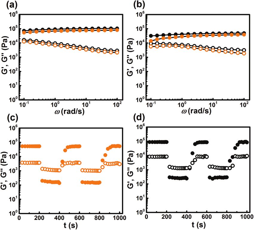
www.advancedsciencenews.com                                                                                                  www.mbs-journal.de

Eagle Medium/Nutrient Mixture F-12 (1:1 DMEM/F-12) (Gibco,               sterilized with 0.2% v/v solution of peracetic acid (Sigma-
Life Technologies, Paisly, UK) supplemented with 10% v/v                 Aldrich, Zwijndrecht, the Netherlands) in 4% v/v ethanol for
fetal calf serum (FCS) (Greiner Bio-One, Alphen aan den Rijn,            45 min, and then extensively rinsed three times with HBSS
the Netherlands), 5 µg mL−1 insulin, 5 µg mL−1 transferrin,              and left in HBSS for additional 24 h. Afterward, small disks
5 µg mL−1 selenium, 35 ng mL−1 hydrocortisone, 10 ng mL−1                of hydrogels were introduced in empty wells of 96-well plates
epidermal growth factor, and 40 pg mL−1 tri-iodothyronine (all           and the l-DOPA (l-3,4-dihydroxyphenylalanine, Sigma-Aldrich,
from Sigma-Aldrich, Zwijndrecht, the Netherlands), creating              Zwijndrecht, the Netherlands) coating was applied on the gels
a complete cell culture medium, as reported previously.[27]              to support cell attachment and growth, based on previously
CiPTEC-OAT1 were cultured up to 60 passages. Since the cells             published studies.[30,31] l-DOPA was dissolved in 10 mm Tris
were conditionally immortalized with a temperature-sensitive             buffer (pH 8.5) at 37 °C for 45 min with occasional mixing,
SV40 large T antigen,[28] cells were cultured at 33 °C and 5% v/v        filter sterilized, and applied on hydrogel surface at 2 mg mL−1
CO2 to allow proliferation, while the differentiation and matura-        final concentration for 5 h at 37 °C, as described previously.[32]
tion was achieved after 7 days of incubation at 37 °C, 5% v/v            Following the coating procedure, hydrogels were washed in
CO2, changing the medium every second day.                               HBSS and used further for cell seeding.

2.12. Preparation of Gel Extracts                                        2.15. Cell Proliferation on Hydrogels

In order to test the effects of hydrogel extracts on cell viability,     For cell proliferation assay, a total of 18 000 cells per hydrogel
elution test method was performed according to ISO 10993–5               sample (surface growth area 0.28 cm2) were seeded and incu-
protocols for cytotoxicity of biomedical devices.[29] Extracts were      bated at 33 °C, 5% v/v CO2. PrestoBlue cell viability reagent was
obtained by incubating all hydrogel samples separately in com-           applied after 1, 4, and 7 days to determine cell proliferation, as
plete cell culture medium for either 1 or 7 days at 37 °C, 5% v/v        described previously for the cell viability assay. Measured fluo-
CO2, respecting the ratio of surface area of the gel to the volume       rescence values for each sample, proportional to the number of
of the medium equal to 3 cm2 mL−1. Latex was used as a nega-             viable cells, were corrected for the background and presented as
tive control. The obtained culture medium was applied on cell            relative a.u. of fluorescence.
monolayers for a period of 24 h and 48 h, during and after which
cells were monitored for morphological changes and viability.
                                                                         2.16. CiPTEC-OAT1 Visualization on Hydrogels

2.13. Cell Viability Assay                                               In order to visualize ciPTEC-OAT1 cells on different hydrogel
                                                                         samples, cells were seeded at a density of 45 000 cells per hydrogel
Cells were seeded on 96-well plates (Costar 3599; Corning, NY,           (surface growth area 0.28 cm2), on 96-well plates, and incubated at
USA) at a density of 55 000 cell cm−2, allowed to adhere and             33 °C, 5% v/v CO2 for 24 h followed by 7 days incubation at 37 °C,
proliferate for 24 h at 33 °C, 5% v/v CO2, and incubated at              5% v/v CO2, as mentioned previously. After 7 days incubation cell
37 °C, 5% v/v CO2 for 7 days to allow maturation. Afterward,             were stained for actin filaments using Phalloidin-FITC (Sigma-
the cells were co-incubated either with PE PEG2000 control and           Aldrich, Zwijndrecht, the Netherlands) to visualize cell distribu-
CNT containing hydrogel samples (disks of 1.5 mm diameter                tion and morphology. Briefly, cells were washed with HBSS three
and 0.85 mm thickness) or with gel extracts (obtained as already         times, fixed with 2% w/v paraformaldehyde in PBS containing
mentioned) for 24 and 48 h, at 37 °C, 5% v/v CO2. Following              4% w/v sucrose (Sigma-Aldrich, Zwijndrecht, the Netherlands)
the incubation period, cell viability was determined using               for 10 min, then washed three times with 0.1% v/v Tween (Sigma-
PrestoBlue cell viability reagent (LifeTechnologies, Paisly, UK)         Aldrich, Zwijndrecht, the Netherlands) solution in PBS and per-
as suggested by the manufacturer. Briefly, 100 µL of the Presto-         meabilized with 0.3% v/v Triton (Merck, Darmstadt, Germany)
Blue reagent (1:10 in complete cell culture medium) was added            solution for 15 min. After another three washing steps with 0.1%
to each well and the cells were incubated for additional 1 h at          v/v Tween-PBS, cells were incubated with phalloidin-FITC (1:250
37 °C, 5% v/v CO2 in the dark. Finally, the fluorescence was             in PBS) for 1 h at RT and in the dark. Finally, hydrogel disks were
measured using a fluorescent microplate reader (Fluoroskan               mounted on the Willco glass bottom dishes (WillCo Wells B.V.,
Ascent FL, Thermo Fisher Scientific, Vantaa, Finland), at                Amsterdam, The Netherlands), using ProLong Gold antifade rea-
excitation wavelength of 530 nm and emission wavelength of               gent containing DAPI (Life Technologies, Eugene, OR, USA), and
590 nm. The obtained fluorescence values were corrected for              cells were imaged using confocal microscope (Leica TCS SP8 X,
the background, normalized to the untreated cells control and            Leica Microsystems CMS GmbH, Wetzlar, Germany). Analysis
plotted as relative cell viability.                                      was performed using Leica Application Suite X software (Leica
                                                                         Microsystems CMS GmbH).

2.14. CiPTEC-OAT1 Culture on Hydrogels
                                                                         2.17. Live/Dead Viability Assay
For cell culture, round-shaped pieces of the hydrogels (diam-
eter 6 mm, thickness 0.85 mm, approximate surface growth                 CiPTEC-OAT1 cells were seeded on hydrogel disks on 96-well
area 0.28 cm2) were cut out from compressed molded samples,              plates at a density of 45 000 cells per hydrogel (surface growth

Macromol. Biosci. 2019, 19, 1800173   1800173 (4 of 12)                © 2018 The Authors, Published by WILEY-VCH Verlag GmbH & Co. KGaA, Weinheim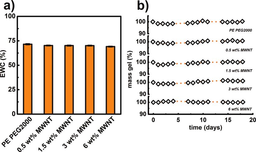
www.advancedsciencenews.com www.mbs-journal.de
area 0.28 cm2) and incubated at 33 °C, 5% v/v CO2 for 24 h. functionalities on the surface and to increase their solubility
Following 7 days maturation at 37 °C, 5% v/v CO2, cells were and dispersibility in water. The prepared ox-MWNT were
washed twice with warm HBSS and incubated with calcein- characterized prior to being incorporated in the PE PEG2000
AM (2 µm; Life Technologies, Eugene, OR, USA) and ethidium polymer matrix. The ox-MWNT were analyzed by several
homodimer-1 (EthD-1; 2 µm; Life Technologies, Eugene, OR, techniques, which confirm their successful functionalization
USA) diluted in HBSS, for 1 h at 37 °C in the dark. Next, cells (Figure S1, Supporting Information). In order to prepare homo-
were washed once with HBSS and hydrogel samples were geneous nanocomposite supramolecular hydrogels, aqueous
mounted on the Willco glass bottom dishes using Dako Fluores- solution of ox-MWNT was added to the acetone solution of
cence mounting medium (DAKO, Carpinteria, CA, USA). Visu- PE PEG2000. Water containing CNTs was able to replace the
alization was performed using confocal microscope (Leica TCS acetone and induce the self-assembly of DFA nanodomains
SP8 X) and analysis using Leica Application Suite X software. of the segmented copolymer. This resulted in hydrogel forma-
tion with CNTs included in the gel matrix. Nanocomposite gels
were prepared at four different concentrations of ox-MWNT:
2.18. Fluorescein Assay for OAT1 Activity Measurement 0.5, 1.5, 3, and 6 wt% relative to the amount of PE PEG2000.
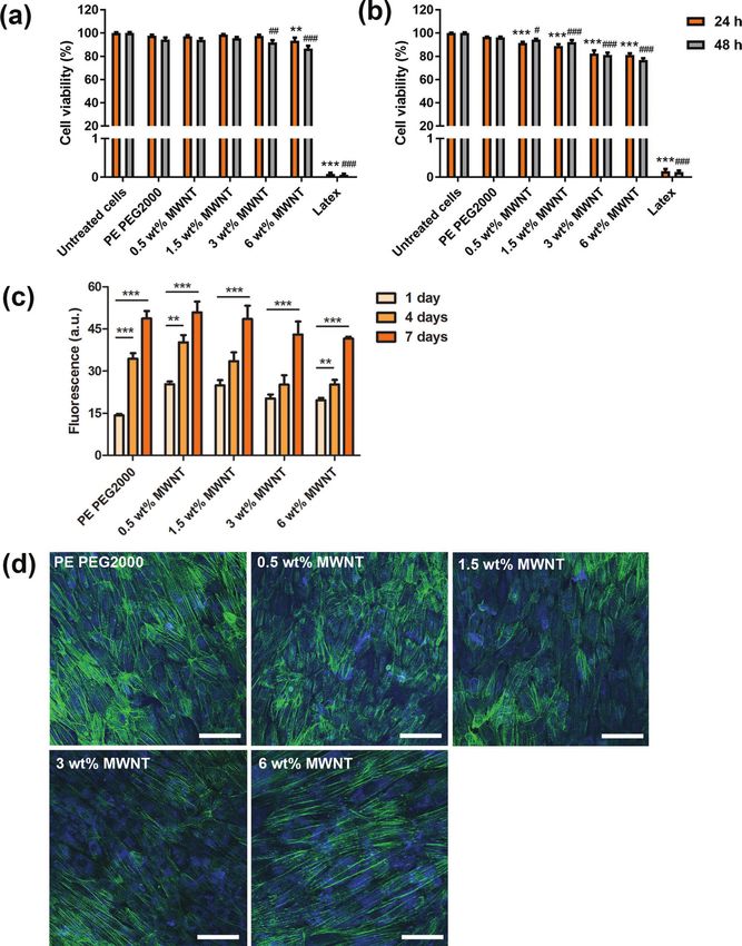
Figure 1a shows the components used to fabricate nanocom-
The fluorescein uptake assay was used to determine the OAT1 posite hydrogels. CNTs are embedded and physically dispersed
activity of ciPTEC-OAT1 cultured on hydrogels. In brief, cells in the hydrogel matrix, with no covalent cross-links or any other
were seeded on hydrogel disks with surface growth area of type of specific interactions between CNTs and segmented
0.28 cm2 (45 000 cells per hydrogel sample) on 96-well plates copolymer, except chain adsorption and hydrophobic inter-
and incubated at 33 °C, 5% v/v CO2 for 24 h, followed by actions (Figure 1b). Dried, pressed films of PE PEG2000
7 days maturation at 37 °C, 5% v/v CO2. Afterward, cells were copolymer and four nanocomposite materials were immersed
washed twice with warm HBSS and incubated with HBSS sup- in a large excess of water for 24 h, until swollen hydrogels were
plemented with HEPES (10 mm; Acros Organics, New Jersey, obtained. PE PEG2000 gave rise to a transparent hydrogel, while
USA), pH 7.4 and containing fluorescein (1 µm; Sigm-Aldrich, all of the nanocomposite hydrogels were completely black, due
Zwijndrecht, the Netherlands), in absence or presence of OAT1 to the incorporation of CNTs. At all CNT concentrations, homo-
inhibitor, probenecid (500 µm; Sigma-Aldrich, Zwijndrecht, the geneous black disks were observed, which confirmed good
Netherlands) for 10 min at 37 °C, 5% v/v CO2. Next, fluorescein dispersion of the nanotubes within the gel matrix (Figure 1c).
was removed, cells washed quickly with ice-cold HBSS to ter- The FT-IR spectrum of the dried PE PEG2000 hydrogel shows
minate the OAT1-mediated transport of fluorescein, and lysed the characteristic peak at 1735 cm−1, corresponding to the ester
with 0.1 m NaOH. Fluorescence was meas-
ured using Fluoroskan Ascent FL fluorescent
microplate reader at excitation wavelength of
492 nm and emission wavelength of 518 nm.
The measured fluorescence values were cor-
rected for the background (NaOH), and
plotted as relative fluorescein uptake.
2.19. Data Analysis
All data are presented as mean ± standard
error of the mean. Statistical analysis was
performed using one-way ANOVA followed
by Dunnett’s multiple comparison test or,
where appropriate, Tukey’s multiple com-
parison test, and a p-value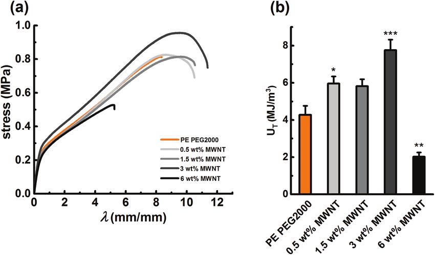
www.advancedsciencenews.com                                                                                                  www.mbs-journal.de

                                                                                                  amount of water at room temperature
                                                                                                  (RT), as determined by the gravimetric
                                                                                                  method (Figure 2a). EWC was deter-
                                                                                                  mined to be between 72 wt% and 74 wt%
                                                                                                  for all samples. This suggests that dis-
                                                                                                  persed nanotubes in the polymer network
                                                                                                  do not significantly alter the network
                                                                                                  structure. Therefore, the EWC is pre-
                                                                                                  dominantly determined by the PEG to
                                                                                                  DFA ratio and by the cross-linking den-
                                                                                                  sity. Since the bare PE PEG2000 hydrogel
                                                                                                  proved to be stable and non-eroding for
                                                                                                  50 days at ambient conditions,[25] the sta-
                                                                                                  bility of the nanocomposite gels was also
                                                                                                  assessed. As expected, the robust nano-
                                                                                                  composite gels showed the same stability
                                                                                                  and solubility resistance as the parent PE
                                                                                                  PEG2000 (Figure S2, Supporting Infor-
Figure 2. Swelling and stability of hydrogels. a) EWC determined for PE PEG2000 and at different  mation). This is due to the very strong
MWNT amounts; b) stability of hydrogels in PBS, at 37 °C, expressed as weight variation in time. hydrophobic interactions between DFA
All samples are indicated in individual panels.                                                   segments and consequently long relaxa-
                                                                                                  tion time.[25] In addition, the same experi-
carbonyl stretch[25] (Figure 1d). The same vibrational stretch             ment was performed at 37 °C with samples immersed in PBS
was observed in all of the nanocomposite hydrogels, confirming             (Figure 2b). The response was the same at 37 °C as at RT
that they are based on PE PEG2000. The reference MWNT                      for all gels, with no significant decrease in weight over a time
spectrum displays the stretching band at 1705 cm−1, which                  period of 20 days. This confirmed the extraordinary stability
indicates the presence of the COOH groups on their surface.                of the present gels under physiological conditions, making
However, this band was too weak to be detected in the nano-                them suitable for biomedical applications where high stability
composite gels, due to the very low concentration of MWNTs in              is desired.[33]
the nanocomposites.

                                                                         3.3. Internal Structure of Hydrogels
3.2. Equilibrium Water Content and Stability
of the Nanocomposite Hydrogels                                           To investigate the presence of CNTs in the material structure
                                                                         and changes caused by their inclusion, the cross sections of
Compared to the PE PEG2000, all nanocomposite gels, regard-              all hydrogels were imaged by SEM (Figure 3). Freeze drying
less of the amount of the CNTs, were able to absorb the same             method was employed to create porous structure of the

Figure 3. SEM micrographs of cross sections of a) PE PEG2000, b) 0.5, c) 1.5, d) 3, e) 6 wt% MWNT hydrogels.

Macromol. Biosci. 2019, 19, 1800173    1800173 (6 of 12)               © 2018 The Authors, Published by WILEY-VCH Verlag GmbH & Co. KGaA, Weinheim
www.advancedsciencenews.com www.mbs-journal.de
samples. Clearly, the PE PEG2000 gel and nanocomposites at interact with DFA aggregates, while still being efficiently dis-
low MWNT content (0.5 and 1.5 wt%) appeared to be porous. persed within the PEG matrix. Therefore, they most likely do
However, the pore size did not seem to be related to the amount not affect cross-link density, which is why the nanocomposite
of MWNT. In addition, the nanocomposites also displayed some hydrogel exhibited the same stiffness as the control. A sim-
filamentous elements, which are typical of the MWNT mor- ilar conclusion was drawn from EWC measurements, which
phology, confirming their presence in the network. Moreover, showed that EWC did not change upon addition of MWNT,
at higher MWNT content (3 and 6 wt%), the structure appeared suggesting that they do not disturb the network structure and
to be somewhat less porous, with an overall denser and more cross-link density. This observation might be related to the
compact architecture. Rougher pore walls were observed, due purely physical nature of the interactions in this nanocom-
to the higher MWNT loading, along with the presence of CNT posite system, whereas most reported studies on CNT-based
aggregates at 6 wt%. It has been reported that CNTs are able to nanocomposites are covalently cross-linked. We do not consider
affect the porosity of hydrogels.[15,34,35] The observed decrease the lack of stiffening by the MWNT as a drawback, because a
of porosity could be related to the hydrophobic nature of stiffness of ≈105 Pa of the nanocomposite is sufficient for many
MWNT, which allows them to interact and aggregate with DFA applications.[40]
domains, displacing PEG components and thus reducing the Subsequently, the ability of the nanocomposite hydrogel to
network density,[15,35] although we do not exclude the possibility recover its original stiffness after cessation of shear was tested.
that the freeze drying could have caused mechanical disruption Recoverability is a very important feature if the gel is to be
of the network structure. Overall, from SEM imaging we can processable. To test this, large amplitude strain response of
conclude that MWNT were indeed successfully incorporated the MWNT 3 wt% and PE PEG2000 hydrogels was performed
and dispersed in the PEG-DFA matrix.[21,35,36] (Figure 4c,d). First, G′ (ω) and G″ (ω) were monitored at a con-
stant strain γ = 0.1% and at a frequency ω = 1 rad s−1 for 200 s.
G′ (ω) was higher than G″ (ω) for both gels, confirming that the
3.4. Viscoelastic Properties and Reversibility of Physical gels were solid-like. Next, the strain was increased to γ = 200%
Interactions and kept constant, while frequency remained unchanged for
200 s. In this phase, the network structure was broken, G″ (ω)
The viscoelastic behavior of PE PEG2000 and MWNT 3 wt% was higher than G′(ω) and the samples were in a liquid-like,
nanocomposite hydrogel was studied at equilibrium swelling viscous state. In the next step, the strain was reduced back to
state and frequency sweep test was performed to evaluate the 0.1% to assess recovery of the stiffness. For the PE PEG2000
general viscoelastic behavior, both at RT and
at 37 °C (Figure 4a,b). For both gels G′ (ω)
was higher than G″ (ω) over the entire fre-
quency range probed, which confirmed the
elastic nature of hydrogels under both condi-
tions. Surprisingly, no significant increase of
stiffness of the nanocomposite, as compared
to the bare hydrogel was observed. According
to the theory of rubber elasticity,[37] inclusion
of any type of solid particles (such as CNTs)
should result in increase of stiffness, as
was seen in other studies.[13,36] In this case,
however, the stiffness of PE PEG2000 and
3 wt% MWNT hydrogel was quite similar (in
the order of ≈105 Pa at 25 °C). Interestingly,
the opposite effect has also been reported,
where upon addition of CNT to the hydrogel
the stiffness was reduced.[15,38] This was
attributed to the high aspect ratio of CNTs,
which created a large surface for interaction
with polymer chains, reducing their mobility
next to CNTs and, consequently, decreasing
crosslinking density.[39]
We hypothesize that in our system MWNT
are embedded very efficiently in the PEG-
DFA matrix. PEG-DFA hydrogel is charac-
terized by apolar nanodomains within the
hydrophilic PEG environment. Being char-
Figure 4. Frequency sweep of PE PEG2000 and 3 wt% MWNT nanocomposite at ω = 1 rad s−1
acterized by COOH hydrophilic groups and and γ = 0.1%, at a) 25 °C and b) 37 °C; large amplitude strain test for c) PE PEG2000 and
by the apolar surface due to extensive CC d) 3 wt% MWNT nanocomposite. Orange symbols, PE PEG2000; black symbols, 3 wt% MWNT
structure, we suggest that MWNT are able to nanocomposite; closed symbols, G′; open symbols, G″.
Macromol. Biosci. 2019, 19, 1800173 1800173 (7 of 12) © 2018 The Authors, Published by WILEY-VCH Verlag GmbH & Co. KGaA, Weinheimwww.advancedsciencenews.com www.mbs-journal.de
the PE PEG2000. The same was observed
for the tensile strength. However, a sig-
nificant increase for both ET and σT was
found for 3 wt% MWNT nanocomposite,
as compared to the PE PEG2000, with
ET increasing from 0.73 to 0.88 MPa
and σT increasing from 0.80 MPa for PE
PEG2000 to 0.96 MPa for 3 wt% MWNT,
respectively. This effect is most likely due
to favorable physical interactions between
MWNT and polymer chains, creating
additional entanglements at 3 wt%. Fur-
thermore, elongation at break (expressed
as displacement λ) increased as a func-
tion of MWNT content, with elongation
at break increasing from 8.27 for PE
PEG2000 to 11.37 at 3 wt% MWNT con-
tent corresponding to an increase of 37%.
Figure 5. Tensile properties of nanocomposites. a) Stress versus displacement of PE PEG2000 and Other mechanical parameters ET, σT, and
nanocomposite hydrogels at different MWNT contents, as indicated in the panel; b) comparison UT showed a similar trend, with tough-
of tensile toughness of PE PEG2000 as a function of MWNT concentration. *p < 0.05, **p < 0.01, ness increasing from 4.28 MJ m−3 for PE
***p < 0.001 as compared to the PE PEG2000 (one-way ANOVA, Dunnett’s multiple comparison PEG2000 (in accordance with previous
test).
results[25]) to 7.76 MJ m−3 in the 3 wt%
MWNT hydrogel, an increase of more
sample, full recovery took ≈100 s, whereas the MWNT 3 wt% than 80% (Figure 5b and Table 1).
nanocomposite recovered after ≈125 s. This behavior was repro- However, when the concentration of MWNT was increased
ducible for the second consecutive deformation cycle for both further to 6 wt%, a significant deterioration of all of the ten-
samples. Based on these findings, we conclude that the pres- sile parameters was observed. The toughness was reduced to
ence of MWNTs did not interfere with the process of reforma- 2 MJ m−3, less than half of the value for the parent PE PEG2000
tion of hydrophobic associations between DFA when the strain gel. Similar CNT-dependent trends were reported for other
was removed. Therefore, nanocomposites prepared in this way nanocomposite materials.[23] We suggest that at higher CNT
are fully reversible and can be processed like PE PEG2000,[25] concentrations, CNTs were less well dispersed in the matrix[24]
which widens their potential applications. and formed bundled aggregates which might have even com-
promised self-assembly of DFA nanodomains, resulting in a
less uniform network. SEM images (Figure 3e) of 6 wt% nano-
3.5. Mechanical Properties and Toughness composite hydrogel indeed show the presence of CNT aggre-
gates. Rather than reinforcing the gel, these aggregates acted as
To assess the effect that CNTs exert on the mechanical prop- defects, which led to stress concentration and material failure
erties and toughness of the hydrogels, uniaxial tensile testing at lower strain. Most likely, the combined effects of hydrogel
was performed on both PE PEG2000 and MWNT containing porosity, amount of CNTs, uniformity of their dispersion and
hydrogels, at equilibrium swelling and at a strain rate of their physical interaction with PE PEG2000 are responsible for
10 mm min−1. The results are plotted as stress versus displace- the observed trends in tensile performance.[23]
ment (Figure 5a). The values of the determined parameters In conclusion, improvement of mechanical performance by
tensile modulus ET, tensile strength σT, displacement λ, and increasing CNT content, as reported in other works on nan-
tensile toughness UT for all samples are listed in Table 1. wocomposites,[12,41,42] is confirmed in the present CNT-based
The tensile modulus for 0.5 wt% MWNT and 1.5 wt% nanocomposite hydrogels, but the effect is lost at a CNT con-
MWNT samples barely showed any improvement compared to tent of more than 3%.
Table 1. Tensile properties of PE PEG2000 and nanocomposite hydrogels
Sample ET σT λ UT
[MPa] [MPa] [mm/mm] [MJ m−3]
PE PEG2000 0.73 ± 0.03 0.80 ± 0.06 8.27 ± 0.29 4.28 ± 0.48
0.5 wt% MWNT 0.75 ± 0.03 0.81 ± 0.03 10.40 ± 0.34 5.96 ± 0.39
1.5 wt% MWNT 0.72 ± 0.07 0.80 ± 0.02 10.29 ± 0.34 5.82 ± 0.37
3 wt% MWNT 0.88 ± 0.02 0.96 ± 0.01 11.37 ± 0.81 7.76 ± 0.56
6 wt% MWNT 0.72 ± 0.04 0.51 ± 0.03 5.35 ± 0.52 2.02 ± 0.23
Macromol. Biosci. 2019, 19, 1800173 1800173 (8 of 12) © 2018 The Authors, Published by WILEY-VCH Verlag GmbH & Co. KGaA, Weinheimwww.advancedsciencenews.com www.mbs-journal.de
To evaluate hydrogels effect on ciPTEC viability, the gel
extract test was performed. According to ISO 10993-5,[29] gels
were incubated at 37 °C, CO2 5% in complete culture medium
(containing 10% FCS) for either 1 or 7 days, allowing for even-
tual elution of hydrogel components in the medium. Prior to
the viability assay, ciPTEC were allowed to mature at 37 °C for
7 days as reported previously.[27,28] Upon maturation, the cells
were exposed to complete cell culture media containing even-
tual gel leachables. Latex was used as negative control. Cell
viability was hardly affected by hydrogel components derived
from a 1 day elution test, regardless of the incubation time (24
or 48 h), with values remaining well above 90% (Figure 7a).
However, when ciPTEC cells were exposed to medium derived
from 7 days elution test, cell viability was slightly reduced, espe-
cially after 48 h of exposure (Figure 7b). Here, a MWNT dose-
dependent trend was observed, with nanocomposites at 3 wt%
and 6 wt% of MWNT reducing cell viability by nearly 20%
Figure 6. Electrical conductivity of PE PEG2000 and nanocomposite
and 25%, respectively. The same viability tests were repeated
hydrogels at different MWNT concentrations. *p < 0.05, ***p < 0.001
as compared to the PE PEG2000 (one-way ANOVA, Dunnett’s multiple with HeLa cells (Figure S3, Supporting Information). HeLa
comparison test). cells did not present any alterations in viability, regardless of
the hydrogel composition, elution time, or exposure time. The
reduction of ciPTEC-OAT1’s viability, but not that of HeLa cells,
3.6. Electrical Conductivity of the Nanocomposites at higher MWNT loading most likely indicates that there might
have been a release of rather small amount of CNTs, which
It is known that CNT-based materials are able to conduct elec- effect on cell viability is cell type specific.
tricity,[14,15,43] due to the formation of conductive pathways of In addition to the elution test indicated by ISO 10993-5 for
dispersed CNTs.[15,16] Therefore, resistivity measurements were cytotoxicity, the viability assay upon direct contact of cells with
performed on the present hydrogels, and the conductivity was hydrogel pieces (1 mm diameter, 0.85 mm thickness) was per-
calculated (Figure 6). As expected, the nanocomposites showed formed. The co-incubation of cells with hydrogels was done for
an increasing trend in conductivity in MWNT concentration- 24 h and 48 h, showing that HeLa cell viability was maintained
dependent manner. The hydrogel without MWNT had a con- at high levels compared to the untreated controls (Figure S4,
ductivity of 2.4–10−3 S m−1, the value due to eventual impurities Supporting Information). However, it should be noted that in
and CO2 dissolved in water from air,[14,44] and increased to this test hydrogel disks might have had exerted external pres-
4.7–10−3 S m−1 upon inclusion of 1.5 wt% of MWNT. At 3 and sure on cell monolayers, thus slightly affecting cell viability.[52]
6 wt% MWNT content, the conductivities were 1.1·10−2 S m−1 Furthermore, to observe cell viability when grown on top
and 1.6·10−2 S m−1, corresponding to a total increase by 358% of the hydrogels, a live/dead assay was carried out. Prior to
and 566%, respectively. The observed conductivity values were cell seeding, all hydrogels were coated with l-DOPA to allow
in line with other work, where similar conductivity was shown ciPTEC cell adhesion on the surface, as previously reported for
to be relevant for supporting growth and improving nerve other types of biomaterials.[30,31,53] After 7 days of maturation,
cell response.[14] This property might be useful for a variety cells were stained with calcein-AM and ethidium homodimer-1
of applications, such as biosensors and electrically conductive to distinguish live (green) from dead (red) cells. Obtained
scaffolds.[14,45–47] images show that none of the gels caused any significant cell
death, as the majority of cells were stained green, confirming
cytocompatibility of all examined hydrogels (Figure S5, Sup-
3.7. Cytocompatibility of the PE PEG2000 and MWNT porting Information).
Nanocomposite Hydrogels Additionally, the ability of cells to proliferate when grown on
hydrogels was assessed. Gels were first coated with l-DOPA, as
In order to determine cytocompatibility of the nanocomposites, discussed previously and cells were seeded at a low density. Cell
ciPTEC and HeLa cells were employed. HeLa cells were used metabolic activity, reflecting the number of cells, was evaluated
as suggested by ISO 10993-5[29,48] because of their wide use in at designated time periods. Figure 7c shows increased fluo-
biocompatibility studies. In addition, we opted for kidney epi- rescent signal and, therefore, increased metabolic activity over
thelial cells as an example of less robust cell line compared to time, indicating the ability of cells to proliferate on hydrogel
HeLa, given their susceptibility to many toxic agents and sub- surface, regardless of the material composition. After 1 day of
stances, including drugs such as cisplatin.[49] Also, ciPTEC rep- culture there was higher activity observed on CNT-based hydro-
resent a good choice considering that CNTs are in part excreted gels, compared to the pure PE PEG2000. Following 4 days of
by kidneys.[50] Moreover, it has been shown that human prox- culture, higher number of cells was observed on all hydrogels,
imal tubule epithelial cells were able to adhere and grow on with the highest values for PE PEG2000 and 0.5 wt% MWNT.
substrates characterized by high stiffness,[51] such as the pre- Anyway, after culturing for 7 days all numbers were nearly
sent hydrogels. similar, suggesting a comparable cell proliferation rate on all
Macromol. Biosci. 2019, 19, 1800173 1800173 (9 of 12) © 2018 The Authors, Published by WILEY-VCH Verlag GmbH & Co. KGaA, Weinheimwww.advancedsciencenews.com www.mbs-journal.de Figure 7. Cytocompatibility of the nanocomposite hydrogels at different MWNT concentrations and cell proliferation. ciPTEC-OAT1 viability after 24 and 48 h exposure to the culture medium containing hydrogel extracts following a) 1 day and b) 7 days elution test; latex is used as a negative control; **p < 0.01, ***p < 0.001, as compared to the corresponding 24 h untreated controls; #p < 0.05, ##p < 0.01, ###p < 0.001, as compared to the cor- responding 48 h untreated controls (one-way ANOVA, Tukey’s multiple comparison test). c) ciPTEC-OAT1 proliferation on l-DOPA coated hydrogels, after 1, 4, and 7 days; d) representative images (25× magnification) of ciPTEC-OAT1 cultured on l-DOPA coated hydrogels for 7 days; Actin filaments (green) and DAPI nuclear staining (blue); scale bar 10 μm; **p < 0.01, ***p < 0.001, compared to the corresponding gel sample with the cell prolifera- tion of 1 day (one-way ANOVA, Dunnett’s multiple comparison test). hydrogels. These results support the findings regarding nano- membrane of PTEC and is responsible for the uptake of many composites cytocompatibility. drugs, xenobiotics, and uremic waste molecules from the Moreover, cells grown on top of the hydrogels were visu- blood compartment.[27,28,54] Its activity can easily be measured alized, showing that all hydrogels were able to support cell using a fluorescent substrate, such as fluorescein, via deter- growth, adhesion, and spreading, leading to a complete surface mination of its intracellular accumulation.[53] This measure- coverage (Figure 7d). ment in the presence or absence of the OAT-1 inhibitor— Finally, in order to show that ciPTEC maintain their probenecid, directly relates to the OAT-1 uptake activity. Cells normal function when grown on hydrogels, OAT-1 activity grown on all gels retained their OAT-1 activity, which was sup- was assessed. As described, OAT-1 is located at the basolateral ported by the reduction of fluorescein uptake in the presence Macromol. Biosci. 2019, 19, 1800173 1800173 (10 of 12) © 2018 The Authors, Published by WILEY-VCH Verlag GmbH & Co. KGaA, Weinheim
www.advancedsciencenews.com                                                                                                    www.mbs-journal.de

                                                                                               flow processability even in the presence of
                                                                                               MWNT, which is an advantageous feature in
                                                                                               combination with an easy and scalable prepa-
                                                                                               ration procedure.
                                                                                                  Furthermore, it was shown that hydrogels
                                                                                               presented increasing electrical conductivity,
                                                                                               due to the presence of CNTs. A remarkable
                                                                                               increase of 80% in toughness was achieved
                                                                                               by incorporating 3 wt% of MWNT.
                                                                                                  Overall, the hydrogels displayed some
                                                                                               highly desirable features, including tun-
                                                                                               able mechanical and electrical properties,
                                                                                               while maintaining other features character-
                                                                                               istic of the parent PEG-DFA supramolec-
                                                                                               ular hydrogel, such as recoverability. These
                                                                                               nanocomposites also exhibited remarkable
                                                                                               biocompatibility, regardless of the CNT con-
                                                                                               tent, suggesting their safe use for biomed-
                                                                                               ical applications. We speculate that due to
                                                                                               very favourable interactions with cells, good
                                                                                               cytocompatibility and electrical conductivity,
                                                                                               these gels could be used in the future as sup-
                                                                                               porting scaffolds for different cell types, in
                                                                                               particular electrically active cells, such as car-
                                                                                               diomyocytes and neurons.

                                                                                               Supporting Information
                                                                                               Supporting Information is available from the Wiley
                                                                                               Online Library or from the author.

Figure 8. Cell function on the nanocomposite hydrogels at different MWNT concentrations.       Acknowledgements
a) OAT1 activity determined by 10 min fluorescein (1 μm) uptake in the absence or pres-       Marko Mihajlovic would like to thank Dr. René
ence of probenecid (500 μm); b) schematic representation of ciPTEC-OAT1 transport activity;   Lafleur for helping with acquisition of TEM
**p < 0.01, ***p < 0.001 (one-way ANOVA, Tukey’s multiple comparison test).                   images and Ingeborg Schreur-Piet for assistance
                                                                                              with SEM imaging. Milos Mihajlovic would like
of probenecid as compared to the uptake in normal conditions              to thank Dr. Silvia Mihaila for helpful discussions. This research was
(Figure 8).                                                               funded by the Marie Curie ITN project SASSYPOL (grant no. 607602,
                                                                          EU-FP7-PEOPLE-2013-ITN) and by the Ministry of Education, Culture,
   Observed results of cytocompatibility are in line with pre-
                                                                          and Science of the Netherlands (Gravity program 024.001.035). Also,
vious literature, showing good compatibility of CNT-based                 Milos Mihajlovic and Rosalinde Masereeuw would like to acknowledge
hydrogels with different cell types, such as neurons, cardio-             financial support of the Marie Curie ITN project BIOART (grant no.
myocytes, and human mesenchymal stem cells (hMSC).[14,55,56]              316690, EU-FP7-PEOPLE-2012-ITN).
Here, we show that our novel nanocomposite hydrogels are
also cytocompatible, support cell growth and proliferation, and
do not compromise cell activity and function, which are highly
                                                                          Conflict of Interest
desired features when hydrogels are intended for biomedical
applications.                                                             The authors declare no conflict of interest.

4. Conclusions                                                            Keywords
                                                                          biocompatibility, electrical conductivity, multi-walled carbon nanotubes,
Physical nanocomposite hydrogels based on CNTs were suc-
                                                                          nanocomposite supramolecular hydrogel, tensile toughness
cessfully fabricated. MWNTs were homogeneously incorpo-
rated in the matrix of supramolecular PEG-DFA hydrogel and                                                                  Received: May 8, 2018
the resulting nanocomposites were fully characterized. Hydro-                                                                 Revised: July 9, 2018
gels presented fully reversible shear thinning, demonstrating                                                     Published online: August 7, 2018

Macromol. Biosci. 2019, 19, 1800173    1800173 (11 of 12)               © 2018 The Authors, Published by WILEY-VCH Verlag GmbH & Co. KGaA, Weinheim
www.advancedsciencenews.com www.mbs-journal.de
[1] N. Annabi, A. Tamayol, J. A. Uquillas, M. Akbari, L. E. Bertassoni, [31] F. Hulshof, C. Schophuizen, M. Mihajlovic, C. van Blitterswijk,
C. Cha, G. Camci-Unal, M. R. Dokmeci, N. A. Peppas, R. Masereeuw, J. de Boer, D. Stamatialis, J. Tissue Eng. Regen. Med.
A. Khademhosseini, Adv. Mater. 2014, 26, 85. 2018, 12, E817.
[2] S. Van Vlierberghe, P. Dubruel, E. Schacht, Biomacromolecules 2011, [32] M. Ni, J. C. M. Teo, M. S. bin Ibrahim, K. Y. Zhang, F. Tasnim,
12, 1387. P. Y. Chow, D. Zink, J. Y. Ying, Biomaterials 2011, 32, 1465.
[3] J. L. Drury, D. J. Mooney, Biomaterials 2003, 24, 4337. [33] K. L. Spiller, S. A. Maher, A. M. Lowman, Tissue Eng. Part B-Rev.
[4] A. S. Hoffman, Adv. Drug Deliv. Rev. 2002, 54, 3. 2011, 17, 281.
[5] N. A. Peppas, J. Z. Hilt, A. Khademhosseini, R. Langer, Adv. Mater. [34] S. R. Shin, S. M. Jung, M. Zalabany, K. Kim, P. Zorlutuna, S. B. Kim,
2006, 18, 1345. M. Nikkhah, M. Khabiry, M. Azize, J. Kong, K. T. Wan, T. Palacios,
[6] K. Haraguchi, Curr. Opin. Solid State Mat. Sci. 2007, 11, 47. M. R. Dokmeci, H. Bae, X. W. Tang, A. Khademhosseini, ACS Nano
[7] F. Song, X. Li, Q. Wang, L. Liao, C. Zhang, J. Biomed. Nanotechnol. 2013, 7, 2369.
2015, 11, 40. [35] Y. S. Chen, P. C. Tsou, J. M. Lo, H. C. Tsai, Y. Z. Wang, G. H. Hsiue,
[8] D. Janas, S. Boncel, K. K. K. Koziol, Carbon 2014, 73, 259. Biomaterials 2013, 34, 7328.
[9] K. K. R. Datta, A. Achari, M. Eswaramoorthy, J. Mater. Chem. A [36] S. R. Shin, H. Bae, J. M. Cha, J. Y. Mun, Y. C. Chen, H. Tekin,
2013, 1, 6707. H. Shin, S. Farshchi, M. R. Dokmeci, S. Tang, A. Khademhosseini,
[10] L. Wei, N. Hu, Y. Zhang, Materials 2010, 3, 4066. ACS Nano 2012, 6, 362.
[11] K. Z. Gao, Z. Q. Shao, X. Wang, Y. H. Zhang, W. J. Wang, F. J. Wang, [37] B. E. J. E. Mark, Rubberlike Elasticity: A Molecular Primer, 2nd ed.,
RSC Adv. 2013, 3, 15058. Cambridge University Press, Cambridge, NY, 2007.
[12] L. Q. Liu, A. H. Barber, S. Nuriel, H. D. Wagner, Adv. Funct. Mater. [38] S. Sathaye, A. Mbi, C. Sonmez, Y. C. Chen, D. L. Blair,
2005, 15, 975. J. P. Schneider, D. J. Pochan, Wiley Interdiscip. Rev.: Nanomed. Nano-
[13] H. U. Rehman, Y. J. Chen, Y. L. Guo, Q. Du, J. Zhou, Y. P. Guo, biotechnol. 2015, 7, 34.
H. N. Duan, H. Li, H. Z. Liu, Composites, Part A 2016, 90, 250. [39] T. Ramanathan, H. Liu, L. C. Brinson, J. Polym. Sci., Part B-Polym.
[14] X. F. Liu, A. L. Miller, S. Park, B. E. Waletzki, A. Terzic, Phys. 2005, 43, 2269.
M. J. Yaszemski, L. C. Lu, J. Mat. Chem. B 2016, 4, 6930. [40] A. K. Gaharwar, N. A. Peppas, A. Khademhosseini, Biotechnol.
[15] K. Shah, D. Vasileva, A. Karadaghy, S. P. Zustiak, J. Mat. Chem. B Bioeng. 2014, 111, 441.
2015, 3, 7950. [41] P. C. Ma, N. A. Siddiqui, G. Marom, J. K. Kim, Composites, Part A
[16] R. A. MacDonald, C. M. Voge, M. Kariolis, J. P. Stegemann, Acta 2010, 41, 1345.
Biomater. 2008, 4, 1583. [42] Y. Z. Bin, M. Mine, A. Koganernaru, X. W. Jiang, M. Matsuo, Polymer
[17] B. L. Guo, P. X. Ma, Biomacromolecules 2018, 19, 1764. 2006, 47, 1308.
[18] M. C. Serrano, M. C. Gutierrez, F. del Monte, Prog. Polym. Sci. [43] Z. X. Deng, Y. Guo, X. Zhao, P. X. Ma, B. L. Guo, Chem. Mat. 2018,
2014, 39, 1448. 30, 1729.
[19] C. M. Homenick, H. Sheardown, A. Adronov, J. Mater. Chem. 2010, [44] R. M. Pashley, M. Rzechowicz, L. R. Pashley, M. J. Francis, J. Phys.
20, 2887. Chem. B 2005, 109, 1231.
[20] M. K. Bayazit, L. S. Clarke, K. S. Coleman, N. Clarke, J. Am. Chem. [45] S. Bosi, A. Fabbro, C. Cantarutti, M. Mihajlovic, L. Ballerini,
Soc. 2010, 132, 15814. M. Prato, Carbon 2016, 97, 87.
[21] W. F. Dong, C. G. Huang, Y. Wang, Y. J. Sun, P. M. Ma, M. Q. Chen, [46] G. Cirillo, S. Hampel, U. G. Spizzirri, O. I. Parisi, N. Picci,
Int. J. Mol. Sci. 2013, 14, 22380. F. Iemma, Biomed. Res. Int. 2014, 17.
[22] V. Saez-Martinez, A. Garcia-Gallastegui, C. Vera, B. Olalde, [47] V. Martinelli, G. Cellot, F. M. Toma, C. S. Long, J. H. Caldwell,
I. Madarieta, I. Obieta, N. Garagorri, J. Appl. Polym. Sci. 2011, 120, L. Zentilin, M. Giacca, A. Turco, M. Prato, L. Ballerini, L. Mestroni,
124. ACS Nano 2013, 7, 5746.
[23] Y. Y. Huang, Y. D. Zheng, W. H. Song, Y. X. Ma, J. Wu, L. Z. Fan, [48] W. J. Li, J. Zhou, Y. Y. Xu, Biomed. Rep. 2015, 3, 617.
Composites, Part A 2011, 42, 1398. [49] M. H. Hanigan, P. Devarajan, Cancer Ther. 2003, 1, 47.
[24] B. Joddar, E. Garcia, A. Casas, C. M. Stewart, Sci. Rep. 2016, 6, 12. [50] A. Ruggiero, C. H. Villa, E. Bander, D. A. Rey, M. Bergkvist,
[25] M. Mihajlovic, M. Staropoli, M. S. Appavou, H. M. Wyss, C. A. Batt, K. Manova-Todorova, W. M. Deen, D. A. Scheinberg,
W. Pyckhout-Hintzen, R. P. Sijbesma, Macromolecules 2017, 50, 3333. M. R. McDevitt, Proc. Natl. Acad.Sci. U. S. A. 2010, 107, 12369.
[26] A. Pistone, A. Ferlazzo, M. Lanza, C. Milone, D. Iannazzo, [51] J. A. Beamish, E. Chen, A. J. Putnam, PLoS One 2017, 12, e0181085.
A. Piperno, E. Piperopoulos, S. Galvagno, J. Nanosci. Nanotechnol. [52] L. Jing, H. Li, R. Y. Tay, B. Sun, S. H. Tsang, O. Cometto, J. Lin,
2012, 12, 5054. E. H. T. Teo, A. I. Y. Tok, ACS Nano 2017, 11, 3742.
[27] T. T. G. Nieskens, J. G. P. Peters, M. J. Schreurs, N. Smits, [53] J. Jansen, M. Fedecostante, M. J. Wilmer, J. G. Peters, U. M. Kreuser,
R. Woestenenk, K. Jansen, T. K. van der Made, M. Roring, P. H. van den Broek, R. A. Mensink, T. J. Boltje, D. Stamatialis,
C. Hilgendorf, M. J. Wilmer, R. Masereeuw, Aaps J. 2016, 18, 465. J. F. Wetzels, L. P. van den Heuvel, J. G. Hoenderop, R. Masereeuw,
[28] M. J. Wilmer, M. A. Saleem, R. Masereeuw, L. Ni, T. J. van der Sci. Rep. 2016, 6, 12.
Velden, F. G. Russel, P. W. Mathieson, L. A. Monnens, L. P. van den [54] F. G. Russel, R. Masereeuw, R. A. van Aubel, Annu. Rev. Physiol.
Heuvel, E. N. Levtchenko, Cell Tissue Res. 2010, 339, 449. 2002, 64, 563.
[29] ISO 10993-5:2009, Biological evaluation of medical devices—Part 5: [55] A. K. Gaharwar, A. Patel, A. Dolatshahi-Pirouz, H. B. Zhang,
Tests for in vitro cytotoxicity, 3rd ed., 2009. K. Rangarajan, G. Iviglia, S. R. Shin, M. A. Hussain,
[30] C. M. S. Schophuizen, I. E. De Napoli, J. Jansen, S. Teixeira, A. Khademhosseini, Biomater. Sci. 2015, 3, 46.
M. J. Wilmer, J. G. J. Hoenderop, L. P. W. Van den Heuvel, [56] S. Pok, F. Vitale, S. L. Eichmann, O. M. Benavides, M. Pasquali,
R. Masereeuw, D. Stamatialis, Acta Biomater. 2015, 14, 22. J. G. Jacot, ACS Nano 2014, 8, 9822.
Macromol. Biosci. 2019, 19, 1800173 1800173 (12 of 12) © 2018 The Authors, Published by WILEY-VCH Verlag GmbH & Co. KGaA, WeinheimYou can also read