Cell-free biogenesis of bacterial division proto-rings that can constrict liposomes - Nature
←
→
Page content transcription
If your browser does not render page correctly, please read the page content below
ARTICLE
                  https://doi.org/10.1038/s42003-020-01258-9               OPEN

                  Cell-free biogenesis of bacterial division proto-rings
                  that can constrict liposomes
                  Elisa Godino1, Jonás Noguera López1, Ilias Zarguit1, Anne Doerr1, Mercedes Jimenez                                      2,   Germán Rivas2 &
                  Christophe Danelon 1 ✉
1234567890():,;

                  A major challenge towards the realization of an autonomous synthetic cell resides in the
                  encoding of a division machinery in a genetic programme. In the bacterial cell cycle, the
                  assembly of cytoskeletal proteins into a ring defines the division site. At the onset of
                  the formation of the Escherichia coli divisome, a proto-ring consisting of FtsZ and its
                  membrane-recruiting proteins takes place. Here, we show that FtsA-FtsZ ring-like structures
                  driven by cell-free gene expression can be reconstituted on planar membranes and inside
                  liposome compartments. Such cytoskeletal structures are found to constrict the liposome,
                  generating elongated membrane necks and budding vesicles. Additional expression of the
                  FtsZ cross-linker protein ZapA yields more rigid FtsZ bundles that attach to the membrane
                  but fail to produce budding spots or necks in liposomes. These results demonstrate that
                  gene-directed protein synthesis and assembly of membrane-constricting FtsZ-rings can be
                  combined in a liposome-based artificial cell.

                  1 Department of Bionanoscience, Kavli Institute of Nanoscience, Delft University of Technology, van der Maasweg 9, 2629HZ Delft, The Netherlands. 2 Centro

                  de Investigaciones Biológicas Margarita Salas, CSIC, 28040 Madrid, Spain. ✉email: c.j.a.danelon@tudelft.nl

                  COMMUNICATIONS BIOLOGY | (2020)3:539 | https://doi.org/10.1038/s42003-020-01258-9 | www.nature.com/commsbio                                             1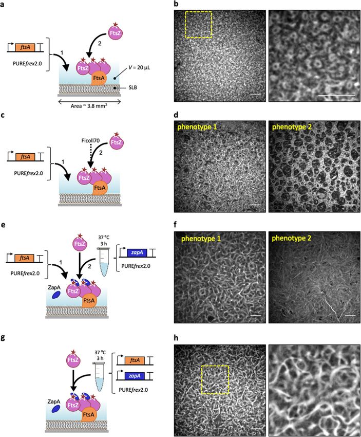
ARTICLE                                                              COMMUNICATIONS BIOLOGY | https://doi.org/10.1038/s42003-020-01258-9

C
         ell-free biology aims at understanding cellular processes by          We show that cell-free expressed FtsA is able to recruit FtsZ
         reconstituting biological functions from their isolated            polymers, forming large-scale two-dimensional networks of
         elementary components in in vitro model systems. Owing             curved and ring-like structures in the absence of bundling factors.
to their openness and easy manipulation, cytoplasmic extracts               When the entire set of reactions is encapsulated inside liposomes,
and systems reconstituted from purified elements are more                    proto-rings of FtsA-FtsZ filaments are found to constrict the
amenable to customized experimental design and quantitative                 vesicle, generating extended membrane necks and budding vesi-
description compared to living cells. Therefore, the minimal                cles, a phenotype that has not been reported before. Co-
requirements to achieve a particular function can be assessed               expression of ZapA, a native stabilizer of FtsZ filaments, yields
more reliably. Many complex biological structures and processes             stiffer FtsZ bundles attached to the membrane that fail to con-
taking place in bacterial or eukaryotic cells have already been             strict into bud necks. FtsZ cytoskeletal structures are also inves-
reconstituted in vitro. Notable achievements include the recon-             tigated with ZipA membrane-anchor protein. We find that in our
stitution of the minimal translation machinery from Escherichia             low-volume supported lipid bilayer (SLB) assays with ZipA and
coli1, the yeast DNA replication apparatus2, filopodial structures3,         ≤3 µM FtsZ, the generic crowding agent Ficoll70 is necessary to
cytoskeleton self-organization and centrosome positioning4, egg             elicit bundle formation. Cell-free expressed ZapA obviates the
cytokinesis signaling5, DNA segregation with Par6, and clathrin-            need of Ficoll70 and promotes formation of cytoskeletal networks
coated buds7. Encouraged by the many cellular pieces that have              with different, likely more physiological, morphology, and protein
already been reconstituted in vitro, synthetic biologists have now          monomer dynamics. The prospects of further improvement
engaged in the construction of an entire cell8–12.                          suggest that the DNA-programmed hierarchical assembly of the
   One of the hallmarks of living systems is their ability to divide.       Z-ring in liposomes is a promising strategy for dividing synthetic
An obvious starting point to conceiving a biology-inspired divi-            cells. In addition, our approach to reconstituting cellular pro-
sion mechanism in artificial cells is to consider the canonical              cesses in PURE system provides a generic platform that fills the
pathways taking place in prokaryotes. In most bacteria, symme-              gap between classical in vitro and in cellulo experiments.
trical cell division proceeds by forming a constriction ring that
eventually splits the mother cell into equally sized daughter cells13.
At an earlier stage of cytokinesis, a proto-ring composed of the            Results
FtsZ, FtsA, and ZipA proteins, assembles on the inner leaflet of the         Cell-free synthesized FtsA drives the formation of curved FtsZ
cytoplasmic membrane at the future division site14–16. The                  filaments. An essential component of the E. coli division proto-
tubulin-related FtsZ is the core constituent of the proto-ring. FtsZ        ring is FtsA, a homolog of actin that anchors FtsZ filaments to the
is a GTPase that can polymerize into protofilaments17,18.                    cytoplasmic membrane. To bypass the difficult purification of
Anchoring of FtsZ protofilaments to the cytoplasmic membrane is              FtsA28, we directly expressed a sequence-optimized ftsAopt gene
mediated by ZipA and the actin homolog FtsA19–21. This process              on an SLB (Fig. 1a). In the presence of 3 µM purified FtsZ-A647,
is regulated by accessory proteins belonging to the Zap family22.           curved filaments and dynamic ring-like structures formed on the
   Earlier attempts to divide cell-like liposomal compartments              membrane (Fig. 1b, Supplementary Fig. 1, Supplementary Note 1,
have focused on the reconstitution of the Z-ring from purified               Movie 1), concurring with previous reports12,28.
proteins23,24. These studies have shown that FtsZ aided by one of              To obtain quantitative insights about the concentration of cell-
its anchoring protein partners—or a chimeric FtsZ bearing a                 free synthesized FtsA, pre-ran PURE system samples were
membrane targeting segment25—can self-organize into filament                 analyzed by liquid chromatography-coupled mass spectrometry
patterns on supported lipid membranes25–28. When encapsulated               (LC-MS) (Supplementary Fig. 2, Supplementary Table 1). Protein
inside vesicles, the elementary cytoskeletal proteins form ring-like        abundance was quantified using an internal standard (QconCAT)
structures that can deform the liposome membrane24,29,30.                   for the most C-terminal peptide detected. We deduced that, after
Whether FtsZ filaments alone exert a contractile force and con-              3 h of expression, FtsA concentration on the SLB was 2.2 ±
tribute to the final stage of division remains a subject of                  0.2 μM (mean ± SD, three biological repeats) (Fig. 1a–d,
debate31,32 and evidence for complete liposome division is still            Supplementary Tables 2 and 3), corresponding to a protein ratio
lacking.                                                                    [FtsZ]:[FtsA] ≈ 1.5:1. In vivo, FtsA concentration is ~0.5 μM and
   A conceptual issue that is inherent to reconstitution assays             the protein ratio [FtsZ]:[FtsA] = 3:1–5:136. However, overlapping
solely relying on purified proteins, is the impossibility to maintain        rings and dynamic filaments on a lipid membrane have also been
steady amounts of cytoskeletal proteins from internal mechan-               observed at protein ratios similar as in our cell-free assay26,28.
isms as the compartment undergoes division. Another problem                 Note that LC-MS data do not report the concentration of active
raised by conventional cell-free assays is the use of oversimplified         protein, which may differ from the measured concentration of
buffer compositions that have been tailored for a particular set of         proteolytic peptides.
enzymatic reactions but fail to reproduce the cytoplasmic                      Promoting lateral interactions of FtsZ protofilaments stimu-
environment.                                                                lates the formation of higher-order cytoskeletal structures
   Herein, we address these issues by encoding E. coli division             in vitro37. However, little is known about how the nature of
proteins on DNA templates. Genetic control over protein pro-                these lateral interactions influences the morphology of the FtsZ
duction offers a general solution to achieve self-replication, as           network. Therefore, we decided to investigate the architecture and
well as self-regulation by establishing feedback loops. In this             dynamics of FtsZ protofilaments in a molecular environment that
context, the PURE system, a minimal gene expression system                  favors lateral interactions. First, we employed Ficoll70, a generic
reconstituted primarily from E. coli constituents1 was employed.            crowding agent known to elicit FtsZ bundle formation (Fig. 1c).
Different types of proteins and biological functions have already           Large SLB areas were covered with curved filaments, rings of
been synthesized de novo with the PURE system, including                    different sizes (most having a diameter of 1–2 µm, phenotype 1)
membrane-associated proteins10–12,33. Moreover, by containing               and large circular patterns (phenotype 2) (Fig. 1d).
all relevant factors for gene expression, the PURE system emu-                 Although Ficoll70 is commonly used as a macromolecular
lates more closely the molecular composition of the bacterial               crowder to mimic cytoplasmic conditions24,33,38,39, we reasoned
cytoplasm than simple buffers. In the present study we utilized             that ZapA, an in vivo regulator of FtsZ polymerization, would
PUREfrex2.0, which provides the best combination of protein                 provide a more targeted and native mechanism to elicit lateral
yield and expression lifespan34,35.                                         interaction, thus conferring physiologically relevant properties of

2                         COMMUNICATIONS BIOLOGY | (2020)3:539 | https://doi.org/10.1038/s42003-020-01258-9 | www.nature.com/commsbio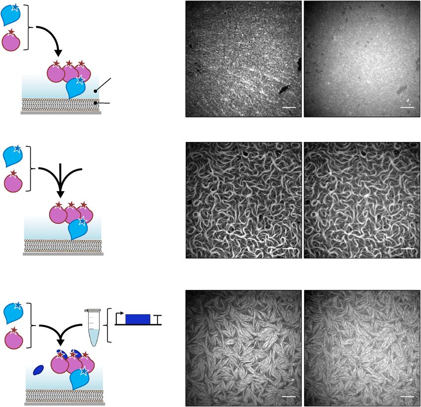
COMMUNICATIONS BIOLOGY | https://doi.org/10.1038/s42003-020-01258-9                                                                  ARTICLE

Fig. 1 Cell-free expressed FtsA recruits FtsZ to an SLB and drives the formation of ring-like structures. a Schematic representation of the SLB assays
with FtsA directly expressed on the membrane. Purified FtsZ-A647 (3 µM) was added. The sequence-optimized construct ftsAopt was used. b Fluorescence
image of FtsZ-A647 forming curved filaments and rings in the presence of in situ synthesized FtsA. The zoom-in image (right) corresponds to the framed
region in the left image. c As in (a) but the solution was supplemented with Ficoll70. d Fluorescence image of FtsZ-A647 forming curved filaments and
rings in the presence of in situ synthesized FtsA and Ficoll70. Two representative filament network morphologies observed on the same SLB are shown,
phenotype 1 being the most prominent. e Schematic illustration of the SLB assays with separately expressed FtsA and ZapA. The constructs ftsAopt and
zapA were used. Purified FtsZ-A647 (3 µM) was included. f Fluorescence images of FtsZ-A647 displaying two representative phenotypes from different
regions of the same SLB, phenotype 1 being the most prominent. g Schematic illustration of the SLB assays with co-expressed FtsA and ZapA from ftsAopt
and zapAopt constructs. Purified FtsZ-A647 (3 µM) was added. h Fluorescence images of FtsZ-A647. The zoom-in image (right) corresponds to the framed
area in the left image. Scale bars indicate 10 µm.

COMMUNICATIONS BIOLOGY | (2020)3:539 | https://doi.org/10.1038/s42003-020-01258-9 | www.nature.com/commsbio                                         3
ARTICLE                                                             COMMUNICATIONS BIOLOGY | https://doi.org/10.1038/s42003-020-01258-9

cytoskeletal patterns. For this reason, ZapA was produced in               cytoskeletal networks (Fig. 2a, b, Supplementary Note 2). This result
PUREfrex2.0 starting from its native gene sequence. Substitution           contrasts with previous observations20,28, highlighting the role of
of Ficoll70 with cell-free synthesized ZapA produced curved                the total reaction volume as a control parameter in the formation of
bundles but also long and straight filaments of FtsZ-A647                   cytoskeletal patterns. The network morphology, as well as the sZipA
recruited to the membrane by cell-free synthesized FtsA (Fig. 1e,          and FtsZ monomer dynamics differ whether lateral interactions are
f). The concentration of cell-free synthesized ZapA in the SLB             promoted by the artificial molecular crowder Ficoll70 or by cell-free
assay was estimated by LC-MS to be 0.2 ± 0.1 µM (mean ± SD,                synthesized ZapA (Fig. 2c–f, Supplementary Figs. 7–11). Moreover,
three biological repeats, Supplementary Table 3), which is                 we found that increasing the expression level of ZapA (Supple-
significantly lower than in E. coli (1.5 µM)40. Remarkably, even            mentary Fig. 4) results in more stable filaments that can even extend
at such a low concentration, ZapA is able to reshape FtsZ                  above the SLB (Supplementary Figs. 8 and 9). Taken together, our
protofilaments (Fig. 1f).                                                   results show that ZapA encourages the formation of membrane-
   Quantitative image analysis revealed that both the curvature            bound sZipA-FtsZ filament network having a different—pre-
and occurrence of branching points of the filament network were             sumably more physiological—morphology and subunit turnover
reduced in the presence of either Ficoll70 or ZapA (Supplemen-             compared with Ficoll70.
tary Fig. 3b). Differences between the two phenotypes observed in             We conclude from these SLB experiments that short, curved
each condition were also quantified (Supplementary Fig. 3c). We             filaments and rings that resemble physiological structures are
speculate that local changes, such as protein concentration (e.g.,         more prominent with FtsA compared with sZipA, and can
owing to edge effects in the chamber) and possible defects in the          develop in the absence of a bundling agent.
membrane, may play a role in the nucleation, morphology, or
dynamics of the cytoskeletal network, therefore driving formation
of one versus the other phenotype on the same SLB.                         FtsZ and internally synthesized FtsA constrict liposomes. The
   We challenged the PURE system to co-express both FtsA and               identification of FtsZ and FtsA as the minimal molecular set to
ZapA in a single reaction. Given the low amount of ZapA                    obtain membrane-anchored curved filaments and rings in the
produced in a single-gene reaction and the extra burden imposed            PURE system prompted us to reconstitute FtsA-FtsZ cytoskeletal
on the biosynthesis machinery when expressing an additional                networks inside liposomes (Fig. 3a). The cell-free gene expression
gene, we attempted to boost ZapA concentration by substituting             solution was supplemented with adenosine triphosphate (ATP,
the native gene with a sequence-optimized zapAopt DNA                      additional 2 mM), guanosine triphosphate (GTP, additional
construct (Supplementary Fig. 4). Co-expression of the ftsAopt             2 mM) and a mixture of highly purified chaperones (DnaK mix).
and zapAopt genes in a one-pot PURE system reaction led also to            Although energy regeneration components are present in the
the formation of bended and straight bundles (Fig. 1g, h,                  PURE system, extra ATP and GTP were provided to compensate
Supplementary Fig. 5) that qualitatively resemble phenotype 1              for the extra demand from FtsA and FtsZ. Purified FtsZ-A647
obtained in separate expression (Fig. 1e, f). Protein quantification        was used to visualize protein localization by laser scanning con-
was performed by LC-MS, and concentration values of 2.4 ± 0.3              focal microscopy. FtsZ-A647 was employed at 3 µM concentra-
µM FtsA and 0.4 ± 0.2 µM ZapA in the SLB assay were                        tion, which is similar to that measured in vivo (~3.5 µM)36.
determined (mean ± SD, three biological repeats, Supplementary             Liposomes were formed by natural swelling, with a composition
Table 3). Quantitative inspection of the protein patterns in co-           of zwitterionic PC and PE phospholipids, anionic PG and car-
expression experiments shows a lower curvature and branch                  diolipin, and a small fraction of TexasRed-conjugated lipid for
point density than in single-gene expression assays (Supplemen-            membrane imaging35. Such a lipid mixture and liposome pre-
tary Fig. 3b), which may be attributed to higher protein                   paration method have proved compatible with the cell-free
concentrations on the SLB when FtsA and ZapA are co-                       synthesis of membrane-associated enzymes10, DNA replication
expressed (Supplementary Table 3). Note again that LC-MS data              proteins11 and division proteins12. Liposome size distribution
do not provide an accurate measure of the concentration of active          ranges from ~1 µm up to over 15 µm in diameter, which provides
protein. Furthermore, usage of PURE system substrates and                  a more relevant bacterial cell-size compartment than >20 µm
cofactors, such as the tRNAs and NTPs, is different in a single-           liposomes produced with other methods23,33. In contrast with
gene or double-gene expression (e.g., nucleotide and codon                 previous studies33, no crowding agent was included during
abundance), which may influence reactions not directly involved             liposome formation. In fact, we found that Ficoll70 impairs for-
in transcription/translation.                                              mation of gene-expressing liposomes with our methodology
   Despite morphological differences observed in the protein               (Supplementary Fig. 12).
patterns between the Ficoll70- and ZapA-containing samples,                   In control experiments where the ftsAopt gene was omitted,
quantitative analysis of FtsZ subunit dynamics by fluorescence              FtsZ was exclusively located in the liposome lumen (Supplemen-
recovery after photobleaching (FRAP) revealed similar recovery             tary Fig. 13). De novo synthesized FtsA successfully recruited
halftime values under the tested conditions (Supplementary Fig. 6).        FtsZ on the membrane as shown by the colocalization of the
                                                                           FtsZ-A647 and membrane dye signals (Fig. 3, Supplementary
                                                                           Fig. 14). Although homogeneous recruitment of FtsZ to the
Bundling enables long sZipA-FtsZ cytoskeletal structure for-               membrane was commonly found within the liposome population,
mation. We then examined the self-organization of FtsZ with the            the majority of the liposomes displayed regions with patches of
membrane-anchor soluble ZipA (sZipA) using purified proteins                FtsZ on the inner surface of the membrane (Fig. 3b). Noticeably,
supplied in a minimal buffer or in PUREfrex2.0 background. The             the membrane spots with clustered FtsZ coincide with different
soluble variant sZipA does not contain the transmembrane region            types of membrane remodeling. In some cases, the recruited FtsZ
by elimination of the hydrophobic N-terminal domain (amino acids           localizes with outward membrane deformation or short protru-
1–25) of the full-length protein. This domain was substituted by a         sions (Fig. 3c, Supplementary Fig. 15). In other instances, the
His6-tag for binding NTA-conjugated lipids. The FtsZ-binding               protrusions developed into vesicles or blebs tethered to the
properties of sZipA are essentially the same as those of the native        parental liposome through a membrane neck coated with FtsZ
protein when incorporated in nanodiscs41,42. We found that in our          (Fig. 3d, Supplementary Fig. 15). Sometimes, the budding neck
low-volume SLB assays with ≤3 µM FtsZ, filament bundling with               extends over a few microns in the form of a tubular structure
either Ficoll70 or ZapA was required to trigger large-scale                containing one or a few FtsA-FtsZ rings (Fig. 3d, e). Interestingly,

4                        COMMUNICATIONS BIOLOGY | (2020)3:539 | https://doi.org/10.1038/s42003-020-01258-9 | www.nature.com/commsbio
COMMUNICATIONS BIOLOGY | https://doi.org/10.1038/s42003-020-01258-9 ARTICLE
a b
1. sZipA
sZipA FtsZ
2. FtsZ
FtsZ V = 20 µL
sZipA
SLB
c d
1. sZipA
sZipA FtsZ
Ficoll70
PUREfrex2.0
2. FtsZ
FtsZ
sZipA
e f
37 ºC
3h
sZipA FtsZ
1. sZipA
zapA
2. FtsZ PUREfrex2.0
ZapA
FtsZ
sZipA
Fig. 2 Purified sZipA and FtsZ form co-filament networks in the PURE system. a Schematic representation of the SLB assays. Purified sZipA-A488 was
first incubated on an SLB. The solution on top of the SLB was replaced by a minimal reaction buffer containing 3 µM purified FtsZ-A647 and 2 mM GTP.
b Fluorescence images of sZipA-A488 (left) and FtsZ-A647 (right) in the minimal reaction buffer without Ficoll70. c Schematic representation of the SLB
assays. Purified sZipA-A488 was first incubated on an SLB. The solution on top of the SLB was replaced by PUREfrex2.0 supplemented with 3 µM purified
FtsZ-A647, 2 mM GTP and 50 g L–1 Ficoll70. d Fluorescence images of sZipA-A488 (left) and FtsZ-A647 (right) in PUREfrex2.0 with Ficoll70. Large-scale
filaments with colocalizing FtsZ-A647 and sZipA-A488 are exclusively observed in the presence of Ficoll70. This conclusion is valid in both the minimal
reaction buffer and in PUREfrex2.0 background. More fields of view are displayed in Supplementary Fig. 7. e Schematic illustration of the SLB assays with
purified FtsZ-A647 (3 µM) and cell-free synthesized ZapA incubated on top of an sZipA-A488-bound SLB. ZapA was expressed from the native gene
zapA. f Fluorescence images of sZipA-A488 (left) and FtsZ-A647 (right) in a sample containing cell-free synthesized ZapA and additional 2 mM GTP.
Different cytoskeletal network phenotypes were observed when ZapA concentration was increased upon expression of the optimized zapAopt construct
(Supplementary Fig. 9). Scale bars indicate 10 µm.
these blebbing structures are dynamic. Events, such as appearance expect that the differences observed at incubation times longer
of new constriction sites, growing vesicles and diffusion of protein than 3 h can be attributed to an increase in protein concentration.
rings along the tube axis were observed (Fig. 3e, Movie 2). Instead, the time-dependent changes could be due to some
Although membrane recruitment of FtsZ in the form of patches delaying factors, such as recruitment of proteins to the
was visible already within 2 h of expression, major liposome- membrane, assembly of filaments and bundles, protein clustering
remodeling events, such as budding spots and elongated blebs into patches and remodeling of the membrane. It is unclear
were observed only after 3–4 h. Moreover, after 6 h expression, whether the FtsZ-coated membrane necks can close to release
small vesicles were found to agglutinate to larger liposomes mature vesicles. Therefore, we cannot ascertain that the small
(Supplementary Fig. 13). FtsA concentration does not signifi- vesicles observed after 6 h are reminiscent to division events. Yet,
cantly increase beyond the first 3 h of expression (Supplementary these aggregated vesicles were not observed when the ftsAopt gene
Fig. 2), in agreement with the kinetic profiles of protein was omitted (Supplementary Fig. 13), indicating that this global
production with the PURE system34. Concentration of synthe- remodeling is dependent on the expression of FtsA.
sized FtsA was compared after 3 and 6 h expression, yielding 4.5 We then decided to investigate how the presence of cell-free
± 0.5 μM and 5.8 ± 1.1 μM, respectively. Therefore, we do not expressed ZapA would modulate the properties of the cytoskeletal
COMMUNICATIONS BIOLOGY | (2020)3:539 | https://doi.org/10.1038/s42003-020-01258-9 | www.nature.com/commsbio 5ARTICLE COMMUNICATIONS BIOLOGY | https://doi.org/10.1038/s42003-020-01258-9
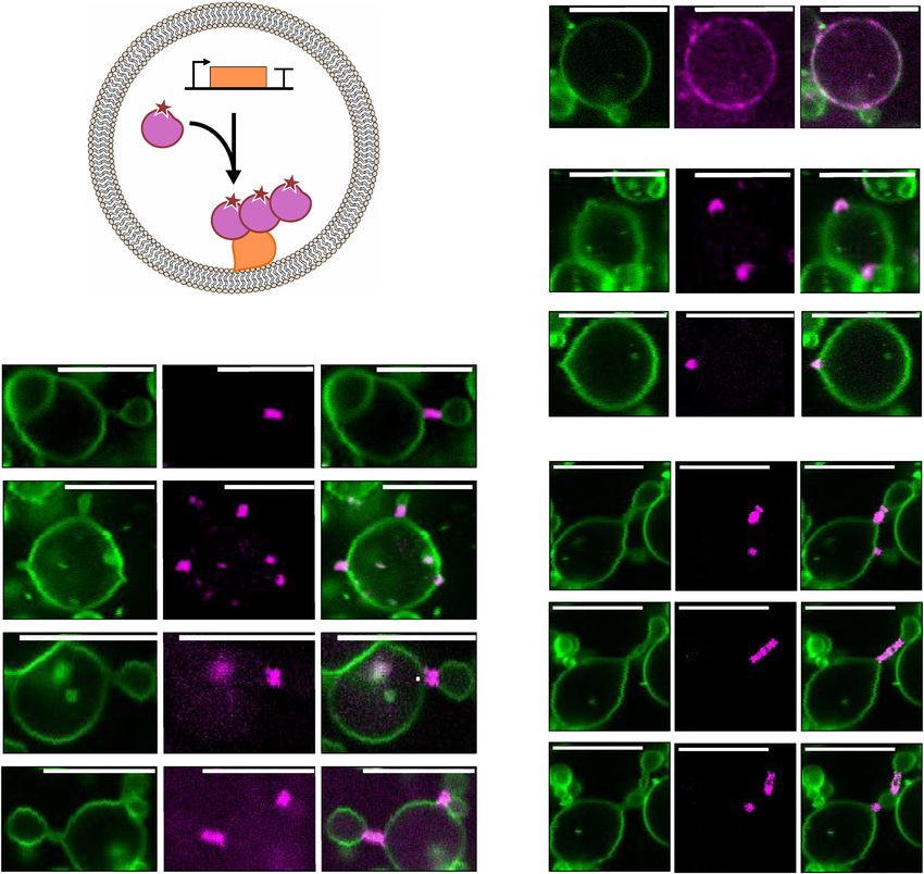
a b Membrane dye FtsZ-A647 Composite
DNase
sA
FtsZ
PUREfrex2.0
c
FtsZ * * *
FtsA
d
* * *
* * *
e
* * * * * *
* * * * * *
* * *
** ** **
Time
* * *
* * *
* * * ** ** **
* * *
Fig. 3 In-liposome synthesized FtsA assembles with FtsZ into ring-like structures that drive vesicle budding. a Schematic illustration of liposome
reconstitution assays. The ftsAopt DNA template was expressed within phospholipid vesicles in the presence of 3 µM purified FtsZ-A647. b–d Confocal
fluorescence images of liposomes exhibiting different morphologies of FtsZ-FtsA cytoskeletal structures and membrane remodeling: recruitment of
proteins to the membrane in the form of clusters with no visible membrane deformation b, budding spots induced by local accumulation of FtsZ-FtsA c, and
budding vesicles from a parental liposome with a clear FtsA-FtsZ-coated membrane neck d. e Time series images showing that a ring-forming protein
cluster localized at a constriction site can split, which induces multiple necks separated by blebbing vesicles (see Movie 2). Timespan is 120 s between the
first and second row of images, and 96 s between the second and third row. Fluorescence from the membrane dye is colored in green and FtsZ-A647 signal
is in magenta. The composite image is the overlay of the two channels. Asterisks indicate budding spots or constriction sites. Scale bars represent 10 µm.
More examples of liposomes are shown in Supplementary Figs. 14 and 15.
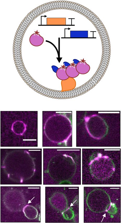
patterns in liposomes (Fig. 4a). Co-expression of ftsAopt and zapAopt presence of ZapA, the small ring-like structures do not form on
DNA constructs induced formation of FtsZ-A647 clusters on the SLB, where longer, curved filaments dominate (Fig. 1). We observed
inner surface of the membrane (Fig. 4b). Liposomes with different a decrease of the filament curvature in the presence of ZapA,
cytoskeletal protein phenotypes were observed, such as homo- especially during co-expression of FtsA and ZapA (Supplementary
geneous coating to the membrane, patches or filaments, and large Fig. 3b), which correlates with a higher concentration of ZapA in
ring-like structures. Bundles of FtsZ polymers adopting apparent the assay (Supplementary Table 3). The straighter cytoskeletal
ring-like structures predominantly localize at the interface of two filaments are likely not able to develop into contractile rings.
liposomes (Fig. 4b), coinciding with a membrane septum (i.e., a Instead, they accommodate to the large compartment and are
membrane separating two adjacent vesicles; it could be a single unable to deform the membrane into narrow necks (Fig. 4b). These
bilayer or two bilayers). However, ZapA abolishes the formation of results indicate that the mechanical properties of FtsZ-ZapA
membrane protrusions, vesicle budding and clustering of FtsZ on bundles impede the formation of membrane-constricting, high-
tubular membrane structures (Fig. 4b). We have seen that, in the curvature cytoskeletal filaments, which suggests that temporal
6 COMMUNICATIONS BIOLOGY | (2020)3:539 | https://doi.org/10.1038/s42003-020-01258-9 | www.nature.com/commsbioCOMMUNICATIONS BIOLOGY | https://doi.org/10.1038/s42003-020-01258-9 ARTICLE
that cannot polymerize44,45. In another report, the FtsZ-sfGFP
a fusion protein was recruited to the membrane of giant liposomes
(diameter 15–100 µm) by FtsA in the presence of Ficoll7033. In
DNase their work, Furusato et al.33 reported a homogenous recruitment
sA of FtsZ to the membrane in the presence of FtsA but no mem-
brane deformation. Local reshaping of liposomes was exclusively
zapA observed with ZipA as the FtsZ membrane anchor, but no
FtsZ constriction sites nor protein ring-like structures were
observed33. Here, we show that wild-type FtsA and FtsZ are
PUREfrex2.0 capable to deform the membrane in PURE system-loaded lipo-
somes with a sizeARTICLE COMMUNICATIONS BIOLOGY | https://doi.org/10.1038/s42003-020-01258-9
fresh prechilled liquid lysogeny broth (LB) medium and incubated for 1 h at 37 °C for 10 min and after cooling to room temperature 0.3 µL of 1 mg mL–1 trypsin
and 250 rpm. The cultures were plated on solid LB medium with ampicillin and (trypsin-ultra, MS-grade, New England Biolabs) was added. Samples were then
grew overnight at 37 °C. Colonies were picked up and cultured in 1 mL of liquid LB incubated at 37 °C overnight. After addition of 0.7 µL 10% trifluoroacetic acid,
medium with 50 µg µL–1 of ampicillin in 1.5-mL Eppendorf tubes for 6 h at 37 °C samples were centrifuged in a table-top centrifuge (5415 R, Eppendorf) for 10 min
and 250 rpm. Plasmid purification was performed using the PureYield™ Plasmid at maximum speed. The supernatant was transferred to a glass vial with small-
Miniprep System (column method, Promega). Plasmid concentration and purity volume insert for LC-MS/MS analysis.
were checked on a Nanodrop. Linear templates for PURE system reactions were
prepared by PCR using the plasmids as substrates with primers 194 and 709
(Supplementary Table 4). Amplification products were checked on a 1% agarose gel LC-MS/MS analysis. LC-MS/MS analysis was performed on a 6460 Triple Quad
and were purified using the Wizard SV Gel kit. DNA concentration and purity LCMS system (Agilent Technologies, USA) using Skyline software52. In all, 7 µL of
were measured using a ND-1000 UV-Vis Spectrophotometer (Nanodrop sample was injected per run to an ACQUITY UPLC® Peptide CSH™ C18 Column
Technologies). (Waters Corporation, USA). The peptides were separated in a gradient of buffer A
The DNA fragment containing the zapA gene (original sequence from E. coli (25 mM formic acid in MilliQ water) and buffer B (50 mM formic acid in acet-
K12 strain) was inserted in a pIDTSMART-AMP plasmid (Integrated DNA onitrile) at a flow rate of 500 µL per minute and at a column temperature of 40 °C.
Technologies). The plasmid was transformed into E. coli TOP10 cells. The column was equilibrated with 98% buffer A. After injection, the gradient was
Transformation, plasmid purification, and production of linear DNA templates changed linearly over 20 min to 70% buffer A, over the next 4 min to 60% buffer A,
were performed as described above. and over the next 30 s to 20% buffer A. This ratio was held for another 30 s and the
The ftsAopt and zapAopt constructs (starting with a T7 promoter and ending column was finally flushed with 98% buffer A to equilibrate for the next run.
with the T7 terminator) were sequence-optimized for codon usage, GC content and Selected peptides were measured by multiple reaction monitoring. For both ZapA
5′ mRNA secondary structures, and were inserted in a pJET1 and pUC57 plasmid, and FtsA, two peptides were present in the QconCAT. In addition, two peptides
respectively (GeneScript). Plasmids were amplified and purified as described above. from ribosomal proteins were also measured as control.
All sequences of the linearized constructs can be found in the Supplementary
Methods. Labeling of in vitro synthesized proteins and gel analysis. PUREfrex2.0 reaction
mixtures were supplemented with 0.5 μL of GreenLys (FluoroTectTM GreenLys,
Purified proteins. Purified FtsZ and sZipA were prepared and labeled with Alexa Promega) and gene expression was performed in a test tube as described above.
Fluor probes according to published protocols24,50,51. The degree of labeling was Samples were treated with RNase (RNaseA Solution, Promega) for 30 min and
0.9 ± 0.2 mol of fluorophore per mol of protein in both cases. FtsZ (150 μM) was proteins were denatured for 10 min at 90 °C in 2× SDS loading buffer with 10 mM
dialyzed against 20 mM Hepes/HCl, pH 8.0, with 50 mM KCl, 5 mM MgCl2, and 1 DTT. Samples were loaded on a 18% sodium dodecyl sulphate-polyacrylamide gel
mM ethylenediaminetetraacetic acid (EDTA). To minimize perturbations on FtsZ electrophoresis gel. Visualization of the fluorescently labeled translation products
assembly properties owing to labeling, the protein was first polymerized at 30 °C was performed on a fluorescence gel imager (Typhoon, Amersham Biosciences)
upon addition of 20 mM CaCl2 and 2 mM GTP. A 20-fold excess of Alexa Fluor using a 488-nm laser and a band pass emission filter of 520 nm.
647 (A647) was added, and the mixture was incubated for 15 min at 30 °C. The
precipitate was resuspended on ice in 50 mM Tris/HCl, pH 7.4, with 100 mM KCl,
and the free fluorescent probe was removed by gel filtration. ZipA (50 μM) was Fabrication and cleaning of the imaging chambers. Home-made glass chambers
labeled by adding 10-fold excess of Alexa Fluor 488 (A488) during 15 min at room were used in both SLB and liposome experiments12. Three microscopy glass slides
temperature in 20 mM Hepes/HCl, pH 8.0, with 50 mM KCl. Labeling reaction was (1-mm thick) were glued on top of each other with NOA 61 glue (Norland Pro-
stopped by the addition of a 1:100 dilution of 1 M Tris buffer. The unreacted probe ducts) and holes with a diameter of 2.5 mm were drilled. A 150 µm-thick coverslip
was removed by gel filtration. FtsZ-A647 (45 µM stock) was stored in a buffer (Menzel-Gläser, Germany) was glued with NOA 61 to cover the apertures, creating
containing 50 mM Tris, 500 mM KCl, 5 mM MgCl2 and 5% glycerol at pH 7.4. the bottom of glass chambers. Cleaning was performed by successive washing steps
sZipA-A488 (14.33 µm stock) was stored in a buffer containing 50 mM Tris, 50 of 10 min each in a bath sonicator (Sonorex digitec, Bandelin), as follows:
mM KCl, and 1 mM EDTA at pH 7.4. chloroform and methanol (1:1 volume ratio), 2% Hellmanex, 1 M KOH, 100%
ethanol and finally MilliQ water. For SLB experiments the glass chambers were
further treated every two to three experiments with Acid Piranha.
Cell-free gene expression. PUREfrex2.0 (GeneFrontier Corporation, Japan) was
utilized following storing and handling instructions provided by the supplier.
Linear DNA templates were used in single-gene expression assays at a final con- Lipids. 1,2-dioleoyl-sn-glycero-3-phosphocholine (DOPC), 1,2-dioleoyl-sn-gly-
centration of 5 nM. In co-expression experiments, both ftsAopt and zapAopt con- cero-3-phosphoethanolamine (DOPE), 1,2-dioleoyl-sn-glycero-3phosphoglycerol
structs were included at 5 nM and 10 nM, respectively, along with 1 µL of DnaK Mix (DOPG), 1′,3′-bis[1,2-dioleoyl-sn-glycero-3-phospho]-glycerol (18:1 CL), 1,2-dis-
(GeneFrontier Corporation). DnaK Mix consists of highly purified E. coli DnaK, tearoyl-sn-glycero-3-phosphoethanolamine-N-[biotinyl(polyethylene glycol)-2000
DnaJ, and GrpE chaperone proteins. Reactions of 20 µL volume were carried out in (DSPE-PEG-biotin), and 1,2-dioleoyl-sn-glycero-3-[(N-(5-amino-1-carboxypentyl)
test tubes for 3 h at 37 °C. When indicated, samples were supplemented with iminodiacetic acid)succinyl] (DGS-NTA) were from Avanti Polar Lipids. Texas
purified proteins (FtsZ-A647, sZipA-A488) and added either on top of an SLB or Red 1,2-dihexadecanoyl-sn-glycero-3-phosphoethanolamine (DHPE-TexasRed)
used for lipid film swelling. was from Invitrogen.
QconCAT purification. QconCAT was designed to contain two specific peptides Preparation of small unilamellar vesicles. Small unilamellar vesicles (SUVs)
for FtsA and two for ZapA (Supplementary Table 1, Supplementary Fig. 2). were used as precursors for the formation of SLBs12. Lipids DOPC (4 µmol),
QconCAT was expressed in BL21(DE3) cells in M9 medium with 15NH4Cl and DOPG (1 µmol) and DGS-NTA (0.25 µmol), all dissolved in chloroform (Avanti
ampicillin. A pre-culture was diluted 1:100 to a 50-mL expression culture. Protein Polar Lipids), were mixed in a glass vial. A lipid film was deposited on the wall of
expression was induced at OD600 = 0.5 with 1 mM isopropyl β-d-1- the vial upon solvent evaporation by applying a gentle flow of argon and was
thiogalactopyranoside and cells were grown for 3 h at 37 °C. Cells were harvested further desiccated for 30 min at room temperature. The lipid film was resuspended
by centrifugation and the pellet was dissolved in 1 mL B-PER. 10 µL of 10 mg mL–1 with 400 µL of SLB buffer (50 mM Tris, 300 mM KCl, 5 mM MgCl2, pH 7.5) and
lysozyme and 10 µL of DNaseI (ThermoScientific, 1 U µL–1) were added and the the solution was vortexed for a few minutes. The final lipid concentration was 1.25
sample was incubated for 10 min at room temperature. The lysate was centrifuged mg mL–1. A two-step extrusion (each of 11 passages) was carried out using the
for 20 min at 16,000 × g and the pellet resuspended in 2 mL of a 1:10 dilution of B- Avanti mini extruder (Avanti Polar Lipids) equipped with 250 µL Hamilton syr-
PER in MilliQ water. The sample was twice again centrifuged, and the pellet was inges (Avant Polar Lipids), filters (drain disc 10 mm diameter, Whatman) and a
resuspended in 2 mL 1:10 diluted B-PER and centrifuged again. The pellet was polycarbonate membrane with a pore size of 0.2 µm (step 1) or 0.03 µm (step 2)
resuspended in 600 µL of 10 mM Tris-HCl pH 8.0, 6 M guanidinium chloride and (Nuclepore track-etched membrane, Whatman).
incubated at room temperature for 30 min. After spinning down unsolubilised
protein the supernatant was loaded onto an equilibrated mini NiNTA spin column
and the flow-through was reloaded twice to maximize protein binding. The column Formation of SLBs. The imaging chamber was treated with oxygen plasma
was washed twice with 600 µL of 10 mM Tris-HCl pH 6.3, 8 M urea and the (Harrick Plasma basic plasma cleaner) for 30 min to activate the glass surface.
QconCAT was eluted with 3 × 200 µL of 10 mM Tris-HCl pH 4.5, 8 M urea, Immediately after plasma cleaning the SUV solution was added to the sample
400 mM imidazole. The eluate was dialyzed overnight and for additional 4 h reservoir at a final lipid concentration of 0.94 mg mL–1 together with 3 mM CaCl2.
against 10 mm Tris-HCl pH 8.0, 100 mM KCl with a 10-kDa cutoff slide-a-lyzer The chamber was closed by sticking a coverslip using a double-sided adhesive
casette (ThermoScientific). silicone sheet (Life Technologies) and the sample was incubated for 30 min at
37 °C. Next, the chamber was opened and the SLB was carefully washed six times
with SLB buffer. Under these conditions, the SLB contains 4.8 molar % of 18:1
Trypsin digest. Per LC-MS injection, 1.5 µL of PURE system reaction was mixed DGS-NTA (Ni2+) lipids, which is within the range studied by ref. 27 (0.5–10 mol
with 3 µL of 100 mM Tris-HCl pH 8.0, 0.3 µL of 20 mM CaCl2, and 0.8 µL MilliQ %), similar as in ref. 28 (1–8 mol%) but higher than in ref. 38 (0.02–0.08 mol% of
water. Samples were incubated at 90 °C for 10 min to stop the reaction. Then, 0.6 full-length ZipA, DGS-NTA lipid was not used in this study) and lower than in
µL of QconCAT (0.3 mg mL–1) was added, the sample was incubated again at 90 °C ref. 50 (10 mol%).
8 COMMUNICATIONS BIOLOGY | (2020)3:539 | https://doi.org/10.1038/s42003-020-01258-9 | www.nature.com/commsbioCOMMUNICATIONS BIOLOGY | https://doi.org/10.1038/s42003-020-01258-9 ARTICLE
Activity assays on supported membranes. In the experiments involving sZipA- SR objective and a 640 nm laser line. The acquisition and reconstruction of the SIM
A488, 1 µM of the purified protein was first incubated on top of an SLB for 10 min images have been performed using the Nikon NIS element software. SIM raw data
at room temperature. The SLB was washed with 10 µL reaction buffer (50 mM Tris- and their corresponding reconstructed images were quality-checked using the Fiji
HCl, 150 mM KCl, 5 mM MgCl2, pH 7.5). Then, 20 µL of sample (composition is plug-in SIMcheck55.
specified where relevant) was added on top of the SLB and the chamber was sealed
by sticking a 20 × 20 mm coverslip with a double-sided adhesive silicone sheet. In
Total internal reflection fluorescence microscopy. FtsA-FtsZ ring dynamic was
the experiments with FtsA, the ftsA or ftsAopt gene was either directly expressed on
investigated using a Nikon TiE inverted fluorescence microscope equipped with an
top of the SLB, or in a test tube and subsequently added onto an SLB as part of the
iLAS2 illumination system, a Plan Apo ×100 oil immersion objective and a 2×
sample. In the earlier configuration, a 20 µL PUREfrex2.0 reaction was carried out
Photometrics EMCCD Evolve Camera. The 640 nm laser line was used in com-
on top of an SLB and 10 µL were removed and replaced by the activity assay
bination with appropriate emission filters to image FtsZ-A647. MetaMorph®
mixture. The exact composition of the sample varies for the different experiments
Microscopy Automation Software was used for image acquisition.
and is specified where appropriate. In all cases, samples contained 2 mM GTP,
supplemented with 2 mM ATP in FtsA experiments. In all assays without ZapA,
Ficoll70 was added to a final concentration of 50 g L–1. No oxygen-scavenging Quantitative image analysis. Image analysis was performed using Mathematica
system was used, unlike in ref. 28 but like in ref. 38. (Wolfram Research, version 11.3). All images were corrected for uneven illumi-
nation by applying a Gaussian filter with radius 70 pixels to each image, fitting a
third-degree polynomial to the filtered image, and dividing the original image
Spinning disk microscopy. SLBs were imaged with an Olympus iX81 inverted pixel-by-pixel with the fitted polynomial. To segment the filaments a ridge filter
fluorescence microscope equipped with a ×100 oil immersion objective (Olympus), with sigma = 1 was applied and the resulting image was binarized using mor-
an iXon3 EMCCD camera (Andor Technology) and a Nipkow spinning disk (CSU- phological binarization with the default parameters. When needed this image was
XI, Yokogawa). FtsZ-A647 and sZipA-A488 were imaged using a 640 nm and 491 convolved with a Laplacian-of-Gaussian filter with radius two pixels and inversed
nm laser line, respectively, and appropriate emission filters (685/40 nm or 525/50 (necessary for images with thin filaments). Filament thicknesses were calculated
nm). The software Andor IQ3 (Andor Technology Ltd.) was used for image from this image as the distance of the centerline of filaments to the edge using the
acquisition and identical settings were used for all experiments. Experiments were distance transform function. Branch point density and filament curvature were
conducted at room temperature. calculated after the thinning operation was applied to the segmented image.
Curvatures were approximated at each pixel along the thinned filament with a
Preparation of lipid-coated beads. Lipid-coated beads were prepared according distance larger than two pixels to the next branch point using Gaussian smoothing
to our published protocol35 with the following lipid composition: DOPC (50 mol on the derivative functions. The image processing steps are illustrated in Supple-
%), DOPE (36 mol%), DOPG (12 mol%), 18:1 CL (2 mol%), DSPE-PEG-biotin (1 mentary Fig. 3.
mass%) and DHPE-TexasRed (0.5 mass%) for a total mass of 2 mg. Lipids dis- Filament dynamic was analyzed by computing kymographs in MatLab version
solved in chloroform were mixed in a round-bottom glass flask. Methanol con- 2018a. A user-defined ellipse was overlaid on the ring-like structures of interest
taining 100 mM rhamnose (Sigma Aldrich) was added to the solution in a based on the first image of the movie and the boundary pixel intensities of all
chloroform-to-methanol volume ratio of 2.5:1. Then, 1.5 g of 212–300 µm glass frames were extracted.
beads (acid washed, Sigma Aldrich) were poured to the lipid-rhamnose mixture
and the organic solvent was removed by rotary evaporation at 200 mbar for 2 h at Statistics and reproducibility. All experiments reported in this study have been
room temperature, followed by overnight desiccation. Lipid-coated beads were reproduced and similar results have been obtained. Microscopy images displayed in
stored under argon at –20 °C until use. the main text figures are representative of the sample properties as analyzed from
larger fields of view in at least three independent biological repeats.
Production and immobilization of gene-expressing liposomes. A PUREfrex2.0
reaction mixture was assembled as described above. Either or both ftsAopt and Reporting summary. Further information on research design is available in the Nature
zapAopt DNA constructs were added at a final concentration of 5 nM and 10 nM, Research Reporting Summary linked to this article.
respectively. The solution was supplemented with (final concentrations indicated):
2 mM GTP, 2 mM ATP, 3 µM FtsZ-A647 and MilliQ to reach a final volume of 20
µL. About 20 mg of lipid-coated beads was added to the solution and liposomes Data availability
were formed by natural swelling of the lipid film for 2 h on ice, protected from All data and custom codes generated during the current study are available from the
light. During incubation, the tube was gently rotated manually a few times. Four corresponding author on reasonable request. Source data underlying the plots shown in
freeze-thaw cycles were then applied by dipping the sample in liquid nitrogen and Supplementary Figures are provided in Supplementary Data 1.
thawing on ice. The sample reservoir of the imaging chamber was functionalized
with 1:1 molar ratio of bovine serum albumin (BSA) and BSA-biotin (1 mg mL–1,
ThermoFisher Scientific), and then with Neutravidin (1 mg mL–1, Sigma Aldrich), Received: 11 March 2020; Accepted: 1 September 2020;
to tether the biotinylated liposomes. About 7 μL of the liposome solution was
carefully pipetted (with a cut tip) into the imaging chamber and supplemented with
RQ1 DNase (0.07 U µL–1) to preclude gene expression outside liposomes. The
chamber was sealed by sticking a 20 × 20 mm coverslip with a double-sided
adhesive silicone sheet. Expression was performed directly on the confocal
microscope at 37 °C for 1.5–6 h. References
1. Shimizu, Y. et al. Cell-free translation reconstituted with purified components.
Nat. Biotechnol. 19, 751–755 (2001).
Confocal microscopy. A Nikon A1R Laser scanning confocal microscope equip- 2. Gros, J., Devbhandari, S. & Remus, D. Origin plasticity during budding yeast
ped with an SR Apo TIRF ×100 oil immersion objective was used to image lipo- DNA replication in vitro. EMBO J. 33, 621–636 (2014).
somes. The 561 nm and 640 nm laser lines were used in combination with
3. Lee, K., Gallop, J. L., Rambani, K. & Kirschner, M. W. Self-Assembly of
appropriate emission filters to image the Texas Red membrane dye and FtsZ-A647,
filopodia-like structures on supported lipid bilayers. Science 329, 1341–1345
respectively. The software NIS (Nikon) was used for image acquisition and iden-
(2010).
tical settings were used for all experiments. Samples were mounted on a
4. Vignaud, T., Blanchoin, L. & Théry, M. Directed cytoskeleton self-
temperature-controlled stage maintained at 37 °C during imaging.
organization. Trends Cell Biol. 22, 671–682 (2012).
5. Nguyen, P. A. et al. Spatial organization of cytokinesis signaling reconstituted
Fluorescence recovery after photobleaching. FRAP experiments were performed in a cell-free system. Science 346, 244–247 (2014).
on an Olympus iX81 spinning disk microscope. Images were acquired using the 6. Garner, E. C., Campbell, C. S., Weibel, D. B. & Mullins, R. D. Reconstitution of
following protocol: 10 frames every s, 10 frames every 250 ms, 10 frames every 2 s, DNA segregation driven by assembly of a prokaryotic actin homolog. Science
10 frames every 4 s. Analysis of the FRAP images was performed with ImageJ53,54 315, 1270–1274 (2007).
using the FRAP profiler plug-in. The intensity of a bleached region of interest (ROI, 7. Dannhauser, P. N. & Ungewickell, E. J. Reconstitution of clathrin-coated bud
29 × 29 pixels) was measured over time and normalized to the intensity of the and vesicle formation with minimal components. Nat. Cell Biol. 14, 634–639
surrounding (250 × 250 pixels area centered on the ROI) to correct for the (2012).
bleaching that occurs during image acquisition. Fitting of the FRAP curves was 8. Focus on the benefits of building life’s systems from scratch. Nature 563,
generated in GraphPad Software Inc. using a one-phase exponential model. At least 155–155 (2018).
three FRAP measurements were performed in each sample analyzed. 9. Nourian, Z., Scott, A. & Danelon, C. Toward the assembly of a minimal
divisome. Syst. Synth. Biol. 8, 237 (2014).
Structured illumination microscopy (SIM). 3D SIM images have been acquired 10. Scott, A. et al. Cell-free phospholipid biosynthesis by gene-encoded enzymes
with a Nikon SIM microscope equipped with a Nikon ×100 and 1.49 NA Apo TIRF reconstituted in liposomes. PLoS ONE 11, e0163058 (2016).
COMMUNICATIONS BIOLOGY | (2020)3:539 | https://doi.org/10.1038/s42003-020-01258-9 | www.nature.com/commsbio 9ARTICLE COMMUNICATIONS BIOLOGY | https://doi.org/10.1038/s42003-020-01258-9
11. van Nies, P. et al. Self-replication of DNA by its encoded proteins in liposome- 40. Small, E. et al. FtsZ Polymer-bundling by the Escherichia coli ZapA
based synthetic cells. Nat. Commun. 9, 1583 (2018). orthologue, YgfE, involves a conformational change in cound GTP. J. Mol.
12. Godino, E. et al. De novo synthesized Min proteins drive oscillatory liposome Biol. 369, 210–221 (2007).
deformation and regulate FtsA-FtsZ cytoskeletal patterns. Nat. Commun. 10, 41. Martos, A. et al. Characterization of self-association and heteroassociation of
4969 (2019). bacterial cell division proteins FtsZ and ZipA in solution by composition
13. Bi, E. & Lutkenhaus, J. FtsZ ring structure associated with division in gradient-static light scattering. Biochemistry 49, 10780–10787 (2010).
Escherichia coli. Nature 354, 161–164 (1991). 42. Hernández-Rocamora, V. M. et al. Dynamic interaction of the Escherichia coli
14. Ma, X., Ehrhardt, D. W. & Margolin, W. Colocalization of cell division cell division ZipA and FtsZ proteins evidenced in nanodiscs. J. Biol. Chem.
proteins FtsZ and FtsA to cytoskeletal structures in living Escherichia coli cells 287, 30097–30104 (2012).
by using green fluorescent protein. Proc. Natl. Acad. Sci. 93, 12998–13003 43. Osawa, M. & Erickson, H. P. Inside-out Z rings - constriction with and
(1996). without GTP hydrolysis. Mol. Microbiol. 81, 571–579 (2011).
15. Hale, C. A. & de Boer, P. A. Recruitment of ZipA to the septal ring of 44. Geissler, B., Elraheb, D. & Margolin, W. A gain-of-function mutation in ftsA
Escherichia coli is dependent on FtsZ and independent of FtsA. J. Bacteriol. bypasses the requirement for the essential cell division gene zipA in
181, 167–176 (1999). Escherichia coli. Proc. Natl. Acad. Sci. 100, 4197–4202 (2003).
16. Walker, B. E., Männik, J. & Mannik, J. Transient membrane-linked FtsZ 45. Pichoff, S., Shen, B., Sullivan, B. & Lutkenhaus, J. FtsA mutants impaired for
assemblies precede Z-ring formation in Escherichia Coli. Curr. Biol. 30, self-interaction bypass ZipA suggesting a model in which FtsA’s self-
499–508 (2019). interaction competes with its ability to recruit downstream division proteins.
17. de Boer, P., Crossley, R. & Rothfield, L. The essential bacterial cell-division Mol. Microbiol. 83, 151–167 (2012).
protein FtsZ is a GTPase. Nature 359, 254–256 (1992). 46. Osawa, M., Anderson, D. E. & Erickson, H. P. Reconstitution of contractile
18. Mukherjee, A. & Lutkenhaus, J. Guanine nucleotide-dependent assembly of FtsZ rings in liposomes. Science 320, 792–794 (2008).
FtsZ into filaments. J. Bacteriol. 176, 2754–2758 (1994). 47. Cabré, E. J. et al. The nucleoid occlusion SlmA protein accelerates the
19. Pichoff, S. & Lutkenhaus, J. Unique and overlapping roles for ZipA and FtsA disassembly of the FtsZ protein polymers without affecting their GTPase
in septal ring assembly in Escherichia coli. EMBO J. 21, 685–693 (2002). activity. PLoS ONE 10, e0126434 (2015).
20. Pichoff, S. & Lutkenhaus, J. Tethering the Z ring to the membrane through a 48. Monterroso, B. et al. The bacterial DNA binding protein MatP involved in
conserved membrane targeting sequence in FtsA. Mol. Microbiol. 55, linking the nucleoid terminal domain to the divisome at midcell interacts with
1722–1734 (2005). lipid membranes. MBio 10, e00376–19 (2019).
21. Hale, C. A. & de Boer, P. A. Direct binding of FtsZ to ZipA, an essential 49. Noireaux, V., Bar-Ziv, R. & Libchaber, A. Principles of cell-free genetic circuit
component of the septal ring structure that mediates cell division in E. coli. assembly. Proc. Natl. Acad. Sci. 100, 12672–12677 (2003).
Cell 88, 175–185 (1997). 50. Mateos-Gil, P. et al. FtsZ polymers bound to lipid bilayers through ZipA form
22. Ortiz, C., Natale, P., Cueto, L. & Vicente, M. The keepers of the ring: dynamic two dimensional networks. Biochim. Biophys. Acta 1818, 806–813
regulators of FtsZ assembly. FEMS Microbiol. Rev. 40, 57–67 (2016). (2012).
23. Osawa, M. & Erickson, H. P. Liposome division by a simple bacterial division 51. González, J. M. et al. Essential cell division protein FtsZ assembles into one
machinery. Proc. Natl Acad. Sci. USA 110, 11000–11004 (2013). monomer-thick ribbons under conditions resembling the crowded
24. Cabré, E. J. et al. Bacterial division proteins FtsZ and ZipA induce vesicle intracellular environment. J. Biol. Chem. 278, 37664–37671 (2003).
shrinkage and cell membrane invagination. J. Biol. Chem. 288, 26625–26634 52. MacLean, B. et al. Skyline: an open source document editor for creating and
(2013). analyzing targeted proteomics experiments. Bioinformatics 26, 966–968
25. Ramirez-Diaz, D. A. et al. Treadmilling analysis reveals new insights into (2010).
dynamic FtsZ ring architecture. PLOS Biol. 16, e2004845 (2018). 53. Schneider, C. A., Rasband, W. S. & Eliceiri, K. W. NIH Image to ImageJ: 25
26. Krupka, M. et al. Escherichia coli FtsA forms lipid-bound minirings that years of image analysis. Nat. Methods 9, 671–675 (2012).
antagonize lateral interactions between FtsZ protofilaments. Nat. Commun. 8, 54. Rueden, C. T. et al. ImageJ2: ImageJ for the next generation of scientific image
15957 (2017). data. BMC Bioinformatics 18, 529 (2017).
27. Krupka, M., Sobrinos-Sanguino, M., Jiménez, M., Rivas, G. & Margolin, W. 55. Ball, G. et al. SIMcheck: a toolbox for successful super-resolution structured
Escherichia coli ZipA organizes FtsZ Polymers into dynamic ring-like illumination microscopy. Sci. Rep. 5, 15915 (2015).
protofilament structures. MBio 9, e01008–18 (2018).
28. Loose, M. & Mitchison, T. J. The bacterial cell division proteins FtsA and FtsZ
self-organize into dynamic cytoskeletal patterns. Nat. Cell Biol. 16, 38–46 Acknowledgements
(2014). We thank Jeremie Capoulade and Duco Blanken for assistance with fluorescence
29. Osawa, M., Anderson, D. E. & Erickson, H. P. Curved FtsZ protofilaments microscopy, and Zohreh Nourian, Sanne Wiersma, Maryse Karsten and Mona Mohseni
generate bending forces on liposome membranes. EMBO J. 28, 3476–3484 Kabir for preliminary experiments. Microscopy measurements were performed at the
(2009). Kavli Nanolab Imaging Center Delft. This work was financially supported by the
30. Szwedziak, P., Wang, Q., Bharat, T. A. M., Tsim, M. & Löwe, J. Architecture of Netherlands Organization for Scientific Research (NWO/OCW) through the ‘BaSyC—
the ring formed by the tubulin homologue FtsZ in bacterial cell division. Elife Building a Synthetic Cell’ Gravitation grant (024.003.019) and the FOM program no. 151,
3, e04601 (2014). and by the Spanish government grant BFU2016-75471-C2-1-P.
31. Söderström, B. et al. Disassembly of the divisome in Escherichia coli: evidence
that FtsZ dissociates before compartmentalization. Mol. Microbiol. 92, 1–9 Author contributions
(2014). C.D. conceived and supervised the research. E.G., J.N., and C.D. designed the experi-
32. Daley, D. O., Skoglund, U. & Söderström, B. FtsZ does not initiate membrane ments. E.G., J.N., A.D., and I.Z. performed the experiments. E.G., J.N., I.Z., A.D., and
constriction at the onset of division. Sci. Rep. 6, 33138 (2016). C.D. analyzed the data. C.D. and E.G. wrote the paper. M.J. and G.R. provided the
33. Furusato, T. et al. De novo synthesis of basal bacterial cell division proteins purified FtsZ and sZipA proteins. All authors discussed the results and gave inputs on the
FtsZ, FtsA, and ZipA inside giant vesicles. ACS Synth. Biol. 7, 953–961 (2018). manuscript.
34. Doerr, A. et al. Modelling cell-free RNA and protein synthesis with minimal
systems. Phys. Biol. 16, 025001 (2019).
35. Blanken, D., van Nies, P. & Danelon, C. Quantitative imaging of gene-expressing Competing interests
liposomes reveals rare favorable phenotypes. Phys. Biol. 16, 045002 (2019). The authors declare no competing interests.
36. Rueda, S., Vicente, M. & Mingorance, J. Concentration and assembly of the
division ring proteins FtsZ, FtsA, and ZipA during the Escherichia coli cell
cycle. J. Bacteriol. 185, 3344–3351 (2003).
Additional information
Supplementary information is available for this paper at https://doi.org/10.1038/s42003-
37. Caldas, P. et al. Cooperative ordering of treadmilling filaments in cytoskeletal
020-01258-9.
networks of FtsZ and its crosslinker ZapA. Nat. Commun. 10, 5744 (2019).
38. Martos, A. et al. FtsZ polymers tethered to the membrane by ZipA are
Correspondence and requests for materials should be addressed to C.D.
susceptible to spatial regulation by Min waves. Biophys. J. 108, 2371–2383
(2015).
Reprints and permission information is available at http://www.nature.com/reprints
39. Rivas, G., Alfonso, C., Jiménez, M., Monterroso, B. & Zorrilla, S.
Macromolecular interactions of the bacterial division FtsZ protein: from
Publisher’s note Springer Nature remains neutral with regard to jurisdictional claims in
quantitative biochemistry and crowding to reconstructing minimal divisomes
published maps and institutional affiliations.
in the test tube. Biophys. Rev. 5, 63–77 (2013).
10 COMMUNICATIONS BIOLOGY | (2020)3:539 | https://doi.org/10.1038/s42003-020-01258-9 | www.nature.com/commsbioYou can also read