Oncolytic adenovirus: A tool for reversing the tumor microenvironment and promoting cancer treatment (Review) - Spandidos Publications
←
→
Page content transcription
If your browser does not render page correctly, please read the page content below
ONCOLOGY REPORTS 45: 49, 2021

         Oncolytic adenovirus: A tool for reversing the tumor
     microenvironment and promoting cancer treatment (Review)
                                 XIAOXI WANG, LIPING ZHONG and YONGXIANG ZHAO

    National Center for International Research of Biological Targeting Diagnosis and Therapy, Guangxi Key Laboratory of
  Biological Targeting Diagnosis and Therapy Research, Collaborative Innovation Center for Targeting Tumor Diagnosis and
          Therapy, Guangxi Medical University, Nanning, Guangxi Zhuang Autonomous Region 530021, P.R. China

                                    Received November 13, 2020; Accepted February 11, 2021

                                                     DOI: 10.3892/or.2021.8000

Abstract. Immunogene therapy can enhance the antitumor               5.	Adenovirus modification combined with stromal cells in
immune effect by introducing genes encoding co‑stimulation               the TME
molecules, cytokines, chemokines and tumor‑associated                6.	Adenovirus modification combined with immune check‑
antigens into treatment cells or human cells through genetic             points in the TME
engineering techniques. Oncolytic viruses can specifically           7. Outlooks
target tumor cells and replicate indefinitely until they kill
tumor cells. If combined with immunogene therapy, oncolytic
viruses can play a more powerful antitumor role. The high            1. Introduction
pressure, hypoxia and acidity in the tumor microenviron‑
ment (TME) provide suitable conditions for tumor cells               Solid tumors are a major cause of mortality in humans (1).
to survive. To maximize the potency of oncolytic viruses,            Due to the advanced proliferative, invasive and migratory
various methods are being developed to promote the reversal          abilities of tumor cells, the prognosis for patients with cancer
of the TME, thereby maximizing transmission of replication           is extremely poor (2). Effective tumor therapy is disrupted
and immunogenicity. The aim of the present review was to             immunosuppression of immune cells in the tumor microenvi‑
discuss the basic mechanisms underlying the effects of onco‑         ronment (TME) and tumor specificity of stromal cells (3). For
lytic adenoviruses on the TME, and suggest how to combine            example, chemotherapy, while killing tumor cells, stimulates
the modification of the adenovirus with the TME to further           other cells in the TME to release signals that promote tumor
combat malignant tumors.                                             growth, ultimately resulting in treatment tolerance (4). Drug
                                                                     development with specific targeting of tumor cells and the
                                                                     TME will be a promising approach to tumor therapy. Thus,
Contents                                                             several studies have focused on the transformation of oncolytic
                                                                     adenoviruses (5,6), which can specifically target tumor cells
1. Introduction                                                      and retain the efficacy of the drug at the tumor site. Gene
2. Oncolytic adenovirus                                              therapy is a novel approach to cancer treatment (7), which
3. TME of solid tumors                                               aimed to target any aspect of tumor occurrence (8). Thus, in
4.	Adenovirus modification combined with immune cells in            terms of genetic modification, several strategies have been
    the TME                                                          adopted to overcome obstacles and reverse the TME.

                                                                     2. Oncolytic adenovirus

Correspondence to: Professor Yongxiang Zhao or Professor             An adenovirus is a non‑enveloped double‑stranded DNA virus
Liping Zhong, National Center for International Research of          with a symmetrical icosahedral structure (9). The genome is
Biological Targeting Diagnosis and Therapy, Guangxi Key              ~36 kb in length and can encode >40 gene products (10). These
Laboratory of Biological Targeting Diagnosis and Therapy Research,   gene products are divided into three subtypes based on their tran‑
Collaborative Innovation Center for Targeting Tumor Diagnosis and
                                                                     scription start time, including early, middle and late stages (11).
Therapy, Guangxi Medical University, Nanning, Guangxi Zhuang
                                                                     Early gene products are predominantly responsible for coding
Autonomous Region 530021, P.R. China
E‑mail: yongxiang_zhao@126.com                                       gene regulation, including the E region, while late gene products
E‑mail: 715065769@qq.com                                             are predominantly responsible for coding structural proteins,
                                                                     including the L region (12,13). Among adenovirus subtypes,
Key words: oncolytic adenovirus, tumor microenvironment, gene        adenovirus serotype 3 (Ad.3) and adenovirus serotype 5 (Ad.5)
therapy, immune checkpoints                                          are the most commonly studied subtypes, and Ad.5 is the most
                                                                     commonly used subtype (14). Following infection, the onco‑
                                                                     lytic adenovirus initially recognizes specific receptors on the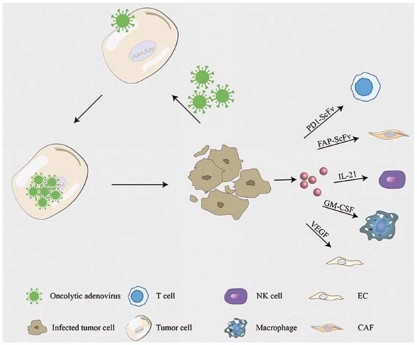
2 WANG et al: ONCOLYTIC ADENOVIRUSES REVERSE THE TUMOR MICROENVIRONMENT
surface of tumor cells and triggers their internalization (15). mast cells are recruited into the tumor, where they release
Subsequently, it enters tumor cells, viral genomes migrate to factors that promote endothelial cell proliferation to promote
the nucleus through microtubules, and early viral proteins in tumor angiogenesis (48‑50). Increasing evidence suggests that
the E1 region immediately begin to be transcribed (16). The microenvironment‑mediated external stimulation plays a key
protein binds to Rb to release the transcription factor, E2F, role in tumor cell survival and drug resistance (28,51). The
which also activates the cell cycle, allowing oncolytic adeno‑ complexity of the TME makes it difficult for the traditional
virus‑infected cells to enter the S phase (6,17). Concurrently, oncolytic virus to reverse the conditions set by the TME while
the E1A protein maintains p53 stability and inhibits tumor targeting tumor cells (27,52). The traditional oncolytic virus
growth by relying on the p53 pathway (18). The release of E2F can only inhibit the growth of tumor cells to a certain extent.
also triggers the coordinated activation of viral genes, which Owing to the constant improvement of genetic engineering
results in the production of new virions, the lysis of infected techniques, it is getting easier to develop oncolytic adenovirus
cells and the spread of viral offspring (19). The oncolytic constructs with required properties. Preclinical trials involve
adenovirus continuously replicates in tumor cells, eventually wild‑type and recombinant oncolytic adenovirus (Table I), aimed
lysing tumor cells and infecting other tumor cells via the to reverse the TME while suppressing tumor cells (6,53) (Fig. 2).
same mechanism of action (20‑23). Due to the large loading Several oncolytic adenoviruses are currently undergoing clinical
capacity of the oncolytic adenovirus vector, therapeutic genes trials as antitumor agents, and notably some progress has been
are commonly inserted into the adenovirus vector (24,25). Due made in reversing the TME (Table II). An ongoing clinical trial
to the continuous replication and accumulation of adenoviruses is testing RGD (Delta‑24‑rgd), a genetically modified oncolytic
in tumor cells, therapeutic genes are expressed and thus spread, adenovirus, as an agent against glioma. The first results obtained
playing a synergistic antitumor role (26). in the phase I trials indicate that 20% of patients showed durable
responses and CD8+ T cells infiltrated the tumor in large quanti‑
3. TME of solid tumors ties (54). TILT‑123 in preclinical studies altered the cytokine
balance in the TME towards Th1 and resulted in a significant
The TME is the internal environment for the growth of tumors, increase of the survival rate in severe combined immunodefi‑
which includes tumor cells (27,28), stromal cells (tumor‑asso‑ ciency (SCID) mice with human tumors (55,56). The currently
ciated vascular endothelial cells and tumor‑associated ongoing phase I trial is recruiting patients with solid tumors to
fibroblasts), immune cells [T lymphocytes and B lymphocytes, evaluate the safety.
tumor‑associated macrophages (TAMs), dendritic cells (DCs)
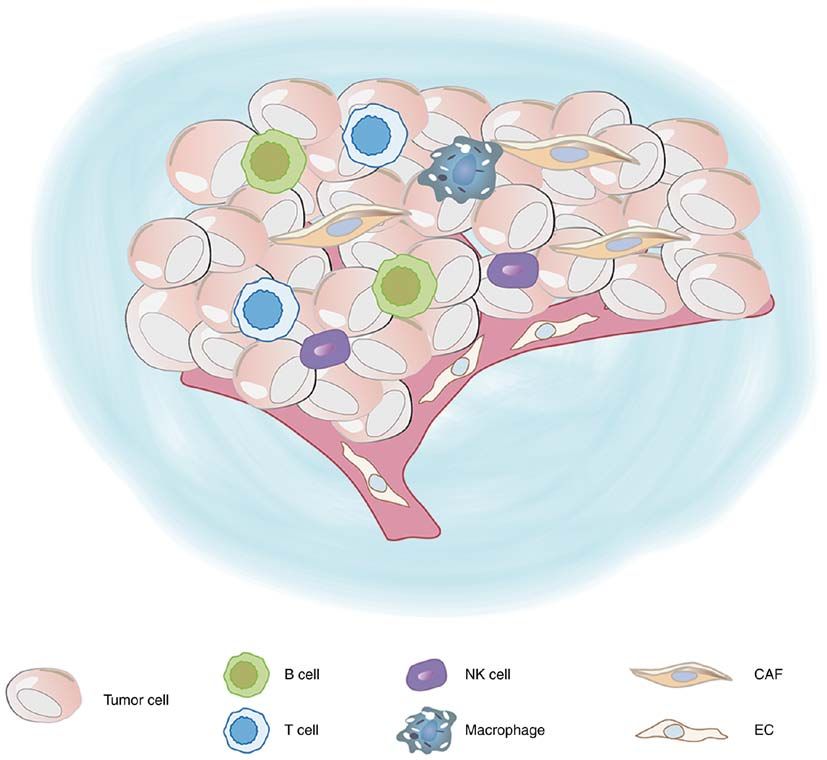
and natural killer (NK) cells], the extracellular matrix (ECM) 4. Adenovirus modification combined with immune cells
and signaling molecules such as IL‑4 and IL‑10 (29‑31). The in the TME
ECM includes various proteins, glycoproteins, proteoglycans
and other biochemical substances, which regulate vascular DCs. DCs are derived from the bone marrow and play a key
endothelial cells and fibroblasts, and promote tumor growth role in inducing and maintaining antitumor immunity (57,58).
and cell migration (32) (Fig. 1). In the TME, tumor blood Infiltration of mature DCs into the tumor can enhance immune
vessels are constantly supplying oxygen and nutrients to activation and increase the recruitment of antitumor immune
support tumor growth (28,32‑34). When the tumor is exposed effector cells and pathways (58,59). However, in the TME,
to hypoxic conditions locally, tumor blood vessels receive the antigen‑presenting function of DCs may be lost or inef‑
signal stimulation and generate branches from existing blood ficient (60). Tumor cells can inhibit the function of DCs or
vessels (35,36). However, the structure of these tumor vessels change the TME by recruiting immunosuppressive DCs (61).
differs from that of normal vessels, with absence of basement CD40 is a member of the tumor necrosis factor receptor
membranes, uneven diameter and size of the tubes, and short family and is expressed in DCs, which is a target for infil‑
circuit of arteries and veins, resulting in tumor interstitial trating T cells (62). CD40L is instantaneously expressed in
hypertension (37,38). Under hypoxic conditions, tumor cells T cells, which activates the maturation of DCs and triggers
undergo glycolysis and produce more lactic acid, which lowers the immune response. Adenovirus delivery of CD40L induces
the pH of the TME (39). Proton transport channels exist in DC activation, thereby inducing a Th1 immune response (63).
all parts of tumor tissues, which transfer the metabolized H+ The oncolytic virus restricts CD40L expression in cancer
out of tumor tissues and maintain the pH in the TME (40‑42). cells, thus decreasing systemic exposure and weakening
However, pH reduction in normal tissues results in necrosis, the systemic immune response (64,65). Currently, there are
which is more conducive to tumor metastasis and growth (43). already two phase I/II clinical trials involving LOAd703 that
Among the myeloid progenitors cells located in the TME, are recruiting patients. One of the studies is recruiting patients
myeloid‑derived suppressor cells (MDSCs), mast cells and with pancreatic cancer to evaluate whether it supports the
most TAMs play key roles in promoting tumor develop‑ current treatment standards for pancreatic cancer and whether
ment (44). MDSCs are immunosuppressive precursors of DCs, it can improve the survival rate of patients. Another study
macrophages and granulocytes (35,45). MDSCs maintain recruited patients with malignant melanoma and monitored
a normal tissue dynamic balance in response to a variety of their tumor response, immune response, virus shedding and
systemic infections and injuries (33). Several animal models survival rate. One of the major virulence factors of bacteria was
have demonstrated that MDSCs can promote tumor angiogen‑ Helicobacter pylori neutrophil‑activating protein (HP‑NAP),
esis and disrupt the main mechanisms of immune surveillance which is a TLR‑2 agonist capable of chemotaxis of neutro‑
by interfering with antigen presentation, T‑cell activation and phils, and monocytes and stimulates them to produce
NK cell killing of DCs (46,47). It has also been reported that reactive oxygen species (66,67). HP‑NAP also inducedONCOLOGY REPORTS 45: 49, 2021 3
Figure 1. Schematic diagram of the tumor microenvironment.
Th1‑polarized immune responses by stimulating the secretion carrying cytomegalovirus promoter cytosine deaminase (CD)
of interleukin (IL)‑12 and IL‑23 and other pro‑inflammatory and GM‑CSF (75). Adenovirus vectors expressing CD and
cytokines such as tumor necrosis factor (TNF)‑ α and GM‑CSF are well tolerated in refractory tumors (5). CD47
IL‑8 (68). Ag‑presenting‑HP‑NAP‑activated DCs effectively is a cell surface transmembrane protein present in normal
amplified Ag specific T cells, an important characteristic of tissues (76). It is highly expressed in malignant tumor cells and
mature DCs (69). HP‑NAP‑activated DCs resulted in Th1 binds to signaling regulatory protein‑α (SIRPα) expressed on
cytokine secretion, with high IL‑12 expression, relatively low macrophages to inhibit macrophage phagocytosis, resulting
IL‑10 secretion and migrated to CCL19 (69). in immune escape. SIRPα‑FC fusion protein inserted into the
oncolytic adenovirus vector blocks the binding of CD47 to
Macrophages. TAMs are divided into specific M1‑like macro‑ macrophages, leading to a large increase in macrophage infiltra‑
phage subsets and specific M2‑like macrophage subsets, and tion in tumor tissues, thus enhancing the antitumor effect (77).
M1‑like macrophage subsets are activated by the classical MMAD‑IL13 loaded with IL13 demonstrated enhanced anti‑
pathway and exert notable antitumor effects (70,71). In the TME, tumor effects by inducing apoptosis in the TME in vivo, and
specific M2‑like macrophage subsets are the most common, decreased the percentage of specific M2‑like macrophages (78).
and their cytokines IL‑6, TNF, IL‑1 and IL‑23 promote tumor Scott et al (79) constructed a set of bivalent and trivalent T‑cell
growth and metastasis and silence T‑cell function (72,73). adapters (BiTEs/TriTEs), which can specifically recognize
Selective removal of specific M2‑like macrophage subsets CD3ε on T cells and the folate receptor or CD206 on specific
has become a research hotspot. Granulocyte‑macrophage M2‑like macrophages. T‑cell adapters were used to specifi‑
colony‑stimulating factor (GM‑CSF) can affect macrophages, cally direct the cytotoxicity of endogenous T cells to M2‑like
promote their rapid differentiation to mature macrophages, macrophages and deplete M2‑like macrophages in tumor
prolong the life of mature macrophages, and enhance their tissues. There was a significant increase in specific M1‑like
cellular immune function (74). By inserting GM‑CSF into macrophage fraction among surviving macrophages, indicating
the Ad5 vector, ONCS‑102 induced notable antitumor immu‑ a reversal of macrophage type in the TME (79).
nity (75). ONCOS‑102 is currently being assessed in two phase I
clinical trials in advanced peritoneal malignancies and malig‑ NKs. NK cells have a notable antitumor effect in the initial stages
nant pleural mesothelioma. Ad‑CD‑GMCSF is an adenovirus of tumors, which can eliminate tumor cells (30). However, at the4 WANG et al: ONCOLYTIC ADENOVIRUSES REVERSE THE TUMOR MICROENVIRONMENT
Table I. Partial oncolytic adenovirus trials to reverse the tumor microenvironment.
Oncolytic adenovirus Gene modification Target cells in TME Target tumor cells (Refs.)
Ad3‑hTERT‑CMV‑hCD40L
CD40L DCs A549 (64)
LOAd703 CD40L DCs A549 (65)
Ad5 [i/ppt‑sNAP] HP‑NAP DCs LNCaP (69)
ONCOS‑102 GM‑CSF Macrophages, CD8+ T cells AB12 (75)
Ad‑CD‑GMCSF GM‑CSF Macrophages, CD8+ T cells Colon cancer cell line (5)
SG635‑SF A signal regulatory Macrophages SK‑OV3, HO8910 (77)
protein‑α (SIRPα)‑IgG1
Fc fusion gene
MMAD‑IL‑13 IL‑13 Macrophages Cal‑27, SCC‑4, Tca8113 (78)
EnAd BiTEs/TriTEs Macrophages DLD‑1 (79)
Ad‑CCL21‑IL21 CCL21 DCs PC‑3M, THP‑1, HeLa, Caco‑2 (82)
Ad‑CCL21‑IL21 IL‑21 NK cells PC‑3M, THP‑1, HeLa, Caco‑2 (82)
Ad‑E2F/IL15 IL‑15 NK cells, CD8+ T cells U87MG, BGC823, SW620, (84)
HCT116
ICO15K‑FBiTE FBiTE CAFs, T cells HT1080, A549 (53)
AdCEAp‑miR126/34a miR‑126, miR‑34a Vascular endothelial cells Pancreatic adenocarcinoma (96)
VEGF‑CRAd VEGF Vascular endothelial cells NCI‑H28, NCI‑H226, (97)
NCI‑H20, NCI‑H2452,
MSTO‑211H
SKL002 CTLA4 T cells HepG2, A549, Lovo, HeLa, (106)
HCT116, SW780
Ad5‑PC PD‑1 CD8+ T cells HCC‑LM3, H22, Hepa1‑6, (105)
A549, B16‑F10, LLC1
TME, tumor microenvironment; DCs, dendritic cells; NK, natural killer; CAFs, cancer‑associated fibroblasts.
advanced tumor stage, NK cells gradually lose their antitumor linked to an anti‑human CD3 single‑chain variable region (scFv)
ability and become dysfunctional (80). IL‑21 is involved in NK and loaded into oncolytic adenovirus. FAP scFv, while specifi‑
cell differentiation (81), and the oncolytic adenovirus equipped cally recognizing and targeting CAFs, activates T cells and
with IL‑21 exerts an obvious inhibitory effect on the prolifera‑ enhances T‑cell‑mediated cytotoxic effects on tumor‑associated
tion of tumor cells (82). Similarly, NK cells can be activated by fibroblasts, thus weakening the cell barrier caused by CAFs and
IL‑15 (83), and oncolytic adenovirus (Ad‑E2F/IL15), which enhancing oncolytic activity (24,53).
expresses IL‑15, can lyse tumor cells and coordinate with
immune cells to enhance the antitumor response (84). Vascular endothelial cells. Blood vessels play a vital role in
the development of tumors, providing nutrition and metastasis
5. Adenovirus modification combined with stromal cells in channels for tumor cells (93). The phenotype of vascular endo‑
the TME thelial cells changes in the TME. Tumor cells secrete vascular
endothelial growth factor (VEGF) and other endothelial growth
Cancer‑associated fibroblasts (CAFs). CAFs are a major compo‑ factors to promote the generation of tumor neovasculariza‑
nent of the tumor stroma, which regulate the TME and influence tion (94). Given that the downstream target gene of microRNA
the behavior of tumor cells, and play crucial roles in the occur‑ (miRNA/miR)‑126 is VEGF (95), miR‑126 is loaded into
rence, development, invasion and metastasis of tumors (85,86). the oncolytic adenovirus, namely ADCEAP‑miR126/34A,
Fibroblasts in the TME secrete growth factors such as hepato‑ which decreases the generation of tumor blood vessels (96).
cyte growth factor, fibroblast growth factor and CXCL12 (87), Concurrently, the VEGF promoter is inserted into the adeno‑
which promote the growth and survival of malignant cells, and virus vector, targeting the tumor vascular endothelial cells
act as chemokines to induce the migration of other cells into the via the same mechanism of action enhancing the oncolysis
TME (88,89). Concurrently, CAFs form a barrier in tumors and of adenovirus (97). IL‑24 is a tumor suppressor molecule
prevent the effective penetration and transmission of oncolytic with broad‑spectrum antitumor activity (98). It inhibits the
virus, thus limiting its efficacy (90,91). By modifying oncolytic growth of tumor cells by inhibiting tumor angiogenesis (99).
adenovirus, the effects of tumor cells and CAFs will be inhib‑ A previous study has demonstrated that while expressing
ited at the same time (92). Fibroblast activation protein‑α (FAP) IL24, CRAd‑IL24 significantly increased the release of virus
is highly expressed in CAFs. The FAP single‑chain antibody is particles and enhanced their antitumor effect (6).ONCOLOGY REPORTS 45: 49, 2021 5
Figure 2. Oncolytic adenovirus mechanism of action. When the oncolytic adenovirus specifically infects tumor cells, it releases new virus particles and
transmits them to other tumor cells. It also delivers cytokines, growth factors, fusion proteins and other substances that act on other corresponding cells in the
tumor microenvironment.
6. Adenovirus modification combined with immune CTLA‑4 antibody was significantly expressed in tumor cells
checkpoints in the TME and its antitumor activity was significantly enhanced (106).
Ad5/3‑Δ24aCTLA4 can express CTLA‑4 human intact mono‑
Checkpoint molecules are regulatory molecules that play an clonal antibody, and in the normal donors and patients with
inhibitory role in the immune system and are critical to maintain advanced solid tumors in the peripheral blood mononuclear
tolerance, prevent an autoimmune response and minimize tissue cells that were tested (107). Ad5/3‑Δ24aCTLA4 significantly
damage by controlling the timing and intensity of the immune enhanced the immune response and activated the pro‑apoptotic
response (100,101). The expression of immunological checkpoint effect of T cells (107).
molecules on immune cells suppresses the immune cell func‑
tion as the host fails to produce an effective antitumor immune 7. Outlook
response. There are numerous receptors on tumorigenic immune
escape T cells, including co‑stimulatory signal receptors that can Recently, the transformation technology of oncolytic adeno‑
stimulate T‑cell proliferation, and co‑inhibitory signal receptors viruses has significantly progressed. However, increasing
that can inhibit T‑cell proliferation (102). Immune checkpoint evidence suggest concerns about the tumor‑promoting effect
molecules are predominantly inhibitory molecules, in which the of the TME. Thus, the transformation of oncolytic viruses
immune checkpoint on T cells suppresses the immune function into TME has become a research hot spot; however, the
of T cells, causing tumor escape (103). However, the clinical in vitro simulation of the TME does not accurately reflect
benefits of monotherapy with immune checkpoint inhibitors, the human microenvironment. Cytokines and growth factors
such as anti‑programmed death‑1 antibody, are limited to were added to the cell culture, either co‑cultured with tumor
small populations (104). Zhang et al (105) designed an adeno‑ cells and other cells, or with three‑dimensional scaffoldings
virus (Ad5‑PC) to express a soluble fusion protein (programmed to simulate the TME. However, due to the complexity of the
cell death protein 1/CD137L), which significantly increased the TME, it remains difficult to completely simulate the human
number of T lymphocytes in the TME and effectively improved TME completely in vitro. Currently, xenotransplantation of
the survival rate of tumor‑bearing mice (105). After loading immunodeficient mice is the most commonly used animal
anti‑cytotoxic T lymphocyte‑associated antigen‑4 (CTLA‑4) experimental method for studying human tumors. Briefly,
antibody into the adenovirus vector, tumor cells were infected. human tumor cells are inserted into mice; however, this fails6 WANG et al: ONCOLYTIC ADENOVIRUSES REVERSE THE TUMOR MICROENVIRONMENT
Table II. Clinical trials of oncolytic adenoviruses affecting the tumor microenvironment (Jan. 2021, ClinicalTrials.gov).
Trial number Oncolytic adenovirus Study title Target Phase
NCT01582516 Delta‑24‑rgd Safety Study of Replication‑competent Recurrent glioblastoma I, II
Adenovirus (Delta‑24‑rgd) in Patients With
Recurrent Glioblastoma
NCT04695327 TILT‑123 TNFα and IL‑2 Coding Oncolytic Adenovirus Solid tumor I
TILT‑123 Monotherapy (TUNIMO)
NCT04217473 TILT‑123 TNFalpha and Interleukin 2 Coding Oncolytic Advanced melanoma I
Adenovirus TILT‑123 During TIL Treatment of
Advanced Melanoma (TUNINTIL)
NCT02705196 LOAd703 LOAd703 Oncolytic Virus Therapy for Pancreatic cancer I, II
Pancreatic Cancer
NCT04123470 LOAd703 A Phase I/II Trial Investigating LOAd703 in Malignant melanoma I, II
Combination With Atezolizumab in Malignant
Melanoma
NCT03514836 ONCOS‑102 A Phase I/II, Safety Clinical Trial of Prostate cancer I, II
DCVAC/PCa and ONCOS‑102 in Men With
Metastatic Castration‑resistant Prostate Cancer
NCT02963831 ONCOS‑102 A Phase 1/2 Study to Investigate the Safety, Advanced peritoneal I, II
Biologic and Anti‑tumor Activity of ONCOS‑102 malignancies
in Combination With Durvalumab in Subjects
With Advanced Peritoneal Malignancies
NCT04053283 NG‑641 First in Human Study With NG‑641, an Metastatic or advanced I
Oncolytic Transgene Expressing Adenoviral epithelial tumours
Vector
NCT03852511 NG‑350A First in Human Study of NG‑350A (an Metastatic or advanced I
Oncolytic Adenoviral Vector Which Expresses epithelial cancer
an Anti‑CD40 Antibody) (FORTITUDE)
NCT04097002 ORCA‑010 First in Man Clinical Study to Evaluate Safety Prostate cancer I, II
and Tolerability of an Oncolytic Adenovirus in
Prostate Cancer Patients.
to fully reflect the human TME. Several factors affect the Scholars and Innovative Research Team in University (grant
oncolytic effect of an oncolytic virus. The strong immune no. IRT_15R13), and the Guangxi Science and Technology
response and severe cytokine storm when the virus first enters Base and Talent Special Project (grant no. AD17129003).
the host may be fatal. Subsequent challenges arise at later
stages during elimination of oncolytic virus by the immune Availability of data and materials
system of the host. It is hypothesized that the selection of
oncolytic viruses will be the focus of future research, and it Not applicable.
will be individualized, with the intent that the oncolytic virus
most suitable for each patient can be selected, based on the Authors' contributions
characteristics of the tumor cells and the TME.
XW and YZ conceived the subject of the review. LZ designed
Acknowledgements the review. XW wrote the manuscript, performed the litera‑
ture research as well as interpreted the relevant literature, and
Not applicable. prepared the figures. LZ analyzed the review critically for
important intellectual content. XW and LZ edited and revised
Funding the manuscript. All authors have read and approved the final
manuscript.
This review was funded by Programs for the National Natural
Scientific Foundation of China (grant no. 81430035), the Ethics approval and consent to participate
Major National Science and Technology Projects‑Major New
Drug Creation (grant no. 2019ZX09301‑132), the Changjiang Not applicable.ONCOLOGY REPORTS 45: 49, 2021 7
Patient consent for publication 21. Rosewell Shaw A and Suzuki M: Recent advances in oncolytic
adenovirus therapies for cancer. Curr Opin Virol 21: 9‑15, 2016.
22. Ran L, Tan X, Li Y, Zhang H, Ma R, Ji T, Dong W, Tong T, Liu Y,
Not applicable. Chen D, et al: Delivery of oncolytic adenovirus into the nucleus
of tumorigenic cells by tumor microparticles for virotherapy.
Biomaterials 89: 56‑66, 2016.
Competing interests 23. Tazawaa H, Kagawab S and Fujiwarab T: Oncolytic adeno‑
virus‑induced autophagy: Tumor‑suppressive effect and molecular
The authors declare that they have no competing interests. basis. Acta Med Okayama 67: 333‑342, 2013.
24. Freedman JD, Duffy MR, Lei‑Rossmann J, Muntzer A, Scott EM,
Hagel J, Campo L, Bryant RJ, Verrill C, Lambert A, et al: An
References oncolytic virus expressing a T‑cell engager simultaneously
targets cancer and immunosuppressive stromal cells. Cancer
Res 78: 6852‑6865, 2018.
1. Giraldo NA, Sanchez‑Salas R, Peske JD, Vano Y, Becht E, 25. Jung KH, Choi IK, Lee HS, Yan HH, Son MK, Ahn HM, Hong J,
Petitprez F, Validire P, Ingels A, Cathelineau X, Fridman WH Yun CO and Hong SS: Oncolytic adenovirus expressing relaxin
and Sautès‑Fridman C: The clinical role of the TME in solid (YDC002) enhances therapeutic efficacy of gemcitabine against
cancer. Br J Cancer 120: 45‑53, 2019. pancreatic cancer. Cancer Lett 396: 155‑166, 2017.
2. Chen Y, Liu J, Wang W, Xiang L, Wang J, Liu S, Zhou H and 26. Yokoda RT, Nagalo BM and Borad MJ: Oncolytic adenoviruses
Guo Z: High expression of hnRNPA1 promotes cell invasion by in gastrointestinal cancers. Biomedicines 6: 33, 2018.
inducing EMT in gastric cancer. Oncol Rep 39: 1693‑1701, 2018. 27. Quail DF and Joyce JA: Microenvironmental regulation of tumor
3. Choi J, Gyamfi J, Jang H and Koo JS: The role of tumor‑associ‑ progression and metastasis. Nat Med 19: 1423‑1437, 2013.
ated macrophage in breast cancer biology. Histol Histopathol 33: 28. Belli C, Trapani D, Viale G, D'Amico P, Duso BA, Della Vigna P,
133‑145, 2018. Orsi F and Curigliano G: Targeting the microenvironment in
4. Sun Y: Tumor microenvironment and cancer therapy resistance. solid tumors. Cancer Treat Rev 65: 22‑32, 2018.
Cancer Lett 380: 205‑215, 2016. 29. Zhong S, Jeong JH, Chen Z, Chen Z and Luo JL: Targeting
5. Akbulut H, Coleri A, Sahin G, Tang Y and Icli F: A bicistronic tumor microenvironment by small‑molecule inhibitors. Transl
adenoviral vector carrying cytosine deaminase and granulo‑ Oncol 13: 57‑69, 2020.
cyte‑macrophage colony‑stimulating factor increases the therapeutic 30. Terren I, Orrantia A, Vitalle J, Zenarruzabeitia O and Borrego F:
efficacy of cancer gene therapy. Hum Gene Ther 30: 999‑1007, 2019. NK cell metabolism and tumor microenvironment. Front
6. Ashshi AM, El‑Shemi AG, Dmitriev IP, Kashentseva EA and Immunol 10: 2278, 2019.
Curiel DT: Combinatorial strategies based on CRAd‑IL24 and 31. Maimela NR, Liu S and Zhang Y: Fates of CD8+ T cells in tumor
CRAd‑ING4 virotherapy with anti‑angiogenesis treatment for microenvironment. Comput Struct Biotechnol J 17: 1‑13, 2018.
ovarian cancer. J Ovarian Res 9: 38, 2016. 32. Najafi M, Farhood B and Mortezaee K: Extracellular matrix (ECM)
7. Athanasopoulos T, Munye MM and Yanez‑Munoz RJ: stiffness and degradation as cancer drivers. J Cell Biochem 120:
Nonintegrating gene therapy vectors. Hematol Oncol Clin North 2782‑2790, 2019.
Am 31: 753‑770, 2017. 33. Guo S and Deng CX: Effect of stromal cells in tumor microenvi‑
8. Salzman R, Cook F, Hunt T, Malech HL, Reilly P, Foss‑Campbell B ronment on metastasis initiation. Int J Biol Sci 14: 2083‑2093, 2018.
and Barrett D: Addressing the value of gene therapy and enhancing 34. Jang I and Beningo KA: Integrins, CAFs and mechanical forces
patient access to transformative treatments. Mol Ther 26: in the progression of cancer. Cancers (Basel) 11: 721, 2019.
2717‑2726, 2018. 35. Jarosz‑Biej M, Smolarczyk R, Cichon T and Kulach N: Tumor
9. Stasiak AC and Stehle T: Human adenovirus binding to host microenvironment as A ‘Game Changer’ in cancer radiotherapy.
cell receptors: A structural view. Med Microbiol Immunol 209: Int J Mol Sci 20: 3212, 2019.
325‑333, 2020. 36. Nishide S, Uchida J, Matsunaga S, Tokudome K, Yamaguchi T,
10. Greber UF and Flatt JW: Adenovirus entry: From infection to Kabei K, Moriya T, Miura K, Nakatani T and Tomita S:
immunity. Annu Rev Virol 6: 177‑197, 2019. Prolyl‑hydroxylase inhibitors reconstitute tumor blood vessels in
11. Hoeben RC and Uil TG: Adenovirus DNA replication. Cold mice. J Pharmacol Sci 143: 122‑126, 2020.
Spring Harb Perspect Biol 5: a013003, 2013. 37. Carretero R, Sektioglu IM, Garbi N, Salgado OC, Beckhove P
12. Gao J, Zhang W and Ehrhardt A: Expanding the spectrum of adeno‑ and Hammerling GJ: Eosinophils orchestrate cancer rejection by
viral vectors for cancer therapy. Cancers (Basel) 12: 1139, 2020. normalizing tumor vessels and enhancing infiltration of CD8(+)
13. Ip WH and Dobner T: Cell transformation by the adenovirus T cells. Nat Immunol 16: 609‑617, 2015.
oncogenes E1 and E4. FEBS Lett 594: 1848‑1860, 2020. 38. Zhao H, Tian X, He L, Li Y, Pu W, Liu Q, Tang J, Wu J, Cheng X,
14. Bradley RR, Maxfield LF, Lynch DM, Iampietro MJ, Liu Y, et al: Apj+ vessels drive tumor growth and represent a
Borducchi EN and Barouch DH: Adenovirus serotype 5‑specific tractable therapeutic target. Cell Rep 25: 1241‑1254.e5, 2018.
neutralizing antibodies target multiple hexon hypervariable 39. Brand A, Singer K, Koehl GE, Kolitzus M, Schoenhammer G,
regions. J Virol 86: 1267‑1272, 2012. Thiel A, Matos C, Bruss C, Klobuch S, Peter K, et al:
15. Niemann J and Kuhnel F: Oncolytic viruses: Adenoviruses. LDHA‑Associated lactic acid production blunts tumor immuno‑
Virus Genes 53: 700‑706, 2017. surveillance by T and NK cells. Cell Metab 24: 657‑671, 2016.
16. Machitani M, Katayama K, Sakurai F, Matsui H, Yamaguchi T, 40. Fischer K, Hoffmann P, Voelkl S, Meidenbauer N, Ammer J,
Suzuki T, Miyoshi H, Kawabata K and Mizuguchi H: Edinger M, Gottfried E, Schwarz S, Rothe G, Hoves S, et al:
Development of an adenovirus vector lacking the expression of Inhibitory effect of tumor cell‑derived lactic acid on human T
virus‑associated RNAs. J Control Release 154: 285‑289, 2011. cells. Blood 109: 3812‑3819, 2007.
17. Zheng Y, Stamminger T and Hearing P: E2F/Rb family proteins 41. Colegio OR, Chu NQ, Szabo AL, Chu T, Rhebergen AM, Jairam V,
mediate interferon induced repression of adenovirus immediate Cyrus N, Brokowski CE, Eisenbarth SC, Phillips GM, et al:
early transcription to promote persistent viral infection. PLoS Functional polarization of tumour‑associated macrophages by
Pathog 12: e1005415, 2016. tumour‑derived lactic acid. Nature 513: 559‑563, 2014.
18. Yamauchi S, Zhong B, Kawamura K, Yang S, Kubo S, Shingyoji M, 42. Ohashi T, Aoki M, Tomita H, Akazawa T, Sato K, Kuze B,
Sekine I, Tada Y, Tatsumi K, Shimada H, et al: Cytotoxicity of Mizuta K, Hara A, Nagaoka H, Inoue N and Ito Y: M2‑like
replication‑competent adenoviruses powered by an exogenous macrophage polarization in high lactic acid‑producing head and
regulatory region is not linearly correlated with the viral infec‑ neck cancer. Cancer Sci 108: 1128‑1134, 2017.
tivity/gene expression or with the E1A‑activating ability but is 43. Urbanska K and Orzechowski A: Unappreciated role of LDHA
associated with the p53 genotypes. BMC Cancer 17: 622, 2017. and LDHB to control apoptosis and autophagy in tumor cells. Int
19. Cervera‑Carrascon V, Quixabeira DCA, Havunen R, Santos JM, J Mol Sci 20: 2085, 2019.
Kutvonen E, Clubb JHA, Siurala M, Heiniö C, Zafar S, 44. Fleming V, Hu X, Weber R, Nagibin V, Groth C, Altevogt P,
Koivula T, et al: Comparison of clinically relevant oncolytic Utikal J and Umansky V: Targeting myeloid‑derived suppressor
virus platforms for enhancing T cell therapy of solid tumors. Mol cells to bypass tumor‑induced immunosuppression. Front
Ther Oncolytics 17: 47‑60, 2020. Immunol 9: 398, 2018.
20. Huang H, Liu Y, Liao W, Cao Y, Liu Q, Guo Y, Lu Y and Xie Z: 45. Hinshaw DC and Shevde LA: The tumor microenviron‑
Oncolytic adenovirus programmed by synthetic gene circuit for ment innately modulates cancer progression. Cancer Res 79:
cancer immunotherapy. Nat Commun 10: 4801, 2019. 4557‑4566, 2019.8 WANG et al: ONCOLYTIC ADENOVIRUSES REVERSE THE TUMOR MICROENVIRONMENT
46. Safarzadeh E, Orangi M, Mohammadi H, Babaie F and 67. Codolo G, Fassan M, Munari F, Volpe A, Bassi P, Rugge M,
Baradaran B: Myeloid‑derived suppressor cells: Important Pagano F, D'Elios MM and de Bernard M: HP‑NAP inhibits the
contributors to tumor progression and metastasis. J Cell growth of bladder cancer in mice by activating a cytotoxic Th1
Physiol 233: 3024‑3036, 2018. response. Cancer Immunol Immunother 61: 31‑40, 2012.
47. Dysthe M and Parihar R: Myeloid‑Derived suppressor cells in the 68. D'Elios MM, Amedei A, Cappon A, Del Prete G and
tumor microenvironment. Adv Exp Med Biol 1224: 117‑140, 2020. de Bernard M: The neutrophil‑activating protein of Helicobacter
48. Ribatti D, Tamma R and Crivellato E: Cross talk between natural pylori (HP‑NAP) as an immune modulating agent. FEMS
killer cells and mast cells in tumor angiogenesis. Inflamm Immunol Med Microbiol 50: 157‑164, 2007.
Res 68: 19‑23, 2019. 69. Ramachandran M, Jin C, Yu D, Eriksson F and Essand M:
49. Albini A, Bruno A, Noonan DM and Mortara L: Contribution to Vector‑encoded Helicobacter pylori neutrophil‑activating protein
tumor angiogenesis from innate immune cells within the tumor promotes maturation of dendritic cells with Th1 polarization and
microenvironment: Implications for immunotherapy. Front improved migration. J Immunol 193: 2287‑2296, 2014.
Immunol 9: 527, 2018. 70. Mantovani A, Marchesi F, Malesci A, Laghi L and Allavena P:
50. Kabiraj A, Jaiswal R, Singh A, Gupta J, Singh A and Samadi FM: Tumour‑associated macrophages as treatment targets in oncology.
Immunohistochemical evaluation of tumor angiogenesis and the Nat Rev Clin Oncol 14: 399‑416, 2017.
role of mast cells in oral squamous cell carcinoma. J Cancer Res 71. Ubil E, Caskey L, Holtzhausen A, Hunter D, Story C and
Ther 14: 495‑502, 2018. Earp HS: Tumor‑secreted Pros1 inhibits macrophage M1 polar‑
51. Shee K, Yang W, Hinds JW, Hampsch RA, Varn FS, Traphagen NA, ization to reduce antitumor immune response. J Clin Invest 128:
Patel K, Cheng C, Jenkins NP, Kettenbach AN, et al: 2356‑2369, 2018.
Therapeutically targeting tumor microenvironment‑mediated 72. Myers KV, Amend SR and Pienta KJ: Targeting Tyro3, Axl and
drug resistance in estrogen receptor‑positive breast cancer. J Exp MerTK (TAM receptors): Implications for macrophages in the
Med 215: 895‑910, 2018. tumor microenvironment. Mol Cancer 18: 94, 2019.
52. Binnewies M, Roberts EW, Kersten K, Chan V, Fearon DF, 73. Huang YJ, Yang CK, Wei PL, Huynh TT, Whang‑Peng J,
Merad M, Coussens LM, Gabrilovich DI, Ostrand‑Rosenberg S, Meng TC, Hsiao M, Tzeng YM, Wu AT and Yen Y: Ovatodiolide
Hedrick CC, et al: Understanding the tumor immune microen‑ suppresses colon tumorigenesis and prevents polarization of M2
vironment (TIME) for effective therapy. Nat Med 24: 541‑550, tumor‑associated macrophages through YAP oncogenic path‑
2018. ways. J Hematol Oncol 10: 60, 2017.
53. de Sostoa J, Fajardo CA, Moreno R, Ramos MD, Farrera‑Sal M 74. Cho H, Seo Y, Loke KM, Kim SW, Oh SM, Kim JH, Soh J,
and Alemany R: Targeting the tumor stroma with an oncolytic Kim HS, Lee H, Kim J, et al: Cancer‑Stimulated CAFs enhance
adenovirus secreting a fibroblast activation protein‑targeted monocyte differentiation and protumoral TAM activation via IL6
bispecific T‑cell engager. J Immunother Cancer 7: 19, 2019. and GM‑CSF secretion. Clin Cancer Res 24: 5407‑5421, 2018.
54. Lang FF, Conrad C, Gomez‑Manzano C, Yung WKA, Sawaya R, 75. Kuryk L, Moller AW, Garofalo M, Cerullo V, Pesonen S, Alemany R
Weinberg JS, Prabhu SS, Rao G, Fuller GN, Aldape KD, et al: and Jaderberg M: Antitumor‑specific T‑cell responses induced by
Phase I Study of DNX‑2401 (Delta‑24‑RGD) Oncolytic oncolytic adenovirus ONCOS‑102 (AdV5/3‑D24‑GM‑CSF) in
Adenovirus: Replication and immunotherapeutic effects in peritoneal mesothelioma mouse model. J Med Virol 90: 1669‑1673,
recurrent malignant glioma. J Clin Oncol 36: 1419‑1427, 2018. 2018.
55. Havunen R, Siurala M, Sorsa S, Grönberg‑Vähä‑Koskela S, 76. Hayat SMG, Biancon V, Pirro M, Jaafari MR, Hatamipour M and
Behr M, Tähtinen S, Santos JM, Karell P, Rusanen J, Sahebkar A: CD47 role in the immune system and application to
Nettelbeck DM, et al: Oncolytic adenoviruses armed with tumor cancer therapy. Cell Oncol (Dordr.) 43: 19‑30, 2020.
necrosis factor alpha and interleukin‑2 enable successful adop‑ 77. Huang Y, Lv SQ, Liu PY, Ye ZL, Yang H, Li LF, Zhu HL, Wang Y,
tive cell therapy. Mol Ther Oncolytics 4: 77‑86, 2016. Cui LZ, Jiang DQ, et al: A SIRPα‑Fc fusion protein enhances the
56. Santos JM, Cervera‑Carrascon V, Havunen R, Zafar S, Siurala M, antitumor effect of oncolytic adenovirus against ovarian cancer.
Sorsa S, Anttila M, Kanerva A and Hemminki A: Adenovirus Mol Oncol 14: 657‑668, 2020.
coding for interleukin‑2 and tumor necrosis factor alpha replaces 78. Zhang KL, Li RP, Zhang BP, Gao ST, Li B, Huang CJ, Cao R,
lymphodepleting chemotherapy in adoptive T cell therapy. Mol Cheng JY, Xie XD, Yu ZH and Feng XY: Efficacy of a new
Ther 26: 2243‑2254, 2018. oncolytic adenovirus armed with IL‑13 against oral carcinoma
57. Lee YS and Radford KJ: The role of dendritic cells in cancer. Int models. Onco Targets Ther 12: 6515‑6523, 2019.
Rev Cell Mol Biol 348: 123‑178, 2019. 79. Scott EM, Jacobus EJ, Lyons B, Frost S, Freedman JD, Dyer A,
58. Palucka K and Banchereau J: Cancer immunotherapy via Khalique H, Taverner WK, Carr A, Champion BR, et al: Bi‑ and
dendritic cells. Nat Rev Cancer 12: 265‑277, 2012. tri‑valent T cell engagers deplete tumour‑associated macrophages
59. Parnas O, Jovanovic M, Eisenhaure TM, Herbst RH, Dixit A, in cancer patient samples. J Immunother Cancer 7: 320, 2019.
Ye CJ, Przybylski D, Platt RJ, Tirosh I, Sanjana NE, et al: A 80. Yao C, Ni Z, Gong C, Zhu X, Wang L, Xu Z, Zhou C, Li S,
genome‑wide CRISPR screen in primary immune cells to dissect Zhou W, Zou C and Zhu S: Rocaglamide enhances NK cell‑medi‑
regulatory networks. Cell 162: 675‑686, 2015. ated killing of non‑small cell lung cancer cells by inhibiting
60. Oh DS and Lee HK: Autophagy protein ATG5 regulates CD36 autophagy. Autophagy 14: 1831‑1844, 2018.
expression and anti‑tumor MHC class II antigen presentation in 81. Davis MR, Zhu Z, Hansen DM, Bai Q and Fang Y: The role of
dendritic cells. Autophagy 15: 2091‑2106, 2019. IL‑21 in immunity and cancer. Cancer Lett 358: 107‑114, 2015.
61. Chen L, Hasni MS, Jondal M and Yakimchuk K: Modification 82. Li Y, Li YF, Si CZ, Zhu YH, Jin Y, Zhu TT, Liu MY and Liu GY:
of anti‑tumor immunity by tolerogenic dendritic cells. CCL21/IL21‑armed oncolytic adenovirus enhances antitumor
Autoimmunity 50: 370‑376, 2017. activity against TERT‑positive tumor cells. Virus Res 220:
62. Karnell JL, Rieder SA, Ettinger R and Kolbeck R: Targeting 172‑178, 2016.
the CD40‑CD40L pathway in autoimmune diseases: Humoral 83. Rhode PR, Egan JO, Xu W, Hong H, Webb GM, Chen X, Liu B,
immunity and beyond. Adv Drug Deliv Rev 141: 92‑103, 2019. Zhu X, Wen J, You L, et al: Comparison of the superagonist
63. Vitale LA, Thomas LJ, He LZ, O'Neill T, Widger J, Crocker A, complex, ALT‑803, to IL15 as cancer immunotherapeutics in
Sundarapandiyan K, Storey JR, Forsberg EM, Weidlick J, et al: animal models. Cancer Immunol Res 4: 49‑60, 2016.
Development of CDX‑1140, an agonist CD40 antibody for cancer 84. Yan Y, Li S, Jia T, Du X, Xu Y, Zhao Y, Li L, Liang K, Liang W,
immunotherapy. Cancer Immunol Immunother 68: 233‑245, 2019. Sun H and Li R: Combined therapy with CTL cells and onco‑
64. Zafar S, Sorsa S, Siurala M, Hemminki O, Havunen R, lytic adenovirus expressing IL‑15‑induced enhanced antitumor
Cervera‑Carrascon V, Santos JM, Wang H, Lieber A, activity. Tumour Biol 36: 4535‑4543, 2015.
De Gruijl T, et al: CD40L coding oncolytic adenovirus allows 85. Kubo N, Araki K, Kuwano H and Shirabe K: Cancer‑associated
long‑term survival of humanized mice receiving dendritic cell fibroblasts in hepatocellular carcinoma. World J Gastroenterol 22:
therapy. Oncoimmunology 7: e1490856, 2018. 6841‑6850, 2016.
65. Eriksson E, Milenova I, Wenthe J, Moreno R, Alemany R and 86. Chen X and Song E: Turning foes to friends: Targeting
Loskog A: IL‑6 signaling blockade during CD40‑mediated cancer‑associated fibroblasts. Nat Rev Drug Discov 18: 99‑115,
immune activation favors antitumor factors by reducing TGF‑β, 2019.
collagen type I, and PD‑L1/PD‑1. J Immunol 202: 787‑798, 2019. 87. Feig C, Jones JO, Kraman M, Wells RJ, Deonarine A, Chan DS,
66. Guo X, Ding C, Lu J, Zhou T, Liang T, Ji Z, Xie P, Liu X and Connell CM, Roberts EW, Zhao Q, Caballero OL, et al: Targeting
Kang Q: HP‑NAP ameliorates OXA‑induced atopic dermatitis CXCL12 from FAP‑expressing carcinoma‑associated fibroblasts
symptoms in mice. Immunopharmacol Immunotoxicol 42: synergizes with anti‑PD‑L1 immunotherapy in pancreatic cancer.
416‑422, 2020. Proc Natl Acad Sci USA 110: 20212‑20217, 2013.ONCOLOGY REPORTS 45: 49, 2021 9
88. Erdogan B, Ao M, White LM, Means AL, Brewer BM, Yang L, 98. Chen QN, Chen X, Chen ZY, Nie FQ, Wei CC, Ma HW, Wan L,
Washington MK, Shi C, Franco OE, Weaver AM, et al: Yan S, Ren SN and Wang ZX: Long intergenic non‑coding
Cancer‑associated fibroblasts promote directional cancer cell RNA 00152 promotes lung adenocarcinoma proliferation via
migration by aligning fibronectin. J Cell Biol 216: 3799‑3816, interacting with EZH2 and repressing IL24 expression. Mol
2017. Cancer 16: 17, 2017.
89. Erdogan B and Webb DJ: Cancer‑associated fibroblasts modu‑ 99. Zhuo B, Wang R, Yin Y, Zhang H, Ma T, Liu F, Cao H and
late growth factor signaling and extracellular matrix remodeling Shi Y: Adenovirus arming human IL‑24 inhibits neuroblastoma
to regulate tumor metastasis. Biochem Soc Trans 45: 229‑236, cell proliferation in vitro and xenograft tumor growth in vivo.
2017. Tumour Biol 34: 2419‑2426, 2013.
90. Arwert EN, Milford EL, Rullan A, Derzsi S, Hooper S, Kato T, 100. Zhang Y and Zheng J: Functions of immune checkpoint molecules
Mansfield D, Melcher A, Harrington KJ and Sahai E: STING beyond immune evasion. Adv Exp Med Biol 1248: 201‑226, 2020.
and IRF3 in stromal fibroblasts enable sensing of genomic stress 101. Li K and Tian H: Development of small‑molecule immune check‑
in cancer cells to undermine oncolytic viral therapy. Nat Cell point inhibitors of PD‑1/PD‑L1 as a new therapeutic strategy for
Biol 22: 758‑766, 2020. tumour immunotherapy. J Drug Target 27: 244‑256, 2019.
91. Puig‑Saus C, Gros A, Alemany R and Cascallo M: Adenovirus 102. Kurachi M, Barnitz RA, Yosef N, Odorizzi PM, DiIorio MA,
i‑leader truncation bioselected against cancer‑associated fibro‑ Lemieux ME, Yates K, Godec J, Klatt MG, Regev A, et al: The
blasts to overcome tumor stromal barriers. Mol Ther 20: 54‑62, transcription factor BATF operates as an essential differentia‑
2012. tion checkpoint in early effector CD8+ T cells. Nat Immunol 15:
92. Jing Y, Chavez V, Ban Y, Acquavella N, El‑Ashry D, Pronin A, 373‑383, 2014.
Chen X and Merchan JR: Molecular effects of stromal‑selective 103. Deng J, Wang ES, Jenkins RW, Li S, Dries R, Yates K,
targeting by uPAR‑retargeted oncolytic virus in breast cancer. Chhabra S, Huang W, Liu H, Aref AR, et al: CDK4/6 inhibition
Mol Cancer Res 15: 1410‑1420, 2017. augments antitumor immunity by enhancing T‑cell activation.
93. Czekierdowska S, Stachowicz N, Chrosciel M and Czekierdowski A: Cancer Discov 8: 216‑233, 2018.
Proliferation and maturation of intratumoral blood vessels in women 104. Johnson DB, Sullivan RJ and Menzies AM: Immune checkpoint
with malignant ovarian tumors assessed with cancer stem cells inhibitors in challenging populations. Cancer 123: 1904‑1911,
marker nestin and platelet derived growth factor PDGF‑B. Ginekol 2017.
Pol 88: 120‑128, 2017. 105. Zhang Y, Zhang H, Wei M, Mou T, Shi T, Ma Y, Cai X, Li Y,
94. Wang R, Chadalavada K, Wilshire J, Kowalik U, Hovinga KE, Dong J and Wei J: Recombinant adenovirus expressing a soluble
Geber A, Fligelman B, Leversha M, Brennan C and Tabar V: fusion protein PD‑1/CD137L subverts the suppression of CD8+
Glioblastoma stem‑like cells give rise to tumour endothelium. T cells in HCC. Mol Ther 27: 1906‑1918, 2019.
Nature 468: 829‑833, 2010. 106. Du T, Shi G, Li YM, Zhang JF, Tian HW, Wei YQ, Deng H and
95. Wang S, Aurora AB, Johnson BA, Qi X, McAnally J, Hill JA, Yu DC: Tumor‑specific oncolytic adenoviruses expressing gran‑
Richardson JA, Bassel‑Duby R and Olson EN: The endothe‑ ulocyte macrophage colony‑stimulating factor or anti‑CTLA4
lial‑specific microRNA miR‑126 governs vascular integrity and antibody for the treatment of cancers. Cancer Gene Ther 21:
angiogenesis. Dev Cell 15: 261‑271, 2008. 340‑348, 2014.
96. Feng SD, Mao Z, Liu C, Nie YS, Sun B, Guo M and Su C: 107. Dias JD, Hemminki O, Diaconu I, Hirvinen M, Bonetti A,
Simultaneous overexpression of miR‑126 and miR‑34a induces a Guse K, Escutenaire S, Kanerva A, Pesonen S, Löskog A, et al:
superior antitumor efficacy in pancreatic adenocarcinoma. Onco Targeted cancer immunotherapy with oncolytic adenovirus
Targets Ther 10: 5591‑5604, 2017. coding for a fully human monoclonal antibody specific for
97. Harada A, Uchino J, Harada T, Nakagaki N, Hisasue J, CTLA‑4. Gene Ther 19: 988‑998, 2012.
Fujita M and Takayama K: Vascular endothelial growth factor
promoter‑based conditionally replicative adenoviruses effec‑
tively suppress growth of malignant pleural mesothelioma. This work is licensed under a Creative Commons
Cancer Sci 108: 116‑123, 2017. Attribution-NonCommercial-NoDerivatives 4.0
International (CC BY-NC-ND 4.0) License.You can also read