The contribution of ebv to the pathogenesis of classical hodgkin lymphoma - Annals of Lymphoma
←
→
Page content transcription
If your browser does not render page correctly, please read the page content below
Review Article
                                                                                                                                        Page 1 of 13

The contribution of ebv to the pathogenesis of classical hodgkin
lymphoma
Katerina Vrzalikova1^, Matthew Pugh1, Lucia Mundo2,3, Paul Murray1,3,4^
1
    Institute of Immunology & Immunotherapy, University of Birmingham, Birmingham, UK; 2Department of Medical Biotechnology, University of
Siena, Siena, Italy; 3Health Research Institute, University of Limerick, Limerick, Ireland; 4Department of Clinical and Molecular Pathology, Institute
of Molecular and Translational Medicine, Faculty of Medicine and Dentistry, Palacky University, Olomouc, Czech Republic
Contributions: (I) Conception and design: All authors; (II) Administrative support: None; (III) Provision of study materials or patients: None;
(IV) Collection and assembly of data: None; (V) Data analysis and interpretation: None; (VI) Manuscript writing: All authors; (VII) Final approval of
manuscript: All authors.
Correspondence to: Paul Murray. Health Research Institute, University of Limerick, Limerick, Ireland. Email: paul.murray@ul.ie.

                  Abstract: Hodgkin lymphoma (HL) exists in two major forms, the so-called classical type (cHL) and the
                  nodular lymphocyte predominant type (NLPHL). Since NLPHL is considered to be an EBV-negative
                  entity, this review focusses only on the cHL form. Although cHL is a curable disease in many cases, we
                  still lack an understanding of its pathogenesis that could lead to kinder treatments for patients, and also
                  to more effective therapies for the smaller subset of patients who are destined to die of their disease. One
                  approach might be to therapeutically target the Epstein-Barr virus (EBV), which is present in up to one-half
                  of cases in resource rich nations, and in almost all cases in some resource-poor countries. However, it has
                  been suggested that EBV might be simply a silent passenger in cHL. In this review, we present evidence in
                  support of a crucial role for EBV in virus-positive cHL. In particular, we highlight important epidemiological
                  differences between EBV-positive and EBV-negative cHL that suggest different aetiologies, as well as
                  genetic differences, including a different profile of somatic mutations pointing to a distinct contribution for
                  EBV in substituting for cellular genetic changes that are required for disease development when the virus is
                  absent. We also focus attention on important roles for the individual latent virus genes in the pathogenesis of
                  cHL. Overall, this review suggests that a better understanding of how EBV contributes to the pathogenesis
                  of cHL may eventually lead to improved stratification of patients and to the development of therapies that
                  specifically target EBV or its latent genes.

                  Keywords: Hodgkin lymphoma; Epstein-Barr virus (EBV); transformation

                  Received: 12 February 2021; Accepted: 16 April 2021.
                  doi: 10.21037/aol-21-8
                  View this article at: http://dx.doi.org/10.21037/aol-21-8

Introduction                                                                  to the pathogenesis of cHL, particularly with respect to the
                                                                              role of the viral latent genes, some of which are now being
The oncogenic Epstein-Barr virus (EBV) is present                             explored as targets of new drug and immunotherapeutic
in a subset of cases of classical Hodgkin lymphoma                            approaches.
(cHL). Despite growing evidence of its importance in
the pathogenesis of the subset of virus-associated cHL,
                                                                              General features and pathogenesis of HL
therapies that specifically target EBV are lacking. We
believe it is now timely to revisit the contribution of EBV                   Hodgkin lymphoma (HL) has an incidence of around

^ ORCID: Katerina Vrzalikova: 0000-0002-8754-6633; Paul Murray 0000-0003-0956-6468.

© Annals of Lymphoma. All rights reserved.                                                Ann Lymphoma 2021 | http://dx.doi.org/10.21037/aol-21-8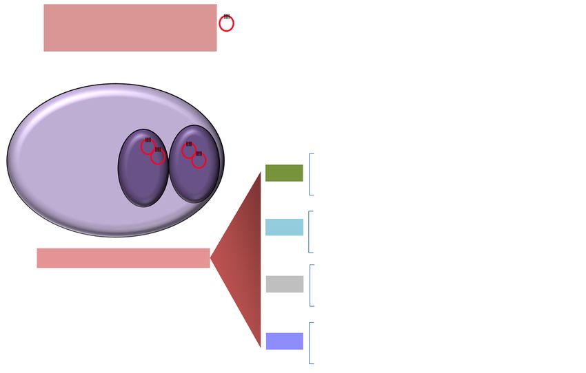
Page 2 of 13                                                                                          Annals of Lymphoma, 2021

3/100,000 per year. Lymph nodes are the most often                   The JAK/STAT signalling pathway is also critically
affected tissues, although extranodal disease does occur.         involved in cHL pathogenesis and may be activated via
Affected tissues show effacement of existing structures           the cytokines which are produced either by HRS cells
which are replaced by rare malignant cells, surrounded by         or by cells of the TME; this in turn elevates the levels of
a florid reactive infiltrate. Based on differences in histology   phosphorylated forms of STATs (e.g., STAT3, STAT5A
and immune phenotype, HL can be classified into two               and STAT6) (20-22). JAK/STAT signalling can also
major types; these are classical HL (cHL) and nodular             be aberrantly stimulated by genetic lesions such as the
lymphocyte predominant (NLP) HL (1). The malignant                amplification of JAK2 or loss-of-function mutations in
cells of cHL are known as Hodgkin/Reed-Sternberg (HRS)            SOCS1 and PTPN1/PTPB1 which are negative regulators
cells, and those of NLPHL as lymphocyte predominant               of these pathways (23-25).
(LP) cells. There are four major subtypes of cHL, known
as mixed cellularity, nodular sclerosis, lymphocyte rich
                                                                  Identification of EBV in cHL
and lymphocyte depleted HL. The tumour cells of HL
are derived from mature B lymphocytes (2), and display            EBV was initially suggested to be involved in the
evidence of somatic hypermutation indicating that they            pathogenesis of cHL after it was shown that patients had
develop from germinal centre, or more likely, post-               raised antibody levels to EBV antigens in their blood (26).
germinal centre B cells (2-5). In approximately one-              Moreover, elevated levels were also shown to be present in
quarter of cases of cHL, the immunoglobulin genes have            patients before the onset of cHL (27).
so called ‘crippling’ mutations which prevent proper                  Southern blotting for EBV DNA was first used to
expression of surface immunoglobulin (3). In fact it is now       identify the presence of EBV DNA in the tissues of cHL
believed that the loss of B cell receptor (BCR) functions is      patients (28). EBV was localized to HRS cells using the
probably directly involved in the pathogenesis of most, if        anti-complement immune fluorescence assay (29), by
not all, cases of cHL.                                            in situ hybridization (ISH) for EBV DNA (30,31), and by
    Most B cells require signalling through the BCR for           ISH for two RNA species known as Epstein-Barr virus
their survival, yet HRS cells do not appear to need these         encoded RNAs (EBER1 and EBER2) (32). An aetiological
survival signals. These observations suggest that a crucial       role for EBV in cHL was supported by the detection of viral
event must be the acquisition of mechanisms that prevent          genomes bearing identical fusion sequences in biopsies.
the apoptosis that would be the normal fate of germinal           These sequences are created when the circular genomes are
centre B cells lacking a functional BCR. Multiple cell            formed from linear virus DNA and are unique to a single
signalling pathways are aberrantly activated in HRS               infection event. Thus, monoclonal viral genomes present
cells, many of which contribute to this anti-apoptotic            in cHL indicate that infection is an early event (30). EBV is
phenotype. For example, HRS cells display constitutive            consistently retained during disease progression, suggesting
activation of a family of transcription factors known as          it is required for maintenance of the tumour phenotype (33).
nuclear factor kappa B (NF-κB) (6). Inhibition of NF-
κB signalling in HL cell lines increases their sensitivity to
                                                                  Epidemiology of EBV-associated cHL
apoptosis and impairs tumourigenicity in severe combined
immunodeficiency mice (7,8). HRS cells express multiple           EBV is associated with cHL, but not with NLPHL.
tumour necrosis factor receptors, including CD30, CD40,           However, not all cases of cHL are EBV-positive. Moreover,
TACI, BCMA and RANK which can induce the activation               the proportion of positive cases varies within different
of NF-κB signalling following their engagement with               populations (34,35). Thus, in resource-rich nations, EBV-
ligands expressed on immune and other cells of the tumour         positive rates range between 20% to 50%; here more EBV-
microenvironment (TME) (9,10). Constitutive NF-κB                 positive cases are seen in older people and in children, and
activation can also be caused by different genetic lesions        fewer in young adults (36,37). In contrast, EBV positive
in HRS cells, including amplification of the gene encoding        rates are often substantially higher in resource-poor
the c-REL subunit of NF-κB, (11-13), mutations in genes           countries (38,39). EBV-positive disease is more common in
encoding inhibitors, IκB alpha and IκB epsilon (14-18),           males, in patients with mixed cellularity disease, and in some
and TNFAIP3/A20, a ubiquitin modifying enzyme that                ethnic groups, even when taking into account potential
inhibits NF-κB signalling (19).                                   confounding factors, such as socioeconomic status (40).

© Annals of Lymphoma. All rights reserved.                                  Ann Lymphoma 2021 | http://dx.doi.org/10.21037/aol-21-8
Annals of Lymphoma, 2021 Page 3 of 13
RISK FACTORS
HLA type: HLA-A*01 (increased risk), HLA-A*02 (reduced risk)
Prior history of symptomatic primary infection (infectious
mononucleosis)
EBV-positive Hodgkin lymphoma,
Elevated antibody levels to the EBV viral capsid and early lytic showing LMP1 expression in HRS
antigens cells
Immune suppression (e.g. HIV infection) and/or immune
senescence (older age)
Figure 1 Risk factors for the development of EBV-positive classical Hodgkin lymphoma.
The incidence of EBV-positive cHL is also more interact with B cells in the normal asymptomatic host.
common in HIV-positive individuals especially when the Infection of resting B lymphocytes by EBV in vitro can
levels of immune impairment are at intermediate levels lead to their transformation, giving rise to continuously
(41,42). Thus, declining levels of EBV-specific immunity growing cell lines, referred to as lymphoblastoid cell lines
in the early stages of HIV infection contribute to an (LCL). If lymphocytes from the blood are used, the T
increased risk of EBV-positive HL. However, this risk of lymphocytes present must be depleted or suppressed by drugs
developing cHL declines as CD4+ T cell numbers fall such as cyclosporin A (54), demonstrating the importance of
further, emphasising the importance of CD4+ T cells in the the T cell response in controlling EBV infection in vivo.
pathogenesis of cHL. EBV encodes a small subset of its genes in the latency
First degree relatives of patients with cHL patients have phase of the virus life cycle which is characterized by the
between a 3-fold and 9-fold increased risk of developing the absence of virus replication. These ‘latent’ genes include
disease (43,44), this rises to 100-fold for monozygotic twins nuclear antigens, known as EBNAs (EBNAs 1, 2, 3A, 3B,
compared with dizygotic twins (45). Susceptibility loci exist 3C, EBNA-LP), latent membrane proteins (LMP1, LMP2),
within the human leukocyte antigen (HLA) region (46,47); as well as two non-coding Epstein-Barr-encoded RNA
HLA-A*01 and HLA-A*02 alleles confer an increased and (EBER1, EBER2), and viral miRNA (55,56). Some latent
decreased risk of EBV-positive cHL, respectively (47-49) genes, for example, EBNA2 and LMP1 are necessary for
(Figure 1). These findings illustrate the potential importance the transformation of B cells, at least in the laboratory (57).
of immune control of the virus in the pathogenesis of the Latency III is the term used to describe the type of latency
EBV-positive form of cHL (50). A prior history of infectious displayed by LCL; here all known latent genes are present.
mononucleosis (IM) is associated with an increased risk Restricted forms of latency are observed during the ‘normal’
of developing EBV-positive, but not EBV-negative, cHL differentiation of EBV-infected B cells in vivo, as well as in
(51-53) and some of the same HLA associations are also EBV-associated malignancies, including cHL. EBV can also
observed for IM (Figure 1). express ‘lytic’ genes when the virus shifts to its replicative
cycle ending in the release of infectious virus particles.
EBV potently transforms B lymphocytes in vitro
EBV infection of B cells is characteristic of the
Before considering how EBV might contribute to the
asymptomatic carrier state
development of cHL, we briefly outline what is known about
the transforming potential of EBV and how the virus might EBV has evolved to survive for the life-time of the
© Annals of Lymphoma. All rights reserved. Ann Lymphoma 2021 | http://dx.doi.org/10.21037/aol-21-8Page 4 of 13 Annals of Lymphoma, 2021
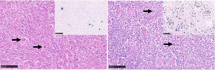
classical Hodgkin lymphoma EBV-positive mucocutaneous ulcer
Figure 2 Morphology and EBER-positivity in classical Hodgkin lymphoma and EBV-positive mucocutaneous ulcer: HRS cells (cHL) and
HRS-like cells (EBV + MCU) are arrowed. EBER staining is shown in the inset.
asymptomatic host, and does so using a highly evolved alternative view of the establishment of the carrier state
mechanism that relies on the colonisation of memory B which is based on the observation that EBV-infected
cells (58). How EBV eventually gets into memory B cells cells in GCs of patients with IM show evidence of
has been difficult to fully define, mainly because primary somatic hypermutation, but without evidence of intra-
infection itself largely goes unnoticed and when IM does clonal diversity; this means that these cells are no longer
occur after primary infection, by the time it becomes undergoing the hypermutation process (63,64). Moreover,
symptomatic, the early events in virus infection have already LMP1 has been shown to be present in EBV-infected cells
taken place. To further exacerbate an already difficult present outside the GC, potentially counteracting the
problem, the virus colonises only a minute fraction of B cells argument that LMP1 is driving a GC reaction (65). Thus,
in asymptomatic carriers (perhaps as low as 1 in 106 B cells). this alternative model proposes that EBV directly infects
Notwithstanding these difficulties, the favoured model of memory B cells. Moreover, ‘non-switched’ memory B cells,
EBV persistence suggests that initial infection of B cells by which do not require GC activity can also harbour EBV
EBV causes them to enter the cell cycle, and to express the (66-68). EBV infection can induce expression of activation-
latency III pattern of viral gene expression; in this respect induced cytosine deaminase (AID) to stimulate somatic
the initially infected B cells are probably similar to LCL. hypermutation (69).
These EBV-infected B cells may then adopt a phenotype
more closely resembling a germinal centre (GC) B cell, this
EBV latent genes are important contributors to
time they adopt a latency II programme in which EBNA1,
the pathogenesis of cHL
but not the other EBNAs, and the latent membrane
proteins are present (59). This pattern of virus expression EBV-infected HRS cells express a restricted pattern of virus
mirrors that seen in cHL. At this stage, LMP1 and LMP2 latency characterised by the presence of EBV’s maintenance
provide the EBV-infected B cells with surrogate CD40 protein, EBNA1, as well as both latent membrane proteins.
and B cell receptor (BCR) signals, respectively, and allow A subset of viral miRNA are also expressed. While the
the EBV-infected GC B cells to survive and subsequently contribution of the EBV latent proteins to the pathogenesis
to differentiate into memory B cells (60,61). The EBV- of cHL is increasingly better understood (70), the roles
infected memory B cells no longer express virus genes (a of the EBV miRNAs have only just begun to be explored
state known as latency 0), but can if necessary switch on (Figure 2).
EBNA1 expression to allow them to proliferate (this stage is
known as latency I) (59). EBV-infected B cells can become
Epstein-Barr virus nuclear antigen-1 (EBNA1)
plasma cells; in this case the lytic cycle is induced, eventually
leading to virion release in oropharyngeal secretions. This EBNA1 is essential for the maintenance of EBV episomes
is probably the major way in which the virus to transmitted in infected cells, as it is a key viral replication factor and is
from host to host (62). responsible for tethering viral genomes to the chromosomes
It is important to also mention here that there is an of the host cell; loss of EBNA1 expression therefore results
© Annals of Lymphoma. All rights reserved. Ann Lymphoma 2021 | http://dx.doi.org/10.21037/aol-21-8Annals of Lymphoma, 2021 Page 5 of 13
in the loss of EBV genomes during cell division (71). to the transcriptional programme of cHL, for example by
EBNA1 is also a transcription factor that can bind to viral reducing the expression of numerous B cell transcription
and cellular promoters (72-74). EBNA1 inhibits TGFβ factors, including EBF1 and E2A (90-93).
signalling, in part by modulating the levels of SMAD2.
EBNA1 also decreases expression of the TGFβ target gene,
EBV-encoded miRNA
PTPRK, which in turn promotes the growth and survival of
HRS cells (75,76). LCL express a subset of BART miRNA (including
approximately half of Cluster 2) as well as 3 of the 4
BHRF1 miRNAs, that are subsequently turned off in GC
Latent membrane protein-1
B cells and memory B cells. In parallel, EBV-infected GC
LMP1 is a constitutively active homologue of CD40 that B cells and memory B cells up-regulate the remaining
has been shown to activate NF-κB, JAK/STAT, AP-1 and BART miRNAs by 5–10 fold (94). In EBV associated
phosphatidylinositol-3 kinase (PI3K)/AKT signalling (77-79). tumours expressing a latency II phenotype, including
While LMP1 might be responsible for activating these cHL, the latency III associated BART but not BHRF1
pathways in EBV-positive cHL, mutations in components miRNAs are up-regulated (95). Among these, miR-
of these signalling pathways appear to be necessary when BART2-5p is expressed in EBV-positive cHL and can act
the virus is absent. For example, TNFAIP3 mutations are as an anti-sense miRNA to the EBV DNA polymerase
more commonly seen in EBV-negative cHL, suggesting that BALF5 to inhibit virus replication (96). miR-BART2-5p
mutational inactivation of TNFAIP3 and EBV infection are also decreases BCR-mediated NF-kappa-B activation (97).
alternative pathways both leading to deregulated NF-κB miR-BART13-3p is one of the most highly expressed viral
signalling (19). The requirement for more genetic changes in miRNA in cHL, and can be released into the circulation
EBV-negative cHL is supported by other studies which show via exosomes (98). miR-BARTs in exosomes derived from
that EBV-positive cHL has significantly fewer chromosome EBV-positive cells have been shown to induce changes in
abnormalities than EBV-negative cHL (80-82). LMP1 also the phenotype of macrophages, including the increased
induces many of the features of the aberrant transcriptional production of cytokines, such TNF-α, and IL-10 (99).
programme characteristic of HRS cells, including the down- EBV also influences host miRNA expression in cHL.
regulation of BCR signalling components, and the increased For example, Navarro et al. observed a subset of 10 host
expression of anti-apoptotic genes such as BCL2 and BFL-1 miRNAs whose expression was influenced by the presence
(83,84). The FLICE-inhibitory protein (c-FLIP), a negative of EBV. Among these, miR-96, miR-128a, miR-128b were
regulator of Fas induced apoptosis (85), is also an LMP1 selectively down regulated in EBV-positive cHL. The
target and could be important in the rescue of pre-apoptotic authors also reported a distinctive signature of 25 miRNAs
GC B cells early in cHL pathogenesis (86). LMP1 also that were differentially expressed between cHL and reactive
increases the expression of Programmed death ligand-1 (PD- lymph nodes. Interestingly, the dysregulation of miR-21
L1) in B cells, which might account at least in part for the that favours cell survival by indirectly up-regulating anti-
sensitivity of cHL to immune checkpoint inhbitors targeting apoptotic genes suggests an important role for cellular
the PD-L1/PD-1 axis, although definitive studies assessing miRNAs in the biology of cHL (100).
the sensitivity of EBV-positive versus EBV-negative cHL to
immune checkpoint blockade have not been done (87).
Co-expression of latent genes
A drawback of many of the studies investigating the
Latent membrane protein-2A (LMP2A)
function of individual virus genes has been the reliance
EBV appears to be crucial for the survival of HRS on single gene over-expression or knockdown/knockout
progenitors harbouring so called ‘crippling’ mutations in experiments; an approach that cannot take account of the
immunoglobulin genes; these mutations are found almost effects of the co-expression of virus genes. This could be
exclusively in EBV positive cases (88). EBV efficiently important, particularly because some studies have shown
immortalises BCR-negative GC B cells in vitro; an effect that some of the latent genes, for example, LMP1 and
that is dependent upon LMP2A which provides a BCR- LMP2A have partially overlapping, as well as counteracting,
like signalling function (61,89). LMP2A also contributes transcriptional programmes (101). Moreover, it has been
© Annals of Lymphoma. All rights reserved. Ann Lymphoma 2021 | http://dx.doi.org/10.21037/aol-21-8Page 6 of 13 Annals of Lymphoma, 2021
shown that when expressed in the B cells of transgenic typical HRS cell morphology and the characteristic cellular
mice, LMP1 is transforming, but not when LMP2A is infiltrate supported by additional immunohistochemistry
co-expressed (102). In another study it was shown that stains for example for recognition of CD30-positive
LMP1 and LMP2A co-expression in mouse B cells led HRS cells. However, a number of EBV-associated
to tumour development, but only if the animals were lymphoproliferative disorders (LPD) can harbour EBV-
immunosuppressed, suggesting that a background of positive HRS-like B-cells, morphologically mimicking EBV-
immune impairment is important (103). Studies exploring positive cHL. Many of these LPD show distinct clinical
the co-expression of virus genes in cHL at single cell behaviour and require radically different management
resolution are required. approaches from cHL, potentially leading to over- or
undertreatment if not recognised by the pathologist. Whilst
cHL is typically a nodal disease, extranodal extension can
EBV and the tumour microenvironment of cHL
be seen in late stage disease (116). Furthermore, extremely
The TME is a defining feature of cHL and there is rare manifestations of primary extra-nodal cHL have been
some evidence that EBV is at least partly responsible for described (117). Similarly, many of the mimics of EBV-
reshaping this TME through expression of the latent positive cHL can present as nodal or extranodal disease and
genes, and also potentially through limited induction of thus have to be considered in the differential diagnosis.
the lytic cycle. For example, LMP1 an increase production EBV-positive diffuse large B-cell lymphoma (DLBCL),
of a diverse array of different chemokines and cytokines NOS, is systemic lymphoma typically presenting with
(104,105). EBNA1 can also influence the transcription prominent extranodal involvement in the elderly, although
of genes encoding key soluble factors, for example the younger patients can also be affected (118). EBV-positive
chemokine CCL20 which is important in the chemo- DLBCL, NOS, can show large pleomorphic HRS-like
attraction of regulatory T cells (106). Conversely, the cHL lesional B-cells which can associated with a prominent
TME of cHL could also modulate viral gene expression inflammatory milieu. On immunophenotyping, CD30
in tumour cells. A good example here is the expression expression is usually seen in the lesional cells and can co-exist
of LMP1 which can be regulated by different cytokines with CD15 expression in 68% of cases (119). Expression
(107,108). The TME could also modify EBV’s oncogenic of CD20 is an important marker in distinguishing EBV+
functions. For example, LMP1 can increase expression of DLBCL, NOS, from EBV+ cHL. However, CD20 can
discoidin domain receptor 1 (DDR1), a collagen receptor show variable expression in EBV+ DLBCL, NOS, as over
leading to the increased survival of lymphoma cells (109). 50% of lesional cells are typically positive.
The function of EBV-specific T cells in the TME of EBV-positive mucocutaneous ulcer (EBVMCU) is a
cHL might be compromised by a variety of immune evasion localised ulcerating EBV-positive LPD affecting the skin
mechanisms. Thus, loss of HLA expression is frequently or mucous membranes (120) (Figure 3). EBVMCU are
observed in HRS cells, but is more commonly found in usually solitary but multi-focal manifestations have been
EBV-negative cHL (110-112); in some cases this is caused described (121). The lesions are typically associated with
by inactivating mutations in the beta-2-microglobulin immunosuppression or in older patients, are assumed to be
gene (113). EBV-positive cases of cHL usually express consequence of immunosenescence (120). EBVMCU is an
normal or sometimes higher levels of HLA expression, and indolent disease and usually resolves upon once the source
contain more activated CTLs than EBV-negative cases of immunosuppression is removed. Rituximab has been used
(110-112,114). EBV-positive cHL also contains more NK with some success in refractory cases.
cells (115). These data suggest that EBV utilises other Some lymphomas, not in themselves associated with
mechanisms to evade anti-tumour immune responses EBV, can show EBV-positive HRS-like B-cells in the
in cHL, including the induction of immune checkpoint background. The archetypal example is angioimmunoblastic
molecules, such as PD-L1 (87). lymphoma (AIL), a T-cell lymphoma with a T-follicular
helper(Tfh) cell phenotype (118). Up to 95% of AIL can
show EBV+ B-cell blasts in the background. In some
Difficulties in the diagnosis of EBV-positive cHL
cases, the B-cell component in the background of AIL can
The diagnosis of cHL is dependent upon morphological progress to EBV+ DLBCL or EBV+ cHL (122). Other
evaluation by a haematopathologist who can recognize the lymphomas accompanied by EBV+ B-cell blasts have also
© Annals of Lymphoma. All rights reserved. Ann Lymphoma 2021 | http://dx.doi.org/10.21037/aol-21-8Annals of Lymphoma, 2021 Page 7 of 13
HRS cells have monoclonal EBV
genomes that are retained during
disease progression
Activates cellular signalling pathways e.g. NF-κB, JAK/STAT, PI3-K/Akt
HRS cell LMP1 Contributes to evasion of T cell immunity e.g. through PD-L1 induction
Recruits tumour microenvironment that supports HRS cell survival
LMP2A Activates cellular signalling pathways e.g. PI3-K/Akt
Allows survival of BCR-negative HRS precursors
HRS cells express latent genes
Ensures viral genomes are retained during cell division
EBNA1 Contributes to viral and cellular transcription
Recruits regulatory T cells
Could suppress lytic cycle and BCR signalling
miRNA
Potential role in immune evasion
Figure 3 Contribution of EBV latent genes to the pathogenesis of classical Hodgkin lymphoma.
been described in the background of other EBV-negative positive LPD extremely difficult.
lymphomas including chronic lymphocytic leukaemia and
mantle zone lymphoma (123,124).
Concluding remarks
EBV-positive manifestations of lymphomas, traditionally
considered not to be associated with EBV, have now A subset of cHL patients harbor EBV in their tumour cells.
been described, thus broadening the range of potential Although the contribution of EBV to the pathogenesis
differential diagnoses. Rare examples of EBV-positive of cHL has been debated, we argue that differences in
nodular lymphocyte predominant Hodgkin lymphoma, the epidemiology, genetics, and biology of EBV-positive
primary mediastinal B-cell lymphoma and EBV-positive compared with EBV-negative cHL strongly suggest a key
mediastinal grey-zone lymphoma (PMGZL) (intermediate role for the virus. Notwithstanding debate over EBV’s exact
features between primary mediastinal lymphoma and contribution to the oncogenic process in cHL, there is
Hodgkin lymphoma) have been described (125,126). undoubtedly an opportunity to exploit the presence of EBV
The diagnosis of mediastinal grey-zone lymphoma is in tumours for patient benefit. In some LPD and in other
further complicated, as both cHL and PMGZL can occur EBV-associated tumours such as nasopharyngeal carcinoma,
in the same vicinity (composite lymphoma), or develop EBV detection in blood and other tissues can be a useful
sequentially, where EBV status has been reported to change biomarker. However, as yet routine blood testing for EBV
from the initial to subsequent biopsy. The diagnosis of these is not employed in the management of the majority of
entities necessitates the presence of all the typical diagnostic EBV-associated malignancies, including cHL. Moreover,
features of the EBV-negative variant to be present in order the development of new approaches to specifically target
to make the diagnosis. Nonetheless, the recognition of these EBV, for example, by reactivating the virus into the lytic
entities can make distinction between the various EBV- cycle (and thereby killing the infected cell, or making it
© Annals of Lymphoma. All rights reserved. Ann Lymphoma 2021 | http://dx.doi.org/10.21037/aol-21-8Page 8 of 13 Annals of Lymphoma, 2021
susceptible to drugs such as ganciclovir) (127-129), gene Classification of Tumours of Haematopoietic and
therapy (130), EBNA1 inhibitors (131,132), and therapeutic Lymphoid Tissues. 4th ed. Lyon, France: IARC Press;
vaccination/T cell therapies (133,134) are provide exciting 2008.
new opportunities to improve outcomes for patients. 2. Kuppers R, Rajewsky K, Zhao M, et al. Hodgkin
disease: Hodgkin and Reed-Sternberg cells picked from
histological sections show clonal immunoglobulin gene
Acknowledgments
rearrangements and appear to be derived from B cells at
We are grateful to the Children’s Cancer and Leukaemia various stages of development. Proc Natl Acad Sci U S A
Group (CCLG; PM, KV), Blood Cancer UK (PM, KV), 1994;91:10962-6.
the Medical Research Council, UK (MP) and an EU Marie 3. Kanzler H, Kuppers R, Hansmann ML, et al. Hodgkin
Curie Sklodowska Fellowship (LM). and Reed-Sternberg cells in Hodgkin's disease represent
Funding: The work was supported in part by a European the outgrowth of a dominant tumor clone derived
Regional Development Fund Project (ENOCH: CZ.02.1.0 from (crippled) germinal center B cells. J Exp Med
1/0.0/0.0/16_019/0000868). 1996;184:1495-505.
4. Vockerodt M, Soares M, Kanzler H, et al. Detection of
clonal Hodgkin and Reed-Sternberg cells with identical
Footnote
somatically mutated and rearranged VH genes in
Provenance and Peer Review: This article was commissioned different biopsies in relapsed Hodgkin's disease. Blood
by the Guest Editors (Christopher P. Fox, Claire Shannon- 1998;92:2899-907.
Lowe) for the series “Lymphoma and Viruses” published 5. Marafioti T, Hummel M, Foss HD, et al. Hodgkin
in Annals of Lymphoma. The article has undergone external and reed-sternberg cells represent an expansion of a
peer review. single clone originating from a germinal center B-cell
with functional immunoglobulin gene rearrangements
Conflicts of Interest: All authors have completed the ICMJE but defective immunoglobulin transcription. Blood
uniform disclosure form (available at http://dx.doi. 2000;95:1443-50.
org/10.21037/aol-21-8). The series “Lymphoma and 6. Bargou RC, Leng C, Krappmann D, et al. High-level
Viruses” was commissioned by the editorial office without nuclear NF-kappa B and Oct-2 is a common feature
any funding or sponsorship. The authors have no other of cultured Hodgkin/Reed-Sternberg cells. Blood
conflicts of interest to declare. 1996;87:4340-7.
7. Izban KF, Ergin M, Huang Q, et al. Characterization of
Ethical Statement: The authors are accountable for all NF-kappaB expression in Hodgkin's disease: inhibition of
aspects of the work in ensuring that questions related constitutively expressed NF-kappaB results in spontaneous
to the accuracy or integrity of any part of the work are caspase-independent apoptosis in Hodgkin and Reed-
appropriately investigated and resolved. Sternberg cells. Mod Pathol 2001;14:297-310.
8. Bargou RC, Emmerich F, Krappmann D, et al.
Open Access Statement: This is an Open Access article Constitutive nuclear factor-kappaB-RelA activation is
distributed in accordance with the Creative Commons required for proliferation and survival of Hodgkin's disease
Attribution-NonCommercial-NoDerivs 4.0 International tumor cells. J Clin Invest 1997;100:2961-9.
License (CC BY-NC-ND 4.0), which permits the non- 9. Carbone A, Gloghini A, Gruss HJ, et al. CD40 ligand is
commercial replication and distribution of the article with constitutively expressed in a subset of T cell lymphomas
the strict proviso that no changes or edits are made and the and on the microenvironmental reactive T cells of
original work is properly cited (including links to both the follicular lymphomas and Hodgkin's disease. Am J Pathol
formal publication through the relevant DOI and the license). 1995;147:912-22.
See: https://creativecommons.org/licenses/by-nc-nd/4.0/. 10. Pinto A, Aldinucci D, Gloghini A, et al. The role of
eosinophils in the pathobiology of Hodgkin's disease. Ann
Oncol 1997;8 Suppl 2:89-96.
References
11. Barth TF, Martin-Subero JI, Joos S, et al. Gains of 2p
1. Swerdlow SH, Campo E, Harris NL, et al. WHO involving the REL locus correlate with nuclear c-Rel
© Annals of Lymphoma. All rights reserved. Ann Lymphoma 2021 | http://dx.doi.org/10.21037/aol-21-8Annals of Lymphoma, 2021 Page 9 of 13
protein accumulation in neoplastic cells of classical of the tumor suppressor gene SOCS-1 in classical
Hodgkin lymphoma. Blood 2003;101:3681-6. Hodgkin lymphoma are frequent and associated with
12. Joos S, Granzow M, Holtgreve-Grez H, et al. Hodgkin's nuclear phospho-STAT5 accumulation. Oncogene
lymphoma cell lines are characterized by frequent 2006;25:2679-84.
aberrations on chromosomes 2p and 9p including REL 25. Gunawardana J, Chan FC, Telenius A, et al. Recurrent
and JAK2. Int J Cancer 2003;103:489-95. somatic mutations of PTPN1 in primary mediastinal
13. Martin-Subero JI, Gesk S, Harder L, et al. Recurrent B cell lymphoma and Hodgkin lymphoma. Nat Genet
involvement of the REL and BCL11A loci in classical 2014;46:329-35.
Hodgkin lymphoma. Blood 2002;99:1474-7. 26. Levine PH AD, Berard CW, Carbone PP, Waggoner DE,
14. Cabannes E, Khan G, Aillet F, et al. Mutations in the IkBa Malan L. Elevated antibody titers to Epstein-Barr virus in
gene in Hodgkin's disease suggest a tumour suppressor Hodgkin's disease. Cancer 1971;7 416-21.
role for IkappaBalpha. Oncogene 1999;18:3063-70. 27. Mueller N, Evans A, Harris NL, et al. Hodgkin's disease
15. Emmerich F, Meiser M, Hummel M, et al. Overexpression and Epstein-Barr virus. Altered antibody pattern before
of I kappa B alpha without inhibition of NF-kappaB diagnosis. N Engl J Med 1989;320:689-95.
activity and mutations in the I kappa B alpha gene in Reed- 28. Weiss LM, Strickler JG, Warnke RA, et al. Epstein-Barr
Sternberg cells. Blood 1999;94:3129-34. viral DNA in tissues of Hodgkin's disease. Am J Pathol
16. Jungnickel B, Staratschek-Jox A, Brauninger A, et al. 1987;129:86-91.
Clonal deleterious mutations in the IkappaBalpha gene in 29. Poppema S, van Imhoff G, Torensma R, et al.
the malignant cells in Hodgkin's lymphoma. J Exp Med Lymphadenopathy morphologically consistent with
2000;191:395-402. Hodgkin's disease associated with Epstein-Barr virus
17. Emmerich F, Theurich S, Hummel M, et al. Inactivating infection. Am J Clin Pathol 1985;84:385-90.
I kappa B epsilon mutations in Hodgkin/Reed-Sternberg 30. Anagnostopoulos I, Herbst H, Niedobitek G, et al.
cells. J Pathol 2003;201:413-20. Demonstration of monoclonal EBV genomes in Hodgkin's
18. Lake A, Shield LA, Cordano P, et al. Mutations of disease and Ki-1-positive anaplastic large cell lymphoma
NFKBIA, encoding IkappaB alpha, are a recurrent finding by combined Southern blot and in situ hybridization.
in classical Hodgkin lymphoma but are not a unifying Blood 1989;74:810-6.
feature of non-EBV-associated cases. Int J Cancer 31. Weiss LM, Movahed LA, Warnke RA, et al. Detection of
2009;125:1334-42. Epstein–Barr Viral Genomes in Reed–Sternberg Cells of
19. Schmitz R, Hansmann ML, Bohle V, et al. TNFAIP3 Hodgkin's Disease. N Engl J Med 1989;320:502-6.
(A20) is a tumor suppressor gene in Hodgkin lymphoma 32. Wu T-C, Mann RB, Charache P, et al. Detection of EBV
and primary mediastinal B cell lymphoma. J Exp Med gene expression in Reed-Sternberg cells of Hodgkin's
2009;206:981-9. disease. Int J Cancer 1990;46:801-4.
20. Hinz M, Lemke P, Anagnostopoulos I, et al. Nuclear factor 33. Coates PJ, Slavin G, D'Ardenne AJ. Persistence of
kappaB-dependent gene expression profiling of Hodgkin's Epstein-Barr virus in Reed-Sternberg cells throughout the
disease tumor cells, pathogenetic significance, and link to course of Hodgkin's disease. J Pathol 1991;164:291-7.
constitutive signal transducer and activator of transcription 34. Glaser SL, Lin RJ, Stewart SL, et al. Epstein-Barr virus-
5a activity. J Exp Med 2002;196:605-17. associated Hodgkin's disease: epidemiologic characteristics
21. Skinnider BF, Elia AJ, Gascoyne RD, et al. Signal in international data. Int J Cancer 1997;70:375-82.
transducer and activator of transcription 6 is frequently 35. Glaser SL, Jarrett RF. The epidemiology of Hodgkin's
activated in Hodgkin and Reed-Sternberg cells of Hodgkin disease. Baillieres Clin Haematol 1996;9:401-16.
lymphoma. Blood 2002;99:618-26. 36. Armstrong AA, Alexander FE, Cartwright R, et al.
22. Kube D, Holtick U, Vockerodt M, et al. STAT3 is Epstein-Barr virus and Hodgkin's disease: further
constitutively activated in Hodgkin cell lines. Blood evidence for the three disease hypothesis. Leukemia
2001;98:762-70. 1998;12:1272-6.
23. Joos S, Kupper M, Ohl S, et al. Genomic imbalances 37. Jarrett RF, Gallagher A, Jones DB, et al. Detection of
including amplification of the tyrosine kinase gene JAK2 Epstein-Barr virus genomes in Hodgkin's disease: relation
in CD30+ Hodgkin cells. Cancer Res 2000;60:549-52. to age. J Clin Pathol 1991;44:844-8.
24. Weniger MA, Melzner I, Menz CK, et al. Mutations 38. Chang KL, Albujar PF, Chen YY, et al. High prevalence
© Annals of Lymphoma. All rights reserved. Ann Lymphoma 2021 | http://dx.doi.org/10.21037/aol-21-8Page 10 of 13 Annals of Lymphoma, 2021
of Epstein-Barr virus in the Reed-Sternberg cells following infectious mononucleosis. Cancer Res
of Hodgkin's disease occurring in Peru. Blood 1974;34:1172-8.
1993;81:496-501. 52. Rosdahl N, Larsen SO, Clemmesen J. Hodgkin's disease
39. Weinreb M, Day PJ, Niggli F, et al. The consistent in patients with previous infectious mononucleosis: 30
association between Epstein-Barr virus and Hodgkin's years' experience. Br Med J 1974;2:253-6.
disease in children in Kenya. Blood 1996;87:3828-36. 53. Hjalgrim H, Smedby KE, Rostgaard K, et al. Infectious
40. Flavell KJ, Biddulph JP, Powell JE, et al. South Asian mononucleosis, childhood social environment, and risk of
ethnicity and material deprivation increase the risk of Hodgkin lymphoma. Cancer Res 2007;67:2382-8.
Epstein-Barr virus infection in childhood Hodgkin's 54. Rickinson AB, Rowe M, Hart IJ, et al. T-cell-mediated
disease. Br J Cancer 2001;85:350-6. regression of "spontaneous" and of Epstein-Barr virus-
41. Biggar RJ, Jaffe ES, Goedert JJ, et al. Hodgkin lymphoma induced B-cell transformation in vitro: studies with
and immunodeficiency in persons with HIV/AIDS. Blood cyclosporin A. Cell Immunol 1984;87:646-58.
2006;108:3786-91. 55. Kerr BM, Lear AL, Rowe M, et al. Three transcriptionally
42. Glaser SL, Clarke CA, Gulley ML, et al. Population- distinct forms of epstein-barr virus latency in somatic cell
based patterns of human immunodeficiency virus-related hybrids: Cell phenotype dependence of virus promoter
Hodgkin lymphoma in the Greater San Francisco Bay usage. Virology 1992;187:189-201.
Area, 1988-1998. Cancer 2003;98:300-9. 56. Pfeffer S. Identification of virus-encoded MicroRNAs.
43. Crump C, Sundquist K, Sieh W, et al. Perinatal and family Science 2004;304:734-6.
risk factors for Hodgkin lymphoma in childhood through 57. Young LS, Yap LF, Murray PG. Epstein-Barr virus: more
young adulthood. Am J Epidemiol 2012;176:1147-58. than 50 years old and still providing surprises. Nat Rev
44. Crump C, Sundquist K, Sieh W, et al. Perinatal and Cancer 2016;16:789-802.
family risk factors for non-Hodgkin lymphoma in early 58. Babcock GJ, Decker LL, Volk M, et al. EBV persistence in
life: a Swedish national cohort study. J Natl Cancer Inst memory B cells in vivo. Immunity 1998;9:395-404.
2012;104:923-30. 59. Babcock GJ, Hochberg D, Thorley-Lawson DA. The
45. Mack TM, Cozen W, Shibata DK, et al. Concordance for expression pattern of Epstein-Barr virus latent genes in
Hodgkin's disease in identical twins suggesting genetic vivo is dependent upon the differentiation stage of the
susceptibility to the young-adult form of the disease. N infected B cell. Immunity 2000;13:497-506.
Engl J Med 1995;332:413-8. 60. Gires O, Zimber-Strobl U, Gonnella R, et al. Latent
46. Kushekhar K, van den Berg A, Nolte I, et al. Genetic membrane protein 1 of Epstein-Barr virus mimics
associations in classical hodgkin lymphoma: a systematic a constitutively active receptor molecule. EMBO J
review and insights into susceptibility mechanisms. Cancer 1997;16:6131-40.
Epidemiol Biomarkers Prev 2014;23:2737-47. 61. Caldwell RG, Wilson JB, Anderson SJ, et al. Epstein-Barr
47. Hjalgrim H, Rostgaard K, Johnson PC, et al. HLA-A virus LMP2A drives B cell development and survival in
alleles and infectious mononucleosis suggest a critical role the absence of normal B cell receptor signals. Immunity
for cytotoxic T-cell response in EBV-related Hodgkin 1998;9:405-11.
lymphoma. Proc Natl Acad Sci U S A 2010;107:6400-5. 62. Laichalk LL, Thorley-Lawson DA. Terminal
48. Niens M, Jarrett RF, Hepkema B, et al. HLA-A*02 is differentiation into plasma cells initiates the Replicative
associated with a reduced risk and HLA-A*01 with an cycle of Epstein-Barr virus in vivo. Journal of Virology
increased risk of developing EBV+ Hodgkin lymphoma. 2004;79:1296-307.
Blood 2007;110:3310-5. 63. Kurth J, Hansmann ML, Rajewsky K, et al. Epstein-Barr
49. Niens M, van den Berg A, Diepstra A, et al. The human virus-infected B cells expanding in germinal centers of
leukocyte antigen class I region is associated with EBV- infectious mononucleosis patients do not participate in
positive Hodgkin's lymphoma: HLA-A and HLA complex the germinal center reaction. Proc Natl Acad Sci U S A
group 9 are putative candidate genes. Cancer Epidemiol 2003;100:4730-5.
Biomarkers Prev 2006;15:2280-4. 64. Kurth J, Spieker T, Wustrow J, et al. EBV-Infected B cells
50. Farrell K, Jarrett RF. The molecular pathogenesis of in infectious Mononucleosis. Immunity 2000;13:485-95.
Hodgkin lymphoma. Histopathology 2011;58:15-25. 65. Mohamed G, Vrzalikova K, Cader FZ, et al. Epstein-
51. Connelly RR, Christine BW. A cohort study of cancer Barr virus, the germinal centre and the development of
© Annals of Lymphoma. All rights reserved. Ann Lymphoma 2021 | http://dx.doi.org/10.21037/aol-21-8Annals of Lymphoma, 2021 Page 11 of 13
Hodgkin's lymphoma. J Gen Virol 2014;95:1861-9. in B cell lymphomas. PLoS Pathog 2007;3:e166.
66. Chaganti S, Ma CS, Bell AI, et al. Epstein-Barr virus 79. Zhang L, Hong K, Zhang J, et al. Multiple signal
persistence in the absence of conventional memory transducers and activators of transcription are induced by
B cells: IgM+IgD+CD27+ B cells harbor the virus in EBV LMP-1. Virology 2004;323:141-52.
X-linked lymphoproliferative disease patients. Blood 80. Wienand K, Chapuy B, Stewart C, et al. Genomic analyses
2008;112:672-9. of flow-sorted Hodgkin Reed-Sternberg cells reveal
67. Agematsu K, Nagumo H, Shinozaki K, et al. Absence complementary mechanisms of immune evasion. Blood
of IgD-CD27(+) memory B cell population in X-linked Adv 2019;3:4065-80.
hyper-IgM syndrome. J Clin Invest 1998;102:853-60. 81. Montgomery ND, Coward WBt, Johnson S, et al.
68. Ma CS, Pittaluga S, Avery DT, et al. Selective Karyotypic abnormalities associated with Epstein-Barr
generation of functional somatically mutated virus status in classical Hodgkin lymphoma. Cancer Genet
IgM+CD27+, but not Ig isotype-switched, memory B 2016;209:408-16.
cells in X-linked lymphoproliferative disease. J Clin 82. Tiacci E, Ladewig E, Schiavoni G, et al. Pervasive
Invest 2006;116:322-33. mutations of JAK-STAT pathway genes in classical
69. Heath E, Begue-Pastor N, Chaganti S, et al. Epstein- Hodgkin lymphoma. Blood 2018;131:2454-65.
Barr virus infection of naive B cells in vitro frequently 83. Henderson S, Rowe M, Gregory C, et al. Induction of
selects clones with mutated immunoglobulin bcl-2 expression by Epstein-Barr virus latent membrane
genotypes: implications for virus biology. PLoS Pathog protein 1 protects infected B cells from programmed cell
2012;8:e1002697. death. Cell 1991;65:1107-15.
70. Murray PG, Young LS. An etiological role for the Epstein- 84. Vockerodt M, Morgan SL, Kuo M, et al. The Epstein-
Barr virus in the pathogenesis of classical Hodgkin Barr virus oncoprotein, latent membrane protein-1,
lymphoma. Blood 2019;134:591-6. reprograms germinal centre B cells towards a Hodgkin's
71. Westhoff Smith D, Sugden B. Potential cellular functions Reed-Sternberg-like phenotype. The Journal of Pathology
of Epstein-Barr Nuclear Antigen 1 (EBNA1) of Epstein- 2008;216:83-92.
Barr Virus. Viruses 2013;5:226-40. 85. Cahir-McFarland ED, Carter K, Rosenwald A, et al.
72. Frappier L. Contributions of Epstein-Barr nuclear antigen Role of NF-kappa B in cell survival and transcription
1 (EBNA1) to cell immortalization and survival. Viruses of latent membrane protein 1 - Expressing or Epstein-
2012;4:1537-47. Barr virus latency III-infected cells. Journal of Virology
73. Frappier L. EBNA1 and host factors in Epstein-Barr virus 2004;78:4108-19.
latent DNA replication. Curr Opin Virol 2012;2:733-9. 86. Dutton A, O'Neil JD, Milner AE, et al. Expression of
74. Frappier L. The Epstein-Barr Virus EBNA1 Protein. the cellular FLICE-inhibitory protein (c-FLIP) protects
Scientifica (Cairo) 2012;2012:438204. Hodgkin's lymphoma cells from autonomous Fas-mediated
75. Flavell JR, Baumforth KR, Wood VH, et al. Down- death. Proc Natl Acad Sci U S A 2004;101:6611-6.
regulation of the TGF-beta target gene, PTPRK, by the 87. Green MR, Rodig S, Juszczynski P, et al. Constitutive AP-1
Epstein-Barr virus encoded EBNA1 contributes to the activity and EBV infection induce PD-L1 in Hodgkin
growth and survival of Hodgkin lymphoma cells. Blood lymphomas and posttransplant lymphoproliferative
2008;111:292-301. disorders: implications for targeted therapy. Clin Cancer
76. Wood VH, O'Neil JD, Wei W, et al. Epstein-Barr virus- Res 2012;18:1611-8.
encoded EBNA1 regulates cellular gene transcription and 88. Brauninger A, Schmitz R, Bechtel D, et al. Molecular
modulates the STAT1 and TGFbeta signaling pathways. biology of Hodgkin's and Reed/Sternberg cells in
Oncogene 2007;26:4135-47. Hodgkin's lymphoma. Int J Cancer 2006;118:1853-61.
77. Floettmann JE, Rowe M. Epstein-Barr virus latent 89. Merchant M, Swart R, Katzman RB, et al. The effects of
membrane protein-1 (LMP1) C-terminus activation the Epstein-Barr virus latent membrane protein 2A on B
region 2 (CTAR2) maps to the far C-terminus and requires cell function. Int Rev Immunol 2001;20:805-35.
oligomerisation for NF-kappaB activation. Oncogene 90. Portis T, Dyck P, Longnecker R. Epstein-Barr Virus (EBV)
1997;15:1851-8. LMP2A induces alterations in gene transcription similar
78. Shair KH, Bendt KM, Edwards RH, et al. EBV latent to those observed in Reed-Sternberg cells of Hodgkin
membrane protein 1 activates Akt, NFkappaB, and Stat3 lymphoma. Blood 2003;102:4166-78.
© Annals of Lymphoma. All rights reserved. Ann Lymphoma 2021 | http://dx.doi.org/10.21037/aol-21-8Page 12 of 13 Annals of Lymphoma, 2021
91. Portis T, Longnecker R. Epstein-Barr virus (EBV) LMP2A importance of mRNA half-life regulation. Int J Cancer
alters normal transcriptional regulation following B-cell 2005;114:598-605.
receptor activation. Virology 2004;318:524-33. 105. Nakagomi H, Dolcetti R, Bejarano MT, et al. The
92. Portis T, Longnecker R. Epstein-Barr virus LMP2A Epstein-Barr virus latent membrane protein-1 (LMP1)
interferes with global transcription factor regulation when induces interleukin-10 production in Burkitt lymphoma
expressed during B-lymphocyte development. J Virol lines. Int J Cancer 1994;57:240-4.
2003;77:105-14. 106. Baumforth KR, Birgersdotter A, Reynolds GM, et al.
93. Vockerodt M, Wei W, Nagy E, et al. Suppression of Expression of the Epstein-Barr virus-encoded Epstein-
the LMP2A target gene, EGR-1, protects Hodgkin's Barr virus nuclear antigen 1 in Hodgkin's lymphoma cells
lymphoma cells from entry to the EBV lytic cycle. J Pathol mediates Up-regulation of CCL20 and the migration of
2013;230:399-409. regulatory T cells. Am J Pathol 2008;173:195-204.
94. Qiu J, Smith P, Leahy L, et al. The Epstein-Barr virus 107. Kis LL, Gerasimcik N, Salamon D, et al. STAT6 signaling
encoded BART miRNAs potentiate tumor growth in vivo. pathway activated by the cytokines IL-4 and IL-13 induces
PLoS Pathog 2015;11:e1004561. expression of the Epstein-Barr virus-encoded protein
95. Qiu J, Cosmopoulos K, Pegtel M, et al. A novel persistence LMP-1 in absence of EBNA-2: implications for the type II
associated EBV miRNA expression profile is disrupted in EBV latent gene expression in Hodgkin lymphoma. Blood
neoplasia. PLoS Pathog 2011;7:e1002193. 2011;117:165-74.
96. Barth S, Pfuhl T, Mamiani A, et al. Epstein-Barr virus- 108. Kis LL, Takahara M, Nagy N, et al. Cytokine mediated
encoded microRNA miR-BART2 down-regulates the induction of the major Epstein-Barr virus (EBV)-
viral DNA polymerase BALF5. Nucleic Acids Res encoded transforming protein, LMP-1. Immunol Lett
2008;36:666-75. 2006;104:83-8.
97. Chen Y, Fachko D, Ivanov NS, et al. Epstein-Barr virus 109. Cader FZ, Vockerodt M, Bose S, et al. The EBV oncogene
microRNAs regulate B cell receptor signal transduction LMP1 protects lymphoma cells from cell death through
and lytic reactivation. PLoS Pathog 2019;15:e1007535. the collagen-mediated activation of DDR1. Blood
98. Pegtel DM, Cosmopoulos K, Thorley-Lawson DA, et al. 2013;122:4237-45.
Functional delivery of viral miRNAs via exosomes. Proc 110. Lee SP, Constandinou CM, Thomas WA, et al. Antigen
Natl Acad Sci U S A 2010;107:6328-33. presenting phenotype of Hodgkin Reed-Sternberg cells:
99. Higuchi H, Yamakawa N, Imadome KI, et al. Role analysis of the HLA class I processing pathway and the
of exosomes as a proinflammatory mediator in the effects of interleukin-10 on epstein-barr virus-specific
development of EBV-associated lymphoma. Blood cytotoxic T-cell recognition. Blood 1998;92:1020-30.
2018;131:2552-67. 111. Murray PG, Constandinou CM, Crocker J, et al.
100. Navarro A, Gaya A, Martinez A, et al. MicroRNA Analysis of major histocompatibility complex class I, TAP
expression profiling in classic Hodgkin lymphoma. Blood expression, and LMP2 epitope sequence in Epstein-Barr
2008;111:2825-32. virus-positive Hodgkin's disease. Blood 1998;92:2477-83.
101. Vrzalikova K, Ibrahim M, Nagy E, et al. Co-Expression 112. Oudejans JJ, Jiwa NM, Kummer JA, et al. Analysis of
of the Epstein-Barr Virus-Encoded Latent Membrane major histocompatibility complex class I expression on
Proteins and the Pathogenesis of Classic Hodgkin Reed-Sternberg cells in relation to the cytotoxic T-cell
Lymphoma. Cancers (Basel) 2018;10:285. response in Epstein-Barr virus-positive and -negative
102. Vrazo AC, Chauchard M, Raab-Traub N, et al. Epstein- Hodgkin's disease. Blood 1996;87:3844-51.
Barr virus LMP2A reduces hyperactivation induced by 113. Reichel J, Chadburn A, Rubinstein PG, et al. Flow
LMP1 to restore normal B cell phenotype in transgenic sorting and exome sequencing reveal the oncogenome
mice. PLoS Pathog 2012;8:e1002662. of primary Hodgkin and Reed-Sternberg cells. Blood
103. Wirtz T, Weber T, Kracker S, et al. Mouse model for 2015;125:1061-72.
acute Epstein-Barr virus infection. Proc Natl Acad Sci U S 114. Oudejans JJ, Jiwa NM, Kummer JA, et al. Activated
A 2016;113:13821-6. cytotoxic T cells as prognostic marker in Hodgkin's
104. Vockerodt M, Pinkert D, Smola-Hess S, et al. The disease. Blood 1997;89:1376-82.
Epstein-Barr virus oncoprotein latent membrane 115. Wu R, Sattarzadeh A, Rutgers B, et al. The
protein 1 induces expression of the chemokine IP-10: microenvironment of classical Hodgkin lymphoma:
© Annals of Lymphoma. All rights reserved. Ann Lymphoma 2021 | http://dx.doi.org/10.21037/aol-21-8Annals of Lymphoma, 2021 Page 13 of 13
heterogeneity by Epstein-Barr virus presence and location PBSCT: Phenotypically distinct but genetically related
within the tumor. Blood Cancer J 2018;8:e622. tumors. Pathol Int 2021;71:96-101.
116. Cheson BD, Fisher RI, Barrington SF, et al. 125. Huppmann AR, Nicolae A, Slack GW, et al. EBV may
Recommendations for initial evaluation, staging, and be expressed in the LP cells of nodular lymphocyte-
response assessment of Hodgkin and non-Hodgkin predominant Hodgkin lymphoma (NLPHL) in both
lymphoma: the Lugano classification. J Clin Oncol children and adults. Am J Surg Pathol 2014;38:316-24.
2014;32:3059-68. 126. Elsayed AA, Satou A, Eladl AE, et al. Grey zone
117. Yang M, Ping L, Liu W, et al. Clinical characteristics lymphoma with features intermediate between diffuse
and prognostic factors of primary extranodal classical large B-cell lymphoma and classical Hodgkin lymphoma: a
Hodgkin lymphoma: a retrospective study. Hematology clinicopathological study of 14 Epstein-Barr virus-positive
2019;24:413-9. cases. Histopathology 2017;70:579-94.
118. Arber DA, Orazi A, Hasserjian R, et al. The 2016 revision 127. Kerr JR. Epstein-Barr virus (EBV) reactivation and
to the World Health Organization classification of myeloid therapeutic inhibitors. J Clin Pathol 2019;72:651-8.
neoplasms and acute leukemia. Blood 2016;127:2391-405. 128. Tikhmyanova N, Paparoidamis N, Romero-Masters J, et
119. Dojcinov SD, Venkataraman G, Pittaluga S, et al. Age- al. Development of a novel inducer for EBV lytic therapy.
related EBV-associated lymphoproliferative disorders in Bioorg Med Chem Lett 2019;29:2259-64.
the Western population: a spectrum of reactive lymphoid 129. Ramos JC, Sparano JA, Rudek MA, et al. Safety and
hyperplasia and lymphoma. Blood 2011;117:4726-35. Preliminary Efficacy of Vorinostat With R-EPOCH in
120. Dojcinov SD, Venkataraman G, Raffeld M, et al. EBV High-risk HIV-associated Non-Hodgkin's Lymphoma
positive mucocutaneous ulcer--a study of 26 cases (AMC-075). Clin Lymphoma Myeloma Leuk
associated with various sources of immunosuppression. Am 2018;18:180-90.e2.
J Surg Pathol 2010;34:405-17. 130. Ricciardelli I, Blundell MP, Brewin J, et al. Towards gene
121. Pugh MR, Leopold GD, Morgan M, et al. Epstein-Barr therapy for EBV-associated posttransplant lymphoma with
Virus-Positive Mucocutaneous Ulcers Complicate Colitis genetically modified EBV-specific cytotoxic T cells. Blood
Caused by Immune Checkpoint Regulator Therapy and 2014;124:2514-22.
Associate With Colon Perforation. Clin Gastroenterol 131. Messick TE, Smith GR, Soldan SS, et al. Structure-based
Hepatol 2020;18:1785-95.e3. design of small-molecule inhibitors of EBNA1 DNA
122. Attygalle AD, Kyriakou C, Dupuis J, et al. Histologic binding blocks Epstein-Barr virus latent infection and
evolution of angioimmunoblastic T-cell lymphoma in tumor growth. Sci Transl Med 2019;11:eaau5612.
consecutive biopsies: clinical correlation and insights into 132. Jiang L, Xie C, Lung HL, et al. EBNA1-targeted
natural history and disease progression. Am J Surg Pathol inhibitors: Novel approaches for the treatment of
2007;31:1077-88. Epstein-Barr virus-associated cancers. Theranostics
123. Kanzler H, Kuppers R, Helmes S, et al. Hodgkin and 2018;8:5307-19.
Reed-Sternberg-like cells in B-cell chronic lymphocytic 133. Taylor GS, Jia H, Harrington K, et al. A recombinant
leukemia represent the outgrowth of single germinal- modified vaccinia ankara vaccine encoding Epstein-
center B-cell-derived clones: potential precursors of Barr Virus (EBV) target antigens: a phase I trial in UK
Hodgkin and Reed-Sternberg cells in Hodgkin's disease. patients with EBV-positive cancer. Clin Cancer Res
Blood 2000;95:1023-31. 2014;20:5009-22.
124. Kanai R, Miyagawa-Hayashino A, Shishido-Hara Y, et al. 134. Ruhl J, Citterio C, Engelmann C, et al. Heterologous
Mantle cell lymphoma with EBV-positive Hodgkin and prime-boost vaccination protects against EBV antigen-
Reed-Sternberg-like cells in a patient after autologous expressing lymphomas. J Clin Invest 2019;129:2071-87.
doi: 10.21037/aol-21-8
Cite this article as: Vrzalikova K, Pugh M, Mundo L, Murray P.
The contribution of ebv to the pathogenesis of classical hodgkin
lymphoma. Ann Lymphoma 2021.
© Annals of Lymphoma. All rights reserved. Ann Lymphoma 2021 | http://dx.doi.org/10.21037/aol-21-8You can also read