Ex Vivo Expansion of Hematopoietic Stem Cells for Therapeutic Purposes: Lessons from Development and the Niche - MDPI
←
→
Page content transcription
If your browser does not render page correctly, please read the page content below
cells
Review
Ex Vivo Expansion of Hematopoietic Stem Cells for
Therapeutic Purposes: Lessons from Development
and the Niche
Parisa Tajer 1,2 , Karin Pike-Overzet 1,2 , Sagrario Arias 3 , Menzo Havenga 3 and
Frank J.T. Staal 1,2, *
 1    Department of Immunohematology and Blood Transfusion, L3-Q Leiden University Medical Center,
      2333 ZA Leiden, The Netherlands; S.P.Tajer@lumc.nl (P.T.); K.Pike-Overzet@lumc.nl (K.P.-O.)
 2    Department of Molecular Cell Biology, Leiden University Medical Center, 2333 ZA Leiden, The Netherlands
 3    Batavia Biosciences, Zernikedreef 16, 2333 CL Leiden, The Netherlands;
      s.ariasrivas@bataviabiosciences.com (S.A.); m.havenga@bataviabiosciences.com (M.H.)
 *    Correspondence: F.J.T.Staal@lumc.nl; Tel.: +31-7152-63800

 Received: 28 December 2018; Accepted: 13 February 2019; Published: 18 February 2019

 Abstract: Expansion of hematopoietic stem cells (HSCs) for therapeutic purposes has been a “holy
 grail” in the field for many years. Ex vivo expansion of HSCs can help to overcome material shortage
 for transplantation purposes and genetic modification protocols. In this review, we summarize
 improved understanding in blood development, the effect of niche and conservative signaling
 pathways on HSCs in mice and humans, and also advances in ex vivo culturing protocols of human
 HSCs with cytokines or small molecule compounds. Different expansion protocols have been tested
 in clinical trials. However, an optimal condition for ex vivo expansion of human HSCs still has
 not been found yet. Translating and implementing new findings from basic research (for instance
 by using genetic modification of human HSCs) into clinical protocols is crucial to improve ex vivo
 expansion and eventually boost stem cell gene therapy.

 Keywords: hematopoietic stem cell; ex vivo expansion; gene therapy; clinics; transplantation

1. Introduction
      HSCs comprise a small heterogeneous pool of cells initially formed during embryogenesis to
maintain the blood system through a regulated process termed hematopoiesis along the lifetime
of an organism [1,2]. HSCs are defined based on the unique dual capacity of self-renewal and
multipotency, while the progenitors have restricted lineage differentiation and lack of self-renewal
capacity. Hence, HSCs have become an attractive source for hematopoietic stem cell transplantations
(HSCT) and regenerative medicine [3–8]. HSC quiescence, self-renewal and differentiation is controlled
through extrinsic modulators largely provided by microenvironment, as well as by stem cell-intrinsic
regulators [9]. One of the main limitations of HSC application for transplantations within the clinic
is the limited quantities of HSCs collected from patients or donors [7,10,11]. A better understanding
of stem cell biology and the mechanisms involved in HSC self-renewal in vivo is crucial for the
development of ex vivo expansion protocols and subsequently for HSC-based gene therapy in
clinical applications.

2. Hematopoietic Stem Cell Hierarchy
     HSCs comprise a molecularly and functionally heterogeneous pool that gives rise to diverse blood
and immune cells in a hierarchical manner. In the classical hierarchy model (Figure 1), multipotent

Cells 2019, 8, 169; doi:10.3390/cells8020169                                      www.mdpi.com/journal/cells
Cells 2019, 8, 169 2 of 15
Cells 2019, 8, x 2 of 15
progenitors
HSCs (MPPs),
are located resulting
at the top of the in hierarchy
short-termand multilineage repopulation
generate short-term HSCs [10,12–15]. The MPPs,
or multipotent at the
progenitors
same time,
(MPPs), give in
resulting rise to lineage-committed
short-term progenitors [10,12–15].
multilineage repopulation of commonThe lymphoid
MPPs, at(CLP) andtime,
the same common
give
myeloid progenitors (CMP). Furthermore, CMP give rise to
rise to lineage-committed progenitors of common lymphoid (CLP) and common myeloid progenitors granulocyte/monocyte and
Megakaryocyte/erythrocyte progenitors (MEP), which differentiate
(CMP). Furthermore, CMP give rise to granulocyte/monocyte and Megakaryocyte/erythrocyte into platelets and red blood cells
[16,17]. However,
progenitors (MEP), recent
which data from cell
differentiate purification
into andred
platelets and functional
blood cellsassays in both
[16,17]. human
However, and mice
recent data
challenge the current model and provide a new roadmap to describe the
from cell purification and functional assays in both human and mice challenge the current model blood hierarchy [14,18–20].
These
and new insights
provide based ontosingle
a new roadmap cell the
describe RNA sequencing
blood hierarchy analyses show
[14,18–20]. common
These featuresbased
new insights between on
Megakaryocyte (Mk) and HSCs. Additionally, a study by Notta
single cell RNA sequencing analyses show common features between Megakaryocyte (Mk) and HSCs.et al. demonstrated a shift in
progenitor classes from embryo to adult. In this study, single cell functional analyses
Additionally, a study by Notta et al. demonstrated a shift in progenitor classes from embryo to adult. showed eminent
granulocyte/monocyte,
In erythrocyte
this study, single cell functional (Er) and
analyses showedMk eminent
in fetalgranulocyte/monocyte,
liver (FL); however, erythrocytemainly Er (Er) and
granulocyte/monocyte-committed progenitors were observed in bone marrow
and Mk in fetal liver (FL); however, mainly Er and granulocyte/monocyte-committed progenitors were (BM). Moreover, they
also showed
observed Mk-Er-committed
in bone marrow (BM).progenitors
Moreover, within theshowed
they also multipotent compartment, progenitors
Mk-Er-committed suggesting that Mk
within
canmultipotent
the differentiatecompartment,
directly from HSC, bypassing
suggesting CMPcan
that Mk [18]. Other studies,
differentiate using
directly fromlimited
HSC,dilution
bypassingand
single cell transplantation in mice, showed an HSC hierarchy model with
CMP [18]. Other studies, using limited dilution and single cell transplantation in mice, showed an HSC different lymphoid and
myeloid output [21,22]. The existence of a platelet-biased HSC was first identified
hierarchy model with different lymphoid and myeloid output [21,22]. The existence of a platelet-biased in mouse model.
It haswas
HSC been
firstsuggested
identifiedthat this population
in mouse model. It has resides
beenat the apexthat
suggested of the
thishierarchy,
populationwith a tendency
resides at the apexfor
short- and long-term reconstitution of platelets in mice [14]. Also, Yomamoto et
of the hierarchy, with a tendency for short- and long-term reconstitution of platelets in mice [14]. Also, al. identified a subset
within phenotypically
Yomamoto et al. identifieddefined
a subsetHSCswithinthatphenotypically
comprised functionally
defined HSCs myeloid-restricted
that comprisedrepopulation
functionally
progenitors (MyRPs). Thus, they demonstrated that HSCs could give
myeloid-restricted repopulation progenitors (MyRPs). Thus, they demonstrated that HSCs could rise directly to MyPRs through
give
a myeloid-bypass pathway (Figure 1) [12].
rise directly to MyPRs through a myeloid-bypass pathway (Figure 1) [12].
Figure 1. Revised
Figure 1. Revised model
model for
for human
human HSC
HSC hierarchy.
hierarchy. In
In the
the classic
classic model
model for
for the
the human
human HSCHSC hierarchy
hierarchy
LT-HSCs
LT-HSCs areare defined
defined by
byCD34+
CD34+ CD38-
CD38- CD45RA-
CD45RA- CD90+CD49f+
CD90+CD49f+ which differentiates
differentiates into
into MPPS,
MPPS,
CMPs,
CMPs, MLPs,
MLPs, GMPs.
GMPs. However, in a revised model,
model, HSCs can differentiate
differentiate directly
directly into
into MEPs
MEPs byby
bypassing CMP (here shown as MEP bypass route). LT-HSC: long-term hematopoietic
bypassing CMP (here shown as MEP bypass route). LT-HSC: long-term hematopoietic stem cell. stem cell. MLP:
multipotent
multipotentprogenitor, CMP:
progenitor, common
CMP: myeloid
common progenitor,
myeloid GMP: granulocyte/macrophage
progenitor, GMP: granulocyte/macrophageprogenitor,
MEP: Megakaryocyte-erythrocyte
progenitor, progenitors. progenitors.
MEP: Megakaryocyte-erythrocyte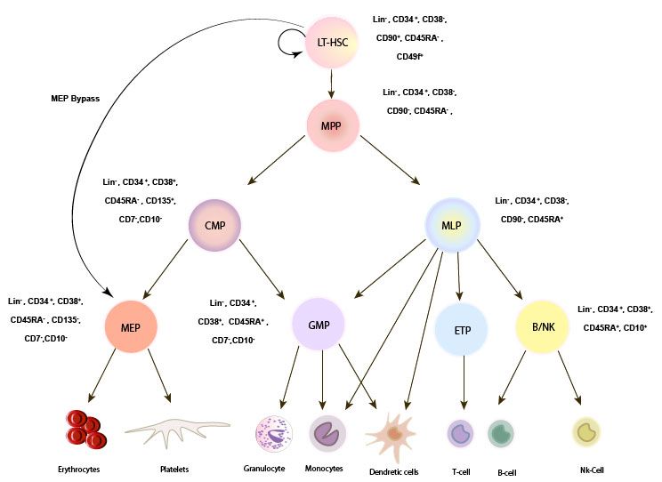
Cells 2019, 8, 169 3 of 15
In addition, current advances in fluorescence-activated cell sorting (FACS) and sorting strategies
provide high-purity isolation and identification of HSCs and progenitors using various cell surface
markers. For instance, CD34, CD38, CD90, CD45RA and CD49f are common surface markers used
for identifying human HSCs and progenitors in vitro and in vivo [7]. However, the expression of
some of these markers such as CD38 of CD90 can change in vitro. Therefore, identifying robust stable
markers that support the identification of HSCs subsets is crucial, especially when testing novel
expansion protocols [23]. Novel surface markers have been suggested for identification of HSCs
subsets; for instance, junction adhesion molecule-2 (Jam2) is highly expressed in a HSC subset that
preferentially generates T cells [24]. Endothelial cell-selective adhesion molecule (ESAM) is another
reliable marker for identification of both murine and human hematopoietic stem cells that are expressed
throughout the lifetime. ESAM is highly expressed in long-term HSCs and MPPs. However, disruption
in ESAM leads to an increased generation of T cells versus B cells. Therefore, ESAM may influence
HSC differentiation paths [25–28]. Recently, endothelial protein C receptor (EPCR) was identified as a
relatively robust surface marker for murine and human LT-HSCs [23].
Broad use of single cell RNA sequencing (scRNA-seq) has also helped in identifying different
cell populations within HSC pools by screening thousands of gene expression profiles of single HSCs
and progenitors [29,30]. This method can be used for different purposes, such as identifying new
progenitor populations, cellular hierarchies in normal and distorted hematopoiesis or distinction
between self-renewal potential and activation of lineage programs [31].
Dormant HSCs have been identified in mice using label-retaining studies and flow cytometry.
Dormant HSCs divide four to five times throughout their lifetime, and they retain multilineage
self-renewal capacity, while remaining quiescent during hematopoiesis. Label-retaining studies suggest
that these cells can switch reversibly from dormancy to self-renewal in response to hematopoietic
stress [13,32]. Therefore, heterogeneity in self-renewal capacity seems to be linked to HSC dormancy.
Several label-retaining assays have been developed to isolate the HSCs based on their division history.
However, one of the major limitations linked to this method is indirect measurement of symmetric
divisions. Thus, distinct cell cycle properties within the HSC pool are intimately linked to HSC function.
Therefore, exploration of new and robust surface markers using FACS and sorting strategies,
as well as single cell RNA sequencing, will provide a better understanding of both the molecular
mechanism and intrinsic programming of HSCs, thus shedding light on their heterogeneity, lineage
choice, functionality and the impact on ex vivo culturing.
3. HSC Self-Renewal Regulation and Niche
How HSC self-renewal is regulated remains one of the main questions of the hematopoietic stem
cell biology field, and answering it will bring new knowledge and possibilities beneficial for clinical
applications. Self-renewal, which is important in maintenance of HSC pool size, is regulated by key
genes and proteins such as transcription factors, epigenetic modifiers and cell cycle regulators, as well
as extrinsic factors from the environment, called niche [33–36]. Niche provides a specialized and tightly
regulated environment that determines the stem cell fate, regulates the proliferation rate and protects
cells from exhaustion and cell death [1,37–39]. Identification of stem cell niches, using advanced
imaging techniques and genetic tools, has shed light on stem cell regulation and hematopoiesis in
murine model (reviewed in [40]). However, little is known about the key cell types and growth factors
involved in communication between human HSCs and the niche, due to challenges of modeling this
network, as well as limited materials in the human system and obvious ethical constraints.
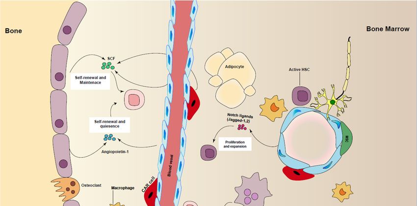
HSCs emerge at several sites at different developmental stages. During embryogenesis, definitive
hematopoiesis in mice starts at the aorta-gonad-mesonephros region (AGM) and then moves to the
fetal liver (FL) and eventually to bone marrow (BM), where the HSCs retain fetal liver characteristics
for three weeks, and finally, they mainly remain in a quiescent state and maintain their pool size by the
regulation of HSC-self renewal and differentiation. They are highly proliferative in FL and undergo
several symmetrical divisions to give rise to the HSC pool required for the lifetime [10,11,41–43]. BM isCells 2019,8,8,169
Cells2019, x 44of
of15
15
[10,11,41–43]. BM is a primary site of hematopoiesis in adult mammals after birth, although it can
a primary site of hematopoiesis in adult mammals after birth, although it can transfer partially into
transfer partially into liver and spleen in response to severe hematopoietic stresses. It has been shown
liver and spleen in response to severe hematopoietic stresses. It has been shown that HSCs in different
that HSCs in different BM regions have different self-renewal capacities [44]. Quiescent HSCs are
BM regions have different self-renewal capacities [44]. Quiescent HSCs are mainly located near
mainly located near perivascular stromal cells and arterioles with low level of oxygen in BM [1,45,46]
perivascular stromal cells and arterioles with low level of oxygen in BM [1,45,46] (Figure 2). Moreover,
(Figure 2). Moreover, Kohler et al. showed the dynamics of young and aged HSCs inside the BM
Kohler et al. showed the dynamics of young and aged HSCs inside the BM cavity, demonstrating that
cavity, demonstrating that the location of HSCs in BM is changed during ageing [47]. Additionally,
the location of HSCs in BM is changed during ageing [47]. Additionally, advanced imaging techniques,
advanced imaging techniques, in combination with conditional depletion of important regulators in
in combination with conditional depletion of important regulators in mouse models, have helped in
mouse models, have helped in identification of key cells and local signals and their role in HSC
identification of key cells and local signals and their role in HSC maintenance [48].
maintenance [48].
Figure2.2. Regulation
Figure Regulation of
of HSCs
HSCsmaintenance
maintenance in
inniche.
niche. Different
Different cell
cell types
types are
areinvolved
involvedin inpromoting
promoting
HSC maintenance, including perivascular stromal cells, endothelial cells (ECs), macrophages,
HSC maintenance, including perivascular stromal cells, endothelial cells (ECs), macrophages, CAR CAR
cells,sympathetic
cells, sympatheticneurons
neuronsby
byproducing
producingcytokines
cytokines and
and growth
growth factors
factors such
such as
as stem
stem cell
cell factor
factor (SCF),
(SCF),
angiopoietin-1,TGF-β
angiopoietin-1, TGF-βandandnotch
notchligands.
ligands.
Besides this,
Besides this, the
the BM
BM niche
niche is
is heterogeneous,
heterogeneous, with
with aa diversity
diversity of
of cell
cell types
types in
in mice
mice and
and humans.
humans.
Endothelial cells,
Endothelial cells, mesenchymal
mesenchymal stromal
stromal cells
cells (MSCs),
(MSCs), megakaryocytes,
megakaryocytes, osteoblasts
osteoblasts and
and nerve
nerve cells
cells
within BM
within BMhave
havebeen
beenshown
shownto toinfluence
influenceself-renewal
self-renewal and
andexpansion
expansion of ofmurine
murine and
andhuman
human HSCs
HSCs
(extensivelyreviewed
(extensively reviewedin in[49,50]),
[49,50]),directly
directlyor
orindirectly.
indirectly.
AA better
better understanding
understanding of ofHSCs
HSCsinteraction
interactionwith
withtheir niche
their willwill
niche helphelp
to overcome the high
to overcome need
the high
for HSCs
need in clinical
for HSCs applications.
in clinical Recently,
applications. a bio-mimic
Recently, 3D model
a bio-mimic 3Dofmodel
HSC niche has niche
of HSC been developed
has been
using hydrogel.
developed usingEngineering niche in 3D is
hydrogel. Engineering promising
niche in 3D isforpromising
culturing and studying the
for culturing andessential
studyingniche
the
factors under
essential nichesteady
factorsstate andsteady
under activestate
conditions of HSCs
and active with better
conditions control
of HSCs withof better
HSC behavior inHSC
control of vitro
[51].
behavior in vitro [51].Cells 2019, 8, 169 5 of 15
4. Wnt Signaling in HSC
The Wnt pathway is a prominent component in self-renewal of adult stem cells and fetal
hematopoiesis. The Wnt signaling cascade has several different signal transduction possibilities,
referred to as canonical (Wnt/β-catenin) and non-canonical pathways [52,53]. Both pathways are
involved in complex processes, such as embryonic development, stem cell maintenance and tissue
homeostasis. The well-understood canonical pathway is involved in cell fate, proliferation and
survival, while the non-canonical pathway is more involved in differentiation, apoptosis regulation,
cell polarity and migration [53]. β-catenin is a key player in the canonical pathway. In the absence
of Wnt ligands, the level of β-catenin remains very low in cytoplasm through the action of a protein
complex called the destruction complex, which actively degrades β-catenin. At the cell membrane, the
interaction of Wnt ligands and Frizzled receptors and co-receptors leads to inactivation of destruction
complex and subsequently to high level of β-catenin and its translocation to the nucleus. In another
way, non-canonical pathways can be activated through Ca2+ signals, JNK kinases or other receptors
such as Ryk, besides Wnt/Frizzled interaction [53–55]. Given the importance of Wnt signaling in
hematopoiesis, there is great interest in exploiting this pathway for ex vivo expansion of HSCs [56].
The potential role for Wnt signaling in the self-renewal of HSC and proliferation of progenitors has
recently been extensively reviewed [57].
Ex vivo culturing of HSCs using non-canonical wnt5A protein showed increased HSC
repopulation in mice [58]. Also, introducing wnt3a proteins increased murine HSC self-renewal
in vitro [59]. Constitutive expression of β-catenin enhanced the HSC self-renewal in mice [60].
Moreover, given that prostaglandin E2 (PGE2) affects β-catenin stability, Zon et al. suggested using
PGE2, which induces canonical Wnt signaling [61] for ex vivo modulation of human cord blood
HSC [62]. Also, activation of Wnt/ β-catenin pathway using glycogen synthase kinase 3 (GSK3-β)
inhibitor in combination with Rapamycin inhibitor of mTOR pathway could increase the number of
murine LT-HSCs in vivo [63]. These studies suggest that similar strategies could be taken for ex vivo
maintenance of human and murine LT-HSC.
The role of wnt/ β-catenin signaling in HSC self-renewal is complex and is dependent on factors
initiated from niche [64]. The function of Wnt signaling is strictly controlled in a dosage-dependent
fashion. High levels of Wnt in HSCs push stem cells into exhaustion and limited reconstitution in
irradiated patients by driving HSC differentiation toward mature blood lineage and loss of proliferation,
while lower doses of Wnt result in better maintenance of immature cells and higher long-term
repopulation capacity.
5. Notch Signaling in HSC
Notch signaling is another crucial conserved pathway in cell fate decisions, development and
hematopoiesis [65]. The role of Notch in the regulation of HSCs in mammalian is still controversial.
In mice, Notch signaling is essential for early HSC development at AGM during embryogenesis,
while its activity is reduced during HSC maturation, and it becomes unnecessary for adult HSC
maintenance in BM [66]. In mice, Notch signaling plays an important role in the development
of early HSCs by establishing the vascular and circulating system, which are essential for HSC
development. Notch signaling also promotes the expression of Notch receptors and ligands to drive
HSC development [67,68]. Notch signaling is based on cell–cell interactions, where the Notch receptors
interact with the transmembrane ligands of Delta and Jagged. This interaction leads to proteolysis of
the receptor and release of the Notch intracellular domain. The released domain is then translocated
to the nucleus, where it forms a protein complex with transcription factors and binds the regulatory
elements of Notch target genes and activates their expression [65,69].
Early studies on Notch signaling in mice using the gain-of-function method also suggested its
role in HSC self-renewal. Overexpression of Notch genes or receptors in HSCs increased the number
of functional HSCs, self-renewal capacity of HSCs, and inhibited differentiation [70–72]. On the other
hand, loss of function experiments, such as conditional knock out of Notch receptors (Notch 1, 2) orCells 2019, 8, 169 6 of 15
ablation of ligands and regulators, did not show any effect on HSC self-renewal [71,73]. Souilhol et al.
showed that both Notch 1 and Notch 2 are involved in early development of HSCs, even though
the Notch 2 signal is weaker than Notch 1, suggesting that modulation of Notch signaling can be
considered for HSC generation from endothelial stem cells or induced pluripotent stem cell for clinical
applications. Moreover, recent studies on human BM have shown that niche signals can activate
Notch signaling in HSCs and progenitors. However, other studies showed embryonic lethality and
impaired hematopoiesis in mice with targeted mutations in Notch 1 or Jagged 1. No impairment
of hematopoiesis was observed in Notch 2, 3, 4 knock outs, suggesting their non-essential role in
hematopoiesis [66]. Therefore, while a functional role for Notch signaling in embryonic development
has been established, the need for Notch signaling in adult hematopoiesis, certainly under hemostasis,
is more controversial [74].
In evolution, these conserved pathways crosstalk to ensure a correct and strong control of gene
expression. In this manner, Notch and Wnt pathways crosstalk on cell fate decisions. Studies in skin
and mammary glands indicated that Wnt signaling controls the stem cell maintenance, whereas Notch
promotes lineage commitment and differentiation [75–78].
6. Ex Vivo Expansion of HSC
Allogeneic HSC transplantation (HSCT) has been used efficiently in the clinic for patients with
severe immunodeficiency diseases with a matched donor. However, for patients without a suitable
donor, genetically modified autologous HSCs have been applied with low risk of graft-versus-host
disease, which is the main cause of transplant morbidity and mortality [79–82]. One of the main
challenges of using HSCs in clinical application is the limited number of cells that can be enriched
from the patient. Thus, expanding HSCs has become important, due to the increasing interest in stem
cells and the gene therapy field, particularly for clinical applications. HSCs undergo symmetrical
and asymmetrical cell divisions in vivo. The frequency of symmetric and asymmetric cell divisions
determines the number of stem cells and differentiated cells in niche. Symmetric cell division leads to
the expansion of HSCs, in numerical terms. Thus, for ex vivo expansion, approaches that will result
in symmetric stem cell division and self-renewal without further differentiation are required [83].
Different combinations of recombinant growth factors and cytokines have been assessed to expand
HSCs ex vivo [84,85], however, limited success in clinical studies has been reported due to unwanted
differentiation of stem cells and progenitor inputs. Currently, combinations of different cytokines
and growth factors, such as SCF, Flt3, TPO, IL-3, and IL-6, are commonly used for supporting HSC
and progenitor survival, proliferation and maintenance during in vitro culturing systems in clinics
(reviewed in [86,87]). The influence of cytokines on lineage commitment and self-renewal has been
studied extensively. A recent study by Knapp et al. indicates that this cocktail may only regulate
short-term (4 days) survival and proliferation of human HSCs, rather than maintenance of functional
long-term HSC in vitro [88]. Unsuccessful attempts to improve the HSC engraftments using current
cytokine-based ex vivo expansion protocols clearly suggest the need for additional factors to support
in vitro HSC maintenance and expansion.
A high-throughput screening of thousands of molecules has been used to find new compounds
by testing their potential for in vitro stem cell expansion. Among those molecules, Prostaglandin
E2 (PGE2), Stemregenin 1 (SR1) (an Aryl hydrocarbon receptor antagonist) and UM171 were found
through library screenings on CD34+ cells and further tested due to their potential in expanding HSCs
in vitro [89–91].
PGE2 has been investigated for optimizing in vitro expansion of human HSCs for clinical
applications. The outcome of clinical trials using optimized and shortened ex vivo expansion of
HSCs with PGE2 resulted in no effect on ex vivo proliferation and colony forming potential. However,
enhanced and rapid engraftment in human cord blood transplantation, as well as enhanced lentivirus
transduction efficiency, was reported in this study. PGE2 can be considered for clinical hematopoietic
stem cell-based gene therapy, as these findings suggest that short-term modulation of HSCs withCells 2019, 8, 169 7 of 15
PGE2 can overcome some challenges observed during ex vivo expansion of HSCs, such as unwanted
differentiation due to long-term expansion of HSCs and high manufacturing costs involved in clinical
trials [62,92–94].
SR1 was first identified for its ability to support expansion of both murine and human CD34+
in vitro, with faster recovery of neutrophil and platelet in vivo [89]. Therefore, the clinical potential of
SR1 has been explored by culturing CD34+ in presence of TPO, SCF, Flt3 and IL-6 [90]. However, recent
clinical and phenotypic/transcriptional studies demonstrated the increase of multipotent progenitors
and erythroid/megakaryocytic in cultured CD34+ with SR1, rather than long-term repopulating stem
cells [90,95].
UM171 is a promising candidate for ex vivo expansion of HSC for allogenic transplantation and
gene therapy, and a clinical trial using UM171 for allogenic stem cell transplantation is currently
ongoing (NCT02668315). This molecule has been shown to improve ex vivo expansion of human cord
blood HSCs with long-term repopulation potentials [91], while also increasing the myeloid progenitors
in ex vivo culture [95]. However, it suppresses megakaryocyte/erythrocyte and granulocyte expansion,
although this suppression effect could be counteracted by adding SR1. In addition, a recent study has
reported the ability of UM171 to enhance gene transfer to HSCs in a dose-dependent manner by up
to 2-fold, with increased recovery of transduced HSC in mice [96]. All of this together suggests the
potential of UM171 and SR1 for ex vivo gene therapy applications.
It is already known that epigenetic regulation such as DNA methylation and post-translational
histone modifications play an important role in the cell-fate decisions of HSCs. These modifications
allow each cell type to acquire unique forms and functions. Distinct epigenetic markers control
gene expression [97–100]. For instance, during development, the DNA methylation patterns are not
conserved. Changes in H3K27me3 and the enhancer H3K27ac are among the well-known epigenetic
changes through embryonic stem cell development [101–103]. H3K4me3 is another epigenetic
modification in promoters associated with gene transcriptional activation. Loss of H3K4me3 leads to
reduced self-renewal and impaired differentiation in murine embryonic stem cells [104,105]. The role
of H3K4me3 in gene expression is not completely clear; however, it is known that H3K4me3 is involved
in the transcriptional machinery assembly and facilitates induction of gene expression in response to
the microenvironment [106,107]. Given the importance of epigenetic regulation in HSC regulation,
multiple studies have demonstrated that small molecule inhibitors of histone deacetylase (HDAC)
and DNA methyltransferase have the capability to support ex vivo expansion of HSCs. Culturing
human cord blood-derived CD34+ with valproic acid, the HDAC inhibitor showed the upregulation
of genes involved in stemness, and increased the SCID repopulating cells [108]. However, some
methyltransferases, such as G9a and G9a-like protein (GLP), support lineage choice and differentiation.
Introducing small molecule inhibitors of methyltransferases and HDACSs can have a synergetic
effect on ex vivo expansion, helping the maintenance—but not the expansion—of HSCs in culture.
For example, combination of the decitabine—a methyltransferase inhibitor—and trichostain A—a
HDAC inhibitor—showed greater ability to maintain HSC activity ex vivo than individual single
agents [109–111].
The discovery of several new small molecules in recent years has taken HSC expansion beyond
traditional hematopoietic cytokine treatment, and these compounds are currently making their way
into clinical trials. These approaches are summarized in Table 1.Cells 2019, 8, 169 8 of 15
Table 1. Summary of current protocols of ex vivo expansion of human HSCs.
Factor Components Supplements Input Cells Culture Time Effects References
20-fold expansion of CD34+ in vitro
- SCF, FLt3, TPO, IL3 CD34+ 7 days Similar frequency of human CD45+ BM cells [112]
Cytokine
vs. fresh cells (NOD/SCID)
supplement
15-fold increase in CFUs, and fourfold
- SCF, FLt3, TPO, IL3, IL-6 CD34+CD38- 4 days [113,114]
enhanced chimera
SCF, TPO, FGF-1,
- CD133+ 11 days 230-fold increase in TNCs in vitro [114]
IGFBP-2, ANGPTL5
Neutrophil recovery and myeloid
Notch ligands CD34+ 14–21 days [56]
engraftment
Enhances neutrophil recovery
PGE2 - CD34+ 24–48 h enhancing homing, survival, and proliferation [93]
of HSCs
Chemical
supplement 65-fold increase in CFUs; 17-fold enhanced
SR1 SCF. Flt3L, TPO, IL-6 CD34+ 7–21 days [89,90]
chimera; Enhances neutrophil recovery
More than 100-fold expansion of LT-HSC, and
UM171 SCF. Flt3L, TPO CD34+ 7–21 days 35-fold enhanced chimera; Inhibiting [91]
erythroid and megakaryocytic differentiation
36-fold increase in SCID-repopulating cells;
Histone deactylase
SCF. Flt3L, TPO, IL-3 CD34+ 7 days improving homing and maintaining [108]
inhibitor (valproic acid)
quiescence
DNA Methyltransferase Maintaining HSC activity blocking formation
SCF. Flt3L, TPO, IL-6 CD34+ 2 weeks [109]
inhibitor (UNC0638) of higher-order chromatin structureCells 2019, 8, 169 9 of 15
7. Concluding Remarks
The number of HSCs that can be obtained for clinical transplantation is limited and influenced by
different factors, such as a low cell number of HSCs isolated from patients. Thus, expansion of HSCs
for gene therapy and regenerative medicine is an unmet medical need. Optimizing current cell culture
methods for clinical applications by using, for instance, proteins from conserved pathways such as
Wnt and Notch ligands, or small molecules such as PGE2, SR-1 or UM171, might improve the ex vivo
expansion of HSCs and also increase the engraftment capacity of gene-modified HSCs [91,93,115].
Enrichment of HSCs for clinical applications is mainly based on CD34+ selection. However, the
CD34+ population is heterogeneous, with a small fraction of LT-HSCs that are crucial in clinical therapy.
Hence, enrichment using only CD34+ is not sufficient. Advances in FACS, sorting strategies and single
cell RNA sequencing have allowed identification and isolation of different high-purity subpopulations
within the HSC pool. Further enrichment of HSCs using well-known established markers such as
CD90+ , CD45RA+ and CD49f+ would help to target desired populations, avoiding unwanted material
loss. Even though the surface phenotype of cultured HSCs differs from that of unmanipulated HSCs,
identifying and including robust surface markers such as EPCR could improve in vitro measurements
of HSCs [23].
Enormous efforts have already been invested in identifying the key cellular players for HSC
self-renewal regulation in the niche. Imaging advances and genetic tools have increased the
general knowledge of the HSC niche. Clarification of the interactions between HSCs and their
microenvironments may help to identify novel clinical approaches and opportunities in the HSCT
field. Therefore, a better understanding of the mechanisms involved in HSC regulation and fate
determination could also help in the development of new strategies for ex vivo expansion. Given
that gene repair approaches (using for instance CRISPR/Cas9), as opposed to than gene addition
approaches, require more extensive ex vivo culturing of HSCs, developing such strategies is important
for clinical implementation of new gene therapy methods.
Funding: This research received no external funding.
Acknowledgments: We thank Willem E. Fibbe for continuous support.
Conflicts of Interest: The authors declare no conflict of interest. The funders had no role in the design of the
study; in the collection, analyses, or interpretation of data; in the writing of the manuscript, or in the decision to
publish the results.
References
1. Morrison, S.J.; Scadden, D.T. The bone marrow niche for haematopoietic stem cells. Nature 2014, 505, 327.
[CrossRef] [PubMed]
2. Seita, J.; Weissman, I.L. Hematopoietic stem cell: Self-renewal versus differentiation. Wiley Interdiscip. Rev.
Syst. Biol. Med. 2010, 2, 640–653. [CrossRef]
3. Bryder, D.; Rossi, D.J.; Weissman, I.L. Hematopoietic stem cells: The paradigmatic tissue-specific stem cell.
Am. J. Pathol. 2006, 169, 338–346. [CrossRef] [PubMed]
4. Kondo, M.; Wagers, A.J.; Manz, M.G.; Prohaska, S.S.; Scherer, D.C.; Beilhack, G.F.; Shizuru, J.A.; Weissman, I.L.
Biology of hematopoietic stem cells and progenitors: Implications for clinical application. Annu. Rev.
Immunol. 2003, 21, 759–806. [CrossRef] [PubMed]
5. Körbling, M.; Estrov, Z. Adult stem cells for tissue repair—A new therapeutic concept? N. Engl. J. Med. 2003,
349, 570–582. [CrossRef] [PubMed]
6. Copelan, E.A. Hematopoietic stem-cell transplantation. N. Engl. J. Med. 2006, 354, 1813–1826. [CrossRef]
[PubMed]
7. Notta, F.; Doulatov, S.; Laurenti, E.; Poeppl, A.; Jurisica, I.; Dick, J.E. Isolation of single human hematopoietic
stem cells capable of long-term multilineage engraftment. Science 2011, 333, 218–221. [CrossRef] [PubMed]Cells 2019, 8, 169 10 of 15
8. Brunstein, C.G.; Gutman, J.A.; Weisdorf, D.J.; Woolfrey, A.E.; Defor, T.E.; Gooley, T.A.; Verneris, M.R.;
Appelbaum, F.R.; Wagner, J.E.; Delaney, C. Allogeneic hematopoietic cell transplantation for hematologic
malignancy: Relative risks and benefits of double umbilical cord blood. Blood 2010, 116, 4693–4699.
[CrossRef]
9. Nakamura-Ishizu, A.; Takizawa, H.; Suda, T. The analysis, roles and regulation of quiescence in hematopoietic
stem cells. Development 2014, 141, 4656–4666. [CrossRef]
10. Sun, J.; Ramos, A.; Chapman, B.; Johnnidis, J.B.; Le, L.; Ho, Y.-J.; Klein, A.; Hofmann, O.; Camargo, F.D.
Clonal dynamics of native haematopoiesis. Nature 2014, 514, 322. [CrossRef]
11. Wilson, A.; Trumpp, A. Bone-marrow haematopoietic-stem-cell niches. Nat. Rev. Immunol. 2006, 6, 93.
[CrossRef] [PubMed]
12. Yamamoto, R.; Morita, Y.; Ooehara, J.; Hamanaka, S.; Onodera, M.; Rudolph, K.L.; Ema, H.; Nakauchi, H.
Clonal analysis unveils self-renewing lineage-restricted progenitors generated directly from hematopoietic
stem cells. Cell 2013, 154, 1112–1126. [CrossRef] [PubMed]
13. Wilson, A.; Laurenti, E.; Oser, G.; van der Wath, R.C.; Blanco-Bose, W.; Jaworski, M.; Offner, S.; Dunant, C.F.;
Eshkind, L.; Bockamp, E.; et al. Hematopoietic stem cells reversibly switch from dormancy to self-renewal
during homeostasis and repair. Cell 2008, 135, 1118–1129. [CrossRef] [PubMed]
14. Sanjuan-Pla, A.; Macaulay, I.C.; Jensen, C.T.; Woll, P.S.; Luis, T.C.; Mead, A.; Moore, S.; Carella, C.;
Matsuoka, S.; Jones, T.B.; et al. Platelet-biased stem cells reside at the apex of the haematopoietic stem-cell
hierarchy. Nature 2013, 502, 232. [CrossRef] [PubMed]
15. Dykstra, B.; Kent, D.; Bowie, M.; McCaffrey, L.; Hamilton, M.; Lyons, K.; Lee, S.-J.; Brinkman, R.; Eaves, C.
Long-term propagation of distinct hematopoietic differentiation programs in vivo. Cell Stem Cell 2007, 1,
218–229. [CrossRef] [PubMed]
16. Hao, Q.-L.; Zhu, J.; Price, M.A.; Payne, K.J.; Barsky, L.W.; Crooks, G.M. Identification of a novel, human
multilymphoid progenitor in cord blood. Blood 2001, 97, 3683–3690. [CrossRef] [PubMed]
17. Manz, M.G.; Miyamoto, T.; Akashi, K.; Weissman, I.L. Prospective isolation of human clonogenic common
myeloid progenitors. Proc. Natl. Acad. Sci. USA 2002, 99, 11872–11877. [CrossRef] [PubMed]
18. Notta, F.; Zandi, S.; Takayama, N.; Dobson, S.; Gan, O.I.; Wilson, G.; Kaufmann, K.B.; McLeod, J.; Laurenti, E.;
Dunant, C.F.; et al. Distinct routes of lineage development reshape the human blood hierarchy across
ontogeny. Science 2016, 351, aab2116. [CrossRef]
19. Huang, H.; Cantor, A.B. Common features of megakaryocytes and hematopoietic stem cells: What’s the
connection? J. Cell. Biochem. 2009, 107, 857–864. [CrossRef] [PubMed]
20. Adolfsson, J.; Månsson, R.; Buza-Vidas, N.; Hultquist, A.; Liuba, K.; Jensen, C.T.; Bryder, D.; Yang, L.;
Borge, O.-J.; Thoren, L.A.M.; et al. Identification of Flt3+ lympho-myeloid stem cells lacking erythro-
megakaryocytic potential: A revised road map for adult blood lineage commitment. Cell 2005, 121, 295–306.
[CrossRef] [PubMed]
21. Pietras, E.M.; Reynaud, D.; Kang, Y.A.; Carlin, D.; Calero-Nieto, F.J.; Leavitt, A.D.; Stuart, J.M.; Göttgens, B.;
Passegué, E. Functionally distinct subsets of lineage-biased multipotent progenitors control blood production
in normal and regenerative conditions. Cell Stem Cell 2015, 17, 35–46. [CrossRef] [PubMed]
22. Muller-Sieburg, C.E.; Cho, R.H.; Karlsson, L.; Huang, J.-F.; Sieburg, H.B. Myeloid-biased hematopoietic stem
cells have extensive self-renewal capacity but generate diminished lymphoid progeny with impaired IL-7
responsiveness. Blood 2004, 103, 4111–4118. [CrossRef] [PubMed]
23. Fares, I.; Chagraoui, J.; Lehnertz, B.; MacRae, T.; Mayotte, N.; Tomellini, E.; Aubert, L.; Roux, P.P.;
Sauvageau, G. EPCR expression marks UM171-expanded CD34+ cord blood stem cells. Blood 2017, 129,
3344–3351. [CrossRef] [PubMed]
24. Radulovic, V.; van der Garde, M.; Sigurdsson, V.; Zriwil, A.; Soboleva, S.; Kaneko, S.; Quinn, E.S.; Miharada, K.
Junctional Adhesion Molecule 2 Represents a Novel Subset of Hematopoietic Stem Cells Poised for T
Lymphopoiesis. Blood 2016, 128, 3862.
25. Ooi, A.G.L.; Karsunky, H.; Majeti, R.; Butz, S.; Vestweber, D.; Ishida, T.; Quertermous, T.; Weissman, I.L.;
Forsberg, E.C. The adhesion molecule Esam1 is a novel hematopoietic stem cell marker. Stem Cells 2009, 27,
653–661. [CrossRef] [PubMed]
26. Yokota, T.; Oritani, K.; Butz, S.; Kokame, K.; Kincade, P.W.; Miyata, T.; Vestweber, D.; Kanakura, Y. The
endothelial antigen ESAM marks primitive hematopoietic progenitors throughout life in mice. Blood 2009,
113, 2914–2923. [CrossRef] [PubMed]Cells 2019, 8, 169 11 of 15
27. Ishibashi, T.; Yokota, T.; Tanaka, H.; Ichii, M.; Sudo, T.; Satoh, Y.; Doi, Y.; Ueda, T.; Tanimura, A.; Hamanaka, Y.;
et al. ESAM is a novel human hematopoietic stem cell marker associated with a subset of human leukemias.
Exp. Hematol. 2016, 44, 269–281. [CrossRef] [PubMed]
28. Roch, A.; Giger, S.; Girotra, M.; Campos, V.; Vannini, N.; Naveiras, O.; Gobaa, S.; Lutolf, M.P. Single-cell
analyses identify bioengineered niches for enhanced maintenance of hematopoietic stem cells. Nat. Comm.
2017, 8, 221. [CrossRef]
29. Warren, L.; Bryder, D.; Weissman, I.L.; Quake, S.R. Transcription factor profiling in individual hematopoietic
progenitors by digital RT-PCR. Proc. Natl. Acad. Sci. USA 2006, 103, 17807–17812. [CrossRef]
30. Athanasiadis, E.I.; Botthof, J.G.; Andres, H.; Ferreira, L.; Lio, P.; Cvejic, A. Single-cell RNA-sequencing
uncovers transcriptional states and fate decisions in haematopoiesis. Nat. Comm. 2017, 8, 2045. [CrossRef]
31. Pina, C.; Fugazza, C.; Tipping, A.J.; Brown, J.; Soneji, S.; Teles, J.; Peterson, C.; Enver, T. Inferring rules of
lineage commitment in haematopoiesis. Nat. Cell Biol. 2012, 14, 287. [CrossRef] [PubMed]
32. Bernitz, J.M.; Kim, H.S.; MacArthur, B.; Sieburg, H.; Moore, K. Hematopoietic stem cells count and remember
self-renewal divisions. Cell 2016, 167, 1296–1309. [CrossRef] [PubMed]
33. Cabezas-Wallscheid, N.; Klimmeck, D.; Hansson, J.; Lipka, D.B.; Reyes, A.; Wang, Q.; Weichenhan, D.;
Lier, A.; von Paleske, L.; Renders, S.; et al. Identification of regulatory networks in HSCs and their immediate
progeny via integrated proteome, transcriptome, and DNA methylome analysis. Cell Stem Cell 2014, 15,
507–522. [CrossRef] [PubMed]
34. Chambers, S.M.; Boles, N.C.; Lin, K.-Y.K.; Tierney, M.P.; Bowman, T.V.; Bradfute, S.B.; Chen, A.J.;
Merchant, A.A.; Sirin, O.; Weksberg, D.C.; et al. Hematopoietic fingerprints: An expression database
of stem cells and their progeny. Cell Stem Cell 2007, 1, 578–591. [CrossRef] [PubMed]
35. Chen, L.; Kostadima, M.; Martens, J.H.A.; Canu, G.; Garcia, S.P.; Turro, E.; Downes, K.; Macaulay, I.C.;
Bielczyk-Maczynska, E.; Coe, S.; et al. Transcriptional diversity during lineage commitment of human blood
progenitors. Science 2014, 345, 1251033. [CrossRef] [PubMed]
36. Laurenti, E.; Doulatov, S.; Zandi, S.; Plumb, I.; Chen, J.; April, C.; Fan, J.-B.; Dick, J.E. The transcriptional
architecture of early human hematopoiesis identifies multilevel control of lymphoid commitment.
Nat. Immunol. 2013, 14, 756. [CrossRef]
37. Passegué, E.; Wagers, A.J.; Giuriato, S.; Anderson, W.C.; Weissman, I.L. Global analysis of proliferation and
cell cycle gene expression in the regulation of hematopoietic stem and progenitor cell fates. J. Exp. Med. 2005,
202, 1599–1611. [CrossRef]
38. Mendelson, A.; Frenette, P.S. Hematopoietic stem cell niche maintenance during homeostasis and
regeneration. Nat. Med. 2014, 20, 833. [CrossRef]
39. Ito, K.; Suda, T. Metabolic requirements for the maintenance of self-renewing stem cells. Nat. Rev. Mol. Cell
Biol. 2014, 15, 243. [CrossRef]
40. Joseph, C.; Quach, J.M.; Walkley, C.R.; Lane, S.W.; Celso, C.L.; Purton, L.E. Deciphering hematopoietic
stem cells in their niches: A critical appraisal of genetic models, lineage tracing, and imaging strategies.
Cell Stem Cell 2013, 13, 520–533. [CrossRef]
41. Orkin, S.H.; Zon, L.I. Hematopoiesis: An evolving paradigm for stem cell biology. Cell 2008, 132, 631–644.
[CrossRef]
42. Manesia, J.K.; Xu, Z.; Broekaert, D.; Boon, R.; van Vliet, A.; Eelen, G.; Vanwelden, T.; Stegen, S.; Van Gastel, N.;
Pascual-Montano, A.; et al. Highly proliferative primitive fetal liver hematopoietic stem cells are fueled by
oxidative metabolic pathways. Stem Cell Res. 2015, 15, 715–721. [CrossRef] [PubMed]
43. Bowie, M.B.; Kent, D.G.; Dykstra, B.; McKnight, K.D.; McCaffrey, L.; Hoodless, P.A.; Eaves, C.J. Identification
of a new intrinsically timed developmental checkpoint that reprograms key hematopoietic stem cell
properties. Proc. Natl. Acad. Sci. USA 2007, 104, 5878–5882. [CrossRef] [PubMed]
44. Haylock, D.N.; Williams, B.; Johnston, H.M.; Liu, M.C.P.; Rutherford, K.E.; Whitty, G.A.; Simmons, P.J.;
Bertoncello, I.; Nilsson, S.K. hemopoietic stem cells with higher hemopoietic potential reside at the bone
marrow endosteum. Stem Cells 2007, 25, 1062–1069. [CrossRef] [PubMed]
45. Arai, F.; Yoshihara, H.; Hosokawa, K.; Nakamura, Y.; Gomei, Y.; Iwasaki, H.; Suda, T. Niche regulation of
hematopoietic stem cells in the endosteum. Ann. NY Acad. Sci. 2009, 36–46. [CrossRef] [PubMed]
46. Bakker, S.T.; Passegué, E. Resilient and resourceful: Genome maintenance strategies in hematopoietic stem
cells. Exp. Hematol. 2013, 41, 915–923. [CrossRef] [PubMed]Cells 2019, 8, 169 12 of 15
47. Köhler, A.; Schmithorst, V.; Filippi, M.-D.; Ryan, M.A.; Daria, D.; Gunzer, M.; Geiger, H. Altered cellular
dynamics and endosteal location of aged early hematopoietic progenitor cells revealed by time-lapse
intravital imaging in long bones. Blood 2009, 114, 290–298. [CrossRef] [PubMed]
48. Song, J.; Kiel, M.J.; Wang, Z.; Wang, J.; Taichman, R.S.; Morrison, S.J.; Krebsbach, P.H. An in vivo model to
study and manipulate the hematopoietic stem cell niche. Blood 2010, 115, 2592–2600. [CrossRef]
49. Birbrair, A.; Frenette, P.S. Niche heterogeneity in the bone marrow. Ann. NY Acad. Sci. 2016, 1370, 82–96.
[CrossRef]
50. Zhao, M.; Perry, J.M.; Marshall, H.; Venkatraman, A.; Qian, P.; He, X.C.; Ahamed, J.; Li, L. Megakaryocytes
maintain homeostatic quiescence and promote post-injury regeneration of hematopoietic stem cells. Nat. Med.
2014, 20, 1321. [CrossRef]
51. Rödling, L.; Schwedhelm, I.; Kraus, S.; Bieback, K.; Hansmann, J.; Lee-Thedieck, C. 3D models of the
hematopoietic stem cell niche under steady-state and active conditions. Sci. Rep. 2017, 7, 4625. [CrossRef]
52. Luis, T.C.; Ichii, M.; Brugman, M.H.; Kincade, P.; Staal, F.J.T. Wnt signaling strength regulates normal
hematopoiesis and its deregulation is involved in leukemia development. Leukemia 2011, 26, 414. [CrossRef]
[PubMed]
53. Staal, F.J.T.; Luis, T.C.; Tiemessen, M.M. WNT signalling in the immune system: WNT is spreading its wings.
Nat. Rev. Immunol. 2008, 8, 581. [CrossRef] [PubMed]
54. Brembeck, F.H.; Rosário, M.; Birchmeier, W. Balancing cell adhesion and Wnt signaling, the key role of
β-catenin. Curr. Opin. Genet. Dev. 2006, 16, 51–59. [CrossRef] [PubMed]
55. Clevers, H. Wnt/β-Catenin Signaling in Development and Disease. Cell 2006, 127, 469–480. [CrossRef]
[PubMed]
56. Delaney, C.; Heimfeld, S.; Brashem-Stein, C.; Voorhies, H.; Manger, R.L.; Bernstein, I.D. Notch-mediated
expansion of human cord blood progenitor cells capable of rapid myeloid reconstitution. Nat. Med. 2010, 16,
232. [CrossRef] [PubMed]
57. Staal, F.J.T.; Chhatta, A.; Mikkers, H. Caught in a Wnt storm: Complexities of Wnt signaling in hematopoiesis.
Exp. Hematol. 2016, 44, 451–457. [CrossRef] [PubMed]
58. Nemeth, M.J.; Topol, L.; Anderson, S.M.; Yang, Y.; Bodine, D.M. Wnt5a inhibits canonical Wnt signaling in
hematopoietic stem cells and enhances repopulation. Proc. Natl. Acad. Sci. USA 2007, 104, 15436–15441.
[CrossRef]
59. Willert, K.; Brown, J.D.; Danenberg, E.; Duncan, A.W.; Weissman, I.L.; Reya, T.; Yates Iii, J.R.; Nusse, R.
Wnt proteins are lipid-modified and can act as stem cell growth factors. Nature 2003, 423, 448. [CrossRef]
60. Reya, T.; Duncan, A.W.; Ailles, L.; Domen, J.; Scherer, D.C.; Willert, K.; Hintz, L.; Nusse, R.; Weissman, I.L.
A role for Wnt signalling in self-renewal of haematopoietic stem cells. Nature 2003, 423, 409. [CrossRef]
61. Goessling, W.; North, T.E.; Loewer, S.; Lord, A.M.; Lee, S.; Stoick-Cooper, C.L.; Weidinger, G.; Puder, M.;
Daley, G.Q.; Moon, R.T.; et al. Genetic interaction of PGE2 and Wnt signaling regulates developmental
specification of stem cells and regeneration. Cell 2009, 136, 1136–1147. [CrossRef]
62. Goessling, W.; Allen, R.S.; Guan, X.; Jin, P.; Uchida, N.; Dovey, M.; Harris, J.M.; Metzger, M.E.;
Bonifacino, A.C.; Stroncek, D.; et al. Prostaglandin E2 enhances human cord blood stem cell xenotransplants
and shows long-term safety in preclinical nonhuman primate transplant models. Cell Stem Cell 2011, 8,
445–458. [CrossRef] [PubMed]
63. Huang, J.; Nguyen-McCarty, M.; Hexner, E.O.; Danet-Desnoyers, G.; Klein, P.S. Maintenance of hematopoietic
stem cells through regulation of Wnt and mTOR pathways. Nat. Med. 2012, 18, 1778. [CrossRef] [PubMed]
64. Luis, T.C.; Naber, B.A.; Roozen, P.P.; Brugman, M.H.; de Haas, E.F.; Ghazvini, M.; Fibbe, W.E.; van Dongen, J.J.;
Fodde, R.; Staal, F.J. Canonical Wnt signaling regulates hematopoiesis in a dosage-dependent fashion.
Cell Stem Cell 2011, 9, 345–356. [CrossRef] [PubMed]
65. Andersson, E.R.; Sandberg, R.; Lendahl, U. Notch signaling: Simplicity in design, versatility in function.
Development 2011, 138, 3593–3612. [CrossRef] [PubMed]
66. Souilhol, C.; Lendinez, J.G.; Rybtsov, S.; Murphy, F.; Wilson, H.; Hills, D.; Batsivari, A.; Binagui-Casas, A.;
McGarvey, A.C.; MacDonald, H.R.; et al. Developing HSCs become Notch independent by the end of
maturation in the AGM region. Blood 2016, 128, 1567–1577. [CrossRef] [PubMed]
67. Butko, E.; Pouget, C.; Traver, D. Complex regulation of HSC emergence by the Notch signaling pathway.
Dev. Biol. 2016, 409, 129–138. [CrossRef] [PubMed]Cells 2019, 8, 169 13 of 15
68. Kumano, K.; Chiba, S.; Kunisato, A.; Sata, M.; Saito, T.; Nakagami-Yamaguchi, E.; Yamaguchi, T.; Masuda, S.;
Shimizu, K.; Takahashi, T.; et al. Notch1 but not Notch2 is essential for generating hematopoietic stem cells
from endothelial cells. Immunity 2003, 18, 699–711. [CrossRef]
69. Bray, S.J. Notch signalling: A simple pathway becomes complex. Nat. Rev. Mol. Cell Biol. 2006, 7, 678.
[CrossRef]
70. Kunisato, A.; Chiba, S.; Nakagami-Yamaguchi, E.; Kumano, K.; Saito, T.; Masuda, S.; Yamaguchi, T.;
Osawa, M.; Kageyama, R.; Nakauchi, H.; et al. HES-1 preserves purified hematopoietic stem cells ex
vivo and accumulates side population cells in vivo. Blood 2003, 101, 1777–1783. [CrossRef]
71. Mancini, S.J.C.; Mantei, N.; Dumortier, A.; Suter, U.; MacDonald, H.R.; Radtke, F. Jagged1-dependent
Notch signaling is dispensable for hematopoietic stem cell self-renewal and differentiation. Blood 2005, 105,
2340–2342. [CrossRef]
72. Varnum-Finney, B.; Brashem-Stein, C.; Bernstein, I.D. Combined effects of Notch signaling and cytokines
induce a multiple log increase in precursors with lymphoid and myeloid reconstituting ability. Blood 2003,
101, 1784–1789. [CrossRef] [PubMed]
73. Duncan, A.W.; Rattis, F.M.; DiMascio, L.N.; Congdon, K.L.; Pazianos, G.; Zhao, C.; Yoon, K.; Cook, J.M.;
Willert, K.; Gaiano, N.; et al. Integration of Notch and Wnt signaling in hematopoietic stem cell maintenance.
Nat. Immunol. 2005, 6, 314. [CrossRef] [PubMed]
74. Lampreia, F.P.; Carmelo, J.G.; Anjos-Afonso, F. Notch signaling in the regulation of hematopoietic stem cell.
Curr. Stem Cell Rep. 2017, 3, 202–209. [CrossRef]
75. Collu, G.M.; Hidalgo-Sastre, A.; Acar, A.; Bayston, L.; Gildea, C.; Leverentz, M.K.; Mills, C.G.; Owens, T.W.;
Meurette, O.; Dorey, K.; et al. Dishevelled limits Notch signalling through inhibition of CSL. Development
2012, 139, 4405–4415. [CrossRef] [PubMed]
76. Zeng, Y.A.; Nusse, R. Wnt proteins are self-renewal factors for mammary stem cells and promote their
long-term expansion in culture. Cell Stem Cell 2010, 6, 568–577. [CrossRef] [PubMed]
77. Famili, F.; Wiekmeijer, A.-S.; Staal, F.J.T. The development of T cells from stem cells in mice and humans.
Future Sci. OA 2017, 3, FSO186. [CrossRef] [PubMed]
78. Bouras, T.; Pal, B.; Vaillant, F.; Harburg, G.; Asselin-Labat, M.-L.; Oakes, S.R.; Lindeman, G.J.; Visvader, J.E.
Notch signaling regulates mammary stem cell function and luminal cell-fate commitment. Cell Stem Cell
2008, 3, 429–441. [CrossRef]
79. Booth, C.; Gaspar, H.B.; Thrasher, A.J. Treating immunodeficiency through HSC gene therapy. Trends
Mol. Med. 2016, 22, 317–327. [CrossRef]
80. Marina, C.; Emmanuelle, S.; Chantal, L.-P.; Isabelle, A.-S.; Salima, H.-B.-A. Gene therapy for X-linked severe
combined immunodeficiency: Where do we stand? Hum. Gene Ther. 2016, 27, 108–116. [CrossRef]
81. Cooke, K.R.; Luznik, L.; Sarantopoulos, S.; Hakim, F.T.; Jagasia, M.; Fowler, D.H.; van den Brink, M.R.M.;
Hansen, J.A.; Parkman, R.; Miklos, D.B.; et al. The biology of chronic graft-versus-host disease: A task force
report from the national institutes of health consensus development project on criteria for clinical trials in
chronic graft-versus-host disease. Biol. Blood Marrow Transplant. 2017, 23, 211–234. [CrossRef]
82. Naldini, L. Gene therapy returns to centre stage. Nature 2015, 526, 351. [CrossRef] [PubMed]
83. Morrison, S.J.; Kimble, J. Asymmetric and symmetric stem-cell divisions in development and cancer. Nature
2006, 441, 1068. [CrossRef] [PubMed]
84. Hofmeister, C.C.; Zhang, J.; Knight, K.L.; Le, P.; Stiff, P.J. Ex vivo expansion of umbilical cord blood stem
cells for transplantation: Growing knowledge from the hematopoietic niche. Bone Marrow Transplant. 2006,
39, 11. [CrossRef] [PubMed]
85. Buza-Vidas, N.; Antonchuk, J.; Qian, H.; Månsson, R.; Luc, S.; Zandi, S.; Anderson, K.; Takaki, S.; Nygren, J.M.;
Jensen, C.T.; et al. Cytokines regulate postnatal hematopoietic stem cell expansion: Opposing roles of
thrombopoietin and LNK. Genes Dev. 2006, 20, 2018–2023. [CrossRef] [PubMed]
86. Sauvageau, G.; Iscove, N.N.; Humphries, R.K. In vitro and in vivo expansion of hematopoietic stem cells.
Oncogene 2004, 23, 7223. [CrossRef] [PubMed]
87. Metcalf, D. Hematopoietic cytokines. Blood 2008, 111, 485–491. [CrossRef] [PubMed]
88. Knapp, D.J.H.F.; Hammond, C.A.; Miller, P.H.; Rabu, G.M.; Beer, P.A.; Ricicova, M.; Lecault, V.; Da Costa, D.;
VanInsberghe, M.; Cheung, A.M.; et al. Dissociation of survival, proliferation, and state control in human
hematopoietic stem cells. Stem Cell Rep. 2017, 8, 152–162. [CrossRef]Cells 2019, 8, 169 14 of 15
89. Boitano, A.E.; Wang, J.; Romeo, R.; Bouchez, L.C.; Parker, A.E.; Sutton, S.E.; Walker, J.R.; Flaveny, C.A.;
Perdew, G.H.; Denison, M.S.; et al. Aryl hydrocarbon receptor antagonists promote the expansion of human
hematopoietic stem cells. Science 2010, 329, 1345–1348. [CrossRef]
90. Wagner, J.E.; Brunstein, C.G.; Boitano, A.E.; DeFor, T.E.; McKenna, D.; Sumstad, D.; Blazar, B.R.; Tolar, J.;
Le, C.; Jones, J.; et al. Phase I/II trial of stem regenin-1 expanded umbilical cord blood hematopoietic stem
cells supports testing as a stand-alone graft. Cell Stem Cell 2016, 18, 144–155. [CrossRef]
91. Fares, I.; Chagraoui, J.; Gareau, Y.; Gingras, S.; Ruel, R.; Mayotte, N.; Csaszar, E.; Knapp, D.J.H.F.; Miller, P.;
Ngom, M.; et al. Pyrimidoindole derivatives are agonists of human hematopoietic stem cell self-renewal.
Science 2014, 345, 1509–1512. [CrossRef]
92. Heffner, G.C.; Bonner, M.; Christiansen, L.; Pierciey, F.J.; Campbell, D.; Smurnyy, Y.; Zhang, W.; Hamel, A.;
Shaw, S.; Lewis, G.; et al. Prostaglandin E2 increases lentiviral vector transduction efficiency of adult human
hematopoietic stem and progenitor cells. Mol. Ther. 2018, 26, 320–328. [CrossRef]
93. Zonari, E.; Desantis, G.; Petrillo, C.; Boccalatte, F.E.; Lidonnici, M.R.; Kajaste-Rudnitski, A.; Aiuti, A.;
Ferrari, G.; Naldini, L.; Gentner, B. Efficient ex vivo engineering and expansion of highly purified human
hematopoietic stem and progenitor cell populations for gene therapy. Stem Cell Rep. 2017, 8, 977–990.
[CrossRef] [PubMed]
94. Cutler, C.; Multani, P.; Robbins, D.; Kim, H.T.; Le, T.; Hoggatt, J.; Pelus, L.M.; Desponts, C.; Chen, Y.-B.;
Rezner, B.; et al. Prostaglandin-modulated umbilical cord blood hematopoietic stem cell transplantation.
Blood 2013, 122, 3074–3081. [CrossRef] [PubMed]
95. Psatha, N.; Georgolopoulos, G.; Phelps, S.; Papayannopoulou, T. Brief report: A differential transcriptomic
profile of ex vivo expanded adult human hematopoietic stem cells empowers them for engraftment better
than their surface phenotype. Stem Cells Transl. Med. 2017, 6, 1852–1858. [CrossRef] [PubMed]
96. Ngom, M.; Imren, S.; Maetzig, T.; Adair, J.E.; Knapp, D.J.H.F.; Chagraoui, J.; Fares, I.; Bordeleau, M.-E.;
Sauvageau, G.; Leboulch, P.; et al. UM171 enhances lentiviral gene transfer and recovery of primitive human
hematopoietic cells. Mol. Ther. Methods Clin. Dev. 2018, 10, 156–164. [CrossRef] [PubMed]
97. Hodges, E.; Molaro, A.; Dos Santos, C.O.; Thekkat, P.; Song, Q.; Uren, P.J.; Park, J.; Butler, J.; Rafii, S.;
McCombie, W.R.; et al. Directional DNA methylation changes and complex intermediate states accompany
lineage specificity in the adult hematopoietic compartment. Mol. Cell 2011, 44, 17–28. [CrossRef] [PubMed]
98. Ji, H.; Ehrlich, L.I.R.; Seita, J.; Murakami, P.; Doi, A.; Lindau, P.; Lee, H.; Aryee, M.J.; Irizarry, R.A.;
Kim, K.; et al. Comprehensive methylome map of lineage commitment from haematopoietic progenitors.
Nature 2010, 467, 338. [CrossRef] [PubMed]
99. Kulis, M.; Merkel, A.; Heath, S.; Queirós, A.C.; Schuyler, R.P.; Castellano, G.; Beekman, R.; Raineri, E.;
Esteve, A.; Clot, G.; et al. Whole-genome fingerprint of the DNA methylome during human B cell
differentiation. Nat. Genet. 2015, 47, 746. [CrossRef]
100. Meissner, A.; Mikkelsen, T.S.; Gu, H.; Wernig, M.; Hanna, J.; Sivachenko, A.; Zhang, X.; Bernstein, B.E.;
Nusbaum, C.; Jaffe, D.B.; et al. Genome-scale DNA methylation maps of pluripotent and differentiated cells.
Nature 2008, 454, 766. [CrossRef]
101. Buecker, C.; Srinivasan, R.; Wu, Z.; Calo, E.; Acampora, D.; Faial, T.; Simeone, A.; Tan, M.; Swigut, T.;
Wysocka, J. Reorganization of enhancer patterns in transition from naive to primed pluripotency.
Cell Stem Cell 2014, 14, 838–853. [CrossRef]
102. Tesar, P.J.; Chenoweth, J.G.; Brook, F.A.; Davies, T.J.; Evans, E.P.; Mack, D.L.; Gardner, R.L.; McKay, R.D.G.
New cell lines from mouse epiblast share defining features with human embryonic stem cells. Nature 2007,
448, 196. [CrossRef] [PubMed]
103. Weinberger, L.; Ayyash, M.; Novershtern, N.; Hanna, J.H. Dynamic stem cell states: Naive to primed
pluripotency in rodents and humans. Nat. Rev. Mol. Cell Biol. 2016, 17, 155. [CrossRef]
104. Ang, Y.-S.; Tsai, S.-Y.; Lee, D.-F.; Monk, J.; Su, J.; Ratnakumar, K.; Ding, J.; Ge, Y.; Darr, H.; Chang, B.; et al.
Wdr5 mediates self-renewal and reprogramming via the embryonic stem cell core transcriptional network.
Cell 2011, 145, 183–197. [CrossRef] [PubMed]
105. Jiang, H.; Shukla, A.; Wang, X.; Chen, W.-y.; Bernstein, B.E.; Roeder, R.G. Role for Dpy-30 in ES cell-fate
specification by regulation of H3K4 methylation within bivalent domains. Cell 2011, 144, 513–525. [CrossRef]
[PubMed]You can also read