Sarcopenia and Cognitive Function: Role of Myokines in Muscle Brain Cross-Talk
←
→
Page content transcription
If your browser does not render page correctly, please read the page content below
life

Review
Sarcopenia and Cognitive Function: Role of Myokines in
Muscle Brain Cross-Talk
Lucia Scisciola, Rosaria Anna Fontanella, Surina, Vittoria Cataldo, Giuseppe Paolisso *
and Michelangela Barbieri

                                           Department of Advanced Medical and Surgical Sciences, University of Campania “Luigi Vanvitelli”,
                                           80138 Naples, Italy; lucia.scisciola@unicampania.it (L.S.); rosariaanna.fontanella@unicampania.it (R.A.F.);
                                           surina@unicampania.it (S.); vittoria.cataldo@studenti.unicampania.it (V.C.);
                                           michelangela.barbieri@unicampania.it (M.B.)
                                           * Correspondence: giuseppe.paolisso@unicampania.it; Tel.: +39-0815665135

                                           Abstract: Sarcopenia is a geriatric syndrome characterized by the progressive degeneration of muscle
                                           mass and function, and it is associated with severe complications, which are falls, functional decline,
                                           frailty, and mortality. Sarcopenia is associated with cognitive impairment, defined as a decline
                                           in one or more cognitive domains as language, memory, reasoning, social cognition, planning,
                                           making decisions, and solving problems. Although the exact mechanism relating to sarcopenia
                                           and cognitive function has not yet been defined, several studies have shown that skeletal muscle
                                           produces and secrete molecules, called myokines, that regulate brain functions, including mood,
                                           learning, locomotor activity, and neuronal injury protection, showing the existence of muscle-brain
                                           cross-talk. Moreover, studies conducted on physical exercise supported the existence of muscle-brain

                                           cross-talk, showing how physical activity, changing myokines’ circulating levels, exerts beneficial
                                    effects on the brain. The review mainly focuses on describing the role of myokines on brain function
Citation: Scisciola, L.; Fontanella,       and their involvement in cognitive impairment in sarcopenia.
R.A.; Surina; Cataldo, V.; Paolisso, G.;
Barbieri, M. Sarcopenia and Cognitive      Keywords: myokines; exerkines; skeletal muscle; sarcopenic; cognitive impairment
Function: Role of Myokines in Muscle
Brain Cross-Talk. Life 2021, 11, 173.
https://doi.org/10.3390/life11020173
                                           1. Sarcopenia
Academic Editors: Dae Won Jun and
                                                Sarcopenia is a common condition in older individuals, characterized by the progres-
Waqar K. Saeed
                                           sive degeneration of muscle mass and function. It is associated with severe complications,
                                           which are falls, functional decline, frailty, and mortality [1]. The prevalence of sarcopenia
Received: 9 February 2021
                                           varies from 9.9% to 40.4%, depending on its definition [2]. Nowadays, there is no consensus
Accepted: 20 February 2021
                                           on defining the cut-off points, making sarcopenia diagnosis challenging.
Published: 23 February 2021
                                                The pathogenesis of sarcopenia remains still poor clear and involves an interplay
                                           between sedentary lifestyle, aging, obesity, inflammation, and oxidative stress that affect
Publisher’s Note: MDPI stays neutral
with regard to jurisdictional claims in
                                           muscle mass and function [3].
published maps and institutional affil-
                                                A sedentary lifestyle, defined as activities that do not increase energy expenditure,
iations.
                                           impacts muscle mass and metabolism. Indeed, only seven days of decubitus resulted in
                                           a loss of muscle mass and a prolonged period, 90–120 days, reduced 30% of the muscle
                                           volume [4,5]. Studies conducted on old immobilized animals have examined the effects of
                                           bed rest on skeletal muscle metabolism, demonstrating a disruption in the balance between
                                           protein synthesis and degradation in favor of catabolism [4,5].
Copyright: © 2021 by the authors.
                                                Interestingly, aging alters both the homeostasis of skeletal muscle, compromising
Licensee MDPI, Basel, Switzerland.
This article is an open access article
                                           the equilibrium between cell regeneration and differentiation [6], and the rate of protein
distributed under the terms and
                                           synthesis and degradation [7]. It is associated with reducing skeletal muscle stem cells
conditions of the Creative Commons
                                           (satellite cells) in type II fiber. Major pathways associated with changes in satellite cells
Attribution (CC BY) license (https://      during aging include Notch and Wnt signaling; the first one is associated with proliferation
creativecommons.org/licenses/by/           while the second with differentiation of muscle cells [8]. Studies demonstrated that the
4.0/).                                     expression of Notch signaling decreased with age during aging [9], and Wnt canonical

Life 2021, 11, 173. https://doi.org/10.3390/life11020173                                                             https://www.mdpi.com/journal/life
Life 2021, 11, 173 2 of 12
pathway switched to not canonical pathway resulting in the inability of satellite cells to
self-renewal [10]. However, the hypothesis that loss of satellite cell activity is the cause
of sarcopenia has been confuted. In male sedentary mice, the depletion of satellite cells,
resulting in impaired muscle regeneration, did not contribute to muscle size or fiber type
composition, despite low regenerative capacity, but contributed to age-related muscle
fibrosis [11].
With advancing age, the intake of amino acids is inadequate, resulting in a decreased
protein synthesis rate and the proteolysis system’s inability (ubiquitination and lysosomal
degradation) to remove oxidized proteins, inducing a progressive decline in skeletal muscle
mass and function [7,12].
Pathogenic inter-relationship between adipose tissue and muscle is also crucial in
sarcopenia and contributes to functional and physiological impairment. Obesity is char-
acterized by increased production of fatty acids (FAs) that are not only stored in adipose
tissue (AT) but can outflow and accumulate ectopically in skeletal muscle [13]. FAs, in
the form of triglycerides (TG), diacylglycerols (DAG), and ceramides, accumulate both
in intermuscular adipose tissue (IMAT) as in intramyocellular lipids (IMCLs), inducing
impaired single-fiber contractility via mitochondrial dysfunction, impaired β-oxidation of
FAs, and increased reactive oxygen species (ROS) production, leading to lipotoxicity and
insulin resistance (IR) [14,15].
These events’ primary outcome is muscle fiber insufficiency with a decline in muscle
mass and function [16]. Indeed, IMCLs attract immune cells, such as M1-type macrophages,
mast cells, Th1, Th17, and other cells, that produce an array of pro-inflammatory cy-
tokines [17–20]. Activated adipocytes produce pro-inflammatory adipokines, like leptin,
osteopontin, chemerin, and a lower expression of SIRT1 in the subcutaneous abdominal
fat [21], creating a pro-inflammatory vicious circle providing local and systemic, chronic
low-grade inflammation [22,23], which is also related to glucose metabolism derange-
ment [24]. Furthermore, this unfavorable adipokines/cytokine profile increases IR and
contributes to ectopic fat distribution [25].
Besides, mitochondria oxidative capacity and NAD+ biosynthesis are reduced in
sarcopenic muscles. A study conducted on 119 sarcopenic individuals demonstrated that
PGC-1α/ERRα signaling, oxidative phosphorylation, and mitochondrial proteostasis genes
are downregulated. These changes decreased mitochondria, mitochondrial respiratory
complex expression and activity, and NAD+ levels via perturbed NAD+ biosynthesis [26].
Moreover, AT inflammation and skeletal muscle functionality are exacerbated by
senescence-associated secretory phenotype (SASP) produced by senescent cells [27,28].
Studies suggested that senescent cells accumulate in skeletal muscle of aged rodents
and elderly people demonstrating the expression of p16Ink4a and positive results of the
senescence-associated β-galactosidase assay [29].
2. Sarcopenia as a Risk Factor for Cognitive Decline
In the literature, it is well documented that sarcopenia increases the risk of cognitive
decline [30]. Despite the contradictory results that could be due to different criteria and
cut-off points to assess used sarcopenia components [31], a recent systemic review and meta-
analysis demonstrated that the association between sarcopenia and cognitive impairment
was independent of the study population, sarcopenia definition, and cognitive impairment
degree (odds ratio 2.2, 95% CI 1.2–4.2) [32].
In particular, a cross-sectional study based on 3025 women aged 75 years and older
demonstrated an association between muscle strength, a central component of sarcope-
nia, and cognitive function. Lower handgrip (HGS), used to measure muscle strength,
was associated with cognitive impairment, measured by a short portable mental status
questionnaire (SPMSQ) (OR 1.81 and 95% confidence interval: 1.33–2.46) [33,34]. Which
cognitive domains are affected by muscle strength are poorly described. A cross-sectional
study, conducted on 1799 participants aged more than 60 years old, demonstrated a higher
digit symbol substitution test (DSST) score, used to measure visuospatial and motor speedLife 2021, 11, 173 3 of 12
was more significant in higher quadriceps strength groups indicating that muscle strength
was associated with frontal lobe executive functions [34]. Another study of 555 participants,
all aged 85 years at baseline, suggested that HGS was associated with processing speed
and memory function [35].
Even muscle mass is considered a predictor of cognitive decline, the link between
muscle mass and cognitive impairment is not consistently documented [31].
Although the exact mechanisms involved have not yet been defined, risk factors
may partially explain the association between cognitive decline and sarcopenia. Direct
cross-talk between muscle and brain, mediated by exercise-induced myokines release, has
been demonstrated [36,37]. Physical activity restores and maintains cognitive functions
and metabolism [38,39] and ameliorates the process of neurological diseases [40], inducing
muscle cells, metabolically active, to produce and release myokines. It was proposed that
all factors released in response to exercise should be termed “exerkines” [41].
3. Role of Physical Exercise in Muscle and Brain Cross-Talk
Physical activity is a non-pharmacological intervention that ameliorates brain func-
tion [42]. It has been reported that exercise increases the volume and intensifies the pre-
frontal cortex’s function, hippocampus, which are neuronal regions related to memory and
cognition [43–46]. Studies conducted on people with AD, the most common form of de-
mentia, have demonstrated that exercise can improve cognitive and physical function [47].
Moreover, activity was associated with a 30–40% reduction in the risk of developing AD
than physically inactive individuals [48].
A longitudinal observational study demonstrated an association between physical
activity and a lower likelihood of cognitive decline (RR 0.65, 95% CI 0.55–0.76) [49]. Similar
results were obtained from another study that demonstrated that the group with cognitive
impairment had more deficient performance gait speed test than the control group [50].
The exercise-induced improvement in cognitive function was also demonstrated in older
adults. A meta-analytic study examined aerobic fitness effects on cognitive vitality of
healthy but sedentary older adults. The study has indicated that physical activity impacts
positively on cognition [51].
Physical exercise mediates the beneficial effects promoting cerebral angiogenesis,
increasing neurogenesis and plasticity of the hippocampus, increasing cerebral blood flow,
diminishing blood-brain barrier (BBB) permeability and function [52], and enhancing
oxygen-rich blood delivery to the brain [53–56].
In skeletal muscle, physical exercise activates compensatory and adaptive mechanisms
to obtain energy that can be reached via metabolic regulation or changes in gene expres-
sion [57]. Exercise regulates myokines’ expression, contributing to autocrine regulation of
metabolism in the muscle and paracrine/endocrine regulation of other adjacent/remote
organs [42]. Studies conducted on exercise showed that physical activity, increasing cir-
culating levels of myokines in the bloodstream, exert beneficial effects on the brain. The
myokines regulate brain functions, including mood, learning, locomotor activity, and
protecting neuronal injury in animal or in vitro models [41,42,55].
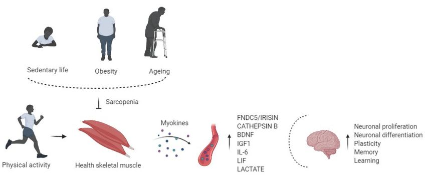
So, altered synthesis and production of myokines due to physical inactivity may
be associated with adverse implications in the brain, such as cognitive impairment and
neurogenerative events [58], showing that muscle may influence the health of the brain
(Figure 1).Life 2021, 11, x FOR PEER REVIEW 4 of 12
Life 2021, 11, 173 4 of 12
Figure 1. Physical activity enhances circulating
Figure 1. circulating levels
levels of myokines in the bloodstream,
bloodstream, affects
affects the brain regulating
regulating neuronal
neuronal
proliferation and
proliferation and differentiation,
differentiation, plasticity,
plasticity, memory,
memory, and
and learning.
learning. Risk factors of sarcopenia, such as physical inactivity,
obesity,and
obesity, andaging,
aging,alter
alterthe
themyokines’
myokines'production
productionand
andrelease,
release,impairing
impairingcognitive
cognitivefunction.
function.
4. Exercise Induced
4. Exercise Induced Myokines
Myokines and and Brain
Brain Function
Function
Myokines
Myokines have provided a new paradigm and
have provided a new paradigm and aa conceptual
conceptual basis
basis for
for understanding
understanding
the cross-talk between muscle and other organs or tissues.
the cross-talk between muscle and other organs or tissues.
The skeletal muscle
The skeletal musclewas
wasidentified
identifiedasas
anan endocrine
endocrine organ
organ with
with a high
a high capacity
capacity to
to pro-
produce, express,
duce, express, andand secrete
secrete various
various factors,
factors, which
which areare classified
classified as myokines
as myokines [59,60].
[59,60].
Myokines are cytokines and other peptides produced following skeletal
Myokines are cytokines and other peptides produced following skeletal muscle con- muscle con-
tractions and exert autocrine, paracrine, and endocrine effects
tractions and exert autocrine, paracrine, and endocrine effects [61]. [61].
Recent
Recent research
research identified
identified over
over 600
600 myokines
myokines [62];
[62]; however,
however, their
their specific
specific bioactivity
bioactivity
remains largely undescribed and poorly understood [63].
remains largely undescribed and poorly understood [63].
Myokines are involved in muscle proliferation, differentiation, and regeneration [64,65],
Myokines are involved in muscle proliferation, differentiation, and regeneration
but also mediate signaling between muscle and liver, gut pancreas, adipose tissue, bone,
[64,65], but also mediate signaling between muscle and liver, gut pancreas, adipose tissue,
brain, vascular bed, skin, and present anticancer effects [61,66–68]. Emerging evidence
bone, brain, vascular bed, skin, and present anticancer effects [61,66–68]. Emerging evi-
indicates that myokines improve human health and ameliorate multiple diseases [69–71].
dence indicates that myokines improve human health and ameliorate multiple diseases
Indeed, myokines regulate systemic glucose homeostasis, lipid metabolism, enhance in-
[69–71]. Indeed, myokines regulate systemic glucose homeostasis, lipid metabolism, en-
sulin sensitivity, and induce white adipose tissue (WAT) browning [72–74].
hance insulin sensitivity, and induce white adipose tissue (WAT) browning [72–74].
Myokine signaling mediates the muscle-brain endocrine loop, promoting relationship
Myokine signaling mediates the muscle-brain endocrine loop, promoting relation-
building between muscle and brain (Table 1) [36,37].
ship building between muscle and brain (Table 1) [36,37].
4.1. FNDC5/Irisin
4.1. FNDC5/Irisin
Fibronectin type III domains containing protein 5 (FNDC5) is a glycosylated type I
Fibronectin
membrane type
protein IIIFollowing
[75]. domains containing
proteolytic protein
cleavage5 of
(FNDC5)
FDN5C,isirisin
a glycosylated
is generatedtype
as aI
membrane protein [75]. Following proteolytic cleavage of FDN5C,
peptide of 112 amino acids (aa 29–140), and it is released into the circulation [76]. irisin is generated as a
peptide of 112 aminoisacids
FNDC5/IRISIN (aa 29–140),
described and it is releasedmyokine;
as an exercise-induced into the circulation [76]. muscle
indeed, skeletal
FNDC5/IRISIN
produces is described
the most quantity as an total
of irisin’s exercise-induced myokine;
circulating levels. indeed,muscle
In skeletal skeletalexercise,
muscle
produces the
inducing the activation
most quantity of irisin's total
of peroxisome circulating levels.receptor
proliferator-activated In skeletal muscle
gamma exercise,
coactivator
inducing
1-alpha the activation
(PGC-1α), a key of peroxisome
regulator proliferator-activated
of skeletal muscle plasticityreceptor gamma
after exercise coactivator
promotes the
synthesis and secretion of irisin [77]. Several studies demonstrated that physical activitythe
1-alpha (PGC-1α), a key regulator of skeletal muscle plasticity after exercise promotes in
synthesis
mice and secretion
and humans of irisin
increases Fndc5 [77].
mRNASeveral studies muscle
in skeletal demonstrated that physical activity
cells [78–80].
in mice and
Irisin humans increases
circulating Fndc5
concentration wasmRNA in skeletal
significantly muscle
higher cells [78–80].
in individuals after endurance
Irisin
exercise circulating concentration was significantly higher in individuals after endur-
[81].
ance Inexercise [81].
the hippocampus, a region involved in memory and spatial awareness, exercise
leadsIn Fndc5 expression in aaPGC-1α-dependent
the hippocampus, region involved in mannermemoryinand mice model.
spatial Pgc1α −/exercise
awareness, − mice
did not present FNDC5 expression [78].
leads Fndc5 expression in a PGC-1α-dependent manner in mice model. Pgc1α −/− mice
did not present FNDC5 expression [78].Life 2021, 11, 173 5 of 12
Also, irisin stimulates neuronal proliferation and differentiation and contributes to
the exercise neuroprotective effects through activation of protein kinase B (PKB) and
extracellular signal-regulated kinases 1/2 (ERK1/2) signaling pathway [82].
Recently, research suggested the role of irisin in regulating synaptic function and
memory in mouse models of AD [83]. In the brain of Fndc5 −/− mice, a mice mole of AD,
synaptic plasticity and long-term potentiation are compromised, while the FNDC5/irisin
re-expression rescued synaptic plasticity and memory impairment [83].
Moreover, irisin increase brain-derived neurotrophic factor (BDNF) expression in the
brain, which is involved in the cognitive function [41].
4.2. Cathepsin B
Cathepsin B (CTSB) belongs to a family of lysosomal cysteine proteases [84].
Exercise induces the Ctsb gene expression in muscle, which promotes BDNF synthesis
in hippocampal stimulating neurogenesis in mice model [85]. These findings in rodents
are supported by results obtained in rhesus macaque and in human, where 4 months
of treadmill exercise elevated CTSB level in plasma [85]. Crossing BBB, skeletal muscle-
derived CTSB induces hippocampal upregulation of BDNF and doublecortin, regulating
synaptic plasticity, cell survival, and neuronal migration [86].
Another study showed that mice treated for 1 week with AICAR, which stimulates
AMP-activated protein kinase (AMPK), presented an improvement of hippocampal neu-
rogenesis and cognitive function [87]. These findings revealed that endurance exercise,
activating AMPK in skeletal muscles, induced a release of CTSB, which may be asso-
ciated with exercise-induced improvement of cognitive function, such as neurogenesis,
memory, and learning [41,87,88]. Cathepsin B knock-out mice showed reduced adult hip-
pocampal neurogenesis and impaired spatial learning and memory. Stimulation of adult
neuro-progenitor cells with recombinant cathepsin B increased neurogenesis [85].
Table 1. Mechanisms of action and effects of myokines on the brain.
Myokine Effects on the Brain Mechanisms of Action
Neuronal proliferation and differentiation, synaptic
FNDC5/Irisin PKB and ERK1/2 signaling pathway [82]
function, memory [75–83]
Neurogenesis, memory,
Cathepsin B BDNF synthesis [86,87]
Learning [84–87]
Synaptic plasticity, neuronal differentiation, cell PI3K and ERK signaling pathway
BDNF
survival, hippocampal function [89–92] [91]
Neurogenesis and neuron survival, neurotrophic,
IGF1 BDNF synthesis [96]
angiogenic, and metabolic proprieties [93–96]
Survival and differentiation
IL-6 Further investigation are needed To be investigated
[97–106]
Astrocyte’s development, oligodendrocytes survival AKT/extracellular signal-regulated-mediated c-fos
LIF
amyloid β-induced neurotoxicity [107–112] induction [112]
BDNF synthesis; Hydroxycarboxylic acid receptor 1
Memory, learning, neuroprotection, neuronal
(HCAR1); VEGF synthesis; NMDA glutamate
L- Lactate plasticity, neuronal metabolism, LTP maintenance,
receptor-mediated signaling; Arc, c-Fos, and Zif268
Angiogenesis [113–118]
synthesis [113–118]
4.3. BDNF
Brain-derived neurotrophic factor (BDNF) belongs to the neurotrophin family, and
it is a crucial mediator of beneficial effects of exercise on the brain [86]. In response to
acute exercise and exercise training, BDNF is abundantly expressed in the brain, but
several studies verify its expression also in skeletal muscle [89]. It is not well understood
if muscle-derived BDNF is released in the bloodstream and mediates muscle-brain cross-
talk. Instead, more evidence suggests that FNDC5 and Cathepsin B, crossing the BBB,
positively induce BDNF expression in the hippocampus via PKB activation and “cAMPLife 2021, 11, 173 6 of 12
response element-binding protein” (CREB) signaling inducing an increment of BDNF [90].
BDNF influences cognitive function activating (Phosphoinositide phospholipase -γ) PLC-
γ, Phosphoinositide 3-kinase (PI3K), and ERK pathways that together affect synaptic
plasticity [91].
BDNF, increasing the growth and proliferation of hippocampal dentate gyrus cells, is
involved in neuronal differentiation, plasticity, cell survival, hippocampal function, show-
ing a dominant role in mediating the effects of physical activity on cognitive changes [86].
Exercise-induced BDNF was shown to decrease the production of toxic amyloid β
peptides, which could be important in treating Alzheimer’s disease (AD). Patients with
neurodegenerative diseases, like AD, Parkinson’s disease, and depression, presented low
serum levels of BDNF [92].
4.4. IGF1
Insulin-like growth factor1 (IGF-1) is an essential factor in brain neurogenesis and
cognitive function; therefore, IGF-1 signaling may play a key role in muscle-brain cross-
talk [37,93]. A primary source of IGF-1 is the liver; however, it is produced by skeletal
muscle during physical activity [94]. Aerobic exercise not only increases the neuronal
uptake of IGF-1 but stimulates IGF-1 signaling pathways inducing PKB-CREB-mediated
BDNF expression, followed by neurogenesis and neuron survival [95].
Studies have demonstrated that, in older adults, aerobic exercise, increasing IGF-1 and
consequently BDNF, significantly increased hippocampal volume and connectivity [96].
IGF-1 is involved in several brain functions, including neurotrophic, angiogenic, and
metabolic proprieties [42].
4.5. IL-6
Interleukin 6 (IL-6) was the first myokine found to be secreted into circulation in a
tumor necrosis factor (TNF)-independent manner [97] in response to muscle contractions,
with a considerable increase in plasma up to 100-fold in response to exercise [98]. During
exercise, circulating IL-6 levels increased without any sign of muscle damage [99]. Physical
activity increased the expression of IL-6 mRNA and protein levels in the brain [100,101].
Two weeks of voluntary wheel running increased IL-6 expression in the hippocampus
in mice, resulting in downregulation of pro-inflammatory cytokines and inflammation,
suggesting that IL-6 may protect the brain, reducing harmful inflammatory responses [102].
In vitro studies showed that IL-6 promotes the survival and differentiation of neural
cells [103,104], protecting against Ca2+ and ROS excitotoxicity [105] Il-6 plays a role in
neurodegenerative diseases such as AD. In vitro studies demonstrated that the addition
of Il-6 in a culture medium increased neurotoxicity caused by Aβ in cortical neurons.
However, in vivo, IL-6 increased astrocytes and microglia cells’ activation, improving
plaque Aβ clearance, showing its neuroprotective properties [106].
Further investigations should be carried out to better identify the role and mechanisms
of myokine IL-6 in cognitive function.
4.6. LIF
Leukemia inhibitory factor (LIF) belongs to the IL-6 family and it is produced by
different organs or tissues, including cardiac muscle, neuronal tissue, and skeletal mus-
cle [107]. LIF is involved in the astrocyte’s development, oligodendrocytes survival, and
recovery processes after injuring the spinal cord in mice [42,108]. LIF is an exercise-induced
myokine and via autocrine or paracrine signaling induces hypertrophy and regeneration
of skeletal muscle [110–112]. It was also demonstrated that LIF, via PKB/extracellular
signal-regulated-mediated c-fos induction, protects against amyloid β-induced neurotoxic-
ity [112].Life 2021, 11, 173 7 of 12
4.7. L-Lactate
L-Lactate is a metabolite from contracting skeletal muscle [113]. L-Lactate is released
from muscles during short-term exercise, even from muscles at rest, during recovery from
short-term exercise, and long-lasting exercise [114].
Extended findings conclude that L-Lactate is a component of the “exercise pill.” L-
Lactate is a hormone involved in memory and neuroprotection, and it seems to be an essen-
tial substrate for neuronal metabolism and long-term potentiation (LTP) maintenance [115].
L-Lactate produced in exercised mice crossed the blood-brain barrier to induce expression
of Bdnf and signaling of the BDNF receptor tropomyosin receptor kinase B (TrkB) in the
hippocampus, resulting in the promotion of learning and memory formation [116].
Acute and high-intensity exercise lactate promotes angiogenesis in the brain by bind-
ing with hydroxycarboxylic acid receptor 1 (HCAR1) [42]. L-lactate’s daily injection,
simulating blood lactate levels observed during exercise, increased vascular endothelial
growth factor (VEGF) levels, and, consequently, microvascular density in the dentate
gyrus [117].
L-Lactate potentiates N-methyl-D-aspartate receptor (NMDA) glutamate receptor-
mediated signaling, playing a central role in neuronal plasticity and memory processes;
L-Lactate activates a cascade of molecular events that end up with stimulation of the
expression of synaptic plasticity-related genes, such as activity regulated cytoskeleton
associated protein (Arc), c-Fos, and Zif268 [118].
5. Conclusions
The remarkable improvements in life expectancy resulted in a longer lifespan that
brings new opportunities for elder people. However, these opportunities and contributions
depend on one factor that is health.
Indeed, aging-chronic diseases negatively impact health and consequently affect the
quality of life and represent major public health concerns. In the last decade, strong empha-
sis was given to sarcopenia, defined as age-related loss of muscle mass and function [6].
Epidemiological evidence suggests that sarcopenia is associated with accelerated
cognitive changes that lead to cognitive impairment [30]. The exact mechanism relat-
ing to sarcopenia and cognitive impairment is not well elucidated, but the existence of
muscle-brain endocrine loop mediated by myokines is demonstrated [31]. Myokines are
muscle-produced factors that improve brain function as cognition, memory, and motor
coordination [36]. Sarcopenia is linked to a reduction in the regenerative capacity of
skeletal muscle stem cells and an altered rate of cellular regenerative and differentia-
tion; therefore, this could result in a compromised production and secretion of myokines
with a negative consequence on brain function [1]. On the contrary, exercise regulates
myokines’ expression contributing to autocrine regulation of metabolism in the muscle
and to paracrine/endocrine regulation of other adjacent/remote organs [37].
Even though specific bioactivity remains undescribed and poorly understood [59],
clarifying better how myokines are secreted and released from muscle fiber, their effect
on the brain will help understand the link between muscle, exercise, and brain function.
It may also be possible to develop small compounds derived from myokines to provide
treatment of neurodegenerative diseases.
Author Contributions: Concept and design, L.S., G.P. and M.B.; drafting of the manuscript: L.S.,
R.A.F., S. and V.C. All authors have read and agreed to the published version of the manuscript.
Funding: This research was funded by the Ministero dell0 Istruzione, dell0 Università e della Ricerca
Scientifica (grants PRIN 2017). POR CAMPANIA FESR 2014/2020- i-CURE.
Institutional Review Board Statement: Not applicable.
Informed Consent Statement: Not applicable.
Data Availability Statement: Not applicable.Life 2021, 11, 173 8 of 12
Acknowledgments: The authors thank Biorender for figures.
Conflicts of Interest: The authors declare no conflict of interest.
References
1. Cruz-Jentoft, A.J.; Bahat, G.; Bauer, J.; Boirie, Y.; Bruyere, O.; Cederholm, T.; Cooper, C.; Landi, F.; Rolland, Y.; Sayer, A.A.; et al.
Writing Group for the European Working Group on Sarcopenia in Older People 2 (EWGSOP2), and the Extended Group for
EWGSOP2. Sarcopenia: revised European consensus on definition and diagnosis. Age Ageing 2019, 48, 16–31. [CrossRef]
2. Kaeberlein, M.; Rabinovitch, P.S.; Martin, G.M. Healthy aging: The ultimate preventative medicine. Science 2015, 350, 1191–1193.
[CrossRef]
3. Molino, S.; Dossena, M.; Buonocore, D.; Verri, M. Sarcopenic obesity: An appraisal of the current status of knowledge and
management in elderly people. J. Nutr. Health Aging 2015, 20, 780–788. [CrossRef]
4. Alkner, B.A.; Tesch, P.A. Knee extensor and plantar flexor muscle size and function following 90 days of bed rest with or without
resistance exercise. Eur. J. Appl. Physiol. 2004, 93, 294–305. [CrossRef]
5. Shackelford, L.C.; Leblanc, A.D.; Driscoll, T.B.; Evans, H.J.; Rianon, N.J.; Smith, S.M.; Spector, E.; Feeback, D.L.; Lai, D. Resistance
exercise as a countermeasure to disuse-induced bone loss. J. Appl. Physiol. 2004, 97, 119–129. [CrossRef]
6. Cruz-Jentoft, A.J.; Sayer, A.A. Sarcopenia. Lancet 2019, 393, 2636–2646. [CrossRef]
7. Murton, A.J.; Marimuthu, K.; Mallinson, J.E.; Selby, A.L.; Smith, K.; Rennie, M.J.; Greenhaff, P.L. Obesity Appears to Be Associated
With Altered Muscle Protein Synthetic and Breakdown Responses to Increased Nutrient Delivery in Older Men, but Not Reduced
Muscle Mass or Contractile Function. Diabetes 2015, 64, 3160–3171. [CrossRef] [PubMed]
8. Brack, A.S.; Conboy, M.J.; Roy, S.; Lee, M.; Kuo, C.J.; Keller, C.; Rando, T.A. Increased Wnt Signaling During Aging Alters Muscle
Stem Cell Fate and Increases Fibrosis. Science 2007, 317, 807–810. [CrossRef] [PubMed]
9. Carey, K.A.; Farnfield, M.M.; Tarquinio, S.D.; Cameron-Smith, D. Impaired Expression of Notch Signaling Genes in Aged Human
Skeletal Muscle. J. Gerontol. Ser. A Boil. Sci. Med. Sci. 2007, 62, 9–17. [CrossRef] [PubMed]
10. Florian, M.C.; Nattamai, K.J.; Dörr, K.; Marka, G.; Überle, B.; Vas, V.; Eckl, C.; Andrä, I.; Schiemann, M.; Oostendorp, R.A.J.;
et al. A canonical to non-canonical Wnt signalling switch in haematopoietic stem-cell ageing. Nat. Cell Biol. 2013, 503, 392–396.
[CrossRef] [PubMed]
11. Fry, C.S.; Lee, J.D.; Mula, J.; Kirby, T.J.; Jackson, J.R.; Liu, F.; Yang, L.; Mendias, C.L.; Dupont-Versteegden, E.E.; McCarthy, J.J.; et al.
Inducible depletion of satellite cells in adult, sedentary mice impairs muscle regenerative capacity without affecting sarcopenia.
Nat. Med. 2015, 21, 76–80. [CrossRef] [PubMed]
12. Marcell, T.J. Review Article: Sarcopenia: Causes, Consequences, and Preventions. J. Gerontol. Ser. A Boil. Sci. Med. Sci. 2003, 58,
M911–M916. [CrossRef]
13. Rutkowski, J.M.; Stern, J.H.; Scherer, P.E. The cell biology of fat expansion. J. Cell Biol. 2015, 208, 501–512. [CrossRef]
14. Kob, R.; Bollheimer, L.C.; Bertsch, T.; Fellner, C.; Djukic, M.; Sieber, C.C.; Fischer, B.E. Sarcopenic obesity: Molecular clues to a
better understanding of its pathogenesis? Biogerontology 2014, 16, 15–29. [CrossRef]
15. Stinkens, R.; Goossens, G.H.; Jocken, J.W.E.; Blaak, EE. Targeting fatty acid metabolism to improve glucose metabolism. Obes. Rev.
2015, 16, 715–757. [CrossRef] [PubMed]
16. Kalinkovich, A.; Livshits, G. Sarcopenic obesity or obese sarcopenia: A cross talk between age-associated adipose tissue and
skeletal muscle inflammation as a main mechanism of the pathogenesis. Ageing Res. Rev. 2017, 35, 200–221. [CrossRef]
17. Rivas, D.A.; McDonald, D.J.; Rice, N.P.; Haran, P.H.; Dolnikowski, G.G.; Fielding, R.A. Diminished anabolic signaling response to
insulin induced by intramuscular lipid accumulation is associated with inflammation in aging but not obesity. Am. J. Physiol.
Integr. Comp. Physiol. 2016, 310, R561–R569. [CrossRef] [PubMed]
18. Apostolopoulos, V.; De Courten, M.P.J.; Stojanovska, L.; Blatch, G.L.; Tangalakis, K.; De Courten, B.; de Courten, M.P.M.P.J. The
complex immunological and inflammatory network of adipose tissue in obesity. Mol. Nutr. Food Res. 2015, 60, 43–57. [CrossRef]
[PubMed]
19. Exley, M.A.; Hand, L.; O’Shea, D.; Lynch, L. Interplay between the immune system and adipose tissue in obesity. J. Endocrinol.
2014, 223, R41–R48. [CrossRef]
20. Tateya, S.; Kim, F.; Tamori, Y. Recent advances in obesity-induced inflammation and insulin resistance. Front. Endocrinol. 2013, 4,
93. [CrossRef]
21. Sardu, C.; Pieretti, G.; D’Onofrio, N.; Ciccarelli, F.; Paolisso, P.; Passavanti, M.B.; Marfella, R.; Cioffi, M.; Mone, P.; Dalise, A.M.;
et al. Inflammatory Cytokines and SIRT1 Levels in Subcutaneous Abdominal Fat: Relationship With Cardiac Performance in
Overweight Pre-diabetics Patients. Front. Physiol. 2018, 9, 1030. [CrossRef]
22. Raschke, S.; Eckel, J. Adipo-Myokines: Two Sides of the Same Coin—Mediators of Inflammation and Mediators of Exercise.
Mediat. Inflamm. 2013, 2013, 1–16. [CrossRef]
23. Rodríguez, A.; Ezquerro, S.; Méndez-Giménez, L.; Becerril, S.; Frühbeck, G. Revisiting the adipocyte: A model for integration of
cytokine signaling in the regulation of energy metabolism. Am. J. Physiol. Metab. 2015, 309, E691–E714. [CrossRef]
24. Paolisso, P.; Foà, A.; Bergamaschi, L.; Donati, F.; Fabrizio, M.; Chiti, C.; Angeli, F.; Toniolo, S.; Stefanizzi, A.; Armillotta, M.; et al.
Hyperglycemia, inflammatory response and infarct size in obstructive acute myocardial infarction and MINOCA. Cardiovasc.
Diabetol. 2021, 20, 1–11. [CrossRef]Life 2021, 11, 173 9 of 12
25. Kim, T.N.; Park, M.S.; Lim, K.I.; Choi, H.Y.; Yang, S.J.; Yoo, H.J.; Kang, H.J.; Song, W.; Choi, H.; Baik, S.H.; et al. Relationships
between sarcopenic obesity and insulin resistance, inflammation, and vitamin D status: The Korean Sarcopenic Obesity Study.
Clin. Endocrinol. 2013, 78, 525–532. [CrossRef] [PubMed]
26. Migliavacca, E.; Tay, S.K.H.; Patel, H.P.; Sonntag, T.; Civiletto, G.; McFarlane, C.; Forrester, T.; Barton, S.J.; Leow, M.K.; Antoun,
E.; et al. Mitochondrial oxidative capacity and NAD+ biosynthesis are reduced in human sarcopenia across ethnicities. Nat.
Commun. 2019, 10, 5808. [CrossRef]
27. Lebrasseur, N.K.; Tchkonia, T.; Kirkland, J.L. Cellular Senescence and the Biology of Aging, Disease, and Frailty. Nestle Nutr. Inst.
Workshop Ser. 2015, 83, 11–18. [PubMed]
28. Kelley, D.E.; Goodpaster, B.H. Stewing in Not-So-Good Juices: Interactions of Skeletal Muscle with Adipose Secretions: Figure 1.
Diabetes 2015, 64, 3055–3057. [CrossRef]
29. Sousa-Victor, P.; Gutarra, S.; García-Prat, L.; Rodriguez-Ubreva, J.; Ortet, L.; Ruiz-Bonilla, V.; Jardí, M.; Ballestar, E.; González, S.;
Serrano, A.L.; et al. Geriatric muscle stem cells switch reversible quiescence into senescence. Nat. Cell Biol. 2014, 506, 316–321.
[CrossRef] [PubMed]
30. Cipolli, G.C.; Yassuda, M.S.; Aprahamian, I. Sarcopenia Is Associated with Cognitive Impairment in Older Adults: A Systematic
Review and Meta-Analysis. J. Nutr. Health Aging 2019, 23, 525–531. [CrossRef] [PubMed]
31. Sui, S.X.; Williams, L.J.; Holloway-Kew, K.L.; Hyde, N.K.; Pasco, J.A. Skeletal Muscle Health and Cognitive Function: A Narrative
Review. Int. J. Mol. Sci. 2020, 22, 255. [CrossRef] [PubMed]
32. Peng, T.-C.; Chen, W.-L.; Wu, L.-W.; Chang, Y.-W.; Kao, T.-W. Sarcopenia and cognitive impairment: A systematic review and
meta-analysis. Clin. Nutr. 2020, 39, 2695–2701. [CrossRef]
33. Van Kan, G.A.; Cesari, M.; Gillette-Guyonnet, S.; Dupuy, C.; Nourhashémi, F.; Schott, A.-M.; Beauchet, O.; Annweiler, C.; Vellas,
B.; Rolland, Y. Sarcopenia and cognitive impairment in elderly women: Results from the EPIDOS cohort. Age Ageing 2012, 42,
196–202. [CrossRef]
34. Chen, W.-L.; Peng, T.-C.; Sun, Y.-S.; Yang, H.-F.; Liaw, F.-Y.; Wu, L.-W.; Chang, Y.-W.; Kao, T.-W. Examining the Association
Between Quadriceps Strength and Cognitive Performance in the Elderly. Medicine 2015, 94, e1335. [CrossRef] [PubMed]
35. Taekema, D.G.; Ling, C.H.Y.; Kurrle, S.E.; Cameron, I.D.; Meskers, C.G.M.; Blauw, G.J.; Westendorp, R.G.J.; De Craen, A.J.M.;
Maier, A.B. Temporal relationship between handgrip strength and cognitive performance in oldest old people. Age Ageing 2012,
41, 506–512. [CrossRef]
36. Severinsen, M.C.K.; Pedersen, B.K. Muscle–Organ Crosstalk: The Emerging Roles of Myokines. Endocr. Rev. 2020, 41, 594–609.
[CrossRef] [PubMed]
37. Chen, W.; Wang, L.; You, W.; Shan, T. Myokines mediate the cross talk between skeletal muscle and other organs. J. Cell. Physiol.
2021, 236, 2393–2412. [CrossRef]
38. Cotman, C.W.; Berchtold, N.C.; Christie, L.-A. Exercise builds brain health: Key roles of growth factor cascades and inflammation.
Trends Neurosci. 2007, 30, 464–472. [CrossRef]
39. Mattson, M.P. Evolutionary aspects of human exercise—Born to run purposefully. Ageing Res. Rev. 2012, 11, 347–352. [CrossRef]
[PubMed]
40. Agudelo, L.Z.; Femenía, T.; Orhan, F.; Porsmyr-Palmertz, M.; Goiny, M.; Martinez-Redondo, V.; Correia, J.C.; Izadi, M.; Bhat,
M.; Schuppe-Koistinen, I.; et al. Skeletal Muscle PGC-1α1 Modulates Kynurenine Metabolism and Mediates Resilience to
Stress-Induced Depression. Cell 2014, 159, 33–45. [CrossRef]
41. Pedersen, B.K. Physical activity and muscle-brain cross-talk. Nat. Rev. Endocrinol. 2019, 15, 383–392. [CrossRef]
42. Kim, S.; Choi, J.-Y.; Moon, S.; Park, D.-H.; Kwak, H.-B.; Kang, J.-H. Roles of myokines in exercise-induced improvement of
neuropsychiatric function. Pflugers Arch. 2019, 471, 491–505. [CrossRef]
43. Colcombe, S.J.; Erickson, K.I.; Scalf, P.E.; Kim, J.S.; Prakash, R.; McAuley, E.; Elavsky, S.; Marquez, D.X.; Hu, L.; Kramer, A.F.
Aerobic Exercise Training Increases Brain Volume in Aging Humans. J. Gerontol. Ser. A Boil. Sci. Med. Sci. 2006, 61, 1166–1170.
[CrossRef] [PubMed]
44. Erickson, K.I.; Prakash, R.S.; Voss, M.W.; Chaddock, L.; Hu, L.; Morris, K.S.; White, S.M.; Wójcicki, T.R.; McAuley, E.; Kramer, A.F.
Aerobic fitness is associated with hippocampal volume in elderly humans. Hippocampus 2009, 19, 1030–1039. [CrossRef]
45. Pereira, A.C.; Huddleston, D.E.; Brickman, A.M.; Sosunov, A.A.; Hen, R.; McKhann, G.M.; Sloan, R.; Gage, F.H.; Brown, T.R.;
Small, S.A. An in vivo correlate of exercise-induced neurogenesis in the adult dentate gyrus. Proc. Natl. Acad. Sci. USA 2007, 104,
5638–5643. [CrossRef]
46. Voss, M.W.; Erickson, K.I.; Prakash, R.S.; Chaddock, L.; Kim, J.S.; Alves, H.; Szabo, A.; Phillips, S.M.; Wójcicki, T.R.; Mailey, E.L.;
et al. Neurobiological markers of exercise-related brain plasticity in older adults. Brain Behav. Immun. 2013, 28, 90–99. [CrossRef]
[PubMed]
47. Vreugdenhil, A.; Cannell, J.; Davies, A.; Razay, G. A community-based exercise programme to improve functional ability in
people with Alzheimer’s disease: A randomized controlled trial. Scand. J. Caring Sci. 2012, 26, 12–19. [CrossRef] [PubMed]
48. Aarsland, D.; Sardahaee, F.S.; Anderssen, S.; Ballard, C. The Alzheimer’s Society the Alzheimer’s Society Systematic Review
group Is physical activity a potential preventive factor for vascular dementia? A systematic review. Aging Ment. Health 2010, 14,
386–395. [CrossRef]
49. Blondell, S.J.; Hammersley-Mather, R.; Veerman, J.L. Does physical activity prevent cognitive decline and dementia?: A systematic
review and meta-analysis of longitudinal studies. BMC Public Health 2014, 14, 510. [CrossRef] [PubMed]Life 2021, 11, 173 10 of 12
50. Amboni, M.; Barone, P.; Hausdorff, J.M. Cognitive contributions to gait and falls: Evidence and implications. Mov. Disord. 2013,
28, 1520–1533. [CrossRef] [PubMed]
51. Kramer, A.F.; Colcombe, S. Fitness Effects on the Cognitive Function of Older Adults: A Meta-Analytic Study—Revisited. Perspect.
Psychol. Sci. 2018, 13, 213–217. [CrossRef] [PubMed]
52. Mokhtarzade, M.; Motl, R.; Negaresh, R.; Zimmer, P.; Khodadoost, M.; Baker, J.S.; Patel, D.; Majdinasab, N.; Ranjbar, R. Exercise-
induced changes in neurotrophic factors and markers of blood-brain barrier permeability are moderated by weight status in
multiple sclerosis. Neuropeptides 2018, 70, 93–100. [CrossRef]
53. Swain, R.; Harris, A.; Wiener, E.; Dutka, M.; Morris, H.; Theien, B.; Konda, S.; Engberg, K.; Lauterbur, P.; Greenough, W. Prolonged
exercise induces angiogenesis and increases cerebral blood volume in primary motor cortex of the rat. Neuroscience 2003, 117,
1037–1046. [CrossRef]
54. Van Praag, H.; Kempermann, G.; Gage, F.H. Running increases cell proliferation and neurogenesis in the adult mouse dentate
gyrus. Nat. Neurosci. 1999, 2, 266–270. [CrossRef]
55. Van Praag, H. Neurogenesis and exercise: Past and future directions. Neuromolecular Med. 2008, 10, 128–140. [CrossRef] [PubMed]
56. Tari, A.R.; Norevik, C.S.; Scrimgeour, N.R.; Kobro-Flatmoen, A.; Storm-Mathisen, J.; Bergersen, L.H.; Wrann, C.D.; Selbæk, G.;
Kivipelto, M.; Moreira, J.B.N.; et al. Are the neuroprotective effects of exercise training systemically mediated? Prog. Cardiovasc.
Dis. 2019, 62, 94–101. [CrossRef] [PubMed]
57. Widmann, M.; Nieß, A.M.; Munz, B. Physical Exercise and Epigenetic Modifications in Skeletal Muscle. Sports Med. 2019, 49,
509–523. [CrossRef] [PubMed]
58. Iizuka, K.; Machida, T.; Hirafuji, M. Skeletal Muscle Is an Endocrine Organ. J. Pharmacol. Sci. 2014, 125, 125–131. [CrossRef]
59. Bortoluzzi, S.; Scannapieco, P.; Cestaro, A.; Danieli, G.A.; Schiaffino, S. Computational reconstruction of the human skeletal
muscle secretome. Proteins Struct. Funct. Bioinform. 2005, 62, 776–792. [CrossRef]
60. Yoon, J.H.; Yea, K.; Kim, J.; Choi, Y.S.; Park, S.; Lee, H.; Lee, C.S.; Suh, P.-G.; Ryu, S.H. Comparative proteomic analysis of the
insulin-induced L6 myotube secretome. Proteomics 2009, 9, 51–60. [CrossRef]
61. Pedersen, B.K.; Febbraio, M.A. Muscles, exercise and obesity: Skeletal muscle as a secretory organ. Nat. Rev. Endocrinol. 2012, 8,
457–465. [CrossRef]
62. Görgens, S.W.; Eckardt, K.; Jensen, J.D.; Drevon, C.A.; Eckel, J. Exercise and Regulation of Adipokine and Myokine Production.
Prog. Mol. Biol. Transl. Sci. 2015, 135, 313–336. [CrossRef]
63. Lee, J.H.; Jun, H.-S. Role of Myokines in Regulating Skeletal Muscle Mass and Function. Front. Physiol. 2019, 10, 42. [CrossRef]
[PubMed]
64. Henningsen, J.; Pedersen, B.K.; Kratchmarova, I. Quantitative analysis of the secretion of the MCP family of chemokines by
muscle cells. Mol. BioSyst. 2010, 7, 311–321. [CrossRef] [PubMed]
65. Henningsen, J.; Rigbolt, K.T.G.; Blagoev, B.; Pedersen, B.K.; Kratchmarova, I. Dynamics of the Skeletal Muscle Secretome during
Myoblast Differentiation. Mol. Cell. Proteom. 2010, 9, 2482–2496. [CrossRef]
66. Benatti, F.B.; Pedersen, B.K. Exercise as an anti-inflammatory therapy for rheumatic diseases—myokine regulation. Nat. Rev.
Rheumatol. 2015, 11, 86–97. [CrossRef]
67. Hojman, P.; Gehl, J.; Christensen, J.F.; Pedersen, B.K. Molecular Mechanisms Linking Exercise to Cancer Prevention and Treatment.
Cell Metab. 2018, 27, 10–21. [CrossRef] [PubMed]
68. Pedersen, L.; Idorn, M.; Olofsson, G.H.; Lauenborg, B.; Nookaew, I.; Hansen, R.H.; Johannesen, H.H.; Becker, J.C.; Pedersen,
K.S.; Dethlefsen, C.; et al. Voluntary Running Suppresses Tumor Growth through Epinephrine- and IL-6-Dependent NK Cell
Mobilization and Redistribution. Cell Metab. 2016, 23, 554–562. [CrossRef]
69. Motl, R.W.; Pilutti, L.A. The benefits of exercise training in multiple sclerosis. Nat. Rev. Neurol. 2012, 8, 487–497. [CrossRef]
70. Pedersen, B.K.; Saltin, B. Evidence for prescribing exercise as therapy in chronic disease. Scand. J. Med. Sci. Sports 2006, 16, 3–63.
[CrossRef] [PubMed]
71. Tanasescu, M.; Leitzmann, M.F.; Rimm, E.B.; Willett, W.C.; Stampfer, M.J.; Hu, F.B. Exercise Type and Intensity in Relation to
Coronary Heart Disease in Men. JAMA 2002, 288, 1994–2000. [CrossRef]
72. Ma, Y.; Gao, M.; Sun, H.; Liu, D. Interleukin-6 gene transfer reverses body weight gain and fatty liver in obese mice. Biochim.
Biophys. Acta 2015, 1852, 1001–1011. [CrossRef]
73. Quinn, L.S.; Strait-Bodey, L.; Anderson, B.G.; Argilés, J.M.; Havel, P.J. WITHDRAWN: Interleukin-15 stimulates adiponectin
secretion by 3T3-L1 adipocytes: Evidence for a skeletal muscle-to-fat signaling pathway. Cell Biol. Int. 2005, 29, 449–457.
[CrossRef] [PubMed]
74. Roberts, L.D.; Bostrom, P.; O’Sullivan, J.F.; Schinzel, R.T.; Lewis, G.D.; Dejam, A.; Lee, Y.K.; Palma, M.J.; Calhoun, S.; Georgiadi,
A.; et al. beta-Aminoisobutyric acid induces browning of white fat and hepatic beta-oxidation and is inversely correlated with
cardiometabolic risk factors. Cell Metab. 2014, 19, 96–108. [CrossRef] [PubMed]
75. Young, M.F.; Valaris, S.; Wrann, C.D. A role for FNDC5/Irisin in the beneficial effects of exercise on the brain and in neurodegen-
erative diseases. Prog. Cardiovasc. Dis. 2019, 62, 172–178. [CrossRef]
76. Schumacher, M.A.; Chinnam, N.; Ohashi, T.; Shah, R.S.; Erickson, H.P. The structure of irisin reveals a novel intersubunit
beta-sheet fibronectin type III (FNIII) dimer: Implications for receptor activation. J. Biol. Chem. 2013, 288, 33738–33744. [CrossRef]Life 2021, 11, 173 11 of 12
77. Bostroem, P.; Wu, J.; Jedrychowski, M.P.; Korde, A.; Ye, L.; Lo, J.C.; Rasbach, K.A.; Bostroem, E.A.; Choi, J.H.; Long, J.Z.; et al. A
PGC1-α-dependent myokine that drives brown-fat-like development of white fat and thermogenesis. Nat. Cell Biol. 2012, 481,
463–468. [CrossRef]
78. Wrann, C.D.; White, J.P.; Salogiannnis, J.; Laznik-Bogoslavski, D.; Wu, J.; Ma, D.; Lin, J.D.; Greenberg, M.E.; Spiegelman, B.M.
Exercise Induces Hippocampal BDNF through a PGC-1α/FNDC5 Pathway. Cell Metab. 2013, 18, 649–659. [CrossRef]
79. Quinn, L.S.; Anderson, B.G.; Conner, J.D.; Wolden-Hanson, T. Circulating irisin levels and muscle FNDC5 mRNA expression are
independent of IL-15 levels in mice. Endocrine 2015, 50, 368–377. [CrossRef] [PubMed]
80. Tiano, J.P.; Springer, D.A.; Rane, S.G. SMAD3 negatively regulates serum irisin and skeletal muscle FNDC5 and peroxisome
proliferator-activated receptor gamma coactivator 1-alpha (PGC-1alpha) during exercise. J. Biol. Chem. 2015, 290, 7671–7684.
[CrossRef]
81. Nygaard, H.; Slettaløkken, G.; Vegge, G.; Hollan, I.; Whist, J.E.; Strand, T.; Rønnestad, B.R.; Ellefsen, S. Irisin in Blood Increases
Transiently after Single Sessions of Intense Endurance Exercise and Heavy Strength Training. PLoS ONE 2015, 10, e0121367.
[CrossRef] [PubMed]
82. Li, D.J.; Li, Y.H.; Yuan, H.B.; Qu, L.F.; Wang, P. The novel exercise-induced hormone irisin protects against neuronal injury via
activation of the Akt and ERK1/2 signaling pathways and contributes to the neuroprotection of physical exercise in cerebral
ischemia. Metabolism 2017, 68, 31–42. [CrossRef]
83. Lourenco, M.V.; Frozza, R.L.; De Freitas, G.B.; Zhang, H.; Kincheski, G.C.; Ribeiro, F.C.; Gonçalves, R.A.; Clarke, J.R.; Beckman,
D.; Staniszewski, A.; et al. Exercise-linked FNDC5/irisin rescues synaptic plasticity and memory defects in Alzheimer’s models.
Nat. Med. 2019, 25, 165–175. [CrossRef]
84. Aggarwal, N.; Sloane, B.F. Cathepsin B: Multiple roles in cancer. Proteom. Clin. Appl. 2014, 8, 427–437. [CrossRef] [PubMed]
85. Moon, H.Y.; Becke, A.; Berron, D.; Becker, B.; Sah, N.; Benoni, G.; Janke, E.; Lubejko, S.T.; Greig, N.H.; Mattison, J.A.; et al.
Running-Induced Systemic Cathepsin B Secretion Is Associated with Memory Function. Cell Metab. 2016, 24, 332–340. [CrossRef]
[PubMed]
86. Miranda, M.; Morici, J.F.; Zanoni, M.B.; Bekinschtein, P. Brain-Derived Neurotrophic Factor: A Key Molecule for Memory in the
Healthy and the Pathological Brain. Front. Cell. Neurosci. 2019, 13, 363. [CrossRef]
87. Kobilo, T.; Yuan, C.; Van Praag, H. Endurance factors improve hippocampal neurogenesis and spatial memory in mice. Learn.
Mem. 2011, 18, 103–107. [CrossRef]
88. Marangos, P.J.; Loftus, T.; Wiesner, J.; Lowe, T.; Rossi, E.; Browne, C.E.; Gruber, H.E. Adenosinergic modulation of homocysteine-
induced seizures in mice. Epilepsia 1990, 31, 239–246. [CrossRef] [PubMed]
89. Wang, T.; Maltez, M.T.; Lee, H.W.; Ahmad, M.; Wang, H.-W.; Leenen, F.H.H. Effect of exercise training on the FNDC5/BDNF
pathway in spontaneously hypertensive rats. Physiol. Rep. 2019, 7, e14323. [CrossRef]
90. Obrietan, K.; Gao, X.-B.; Pol, A.N.V.D. Excitatory actions of GABA increase BDNF expression via a MAPK-CREB-dependent
mechanism—A positive feedback circuit in developing neurons. J. Neurophysiol. 2002, 88, 1005–1015. [CrossRef]
91. Kowiański, P.; Lietzau, G.; Czuba, E.; Waśkow, M.; Steliga, A.; Moryś, J. BDNF: A Key Factor with Multipotent Impact on Brain
Signaling and Synaptic Plasticity. Cell. Mol. Neurobiol. 2018, 38, 579–593. [CrossRef] [PubMed]
92. Giacobbo, B.L.; Doorduin, J.; Klein, H.C.; Dierckx, R.A.J.O.; Bromberg, E.; de Vries, E.F.J. Brain-Derived Neurotrophic Factor in
Brain Disorders: Focus on Neuroinflammation. Mol. Neurobiol. 2018, 56, 3295–3312. [CrossRef] [PubMed]
93. LeWitt, M.S.; Boyd, G.W. The Role of Insulin-Like Growth Factors and Insulin-Like Growth Factor–Binding Proteins in the
Nervous System. Biochem. Insights 2019, 12, 1178626419842176. [CrossRef]
94. Berg, U.; Bang, P. Exercise and Circulating Insulin-Like Growth Factor I. Horm. Res. Paediatr. 2004, 62 (Suppl. 1), 50–58. [CrossRef]
95. Zheng, W.-H.; Quirion, R. Comparative signaling pathways of insulin-like growth factor-1 and brain-derived neurotrophic factor
in hippocampal neurons and the role of the PI3 kinase pathway in cell survival. J. Neurochem. 2004, 89, 844–852. [CrossRef]
96. Leckie, R.L.; Oberlin, L.E.; Voss, M.W.; Prakash, R.S.; Szabo-Reed, A.; Chaddock-Heyman, L.; Phillips, S.M.; Gothe, N.P.; Mailey,
E.; Vieira-Potter, V.J.; et al. BDNF mediates improvements in executive function following a 1-year exercise intervention. Front.
Hum. Neurosci. 2014, 8, 985. [CrossRef] [PubMed]
97. Keller, C.; Hellsten, Y.; Steensberg, A.; Pedersen, B.K. Differential regulation of IL-6 and TNF-α via calcineurin in human skeletal
muscle cells. Cytokine 2006, 36, 141–147. [CrossRef]
98. Pedersen, B.K.; Febbraio, M.A. Muscle as an Endocrine Organ: Focus on Muscle-Derived Interleukin-6. Physiol. Rev. 2008, 88,
1379–1406. [CrossRef] [PubMed]
99. Fischer, C.P. Interleukin-6 in acute exercise and training: What is the biological relevance? Exerc. Immunol. Rev. 2006, 12, 6–33.
100. Nybo, L.; Nielsen, B.; Pedersen, B.K.; Møller, K.; Secher, N.H. Interleukin-6 release from the human brain during prolonged
exercise. J. Physiol. 2002, 542, 991–995. [CrossRef]
101. Rasmussen, P.; Vedel, J.-C.; Olesen, J.; Adser, H.; Pedersen, M.V.; Hart, E.; Secher, N.H.; Pilegaard, H. In humans IL-6 is released
from the brain during and after exercise and paralleled by enhanced IL-6 mRNA expression in the hippocampus of mice. Acta
Physiol. 2010, 201, 475–482. [CrossRef]
102. Funk, J.A.; Gohlke, J.; Kraft, A.D.; McPherson, C.A.; Collins, J.B.; Harry, G.J. Voluntary exercise protects hippocampal neurons
from trimethyltin injury: Possible role of interleukin-6 to modulate tumor necrosis factor receptor-mediated neurotoxicity. Brain
Behav. Immun. 2011, 25, 1063–1077. [CrossRef] [PubMed]Life 2021, 11, 173 12 of 12
103. Zhang, P.-L.; Levy, A.M.; Ben-Simchon, L.; Haggiag, S.; Chebath, J.; Revel, M. Induction of neuronal and myelin-related gene
expression by IL-6-receptor/IL-6: A study on embryonic dorsal root ganglia cells and isolated Schwann cells. Exp. Neurol. 2007,
208, 285–296. [CrossRef] [PubMed]
104. Cao, Z.; Gao, Y.; Bryson, J.B.; Hou, J.; Chaudhry, N.; Siddiq, M.; Martinez, J.; Spencer, T.; Carmel, J.; Hart, R.B.; et al. The Cytokine
Interleukin-6 Is Sufficient But Not Necessary to Mimic the Peripheral Conditioning Lesion Effect on Axonal Growth. J. Neurosci.
2006, 26, 5565–5573. [CrossRef] [PubMed]
105. Rothaug, M.; Becker-Pauly, C.; Rose-John, S. The role of interleukin-6 signaling in nervous tissue. Biochim. Biophys. Acta 2016,
1863, 1218–1227. [CrossRef] [PubMed]
106. Erta, M.; Quintana, A.; Hidalgo, J. Interleukin-6, a Major Cytokine in the Central Nervous System. Int. J. Biol. Sci. 2012, 8,
1254–1266. [CrossRef]
107. Nicola, N.A.; Babon, J.J. Leukemia inhibitory factor (LIF). Cytokine Growth Factor Rev. 2015, 26, 533–544. [CrossRef]
108. Deverman, B.E.; Patterson, P.H. Exogenous Leukemia Inhibitory Factor Stimulates Oligodendrocyte Progenitor Cell Proliferation
and Enhances Hippocampal Remyelination. J. Neurosci. 2012, 32, 2100–2109. [CrossRef] [PubMed]
109. Broholm, C.; Mortensen, O.H.; Nielsen, S.; Akerstrom, T.; Zankari, A.; Dahl, B.; Pedersen, B.K. Exercise induces expression of
leukaemia inhibitory factor in human skeletal muscle. J. Physiol. 2008, 586, 2195–2201. [CrossRef] [PubMed]
110. Broholm, C.; Laye, M.J.; Brandt, C.; Vadalasetty, R.; Pilegaard, H.; Pedersen, B.K.; Schéele, C. LIF is a contraction-induced myokine
stimulating human myocyte proliferation. J. Appl. Physiol. 2011, 111, 251–259. [CrossRef] [PubMed]
111. Broholm, C.; Pedersen, B.K. Leukaemia inhibitory factor—An exercise-induced myokine. Exerc. Immunol. Rev. 2010, 16, 77–85.
[PubMed]
112. Guo, A.; Li, K.; Xiao, Q. Sarcopenic obesity: Myokines as potential diagnostic biomarkers and therapeutic targets? Exp. Gerontol.
2020, 139, 111022. [CrossRef]
113. Brooks, G.A. Lactate as a fulcrum of metabolism. Redox Biol. 2020, 35, 101454. [CrossRef] [PubMed]
114. Proia, P.; Di Liegro, C.M.; Schiera, G.; Fricano, A.; Di Liegro, I. Lactate as a Metabolite and a Regulator in the Central Nervous
System. Int. J. Mol. Sci. 2016, 17, 1450. [CrossRef]
115. Steinman, M.Q.; Gao, V.; Alberini, C.M. The Role of Lactate-Mediated Metabolic Coupling between Astrocytes and Neurons in
Long-Term Memory Formation. Front. Integr. Neurosci. 2016, 10, 10. [CrossRef]
116. El Hayek, L.; Khalifeh, M.; Zibara, V.; Abi Assaad, R.; Emmanuel, N.; Karnib, N.; El-Ghandour, R.; Nasrallah, P.; Bilen, M.;
Ibrahim, P.; et al. Lactate mediates the effects of exercise on learning and memory through SIRT1-dependent activation of
hippocampal brain-derived neurotrophic factor (BDNF). J. Neurosci. 2019, 39, 2369–2382. [CrossRef]
117. Morland, C.; Andersson, K.A.; Haugen, Ø.P.; Hadzic, A.; Kleppa, L.; Gille, A.; Rinholm, J.E.; Palibrk, V.; Diget, E.H.; Kennedy,
L.H.; et al. Exercise induces cerebral VEGF and angiogenesis via the lactate receptor HCAR1. Nat. Commun. 2017, 8, 15557.
[CrossRef] [PubMed]
118. Yang, J.; Ruchti, E.; Petit, J.-M.; Jourdain, P.; Grenningloh, G.; Allaman, I.; Magistretti, P.J. Lactate promotes plasticity gene
expression by potentiating NMDA signaling in neurons. Proc. Natl. Acad. Sci. USA 2014, 111, 12228–12233. [CrossRef]You can also read