Neuroprotective Effect of Turmeric Extract in Combination with Its Essential Oil and Enhanced Brain Bioavailability in an Animal Model
←
→
Page content transcription
If your browser does not render page correctly, please read the page content below
Hindawi
BioMed Research International
Volume 2021, Article ID 6645720, 12 pages
https://doi.org/10.1155/2021/6645720

Research Article
Neuroprotective Effect of Turmeric Extract in Combination with
Its Essential Oil and Enhanced Brain Bioavailability in an
Animal Model

          David Banji ,1 Otilia J. F. Banji,1 and Kavati Srinivas2
          1
              Pharmacy Practice Research Unit, Department of Clinical Pharmacy, College of Pharmacy, Jazan University, Saudi Arabia
          2
              Nalanda College of Pharmacy, Nalgonda, India

          Correspondence should be addressed to David Banji; davidbanji@gmail.com

          Received 12 October 2020; Revised 28 December 2020; Accepted 17 January 2021; Published 27 January 2021

          Academic Editor: Quintino Giorgio D'alessandris

          Copyright © 2021 David Banji et al. This is an open access article distributed under the Creative Commons Attribution License,
          which permits unrestricted use, distribution, and reproduction in any medium, provided the original work is properly cited.

          Purpose. The study evaluated the neuroprotective effect and pharmacokinetic profile of turmeric extract and their metabolites in the
          blood and brain in an aluminum-induced neurotoxic animal model. Methods. Swiss albino mice received turmeric extract (TE), TE-
          essential oil combination (TE+EO) at doses of 25 and 50 mg/kg/day orally, vehicle (control), and a positive control group.
          Neurotoxicity was induced by injecting aluminum chloride (40 mg/kg/day, i.p.), and the effect of the intervention was studied
          for 45 days. The pharmacokinetic and behavioral biochemical markers of brain function and brain histopathological changes
          were evaluated. Results. The AUC 0-t showed a 30.1 and 54.2 times higher free curcumin concentration in plasma with
          25 mg/kg and 50 mg/kg of TE+EO vs. TE, respectively. The concentration of free curcumin in the brain was 11.01 and 13.71-
          fold higher for 25 mg/kg and 50 mg/kg of TE+EO vs. TE, respectively. Aluminum impairs spatial learning and memory, which
          was significantly reversed with TE+EO by 28.6% (25 mg/kg) and 39.4% (50 mg/kg). In the elevated plus maze test, 44.8%
          (25 mg/kg) and 67.1% (50 mg/kg) improvements were observed. A significant reduction in aluminum-induced lipid
          peroxidation was observed. Also, the levels of glutathione, acetylcholinesterase, and catalase were improved with TE+EO.
          Damage to the hippocampal pyramidal cells was averted with TE+EO. Conclusion. The neuroprotective and antioxidant
          response confirms the benefits of TE+EO against aluminum-induced neurotoxicity. The presence of free curcumin and its
          metabolites in the brain and plasma establishes its improved bioavailability and tissue distribution. Therefore, the benefits of TE
          +EO could be harnessed in neurodegenerative diseases.

1. Introduction                                                          proinflammatory cytokines and raising the risk of neuronal
                                                                         damage [5]. Aluminum is implicated in the pathogenesis of
Human exposure to aluminum (Al) occurs through the envi-                 several neurodegenerative disorders like Alzheimer’s disease,
ronment, diet, and occupation [1]. The major sources of con-             Parkinson’s disease, and multiple sclerosis [1].
tact are cookware, cosmetics, and pharmaceutical products.                   The accumulation of Al in the brain increases with age
Aluminum can enter the systemic circulation through the                  and the duration of exposure [6]. High concentrations of Al
gastrointestinal tract and lungs [2]. Daily, nearly 10 mg of             increase amyloid aggregation and deposition, which is the
Al is taken into the human body, and up to 1% of Al                      main feature of Alzheimer’s disease [3]. Iron metabolism is
undergoes absorption. Aluminum readily traverses through                 impaired by Al3+, leading to its accumulation in neurons,
the blood-brain barrier exerting a detrimental impact on                 resulting in oxidative damage [7].
the central nervous system [3]. Elevated concentrations of                   Maintaining brain health is the most important as expo-
Al have been observed in the hippocampus, cortex, and cor-               sure to neurotoxins is unavoidable in everyday life. Plant-
pus callosum [4]. Chronic use of Al actuates the neuroin-                derived supplements are gaining popularity for improving
flammatory process by elevating the levels of                             brain health as these natural products are safer than synthetic
2 BioMed Research International
drugs and can combat age-related oxidative processes [8]. tion (ESI) technique with positive ion polarity. Data
Turmeric has been used for centuries throughout Asia as a acquisition and quantification were made using Mass Lynx
food additive and traditional herbal medicine. Epidemiolog- software version 4.1. The assay was performed using the
ical evidence supports a link between better cognitive func- USP reference standards of individual curcuminoids (CAS
tion in elderly Asians and curry consumption with turmeric numbers: 458-37-7, 24939-17-1, and 24939-16-0).
[9]. Curcumin is the major yellow polyphenol present in The essential oil of turmeric was separated by hydrodis-
the rhizomes of turmeric (Curcuma longa). Studies indicate tillation and characterized using gas chromatography-mass
that curcumin binds redox-active metals and has neuropro- spectrometry (GC-MS). The major components present in
tective potential [10]. However, the health benefits of curcu- the essential oil were characterized by Shimadzu GCMS-QP
min are blunted by its low water solubility, rapid metabolism, 2010 Ultra with EI source, coupled to Shimadzu Gas chro-
and quick elimination [11]. Moreover, curcumin readily con- matograph GC-2010 equipped with RXI-5SI MS capillary
verts to hydrophilic metabolites which can hamper their column (30 m × 0:25 mm; film thickness 0.25 μm). To begin
absorption [12]. Several studies also report that curcumin with, the oven was programmed with an initial temperature
does not readily cross the blood-brain barrier and needs to of 80°C, which was sustained for 2 min, and the temperature
be combined with bioavailability enhancer such as piperine was raised at a rate of 4°C/min until it reached a temperature
[13, 14]. of 240°C, and then maintained at this temperature for 5 min.
The presence of curcumin in the blood does not guaran- The injection port temperature was ensured to be at 250°C,
tee curcumin delivery in the brain, which is critical to combat the flow rate of helium was 1.2 ml/min, and the detection
neurological disorders. Therefore, turmeric extract-essential voltage was 0.2 kV. Injection of the sample was done in split
oil combination has been utilized to assess the availability mode as 12 : 1, and the mass spectral scan range was fixed at
of curcumin in the brain. This study is unique as it assesses 40-450 (m/z). Mass spectral data from the NIST11 library
the efficacy, pharmacokinetics, and tissue distribution of tur- served as a reference to identify compounds in the sample.
meric extracts in the Al-induced neurotoxicity model. Also,
the ability of turmeric extracts to circumvent oxidative dam- 2.3. Animals and Treatment. The selected animals were
age and memory deficits was ascertained. housed in cages and maintained in a room at a temperature
of 22 ± 2° C and on a 12 : 12 h dark-light cycle. Food and water
2. Materials and Methods were provided ad libitum. The study was conducted as per
the framework of CPCSEA (Committee for the Purpose of
2.1. Materials. Rhizomes of turmeric (Curcuma longa L., Zin- Control and Supervision of Experiments on Animals) guide-
giberaceae) identified by a qualified botanist were extracted lines after getting approval from the institutional animal
with ethyl acetate to obtain the turmeric oleoresin. Curcumi- ethics committee (NCOP/IAEC/Approved/70/2014 dated
noids were crystallized from the oleoresin to obtain turmeric 15/3/2014).
extract (TE), which contains 95% curcuminoids. The rhi- Swiss albino mice weighing 22-28 g of both sexes were
zomes were steam distilled to get turmeric essential oil, which selected (n = 6). Pharmacokinetics and organ distribution
was blended with TE to form turmeric extract and essential were ascertained for turmeric extract (TE) and turmeric
oil combination (TE+EO). Aluminum chloride (AlCl3) was extract-essential oil combination (TE+EO). Aluminum chlo-
obtained from Central Drug House, New Delhi, India. In ride was used in a dose of 40 mg/kg/day and administered by
the pharmacokinetic analysis, reference standards such as the intraperitoneal (i.p.) route [15]. The neurotoxicity study
curcumin (98%, HWI pharma services GmbH, Germany), was done with the following groups: (1) vehicle control
demethoxycurcumin and bisdemethoxycurcumin (95%, [0.3% carboxymethyl cellulose (CMC)], (2) positive control
Sigma-Aldrich, USA), curcumin glucuronide (94.6%, TLC (AlCl3-treated; 40 mg/kg, i.p.), (3) TE+EO (25 mg/kg, ora-
Pharmaceutical Standards Ltd., Canada), curcumin sulfate l)+AlCl3-treated (40 mg/kg, i.p.), and (4) TE+EO (50 mg/kg,
(87.1%, TLC Pharmaceutical Standards Ltd., Canada), hexa- oral)+AlCl3-treated (40 mg/kg, i.p.). TE+EO was suspended
hydrocurcumin (90%, Fluka Chemie, Switzerland), and tetra- in 0.3% CMC and fed to mice orally for 45 days. The dose
hydrocurcumin (95%, Fluka Chemie, Switzerland) were of TE+EO was decided based on a previous study wherein
used. All chemicals used for the biochemical estimations it was administered at a dose of 50 mg/kg body weight [16].
were of analytical reagent grade. Aluminum chloride was dissolved in distilled water and
injected intraperitoneally for the same period.
2.2. Phytochemical Analysis. The characterization and quan-
tification of curcuminoids were established using UPLC- 2.4. Behavioral Studies
MS/MS (Waters Acquity H class UPLCMS/MS TQD sys-
tem). An ambient BEH (Ethylene Bridged Hybrid) C18 2.4.1. Morris Water Maze Test. To assess spatial learning and
(2:1 × 50 mm), 1.7 μ reversed-phase column, run in an iso- memory, a water maze that consisted of a black circular water
cratic mode, was used for chromatographic separation with pool filled with water (25°C) to a depth of 20 cm was used.
the mobile phase comprising of acetonitrile/water containing Four equally distant points along the pool’s perimeter served
0.1% formic acid (45 : 55, v/v). Injection volumes were 5 μl, as the starting locations where a small platform (19 cm
the flow rate was 0.2 ml/min, and the sample was detected height) was placed in the center of one of the quadrants
at 420 nm. The TQD-MS was operated under multiple reac- [17]. The platform was visible and kept in the same position
tion monitoring (MRM) mode using the electrospray ioniza- during the training days, and the animal had to swim to findBioMed Research International 3
the hidden platform. External spatial visual cues remained recovery was around 85% for curcumin, its derivatives, and
unchanged throughout the study and could be used by the 87% for metabolites. Method selectivity and sample stability
mice for spatial orientation. The water maze task was carried during the storage and analytical process were appropriate.
out for each mouse on three consecutive days with four con- The repeatability was established over 80-120% of sample
secutive daily training trials. Each trial had a maximum time concentration and confirmed with three quality control sam-
of 90 sec and a trial interval of around 30 seconds. For each ples with six replicates.
trial, mice were placed in water at one of the four starting
positions. During test trials, mice were placed in the pool at 2.6. Biochemical Assessments. After mice were sacrificed, the
the same starting point, with their heads facing the wall, hippocampus was immediately removed, and the brain
and they had to swim until it reached the platform. After homogenate was prepared. Lipid peroxidation, total protein,
climbing onto the platform, they could occupy the platform glutathione (GSH), superoxide dismutase (SOD), catalase
for 20 s before initiating the next trial. If the mice failed to (CAT), and acetylcholinesterase (AchE) levels were assessed.
locate the escape platform within a cut-off time of 90 s, it Lipid peroxidation was assessed by the method of Högberg
was gently placed on the platform and allowed to remain et al. [20], and the results were expressed as nM malondialde-
there for the same amount of time. Four days later, the time hyde (MDA) released/mg of protein. The total protein con-
a mouse takes to reach the submerged platform (escape tent of the homogenates was determined by the method of
latency in seconds) was measured. Lowry et al. [21]. Glutathione concentration was calculated
using the method described by Moron et al. and expressed
2.4.2. Elevated Plus Maze Test. The elevated plus maze con- as mmol/mg protein [22]. Superoxide dismutase was deter-
sisted of two opposite black open arms (50 × 10 cm), crossed mined following the procedures described by Misra and Fri-
with two closed walls of the same dimensions with 40 cm dovich, and the SOD activity was expressed as U/mg protein
high walls. The arms were connected to a central square of [23]. The CAT activity was measured by assessing the degra-
dimensions 10 × 10 cm, and the maze was elevated 50 cm dation of hydrogen peroxide (H2O2) described by Aebi
above the ground. Mice were placed individually at one end (1984) and reported as U/mg protein [24]. Acetylcholinester-
of the open arm facing away from the central square. The ase (AchE) levels were determined by the method of Ellman
time the animals take to move from the open arm to the et al. (1961) method [25].
closed arm was recorded on day 42. After twenty-four hours,
the time of first and second retentions was assessed to deter-
2.7. Histopathology. A portion of the hippocampus was fixed
mine the acquisition and retention memory.
in formalin buffer (10%) for 24 hours, and then washed in
saline and dehydrated using serial dilutions of alcohol. Spec-
2.5. Analysis of Curcumin, Derivatives, and Metabolites.
imens were cleared in xylene, embedded in paraffin, and sec-
Blood samples were collected from each mouse at 1, 2, 3, 4,
tioned at five microns using a microtome. The tissue sections
and 6 h time points via retroorbital plexus into K3EDTA-
were collected on a glass slide, deparaffinized, stained with
coated tubes, and the separated plasma was stored at -80°C
hematoxylin and eosin, and observed under a light
until analysis. After the 6th hour of blood collection, mice
microscope.
were sacrificed by cervical dislocation; the brain was har-
vested and stored at -80°C for further studies. Plasma levels
of curcuminoids and its metabolites were determined by 2.8. Statistical Analysis. Pharmacokinetic analysis was con-
liquid-liquid extraction with ethyl acetate/methanol [18] ducted using the PKNCA package in the statistical tool R
using salbutamol 200 ng/ml as the internal standard [19]. 4.0. Data of pharmacokinetic analysis, behavioral studies,
The brain samples were homogenized in Ringer’s HEPES and biochemical assessment were shown as the mean ±
buffer (RHB) and then extracted with ethyl acetate. The standard error of the mean (SEM) and were compared by
UPLC system with ESCI-MS/MS (Waters, USA) was used one-way analysis of variance (ANOVA) using SPSS version
for analysis. The chromatographic separation was achieved 21. p < 0:05 was considered as statistically significant.
by a C18 column (120Ao, 1:0 × 100 mm, 5.0 μm); analytes
were eluted using acetonitrile and 0.1% formic acid in water 3. Results
(50 : 50, v/v). The primary stock solution was prepared for
each standard, and appropriate dilutions were done to make 3.1. Phytochemical Analysis. The calibration curves of curcu-
working stock solutions. Calibration standards and control min, dimethoxycurcumin, and bisdemethoxycurcumin were
samples were prepared by spiking plasma with the appropri- linear over the concentration range of 1–1000 ppb. The major
ate working mix solution of curcumin derivatives and metab- product ions in the positive mode were at m/z 369.2, 339.2,
olites. The analysis of curcumin from the plasma [18] and and 309.2, respectively (Figure 1). Curcumin, demethoxy
brain [19] was partially validated before analysis. The detec- curcumin, and bisdemethoxycurcumin were eluted at 3.39,
tion limit was 0.01 ng/ml, and quantification limit was estab- 3.10, and 2.82 min, respectively. TE contained 95% total cur-
lished at 0.1 ng/ml. The concentration of compounds was cuminoids, and TE+EO contained 89.86% total curcumi-
linear within a range of 50% to 150% of the sample concen- noids and 7% essential oil of turmeric. GC-MS confirmed
tration, and the y-intercept did not show a significant devia- the presence of Ar-turmerone at retention time 33.337 min.
tion from zero. The accuracy and precision were determined Ar-turmerone was the major component present in the
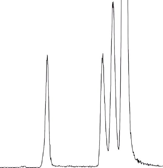
based on 80-120% of sample concentration. The extraction essential oil with a percentage area of 48.48% (Figure 2).4 BioMed Research International
100 3.39 TIC
1.25e6
%
3.10
1.29 2.82
0 Time
1.00 2.00 3.00 4.00 5.00 6.00 7.00 8.00 9.00
Figure 1: The LC-MS/MS total ion chromatogram of TE+EO.
TIC
22,981,322
33.337
Ar-turmerone
33.522
34.720
26.199 26.329
26.869
27.981
37.206
22.374
27.368
36.207
23.961
30.971
7.686
30.528
37.856
3.769
4.745
4.024
5.344
6.027
10.0 20.0 30.0 40.0 44.0
min
Figure 2: Gas chromatography-mass spectrum (GC-MS) of essential oil of turmeric.BioMed Research International
Table 1: Impact of turmeric extract and essential oil combination on cognitive performance and biochemical parameters in aluminum chloride-treated mice.
Morris water maze Elevated plus maze
Total proteins MDA (nM GSH (μM/mg SOD (U/mg CAT (U/mg AchE (nM/mg
Treatment Escape latency 1st retention 2nd retention
(mg/g tissue) released/mg protein) tissue) protein) protein) protein)
(sec) (sec) (sec)
Control (vehicle) 33:17 ± 1:30bc 35:17 ± 1:38bc 15:67 ± 0:67∗ bc 72:76 ± 1:78bcd 45:57 ± 2:07b 309:56 ± 6:69bc 0:58 ± 0:04bcd 42:72 ± 1:76b 3:82 ± 0:09bcd
Untreated control
63:00 ± 1:59acd 64:17 ± 3:89acd 59:83 ± 2:65acd 35:37 ± 0:81acd 97:33 ± 2:50acd 103:91 ± 5:09acd 0:89 ± 0:02acd 28:66 ± 3:11ad 0:34 ± 0:05acd
(AlCl3)
AlCl3+TE+EO-25 45:00 ± 2:38ab 52:00 ± 0:86abd 33:00 ± 0:97∗ abd 58:17 ± 2:48ab 56:23 ± 3:64b 246:93 ± 10:96abd 0:77 ± 0:03ab 37:63 ± 3:57 1:95 ± 0:13abd
b bc ∗ bc ab b bc ab b
AlCl3+TE+EO-50 38:17 ± 1:92 35:33 ± 0:76 19:67 ± 0:67 60:61 ± 1:80 54:72 ± 2:98 287:39 ± 5:65 0:69 ± 0:02 41:92 ± 1:92 2:83 ± 0:22abc
Values are expressed as the mean ± standard error of the mean. ap < 0:05 compared with the control. bp < 0:05 compared with untreated control (AlCl3). cp < 0:05 compared with the AlCl3+TE+EO-treated group
(25 mg/kg); dp < 0:05 compared with the AlCl3+TE+EO-treated group (50 mg/kg). ∗ p < 0:05 comparing second retention with first retention by paired t-test. AlCl3: aluminum chloride; TE+EO: turmeric extract
and essential oil combination.
56 BioMed Research International
120
100
80
60
40
Percentage change
20
0
–20
–40
–60
–80
–100
–120
AICI3 alone AICI3+TE+EO - 25 AICI3+TE+EO - 50
Total proteins SOD
MDA CAT
GSH AchE
Figure 3: Influence on biomarkers of the mouse brain homogenate of the aluminum and TE+EO groups when compared to the control
group.
3.2. Morris Water Maze Test. The experimental mice compared with vehicle control. Simultaneous oral administra-
assigned to the control group learn to locate the hidden plat- tion of TE+EO restored the total protein level and AchE level,
form using visual cues. There was a significant increase and the change was significant compared with Al-treated con-
(p < 0:05) in escape latency time in Al-treated mice com- trol mice (p < 0:05). Similarly, chronic administration of Al
pared to the control animals. A low dose, as well as a high significantly elevated lipid peroxide (MDA) levels and
dose, of TE+EO significantly reduced the time taken by the decreased the levels of GSH and the activity of CAT when
animals to find the platform compared with the aluminum- compared to control animals (p < 0:05). Coadministration of
treated group (p < 0:05). The latency time for the TE+EO 25 and 50 mg/kg of TE+EO with Al significantly reduced lipid
(50 mg/kg) group was almost identical to the vehicle control peroxide levels and was comparable with the vehicle control
group (Table 1). group. Aluminum exposure increased the activity of SOD sig-
nificantly (p < 0:05) in comparison to vehicle control group
3.3. Elevated Plus Maze Test. The first and second retention animals, and coadministration of TE+EO significantly
times representing the time taken to move from the open arms reduced the effect of aluminum exposure. A higher dose of
to the enclosed arms of the maze were significantly increased TE+EO normalized GSH and CAT activity and was compara-
in mice treated with Al (p < 0:05) compared to the vehicle ble with the vehicle control group (Table 1 and Figure 3).
control. A statistically significant difference in the first and sec-
ond retention times was observed with TE+EO (25 mg/kg and
50 mg/kg) when compared with the aluminum-treated groups 3.5. Histopathology of the Brain. Chronic Al-treated animals
(p < 0:05). Treatment of mice with 50 mg/kg TE+EO signifi- showed a marked deterioration in the hippocampal region
cantly reversed Al-induced changes, as evidenced by a as visualized by extensive cytoplasmic vacuolation and dam-
decrease in the first and second retention times (Table 1). age to the pyramidal cells compared with vehicle-treated ani-
mals (Figure 4). Treatment with TE+EO alone exhibited a
3.4. Biochemical Parameters in the Brain. After 45 days of Al similar effect as the control group. TE+EO (25 and 50 mg/kg)
administration, a significant reduction (p < 0:05) in total pro- minimized damage to pyramidal cells and vacuolization
tein level and AchE activity in the mouse brain was observed induced by Al treatment.BioMed Research International 7
(a) (b)
(c) (d)
Figure 4: H&E-stained slides of the hippocampal CA1 area in mice exposed to aluminum and simultaneously treated with TE+EO. (a)
Vehicle control showing normal cells. (b) Aluminum chloride- (AlCl3-) treated showing shrunken, vacuolated, and degenerated neurons.
(c) AlCl3+TE+EO-25 showing improvement. (d) AlCl3+TE+EO-50 resulted in reduction in vacuolization.
3.6. Pharmacokinetic Study. This is the first study to measure increase in all analyte levels in the plasma and brain com-
the efficacy, pharmacokinetics, and tissue distribution of tur- pared to TE alone.
meric extracts in an Al-induced neurotoxicity model to TE+EO-25 provided 37.68-fold, 45.85-fold, 8.22-fold,
understand if plasma bioavailability and brain distribution and 9.45-fold higher AUC (0 − α) of curcumin, curcumi-
of curcumin are related to its neuroprotective effects. The noids, conjugate metabolites, and hydrogenated metabolites,
plasma concentration-time profiles of curcumin, curcumi- respectively, compared to TE-25. TE+EO-50 provided
noids, conjugated metabolites, and hydrogenated metabolites 121.86-fold, 114.29-fold, 10.38-fold, and 12-fold higher
are presented in Table 2, Figure 5, and Figure 6. The curcu- AUC (0 − α) of curcumin, curcuminoids, conjugate metabo-
minoids identified were curcumin, demethoxycurcumin lites, and hydrogenated metabolites, respectively, compared
(DMC), and bisdemethoxycurcumin (BDMC). The conju- to TE-50. TE+EO-50 provided 4.23-fold, 4.14-fold, 1.51-fold,
gated metabolites included curcumin glucuronide (CG) and and 1.69-fold higher AUC (0 − α) of curcumin, curcumi-
curcumin sulfate (CS). Also, hydrogenated or reduced noids, conjugate metabolites, and hydrogenated metabolites,
metabolites included tetrahydrocurcumin (THC) and respectively, compared to TE+EO-25. TE-50 provided 1.31-
hexahydrocurcumin (HHC). TE+EO showed a significant fold, 1.66-fold, 1.19-fold, and 1.33-fold higher AUC (0 − α)8 BioMed Research International
Table 2: Pharmacokinetic parameters of curcumin, curcuminoids, and metabolites and its concentration in brain tissue.
Curcuminoids (curcumin+ Conjugate metabolites Hydrogenated metabolites
Curcumin
Parameters Group DMC+BDMC) (CG+CS) (HHC+THC)
Mean ± SEM Mean ± SEM Mean ± SEM Mean ± SEM
TE+EO-25 3:31 ± 0:51b 3:20 ± 0:46bcd 1:91 ± 0:16cd 1:68 ± 0:14d
TE+EO-50 8:04 ± 1:70acd 6:01 ± 1:17acd 1:56 ± 0:17cd 1:68 ± 0:16d
t1/2
TE-25 2:05 ± 0:43b 0:39 ± 0:03ab 0:86 ± 0:06ab 1:26 ± 0:13d
TE-50 2:06 ± 0:52b 0:52 ± 0:05ab 0:87 ± 0:05ab 0:40 ± 0:02abc
TE+EO-25 3:50 ± 0:22bcd 3:50 ± 0:22bcd 1:00 ± 0:00 1:83 ± 0:17
a
TE+EO-50 2:17 ± 0:17 2:33 ± 0:21a 1:00 ± 0:00 1:83 ± 0:17
T max a a
TE-25 1:83 ± 0:17 2:00 ± 0:00 2:00 ± 0:00 2:17 ± 0:17
TE-50 2:33 ± 0:21a 2:17 ± 0:17a 2:00 ± 0:00 2:17 ± 0:17
bcd bcd bcd
TE+EO-25 32:00 ± 2:22 34:23 ± 2:19 297:91 ± 9:86 16:75 ± 0:74bcd
TE+EO-50 79:60 ± 6:91acd 99:74 ± 4:83acd 471:77 ± 12:85acd 28:11 ± 1:20acd
C max
TE-25 1:27 ± 0:20ab 1:41 ± 0:19ab 47:81 ± 5:88ab 2:43 ± 0:26ab
TE-50 1:59 ± 0:43ab 2:48 ± 0:35ab 56:63 ± 3:73ab 3:45 ± 0:38ab
TE+EO-25 141:86 ± 9:40bcd 152:26 ± 9:32bcd 904:17 ± 43:70bcd 60:36 ± 1:92bcd
TE+EO-50 357:24 ± 32:51acd 439:64 ± 26:88acd 1429:96 ± 35:44acd 101:63 ± 3:24acd
AUC 0-t
TE-25 4:72 ± 0:81ab 5:37 ± 0:82ab 124:98 ± 15:88ab 6:53 ± 0:29ab
TE-50 6:59 ± 1:81ab 8:90 ± 1:93ab 149:84 ± 8:61ab 9:81 ± 0:46ab
TE+EO-25 234:34 ± 22:21b 247:11 ± 23:12b 1045:76 ± 54:11bcd 70:01 ± 3:12bcd
991.90
TE+EO-50 1024.00 ± 177.14acd 1581.53 ± 51.15acd 118.24 ± 5.84acd
AUC 0-inf ± 187.40acd
TE-25 6:22 ± 1:10b 5:39 ± 0:82b 127:18 ± 16:21ab 7:41 ± 0:41ab
TE-50 8:14 ± 1:70b 8:96 ± 1:94b 152:30 ± 8:87ab 9:85 ± 0:46ab
TE+EO-25 6:21 ± 0:68b 6:06 ± 0:60b 3:32 ± 0:16cd 3:46 ± 0:14d
TE+EO-50 12:43 ± 2:47acd 9:65 ± 1:67acd 2:99 ± 0:15 3:47 ± 0:18d
MRT
TE-25 3:96 ± 0:50b 2:69 ± 0:04b 2:69 ± 0:03a 3:07 ± 0:15
b b a
TE-50 4:54 ± 0:75 2:79 ± 0:07 2:62 ± 0:04 2:73 ± 0:04ab
TE+EO-25 65:75 ± 4:39bcd 89:86 ± 5:51bcd 0:24 ± 0:16d 1:35 ± 0:14c
Concentration TE+EO-50 140:07 ± 8:19acd 198:53 ± 10:12acd 0:71 ± 0:31 1:88 ± 0:52cd
in the brain TE-25 5:97 ± 0:94 ab
6:49 ± 0:93 ab
0:52 ± 0:09 0:17 ± 0:10ab
TE-50 10:22 ± 0:81ab 16:98 ± 1:20ab 1:12 ± 0:16a 0:48 ± 0:11b
t1/2: terminal phase half-life; T max: time to maximum plasma concentration; C max: maximum concentration; AUC (0-6 h): area under the curve up to 6 h;
AUC (0-∞): AUC up to infinite time; MRT: mean residence time. ap < 0:05 when compared to TE+EO-25. bp < 0:05 when compared to TE+EO-50. cp < 0:05
when compared to TE-25. dp < 0:05 when compared to TE-50.
of curcumin, curcuminoids, conjugate metabolites, and cuminoids, conjugate metabolites, and hydrogenated metab-
hydrogenated metabolites, respectively, compared to TE-25. olites, respectively, in the brain homogenate compared to
The curcuminoids to conjugate metabolite AUC (0 − α) TE-25. TE+EO-50 provided 13.71-fold, 11.69-fold, 0.63-fold,
ratio of TE+EO-25 and TE+EO-50, respectively, were and 3.92-fold concentrations of curcumin, curcuminoids,
1 : 4.2 : 1 : 1.6, while the similar ratios of TE-25 and TE-50, conjugate metabolites, and hydrogenated metabolites,
respectively, are 1 : 23.6 and 1 : 16.9. These results clearly illus- respectively, in the brain homogenate compared to TE-50.
trate the characteristic drawbacks of the oral administration TE+EO-50 provided 2.13-fold, 2.21-fold, 2.96-fold, and
of turmeric extract. TE+EO (50 mg/kg) displayed the optimal 1.39-fold higher concentrations of curcumin, curcuminoids,
pharmacokinetic profile of curcuminoids with C max of 99:74 conjugate metabolites, and hydrogenated metabolites,
± 4:83 ng/gm and AUC 0-6 h of 439:64 ± 26:88 h ng/gm. respectively, in the brain homogenate compared to TE+EO-
Curcumin, curcuminoids, and metabolites of curcumin 25. TE-50 provided 1.71-fold, 2.62-fold, 2.15-fold, and 2.82-
distributed in whole-brain homogenate were estimated at fold higher concentrations of curcumin, curcuminoids, con-
6 h for the TE+EO and TE groups. Table 2 shows the distri- jugate metabolites, and hydrogenated metabolites, respec-
bution of curcumin and its metabolites present in the brain tively, in brain homogenate compared to TE-25.
fractions. TE+EO-25 provided 11.01-fold, 13.85-fold, 0.46- The curcuminoids to conjugate metabolite concentration
fold, and 7.94-fold higher concentrations of curcumin, cur- ratio of TE+EO-25 and TE+EO-50, respectively, in brainBioMed Research International 9
100 levels of curcumin in the brain compared to TE alone. This
is probably the first study reporting the simultaneous quanti-
Curcumin concentration (ng/g)
80 fication of curcumin, curcuminoids, and primary and sec-
⁎
ondary metabolites in the brain and plasma after oral
AUC 0-t = 357.24 ng/g h
60
administration. The enhancement of brain and plasma bio-
availability even at low dose (HED 142 mg in 70 kg adult)
was due to the presence of turmerones in TE+EO, which acts
40
as inhibitors of the p-glycoprotein pathway, the significant
⁎
bottleneck in curcumin absorption [29]. The oral intake of
AUC 0-t = 141.85 ng/g h
20 bioactive turmeric extract could deliver sustained levels of
⁎
AUC 0-t = 6.59 ng/g h
⁎
AUC 0-t = 4.72 ng/g h
free curcumin and curcuminoids. Moreover, the significantly
0 high mean residence time (MRT), area under the curve
0 1 2 3 4 5 6 7 (AUC), and t-half (p < 0:05) with TE+EO explain their neu-
Time (hours) roprotective effects. The increased bioavailability is in line
TE+EO - 25 mg TE - 25 mg with the study done on curcumin alone vs. curcuminoids
TE+EO - 50 mg TE - 50 mg with turmerones, which showed a sixfold increase in the bio-
availability with the latter [30], and another study proposing
Figure 5: Plasma concentration-time curve of curcumin after the use of turmerones along with curcumin instead of curcu-
administration of TE+EO (25 and 50 mg/kg) and TE (25 and
min alone for better therapeutic utility [29]. The administra-
50 mg/kg).
tion of TE+EO also led to higher levels of THC and HHC in
blood plasma and the brain, which also has pharmacological
homogenate, were 374.4 : 1 and 279.6 : 1, and those of TE-25 activity [31, 32]. Free curcumin was predominantly higher in
and TE-50, respectively, were 12.5 : 1 and 15.2 : 1. The results the brain than its metabolites, while metabolites were higher
showed that free curcumin and curcuminoids from TE+EO in the blood. The low levels of curcumin and metabolites in
were chiefly distributed in the brain tissue and significantly the brain in the TE group also reflected its poor pharmacoki-
higher than that of TE. netic profile.
Chronic Al exposure leads to its accumulation in the
4. Discussion brain as it is slowly removed, owing to its long half-life. Alu-
minum can suppress or interfere with the expression of anti-
The present study demonstrated the potential of formulated oxidants [33–35]. We found that Al treatment caused
turmeric extract and essential oil combination in attenuating elevation in lipid peroxide levels in brain tissue and signifi-
Al-induced cognitive deficits and toxicity in mice. Curcumin cant memory decline in animals. This implies an alteration
targets pathways involved in the pathophysiology of neuro- in homeostasis and damage to neuronal biomolecules [36],
degenerative diseases such as the β-amyloid cascade, tau leading to dysfunction. In the current study, although lipid
phosphorylation, neuroinflammation, and oxidative stress. hydroperoxides and lipid peroxidation have increased in
These findings suggest that curcumin can be considered as the tissues, there was a corresponding increase in the protec-
a therapeutic option to limit neuroinflammation in neurode- tive antioxidant enzymes denoting that brain tissue could
generative disorders. However, low bioavailability and per- quench increased ROS and oxidative stress with TE+EO.
meability across the blood-brain barrier limits the GSH depletion can enhance oxidative stress and may also
therapeutic potential of curcumin. The delivery of free curcu- increase the levels of excitotoxic molecules. GSH functions
min to target tissues can be achieved by improving the bio- as a free radical scavenger, particularly effective against the
availability in the plasma and brain to obtain the adequate OH radical. The ability of GSH to nonenzymatically scavenge
therapeutic outcomes. both singlet oxygen and OH provides the first line of antiox-
Curcuminoids are the phenolic yellowish pigments of idant defense. SOD is an antioxidant enzyme which gets acti-
turmeric present as curcumin (CUR), demethoxycurcumin vated when there is oxidative stress and ROS formation, in
(DMC), and bisdemethoxycurcumin (BDMC). Orally case of our study due to Al. GSH reacts nonenzymatically
administered curcuminoids are absorbed in the gut, carried with ROS. Oxidative stress increases the activity of GSHPx,
to the liver, and are rapidly metabolized. Curcuminoids are which catalyzes the reduction of hydrogen peroxide (H2O2)
metabolized in the liver and the intestinal mucosa to tetrahy- by oxidizing GSH, leading to its depletion. Conditions of oxi-
drocurcumin (THC) and hexahydrocurcumin (HHC) by dative stress, such as GSH depletion and increased H2O2
phase I metabolism. Curcuminoids and their reductive prod- steady-state levels, may favor the catalase pathway for H2O2
ucts are extensively conjugated with glucuronic acid and sul- removal. Aluminum in the presence of transition metals like
fate by phase II metabolism and form curcumin glucuronide Fe causes lipid peroxidation generating H2O2 which is
and curcumin sulfate [26]. It is perceived that curcumin con- quenched by GSHPx and catalase. Furthermore, compro-
centration in the brain was deficient due to the low systemic mised antioxidant enzymes would undoubtedly have an
absorption and poor transport across the blood-brain barrier unfavorable impact on the central nervous system. The find-
[27, 28]. Thus, the remedial measure was to enhance the dose ings of the current study indicate that the use of 50 mg/kg TE
of curcumin to elicit a neuroprotective response. However, +EO counterbalances aluminum-induced free radical-
the use of TE+EO achieved significantly higher (p < 0:05) mediated injury of neural tissue. Curcumin, a quencher of10 BioMed Research International
3000
2500
AUC 0 - infinity (ng/g⁎h)
2000
1500
1000
500
0
TE+EO_25 TE+EO_50 TE_25 TE_50
THC BDMC
HHC DMC
CS Curcumin
CG
Figure 6: Mean of AUC (0 to α) of curcuminoids and its metabolites from the TE and TE+EO groups.
ROS, modulates oxidative stress by normalizing SOD the use of 50 mg/kg of TE+EO as evidenced through the Mor-
enzyme activity and catalase levels plausibly by forming a ris water maze and elevated plus maze task; however,
ligand with Fe. Our study results show that GSH levels 25 mg/kg exhibited a significant response in improving spa-
decreased in response to Al, indicating its role in removing tial memory but did not elicit improvement in transfer
H2O2. On administering TE+EO, the levels of GSH were ele- latency with the elevated plus maze. The role of TE+EO in
vated to normal due to the inhibition of lipid peroxidation. improving cognitive function could mechanistically be
The results of the current study are supported by the findings explained by its protection of biomembranes owing to its
of AlBasher et al., 2020, which show that curcumin along activation of antioxidant systems and reduced formation of
with resveratrol protects against fipronil-induced oxidative free radicals.
stress in the brain [37]. Also, curcumin along with diallyl sul- Learning and memory are correlated mainly with the
fide boosts the cellular antioxidant status [38]. The essential cholinergic system and the availability of acetylcholine. Ace-
oil of turmeric contains terpenes which facilitate the passage tylcholinesterase plays a vital part in the functioning of
of curcumin in the brain as observed through our pharmaco- nerves, and severe physioIogica1 damage arises from its
kinetic findings. Also, the constituents present in the essen- blockage. When the action of acetylcholinesterase is blocked,
tial oil might synergize with curcumin in displaying its acetylcholine accumulates, leading to endogenous poisoning.
antioxidant effects. Ar-turmerone, an important constituent Our study results show that the levels of AChE decreased in
present in the essential oil, is reported to have antioxidant response to Al, and TE+EO elevated it to the normal levels.
action [39]. Only 50 mg/kg of TE+EO was effective in increasing the
In vitro studies show that aluminum significantly acceler- levels of acetylcholinesterase, and the same dose also
ates iron-mediated lipid peroxidation under acidic and neutral improved memory assessed through the behavioral models
conditions. The discovery of elevated levels of lipid hydroper- confirming the efficacy of TE+EO at this dose level. More-
oxide in the brain tissues of mice substantiates the higher over, 50 mg/kg of TE+EO protected the hippocampus from
phase of lipid peroxidation and oxidative stress following Al-induced damage plausibly by improving the defense sys-
exposure to Al. Administration of TE+EO in a higher dose tem, suppressing lipid peroxide levels, and increasing the
reversed the deleterious effect of Al, resulting in a significant availability of curcuminoids in the presence of turmerones
increase in the levels of GSH, CAT, SOD, and total proteins. in the formulation. Furthermore, the expression of transcrip-
Aluminum impacts the hippocampus [40] and com- tion factors could be indirectly impacted, shielding neuronal
plexes with acetylcholine [41], resulting in irreversible mem- structures against peroxidative damage. Imbalances in the
ory deficits [42, 43]. The potential of TE+EO in protecting oxidant-antioxidant status can be a trigger for inflammation.
neural tissues is further proved through the response TE+EO might prevent inflammatory damage of neurons.
observed in behavioral models. Aluminum increases latency The anti-inflammatory effects of TE+EO have been reported
to find the platform or transit from the open to the closed in colitis induced by dextran sulfate [44].
arms, implying the poor functioning of neurons involved in Histopathological changes of the hippocampus in Al-
learning and memory. Aluminum-exposed animals exhibited treated animals have further attested to the biochemical and
cognitive impairment, which was significantly reversed with behavioral results characterized by damage to pyramidalBioMed Research International 11
cells, which are in line with other studies [2, 45]. CA3 pyra- Sciences : The Official Journal of Isfahan University of Medical
midal neurons play an important role in conveying neural Sciences, vol. 23, no. 1, p. 51, 2018.
information to the CA1 neurons; hence, damage to this field [2] P. Sethi, A. Jyoti, E. Hussain, and D. Sharma, “Curcumin
could affect recall of memories recently learned. We observed attenuates aluminium-induced functional neurotoxicity in
that the 50 mg/kg of TE+EO protects the hippocampal neu- rats,” Pharmacology, Biochemistry, and Behavior, vol. 93,
rons, enabling the circuit to work efficiently resulting in a no. 1, pp. 31–39, 2009.
favorable behavioral outcome. [3] C. Exley, Aluminium and Alzheimer’s Disease: The Science that
Describes the Link, Elsevier, 2011.
5. Conclusion [4] B. Platt, G. Fiddler, G. Riedel, and Z. Henderson, “Aluminium
toxicity in the rat brain: histochemical and immunocytochem-
Turmeric extract and essential oil combination works in uni- ical evidence,” Brain Research Bulletin, vol. 55, no. 2, pp. 257–
son to reverse the effects produced by the chronic exposure of 267, 2001.
Al, firstly, by enabling more plasma bioavailability and access [5] A. Jangra, P. Kasbe, S. N. Pandey et al., “Hesperidin and silibi-
of curcuminoids into the brain and, secondly, by upregulat- nin ameliorate aluminum-induced neurotoxicity: modulation
ing the expression of antioxidants, thereby minimizing of antioxidants and inflammatory cytokines level in mice hip-
microglia activation and subsequent neuronal damage. The pocampus,” Biological Trace Element Research, vol. 168, no. 2,
pp. 462–471, 2015.
observed effect of TE+EO could be linked with its ability to
cross the blood-brain barrier, bind to redox metal ions, and [6] L. Wang, “Entry and deposit of aluminum in the brain,” in
Advances in Experimental Medicine and Biology, pp. 39–51,
neutralize free radicals. This makes TE+EO a promising ther-
Springer, 2018.
apeutic option for prophylaxis and treatment of neurodegen-
[7] E. C. Hirsch, J. P. Brandel, P. Galle, F. Javoy-Agid, and Y. Agid,
erative diseases as it could substantially reduce
“Iron and aluminum increase in the substantia nigra of
neuroinflammation and oxidative stress and improve mem- patients with Parkinson’s disease: an X-ray microanalysis,”
ory in mice exposed to aluminum. Journal of Neurochemistry, vol. 56, no. 2, pp. 446–451, 1991.
[8] A. Y. Sun, Q. Wang, A. Simonyi, and G. Y. Sun, “Botanical
Data Availability phenolics and brain health,” Neuromolecular Medicine,
vol. 10, no. 4, pp. 259–274, 2008.
The datasets used and/or analysed during the current study
[9] T. P. Ng, P. C. Chiam, T. Lee, H. C. Chua, L. Lim, and E. H.
are available from the corresponding author on reasonable Kua, “Curry consumption and cognitive function in the
request. elderly,” American Journal of Epidemiology, vol. 164, no. 9,
pp. 898–906, 2006.
Ethical Approval [10] M. Garcia-Alloza, L. A. Borrelli, A. Rozkalne, B. T. Hyman,
and B. J. Bacskai, “Curcumin labels amyloid pathology
All procedures performed in studies involving animals were in vivo, disrupts existing plaques, and partially restores dis-
in accordance with CPCSEA (Committee for the Purpose of torted neurites in an Alzheimer mouse model,” Journal of Neu-
Control and Supervision of Experiments on Animals) guide- rochemistry, vol. 102, no. 4, pp. 1095–1104, 2007.
lines. The study was approved by the Institutional Animal [11] S. Hewlings and D. Kalman, “Curcumin: a review of its’ effects
Ethical Committee. on human health,” Food, vol. 6, no. 10, pp. 92–92, 2017.
[12] R. Jäger, R. P. Lowery, A. V. Calvanese, J. M. Joy, M. Purpura,
Conflicts of Interest and J. M. Wilson, “Comparative absorption of curcumin for-
mulations,” Nutrition Journal, vol. 13, no. 1, p. 11, 2014.
The authors have no conflicts of interest to declare. [13] D. Banji, O. J. F. Banji, S. Dasaroju, and K. Kumar CH, “Cur-
cumin and piperine abrogate lipid and protein oxidation
Authors’ Contributions induced by D-galactose in rat brain,” Brain Research,
vol. 1515, pp. 1–11, 2013.
DB conceived, designed, and wrote the initial draft of the [14] C. A. Martins, G. Leyhausen, J. Volk, and W. Geurtsen, “Cur-
manuscript. OB and KS conducted the experiments, analyzed cumin in combination with piperine suppresses osteoclasto-
data, and refined the manuscript. All authors read and genesis in vitro,” Journal of Endodontia, vol. 41, no. 10,
approved the manuscript. The authors declare that all data pp. 1638–1645, 2015.
were generated in-house and that no paper mill was used. [15] A. A. Shati, F. G. Elsaid, and E. E. Hafez, “Biochemical and
molecular aspects of aluminium chloride-induced neurotox-
Acknowledgments icity in mice and the protective role of Crocus sativus L.
extraction and honey syrup,” Neuroscience, vol. 175,
The authors acknowledge Arjuna Natural Private Ltd., Aluva, pp. 66–74, 2011.
Kerala, India, for providing the gift samples of TE and TE [16] L. Liu, Y. L. Liu, G. X. Liu et al., “Curcumin ameliorates dex-
+EO (BCM-95®/Curcugreen®). tran sulfate sodium-induced experimental colitis by blocking
STAT3 signaling pathway,” International Immunopharmacol-
References ogy, vol. 17, no. 2, pp. 314–320, 2013.
[17] C. V. Vorhees and M. T. Williams, “Morris water maze: proce-
[1] E. Inan-Eroglu and A. Ayaz, “Is aluminum exposure a risk fac- dures for assessing spatial and related forms of learning and
tor for neurological disorders?,” Journal of Research in Medical memory,” Nature Protocols, vol. 1, no. 2, pp. 848–858, 2006.12 BioMed Research International
[18] C. Schiborr, G. P. Eckert, G. Rimbach, and J. Frank, “A vali- [33] Z. Wu, Y. Du, H. Xue, Y. Wu, and B. Zhou, “Aluminum
dated method for the quantification of curcumin in plasma induces neurodegeneration and its toxicity arises from
and brain tissue by fast narrow-bore high-performance liquid increased iron accumulation and reactive oxygen species
chromatography with fluorescence detection,” Analytical and (ROS) production,” Neurobiology of Aging, vol. 33, no. 1,
Bioanalytical Chemistry, vol. 397, no. 5, pp. 1917–1925, 2010. pp. 199.e1–199.e12, 2012.
[19] P. Ramalingam and Y. T. Ko, “A validated LC-MS/MS method [34] V. Kumar and K. D. Gill, “Oxidative stress and mitochondrial
for quantitative analysis of curcumin in mouse plasma and dysfunction in aluminium neurotoxicity and its amelioration:
brain tissue and its application in pharmacokinetic and brain a review,” Neurotoxicology, vol. 41, pp. 154–166, 2014.
distribution studies,” Journal of Chromatography, B: Analytical [35] K. Klotz, W. Weistenhöfer, F. Neff, A. Hartwig, C. Van Thriel,
Technologies in the Biomedical and Life Sciences, vol. 969, and H. Drexler, “The health effects of aluminum exposure,”
pp. 101–108, 2014. Deutsches Ärzteblatt International, vol. 114, no. 39, pp. 653–
[20] J. Högberg, R. E. Larson, A. Kristoferson, and S. Orrenius, 659, 2017.
“NADPH-dependent reductase solubilized from microsomes [36] C. Exley, “The pro-oxidant activity of aluminum,” Free Radical
by peroxidation and its activity,” Biochemical and Biophysi- Biology & Medicine, vol. 36, no. 3, pp. 380–387, 2004.
cal Research Communications, vol. 56, no. 3, pp. 836–842,
[37] G. AlBasher, M. M. Abdel-Daim, R. Almeer et al., “Synergistic
1974.
antioxidant effects of resveratrol and curcumin against
[21] O. H. Lowry, N. J. Rosebrough, A. L. Farr, and R. J. Randall, fipronil-triggered oxidative damage in male albino rats,” Envi-
“Protein measurement with the Folin phenol reagent,” The Jour- ronmental Science and Pollution Research, vol. 27, no. 6,
nal of Biological Chemistry, vol. 193, no. 1, pp. 265–275, 1951. pp. 6505–6514, 2020.
[22] M. Moron, J. Depierre, and B. Mannervik, “Levels of glutathi- [38] M. M. Abdel-Daim and R. H. Abdou, “Protective effects of dia-
one, glutathione reductase and glutathione S-transferase activ- llyl sulfide and curcumin separately against thallium-induced
ities in rat lung and liver,” Biochimica et Biophysica Acta toxicity in rats,” Cell Journal, vol. 17, no. 2, pp. 379–388, 2015.
(BBA)-General Subjects, vol. 582, no. 1, pp. 67–78, 1979.
[39] R. Kuttan, V. B. Liju, and K. Jeena, “An evaluation of antioxi-
[23] H. P. Misra and I. Fridovich, “The role of superoxide anion in dant, anti-inflammatory, and antinociceptive activities of
the autoxidation of epinephrine and a simple assay for super- essential oil from Curcuma longa. L,” Indian Journal of Phar-
oxide dismutase,” The Journal of Biological Chemistry, macology, vol. 43, no. 5, pp. 526–531, 2011.
vol. 247, no. 10, pp. 3170–3175, 1972.
[40] A. Kaur, K. Joshi, R. W. Minz, and K. D. Gill, “Neurofilament
[24] H. Aebi, “Catalase in vitro,” in Methods in Enzymology, phosphorylation and disruption: a possible mechanism of
pp. 121–126, Elsevier, 1984. chronic aluminium toxicity in Wistar rats,” Toxicology,
[25] G. L. Ellman, K. D. Courtney, V. Andres, and R. M. Feather- vol. 219, no. 1–3, pp. 1–10, 2006.
stone, “A new and rapid colorimetric determination of acetyl- [41] P. Matlaba, S. Daya, and T. Nyokong, “Interaction of the neu-
cholinesterase activity,” Biochemical Pharmacology, vol. 7, rotransmitter acetylcholine with aluminium, calcium and
no. 2, pp. 88–95, 1961. sodium,” Pharmacy and Pharmacology Communications,
[26] K. Wang and F. Qiu, “Curcuminoid metabolism and its contri- vol. 6, no. 5, pp. 201–205, 2000.
bution to the pharmacological effects,” Current Drug Metabo-
[42] C. M. Nday, B. D. Drever, T. Salifoglou, and B. Platt, “Alumin-
lism, vol. 14, no. 7, pp. 791–806, 2013.
ium interferes with hippocampal calcium signaling in a
[27] Y. M. Tsai, C. F. Chien, L. C. Lin, and T. H. Tsai, “Curcumin species-specific manner,” Journal of Inorganic Biochemistry,
and its nano-formulation: the kinetics of tissue distribution vol. 104, no. 9, pp. 919–927, 2010.
and blood-brain barrier penetration,” International Journal
[43] Z. Cao, X. Yang, H. Zhang et al., “Aluminum chloride induces
of Pharmaceutics, vol. 416, no. 1, pp. 331–338, 2011.
neuroinflammation, loss of neuronal dendritic spine and cog-
[28] P. Anand, A. B. Kunnumakkara, R. A. Newman, and B. B. nition impairment in developing rat,” Chemosphere, vol. 151,
Aggarwal, “Bioavailability of curcumin: problems and prom- pp. 289–295, 2016.
ises,” Molecular Pharmaceutics, vol. 4, no. 6, pp. 807–818, 2007.
[44] S. Toden, A. L. Theiss, X. Wang, and A. Goel, “Essential tur-
[29] G. G. L. Yue, S. W. Cheng, H. Yu et al., “The role of turmerones meric oils enhance anti-inflammatory efficacy of curcumin in
on curcumin transportation and P-glycoprotein activities in dextran sulfate sodium-induced colitis,” Scientific Reports,
intestinal caco-2 cells,” Journal of Medicinal Food, vol. 15, vol. 7, no. 1, p. 814, 2017.
no. 3, pp. 242–252, 2012.
[45] C. Y. Yuan, Y. J. Lee, and G. S. W. Hsu, “Aluminum overload
[30] B. Antony, B. Merina, V. Iyer, N. Judy, K. Lennertz, and increases oxidative stress in four functional brain areas of neo-
S. Joyal, “A pilot cross-over study to evaluate human oral bio- natal rats,” Journal of Biomedical Science, vol. 19, no. 1, pp. 51–
availability of BCM-95® CG (Biocurcumax™), a novel bioen- 59, 2012.
hanced preparation of curcumin,” Indian Journal of
Pharmaceutical Sciences, vol. 70, no. 4, pp. 445–449, 2008.
[31] Y. Huang, S. Cao, Q. Zhang et al., “Biological and pharmaco-
logical effects of hexahydrocurcumin, a metabolite of curcu-
min,” Archives of Biochemistry and Biophysics, vol. 646,
pp. 31–37, 2018.
[32] L. Pari and P. Murugan, “Effect of tetrahydrocurcumin on
blood glucose, plasma insulin and hepatic key enzymes in
streptozotocin induced diabetic rats,” Journal of Basic and
Clinical Physiology and Pharmacology, vol. 16, no. 4,
pp. 257–274, 2005.You can also read