Investigating real life emotions in romantic couples: a mobile EEG study - Nature
←
→
Page content transcription
If your browser does not render page correctly, please read the page content below
www.nature.com/scientificreports
OPEN Investigating real‑life emotions
in romantic couples: a mobile EEG
study
Julian Packheiser1,2*, Gesa Berretz1,2, Noemi Rook1, Celine Bahr1, Lynn Schockenhoff1,
Onur Güntürkün1 & Sebastian Ocklenburg1
The neural basis of emotional processing has been largely investigated in constrained spatial
environments such as stationary EEGs or fMRI scanners using highly artificial stimuli like standardized
pictures depicting emotional scenes. Typically, such standardized experiments have low ecological
validity and it remains unclear whether their results reflect neuronal processing in real-life affective
situations at all. Critically, emotional situations do not only encompass the perception of emotions,
but also behavioral components associated with them. In this study, we aimed to investigate real-
life emotions by recording couples in their homes using mobile EEG technology during embracing,
kissing and emotional speech. We focused on asymmetries in affective processing as emotions have
been demonstrated to be strongly lateralized in the brain. We found higher alpha and beta power
asymmetry during kissing and embracing on frontal electrodes during emotional kisses and speech
compared to a neutral control condition indicative of stronger left-hemispheric activation. In contrast,
we found lower alpha power asymmetry at parieto-occipital electrode sites in the emotional compared
to the neutral condition indicative of stronger right-hemispheric activation. Our findings for alpha
power asymmetries are in line with models of emotional lateralization that postulate a valence-
specific processing over frontal cortices and right-hemispheric dominance in emotional processing in
parieto-occipital regions. In contrast, beta power asymmetries pointed more towards valence-specific
processing indicating that, while alpha and beta frequencies seem to be functionally associated, they
are not reflecting identical cognitive processing.
One of the most intriguing questions in neuroscience revolves around how and where emotions are processed in
the brain. More than 100 years ago, studies in patients with unilateral right-hemispheric lesions demonstrated
that emotional processing seems to be lateralized in the brain as these patients were impaired in their ability to
express their e motions1. These results were complemented by a large number of behavioral results both from
healthy and patient cohorts supporting the notion that emotions are asymmetrically processed in the b rain2–5.
6–8
With the emergence of brain recording and neuroimaging techniques such as the EEG or fMRI , more specific
statements about where affective states are processed within cortical and subcortical regions could be made.
For cortical emotional processing, especially frontal alpha power has been strongly associated with changes in
affective state and emotional regulation9,10. For example, Hannesdóttir et al.11 investigated relative left frontal
asymmetry (rLFA) and found that reduced rLFA in children was predictive of impaired emotional regulation
and stronger physiological responses to emotional stimuli. Furthermore, rLFA serves as a suitable predictor for
individual differences in emotional expression as well as r egulation12,13.
Regarding the nature of emotional lateralization, two theories have been dominant in research on asymmetries
of emotion processing. These theories are known as the right hemisphere hypothesis (RHH) and the valence
model (VM) of emotional processing. The RHH postulates that all emotions regardless of valence are processed
in the right h emisphere14. In contrast, the VM claims that positive emotions are dominantly processed in the
left hemisphere whereas negative emotions are processed in the right hemisphere15. Both theories have received
much support from behavioral, electrophysiological as well as neuroimaging s tudies16. The nature of emotional
lateralization in the brain therefore remains rather inconclusive to this day. Despite the richness of the neuro-
scientific literature on emotions, the vast majority of published papers in this field share a common issue: It is
largely unclear to what extent the used paradigms elicit neuronal processes that actually resemble the processes
1
Institute of Cognitive Neuroscience, Department of Psychology, Ruhr-University Bochum, Universitätsstraße 150,
44780 Bochum, Germany. 2These authors contributed equally: Julian Packheiser and Gesa Berretz. *email: julian.
packheiser@rub.de
Scientific Reports | (2021) 11:1142 | https://doi.org/10.1038/s41598-020-80590-w 1
Vol.:(0123456789)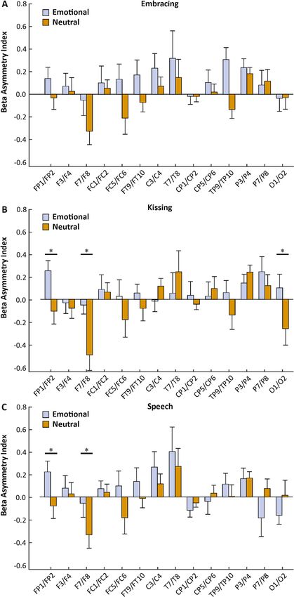
www.nature.com/scientificreports/
during real-life emotional encounters. A potential reason for this heterogeneity in the literature might be the
lack of ecological validity. Recent systematic review articles have pointed out the necessity for ecologically valid
research in neuroscience17,18. This holds especially true for emotion research as the most prevalent method of
positive or negative emotional induction is via movies, pictures or music19–22. Thus, emotions are largely only
perceived during experimental paradigms. However, merely perceiving emotions might not be sufficient for a
valid measurement of the underlying neural substrates. Real-life emotions comprise both the feeling itself as well
as a preparation for and performance of an adequate action associated with the felt emotion23. This for example
involves a behavioral expression such as avoidance behavior if someone experiences fear, or approach behavior
if someone experiences happiness24. The lack of a behavioral component in laboratory settings challenges the
external validity of experimental results and makes it difficult to transfer findings to, for example, mood disorders
for which the associated behavior is of paramount importance.
In conventional settings, this behavioral component is unfortunately difficult to realize either due to the
experimental design or the environment in which it takes place, for example in an fMRI scanner or during EEG
recordings. The past decade however brought forth novel techniques such as mobile E EGs25,26, mobile f NIRS27,28
29–31
and less constraining MEGs . These technological advancements have been developed and optimized to
allow for the investigation of neural correlates in settings of high ecological validity as the participants can move
freely and be tested outside the lab. While the number of publications using these techniques are still sparse, a
growing body of research is being generated converging neuroscientific research with real-life activities such as
cycling32,33, walking34, navigating over obstacles35, real world driving36, viewing real-life faces in natural settings37
or skateboarding38. Recently, Packheiser and c olleagues39 investigated the neural basis of hand and foot use while
the participants were wearing a mobile EEG. They found that both alpha and beta frequency asymmetries were
predictive of the participants’ handedness and footedness and that the neural signals could distinguish between
limb preferences of individuals. Importantly, they found that the neural signals were unaffected by movement
parameters during activities such as jumping or throwing and kicking balls. Thus, mobile EEGs provide a valuable
tool to investigate the neural basis of human emotions and their associated behaviors in more natural settings.
A very prominent human behavior that is usually executed in emotional settings is social touch. To convey our
social intentions or emotional states to other humans, we strongly rely on the use of a variety of tactile interac-
tions, especially in very intimate social relationships40,41. Touch is the earliest sensory modality to fully develop
during the lifespan[42 and is experienced from birth onwards by being cradled in the mother’s a rms43. For that
reason, social touch has been strongly associated with human development, shaping attachment, emotional
regulation and cognitive m aturation44. Affective social touch has been demonstrated to be highly beneficial for
the well-being and physical as well as mental health in humans as it reduces stress, blood pressure and can even
protect from viral infections and allergic r esponses45–48. Studies on the neural basis of social touch have indicated
that somatosensory cortices49 but also limbic or orbitofrontal brain regions are activated when an experimenter
or the romantic partner applies non-sexual pleasant tactile stimulation50–52. Thus, there seems to be a strong
overlap in cerebral processing of social touch and emotions indicating that social touch carries a strong affective
component. However, as for studies investigating emotions, the experimental designs investigating the neural
basis of social touch lack ecological validity as the application of gentle touch to for example the legs while lying
perfectly still rarely occurs in real-life settings.
The aim of the present study was to investigate emotional lateralization in a setting with high ecological
validity. To this end, we tested romantic partners in their home during both embracing and kissing while the
participants were recorded using a mobile EEG system. We also investigated the neural correlates of emotional
speech as this type of social interaction is fundamental to maintain a healthy and long-lasting r elationship53.
We focused on differences in asymmetrical processing in the alpha frequency band due to the pronounced role
of frontal alpha asymmetries in emotional processing. Since beta power asymmetries have been demonstrated
to be highly comparable in function to alpha power asymmetries in studies investigating motor p references39
54
and resting state oscillations , we also included beta power asymmetries as a dependent variable in our study.
For each behavioral task (embracing, kissing and speech), we employed a positive and a neutral condition. We
hypothesize that emotional lateralization differs between the emotional and neutral condition. If the right-
hemisphere hypothesis holds true, we expect stronger right-hemispheric activity in the emotional compared to
the neutral condition. If the valence hypothesis holds true, we expect stronger left-hemispheric activity in the
emotional compared to the neutral condition.
Methods
Participants. A total of 32 individuals (16 females) took part in this study. The sample size was determined
based on prior studies investigating frontal alpha asymmetries using within-subject designs producing reli-
able and large effects (Cohen’s d = 1.0455; ηp2 = 0.3656). There was no restriction regarding the sexuality of the
participants, but all couples were heterosexual in the present study. Age of the participants ranged between
19 and 63 years (mean age = 29 years, SD = 14 years). Participants with neurological or psychiatric disorders
were excluded from the study. The study was conducted in accordance with the declaration of Helsinki and
was approved by a local ethics committee of the psychological faculty at Ruhr University Bochum. All partici-
pants gave written informed consent. Five participants were excluded from the PANAS analysis of the emotional
induction and one participant was excluded from the EEG analysis of the behavioral tasks due to technical
issues. In total, this left 27 data sets for the PANAS and 31 data sets eligible for EEG data analysis in the final
analysis sample. There was no overlap between excluded participants from the PANAS and the EEG data.
Experimental task. The experimental paradigm consisted of conducting three behavioral tasks, i.e.
embracing, kissing and listening to speech in both an emotional and neutral condition. The neutral condition
Scientific Reports | (2021) 11:1142 | https://doi.org/10.1038/s41598-020-80590-w 2
Vol:.(1234567890)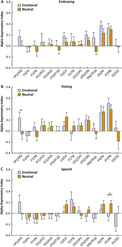
www.nature.com/scientificreports/
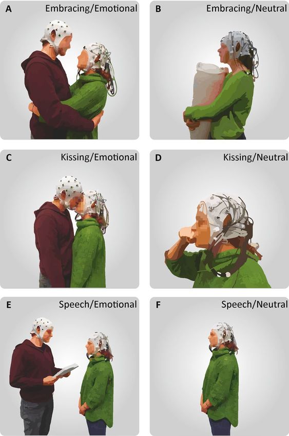
Figure 1. Depiction of the behavioral tasks. (A) During the emotional condition, the participant wearing
the mobile EEG system embraced his/her partner in a frontal embrace. (B) During the neutral condition, the
participant embraced a pillow with the partner absent. (C) In the emotional kissing condition, the partners
kissed each other without using their tongue. (D) During the neutral condition, the recorded participant
performed a kiss with his or her own hand. (E) In the emotional speech condition, the recorded participant
listened to an emotional text written by their partner prior to the session about shared life experiences. (F) In
the neutral speech condition, the recorded participant listened to a pre-recorded weather report without the
partner.
served as a control condition and was conducted using identical movements to control for motor effects in the
experiment. Testing took place in the participants’ homes to provide as much natural setting and ecological
validity as possible. One participant was set up with the mobile EEG system while the other participant filled
out demographic questionnaires, the relationship assessment scale (RAS) and prepared an emotional text about
fond memories and experiences with their partner. Importantly, the partner, that was not recorded, was also
equipped with an electrode cap to reduce the awkwardness of only one partner wearing the cap. Next, the par-
ticipants were instructed about the behavioral tasks and how to perform them appropriately by the experimenter
and an illustrating photograph. After instructing the participants, the experimenter left the room to allow for
privacy for the entire experimental procedure. The behavioral tasks looked as follows:
1. During the embracing condition, the participants were either asked to embrace each other from the front in
the emotional condition (Fig. 1A), or embrace a body pillow in the neutral condition with the partner being
absent (Fig. 1B). The participants were further instructed to avoid touching the electrode cap and move as
little as possible during the embrace.
2. During the kissing condition, the participants were either asked to kiss each other on the lips in the emotional
condition (Fig. 1C), or kiss their own hand during the neutral condition by forming a lip-like structure
with the thumb and index finger with the partner being absent (Fig. 1D). Importantly, the participants were
instructed not to use their tongue during the kiss in either condition to avoid strong motor artifacts.
3. During the speech condition, the participant wearing the mobile EEG system was either listening to the
partner reading the previously prepared emotional text in the emotional condition (Fig. 1E), or was listen-
Scientific Reports | (2021) 11:1142 | https://doi.org/10.1038/s41598-020-80590-w 3
Vol.:(0123456789)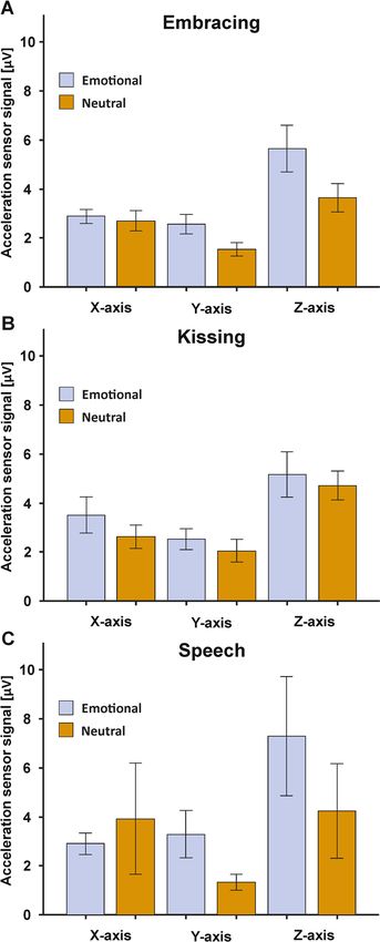
www.nature.com/scientificreports/

                                                ing to a weather report that was recorded prior to the experimental session in the neutral condition with the
                                                partner being absent (Fig. 1F).

                                                The experimental design was counterbalanced so that the experiment could start with the neutral or emotional
                                            condition and with the male or female partner being recorded initially. Furthermore, during the emotional and
                                            neutral condition, the order of behavioral tasks was fully randomized. Each behavioral task was performed for
                                            1 min in total and the tasks were separated by an intertrial interval (ITI) of 30 s in accordance with the procedures
                                            used in Packheiser et al.39. During the ITI, the participant(s) received an additional pre-recorded auditory and
                                            visual instruction about the upcoming behavioral task. This was necessary as it was unknown to the participant(s)
                                            if the procedure started with embracing, kissing or speech due to the randomization procedure. The instruc-
                                            tions were presented using the Presentation software (Neurobehavioral Systems Inc., CA, USA). There was a
                                            5 min break between the emotional and neutral condition to fill out the Positive and Negative Affect Schedule
                                            (PANAS), a questionnaire evaluating their positive and negative affective states by indicating the current emo-
                                            tional state on 10 positive and 10 negative items on a scale from 1 (Very slightly or not at all) to 5 (Extremely)57.
                                            The break was also used to allow for the partner to leave or to join (depending whether the recorded partner
                                            was in the emotional or neutral condition, respectively). We deliberately chose to let the partner leave the room
                                            in the neutral conditions to account for potential confound variables such as social support and non-verbal
                                            communication between the partners.
                                                After one participant had completed the experimental procedure (three emotional and three neutral tasks),
                                            the recorded partner again filled out the PANAS so that the affective state was measured after both the emotional
                                            and neutral condition. Afterward, the roles of the partners switched, and the non-recorded partner went through
                                            the identical experimental protocol.

                                            EEG recording, preprocessing and analysis. EEG signals were obtained with a mobile EEG recording
                                            system (LiveAmp 32, Brain Products GmbH, Gilching, Germany). The LiveAmp 32 comprises 32 Ag–AgCL
                                            electrodes arranged in the international 10–20 system (C3/C4, FP1/FP2, Fz, F3/F4, F7/F8, FCz, FC1/FC2, FC5/
                                            FC6, FT9/FT10, T7/T8, CP1/CP2, CP5/CP6, TP9/TP10, Pz, P3/P4, P7/P8, Oz and O1/O2). The FCz electrode
                                            served as reference signal during data recording. All signals were amplified using a wireless amplifier (analog-
                                            to-digital conversion: 24-bit) and recorded using the Brain Vision Analyzer software at a sampling rate of 1 kHz.
                                            Impedances were lowered to under 10 kHz prior to the recording session to ensure good signal quality. The EEG
                                            system furthermore comprised three acceleration sensors in the X (mediolateral axis), Y (anteroposterior axis)
                                            and Z (dorsoventral axis) direction located at the rear of the skull that recorded movements of the participants’
                                            head.
                                                Following data acquisition, the EEG signals were preprocessed offline in Brain Vision Analyzer (Brain Prod-
                                            ucts GmbH, Gilching, Germany). The raw data files were band-pass filtered from 0.1 Hz (high pass) to 30 Hz (low
                                            pass) at 24 dB (octave). All signals were manually inspected for technical artifacts and channels of poor recording
                                            quality. Systematic artifacts, i.e. horizontal or vertical eye movements as well as pulse-associated signals, were
                                            removed via the application of an infomax independent component analysis (ICA). The reference channel (FCz)
                                            and channels of insufficient signal quality were recalculated via topographic interpolation.
                                                After preprocessing, the individual tasks were first segmented across the entire trial duration (60 s) and
                                            then baseline corrected. The 500 ms prior to task onset were used as baseline signal. The large trial segment was
                                            then further divided into 58 non-overlapping segments of 1024 ms duration each. Individual segments were
                                            excluded via an automatic artifact rejection if any of the following criteria were met: (1) voltage steps of 50 µV /
                                            ms, (2) amplitude differences of more than 200 µV within a 200 ms interval and (3) signal strength below 0.5 µV
                                            within a 100 ms interval. In a next step, we applied a current source density (CSD)58 transformation to remove
                                            the reference potential from the filtered and segmented data. Finally, we used a Fast-Fourier transformation to
                                            decompose the oscillatory data into its different frequency bands (Hammond window of 10%). Alpha oscillations
                                            were defined in the 8–13 Hz range. Beta frequencies were defined in the 13–30 Hz range. We then calculated the
                                            average power density (power per unit bandwidth) per electrode with a bilateral arrangement (C3/C4, FP1/FP2,
                                            F3/F4, F7/F8, FC1/FC2, FC5/FC6, FT9/FT10, T7/T8, CP1/CP2, CP5/CP6, TP9/TP10, P3/P4, P7/P8 and O1/O2)
                                            and extracted it for the three tasks across both conditions individually. In a final step, asymmetry indices (AIs)
                                            were computed between the electrode pairs using the following formula in accordance with Ocklenburg et al.54:

                                                                                   AI = ln power right − ln power left

                                            Statistical analysis. Statistical analyses were conducted using SPSS (version 21, Chicago, Ilinois, USA).
                                            The PANAS scores were evaluated using a two-factorial repeated measures ANOVA with the factor valence (two
                                            levels: average score for all positive and all negative items) and the factor condition (two factors: emotional and
                                            neutral). Post hoc testing was performed using a Bonferroni correction. Neural data was analyzed separately for
                                            the three behavioral tasks. We investigated differences in AIs in the alpha and beta frequency band on all elec-
                                            trode-pairs for which they could be computed, i.e. for all non-central electrodes. We computed a two-factorial
                                            repeated measures ANOVA with each individual electrode pair as the first factor (14 levels: C3/C4, FP1/FP2, F3/
                                            F4, F7/F8, FC1/FC2, FC5/FC6, FT9/FT10, T7/T8, CP1/CP2, CP5/CP6, TP9/TP10, P3/P4, P7/P8 and O1/O2)
                                            and the experimental condition as second factor (two levels: emotional and neutral). Again, post hoc compari-
                                            sons were conducted using a Bonferroni correction. For all analyses, we also used sex as a between-subject vari-
                                            able to identify sex-related interactions. Since partners were tested consecutively rather than in parallel, we also
                                            used the sequence of testing as a between-subject variable to exclude any effects of testing order. If a significant

          Scientific Reports |   (2021) 11:1142 |                https://doi.org/10.1038/s41598-020-80590-w                                                    4

Vol:.(1234567890)
www.nature.com/scientificreports/
Figure 2. PANAS ratings indicating the momentary emotional state following the emotional or the neutral
experimental condition. Error bars represent SEM.
difference between the emotional and neutral condition could be detected on a specific electrode pair, we fur-
thermore correlated this difference with the affectivity score from the RAS questionnaire. To identify movement-
related differences between the conditions, we extracted the acceleration sensor signals on the X-,Y- and Z-axis
during the emotional and neutral condition. A two-factorial repeated measures ANOVA was performed with
the factor orientation (three levels, X,Y and Z) and condition (two levels: emotional and neutral).
Results
Emotional induction. First, we investigated whether our emotional condition elicited more positive affec-
tive states compared to the neutral conditions using the PANAS scores. The results of the PANAS questionnaire
were evaluated by comparing the average value of all positive and all negative items between the emotional and
neutral condition in a 2 × 2 ANOVA. We found significant main effects of valence (F(1,26) = 329.72, p < 0.001,
ηp2 = 0.93) and condition (F(1,26) = 72.42, p < 0.001, ηp2 = 0.74) with the positive items being rated higher (mean
score = 2.75) than the negative items (mean score = 1.24) and the emotional condition receiving higher ratings
(mean score = 2.35) compared to the neutral condition (mean score = 1.64). We found a significant interaction
between item valence and the experimental conditions (F(1,26) = 54.07, p < 0.001, ηp2 = 0.68, see Fig. 2). Bonfer-
roni-corrected post-hoc tests revealed significantly higher positive affect in the emotional (mean score = 3.46,
SEM = 0.14) compared to the neutral condition (mean score = 2.05, SEM = 0.13, p < 0.001). Negative affect did
not differ between the conditions (p > 0.250) and was basically absent in both the emotional (mean score = 1.23,
SEM = 0.05) and the neutral condition (mean score = 1.25, SEM = 0.07).
Alpha power asymmetries. To investigate differences in neural processing between the emotional and
neutral condition, we investigated changes in AIs between the conditions for all three behavioral tasks individu-
ally in a 2 (factor condition) × 14 (factor electrode pair) ANOVA. For the embracing condition, we found neither
a significant main effect of condition ( F(1,30) = 2.39, p = 0.133, ηp2 = 0.07), nor a significant interaction between
condition and electrode pairs (F(13,390) = 0.61, p > 0.250, ηp2 = 0.02, Fig. 3A). The interaction between condition,
electrode pairs and sex did not reach significance ( F(13,377) = 0.73, p > 0.250, ηp2 = 0.03). For the kissing condition,
we found no significant main effect of condition (F(1,30) = 0.38, p > 0.250, ηp2 = 0.01), but a significant interac-
tion between condition and electrode pairs (F(13,390) = 1.76, p = 0.048, ηp2 = 0.06). Bonferroni corrected post hoc
testing revealed a significantly higher asymmetry index on the FP1/FP2 electrode pair in the emotional (mean
µV2/Hz = 0.24, SEM = 0.10) compared to the neutral condition (mean µV2/Hz = − 0.05, SEM = 0.10, p = 0.043,
Fig. 3B). The interaction between condition, electrode pairs and sex did not reach significance (F(13,377) = 1.55,
p = 0.096, ηp2 = 0.05). For the speech condition, we found no significant main effect of condition ( F(1,30) = 0.94,
p > 0.250, ηp2 = 0.03), but a significant interaction between condition and electrode pair (F(13,390) = 2.28, p = 0.007,
ηp2 = 0.07). Bonferroni corrected post hoc testing revealed a significantly lower asymmetry index on the P7/P8
electrode pair in the emotional (mean µV2/Hz = − 0.06, SEM = 0.08) compared to the neutral condition (mean
µV2/Hz = 0.18, SEM = 0.06, p = 0.022, Fig. 3C). The interaction between condition, electrode pairs and sex did not
reach significance (F(13,377) = 1.57, p = 0.091, ηp2 = 0.05). There were no significant results for sequence. Correla-
tions with RAS scores did not reach significance for any behavioral task.
Beta power asymmetries. We repeated the analysis conducted for the alpha frequency band in
the beta frequency band. For the embracing condition, we found neither a significant main effect of condi-
tion (F(1,30) = 3.58, p = 0.068, ηp2 = 0.11), nor a significant interaction between condition and electrode pairs
(F(13,390) = 1.49, p = 0.118, ηp2 = 0.05, Fig. 4A). The interaction between condition, electrode pairs and sex reached
significance (F(13,377) = 1.98, p = 0.021, ηp2 = 0.06). Here, males showed a higher asymmetry index on the F7/F8
and the FT9/FT10 electrode pairs in the emotional (mean µV2/Hz = 0.21, SEM = 0.18) compared to the neutral
condition (mean µV2/Hz = − 0.40, SEM = 0.17, p = 0.005). Similarly, males showed a higher asymmetry index on
the FT9/FT10 electrode pair on the emotional (mean µV2/Hz = 0.17, SEM = 0.13) compared to the neutral con-
Scientific Reports | (2021) 11:1142 | https://doi.org/10.1038/s41598-020-80590-w 5
Vol.:(0123456789)www.nature.com/scientificreports/
Figure 3. Electrode-specific analysis of alpha power asymmetries during the three behavioral tasks (A:
Embracing, B: Kissing, C: Speech) in the emotional and neutral conditions. Error bars represent SEM.
dition (mean µV2/Hz = − 0.07, SEM = 0.09, p = 0.017). No difference could be detected for female participants.
For the kissing condition, we found no significant main effect of condition (F(1,30) = 1.89, p = 0.189, ηp2 = 0.06),
but a significant interaction between condition and electrode pairs ( F(13,390) = 2.57, p = 0.002, ηp2 = 0.08). Bonfer-
roni corrected post hoc testing revealed a significantly higher asymmetry index on the FP1/FP2 electrode pair
in the emotional (mean µV2/Hz = 0.25, SEM = 0.09) compared to the neutral condition (mean µV2/Hz = − 0.10,
SEM = 0.09, p = 0.007). Furthermore, there was a significantly higher asymmetry index on the F7/F8 electrode
pair in the emotional (mean µV2/Hz = − 0.04, SEM = 0.10) compared to the neutral condition (mean µV2/
Hz = − 0.49, SEM = 0.14, p = 0.011, Fig. 4B). Finally, we found a significantly higher asymmetry index on the O1/
O2 electrode pair in the emotional (mean µV2/Hz = 0.11, SEM = 0.12) compared to the neutral condition (mean
µV2/Hz = − 0.25, SEM = 0.15, p = 0.024). The interaction between condition, electrode pairs and sex did not reach
significance (F(13,377) = 1.57, p = 0.092, ηp2 = 0.05).For the speech condition, we found no significant main effect of
condition (F(1,30) = 0.92, p > 0.250, ηp2 = 0.03), but a significant interaction between condition and electrode pair
(F(13,390) = 2.41, p = 0.004, ηp2 = 0.07). Bonferroni corrected post hoc testing revealed a significantly higher asym-
metry index on the FP1/FP2 electrode pair in the emotional (mean µV2/Hz = 0.22, SEM = 0.10) compared to the
neutral condition (mean µV2/Hz = − 0.08, SEM = 0.11, p = 0.016). Furthermore, there was a significantly higher
asymmetry index on the F7/F8 electrode pair in the emotional (mean µV2/Hz = − 0.06, SEM = 0.13) compared to
Scientific Reports | (2021) 11:1142 | https://doi.org/10.1038/s41598-020-80590-w 6
Vol:.(1234567890)www.nature.com/scientificreports/
Figure 4. Electrode-specific analysis of beta power asymmetries during the three behavioral tasks (A:
Embracing, B: Kissing, C: Speech) in the emotional and neutral conditions. Error bars represent SEM.
the neutral condition (mean µV2/Hz = − 0.34, SEM = 0.12, p = 0.047, Fig. 4C). The interaction between condition,
electrode pairs and sex did not reach significance ( F(13,377) = 1.09, p > 0.250, ηp2 = 0.04). There were no significant
results for sequence. Correlations with RAS scores did not reach significance for any behavioral task.
Acceleration sensors. To identify whether the emotional condition was associated with stronger move-
ment, we compared the acceleration sensor signals between the emotional and neutral condition for each
behavioral task. To this end, we computed 2 × 3 ANOVA with the factors condition (two levels: emotional and
neutral) and orientation (three levels: X, Y, and Z-axis). For embracing, we found no main effect of condition
(F(1,30) = 4.15, p = 0.051, ηp2 = 0.12), nor an interaction with the movement orientation (F(2,60) = 1.93, p = 0.154,
ηp2 = 0.06, see Fig. 5A). For kissing, the results were comparable as we also did not detect a main effect of condi-
tion (F(1,30) = 1.01, p > 0.250, ηp2 = 0.03), nor an interaction with the movement orientation (F(2,60) = 0.08, p > 0.250,
ηp2 = 0.003, see Fig. 5B). Finally, the speech condition also did not yield any significant main effect of condition
(F(1,30) = 0.57, p > 0.250, ηp2 = 0.02), nor an interaction with the movement orientation (F(2,60) = 1.20, p > 0.250,
ηp2 = 0.04, see Fig. 5C). Grand averages of the movement signals split by frequency band (alpha, beta, gamma,
delta) for the three behavioral tasks are depicted in SI Fig. 1.
Scientific Reports | (2021) 11:1142 | https://doi.org/10.1038/s41598-020-80590-w 7
Vol.:(0123456789)www.nature.com/scientificreports/
Figure 5. Acceleration sensor signals in the embracing (A), kissing (B) and speech condition (C) for the X-.Y-
and Z-axis orientation. Error bars represent SEM.
Artifact rejection. To assess whether there was a difference in rejected segments between the emotional
and neutral condition, we conducted a 2 × 3 ANOVA with the factors condition (two levels: emotional and
neutral) and behavioral task (three levels: embracing, kissing and speech). We found a significant main effect of
condition (F(1,30) = 31.57, p < 0.001, ηp2 = 0.51) with the emotional condition having less segments left for analysis
following the automatic artifact rejection (mean segments = 45.24, SEM = 1.69) compared to the neutral con-
dition (mean segments = 54.53, SEM = 0.88). Furthermore, we found an interaction between the factors con-
dition and behavioral task (F(2,60) = 4.03, p = 0.023, ηp2 = 0.12). There were less segments left to analyze for the
emotional speech (mean segments = 41.07, SEM = 2.98) compared to emotional embrace condition (mean seg-
ments = 48.00, SEM = 1.61, p = 0.028).
Scientific Reports | (2021) 11:1142 | https://doi.org/10.1038/s41598-020-80590-w 8
Vol:.(1234567890)www.nature.com/scientificreports/
Discussion
In the present study, we used a mobile EEG to record brain activity of romantic partners during affective social
touch and emotional speech in their everyday environment to provide high ecological validity. We specifically
focused on asymmetries in our analysis due to the pronounced lateralization of emotional processing in the brain.
We found that the participants were in a more positive mood after they executed the behavioral tasks with their
respective partner. On the neural level, we found a higher alpha AIs on the FP1/FP2 electrode pair in the emo-
tional compared to the neutral condition during kissing. For speech, we found a lower alpha AI in the emotional
compared to the neutral condition on the P7/P8 electrode pair. In the beta frequency band, we found higher
AIs in the emotional compared to the neutral condition on the F7/F8 and FT9/FT10 electrodes only for males
in the embracing condition. Across both sexes, we found higher Ais in the emotional compared to the neutral
condition on the FP1/FP2 and F7/F8 electrode pair during both kissing and emotional speech. Furthermore,
there was an increased AI on the O1/O2 electrode pair during the emotional compared to the neutral condition
during emotional kisses. Movement signals did not differ between the emotional and neutral condition.
Increases in oscillatory alpha power have been strongly associated with functional inhibition, for example
during visuospatial a ttention59,60, face r ecognition61 and working memory t asks62. Alpha oscillations are hypoth-
esized to be generated by rhythmic burst of local inhibitory GABAergic interneurons62. Increases in AIs are
therefore indicative of stronger right-hemispheric inhibition or increased left frontal activity whereas decreases
in AIs reflect stronger left hemispheric inhibition or right-hemispheric activation. Thus, the frontal increase in
alpha power asymmetries during emotional compared to neutral kisses indicates that frontal regions of the left
hemisphere were more strongly activated in the presence of strong positive affect. These results are in line with the
VM of emotional lateralization which postulates that positive emotions are processed in the left hemisphere and
oppose predictions made by the RHH claiming that all emotions are processed in the right hemisphere irrespec-
tive of valence. Interestingly, previous behavioral research on the effects of emotional context on the lateralization
of social behavior has indicated that the RHH provides the overall best prediction to explain changes in laterality
in emotional compared to neutral s ituations63–65. Prete and c olleagues66 similarly found that behavioral and neural
findings regarding hemispheric asymmetries were incongruent and do not necessarily correspond. It should be
noted however that the VM and RHH are not necessarily mutually e xclusive67. Killgore and Yurgelun-Todd68
have proposed an integrative model postulating that the VM provides accurate predictions for anterior or frontal
asymmetries whereas posterior or parietal asymmetries are more in line with predictions of the RHH. Since
we could find lower asymmetry scores during emotional compared to neutral speech on the P7/P8 electrode
pair indicating stronger right-hemispheric activation in the emotional condition, our results corroborate that
emotional lateralization does not seem to be uniform across cortical brain regions but is rather region-specific.
For asymmetries in the beta frequency band, we found mostly comparable and sometimes even larger effects
compared to the alpha band. Opposed to oscillatory alpha power, the functional role of the beta frequency band
has been rather inconclusive. While some studies have suggested that beta activity is indicative of cognitive
activation69,70, beta power has also been suggested to be associated with the function of inhibitory interneuron
networks indicating that alpha and beta activity share similar c haracteristics71. In a previous mobile EEG study
investigating alpha and beta asymmetries during motor execution, we also found that alpha and beta asymmetries
were functionally similar and associated with i nhibition39. Ocklenburg et al.54 even found that alpha, beta, delta
and theta oscillations were all significantly correlated indicating that there might be some common function
underlying rhythmic brain activity in general. In contrast to alpha power however, beta asymmetries did not
reveal decreased AIs on occipital electrodes during the emotional kiss, but rather increased AIs comparable
to frontal electrodes. Thus, while there are evidently similarities between alpha and beta power, they are not
simply functionally identical. These findings suggest that beta power asymmetries should be investigated more
thoroughly in the future to clearly identify the functional role of beta asymmetries in the brain.
Opposed to the kissing and speech condition, we could not find any overall effects in the alpha or beta
frequency band for the embracing condition. One potential explanation for this lack of a finding relates to the
fact that in contrast to long lasting kisses or emotional speech, embraces take place frequently between platonic
friends and even unfamiliar individuals43. Thus, the emotional condition might have lacked a strong affective
component as embraces are not a partner-specific interaction eliciting strong emotional responses. Furthermore,
embraces are usually shorter than the 1-min interval employed in our e xperiment72, which was necessary for
reliable data acquisition. The unusually long duration could have negatively influenced our results in this experi-
ment. Interestingly, we however found a sex-specific effect in the beta frequency band with only males displaying
higher asymmetry indices over frontal electrode sites. It could be speculated that males experienced the embrace
more emotionally because they engage in embraces less often in everyday life as compared to females, especially
in close male-male relationships73.
An important issue that needs to be addressed are potential effects of eye movements that might have affected
our present results since eye movements were not captured by the acceleration sensors. While ICA algorithms
can reduce eye movement artefacts, results over FP1/FP2 electrodes still raise suspicions. We however firmly
believe that eye movements did not influence the present findings for two major reasons: first, the calculation
of asymmetry indices likely cancels out all ocular artefacts due to eye movements and occurring for both eyes
simultaneously during blinks. Since signals from the left hemisphere are subtracted from right hemisphere sig-
nals, all residual artefacts should have been eliminated by this computation. Second, our individual segments
were not time-locked to a trial start since we investigated 1 min of oscillatory data for each behavioral task in
each condition. We then segmented this data and averaged it. This procedure likely averages out any remaining
ocular signals as they occur randomly across the trial length.
Given the relative novelty of the approach and the experimental paradigm, there are several limitations asso-
ciated with the present study. First, the sample size of the study was rather low even for a within-subject design.
Scientific Reports | (2021) 11:1142 | https://doi.org/10.1038/s41598-020-80590-w 9
Vol.:(0123456789)www.nature.com/scientificreports/
Thus, it might have prevented the detection of smaller effects due to insufficient power. Second, the present
paradigm employed no negative emotional condition due to both practical reasons (difficulty to artificially induce
negative emotions during embraces and kisses) and ethical reasons (possible tension arising in the relationship).
Unfortunately, the lack of a negative emotional condition does not allow to conclusively embed our results into
theories of emotional lateralization as the valence hypothesis distinguishes between positive and negative rather
than positive and neutral affective states. Finally, we could not investigate brain-to-brain synchrony between
both partners as there was only one mobile EEG system available.
In conclusion, we found differences in alpha power asymmetries during emotional compared to neutral
conditions in highly ecological situations that are congruent with models of emotional lateralization integrating
frontal valence and posterior right-hemispheric processing. To provide conclusive evidence in this regard, future
studies should however conceive a similar experiment and include a negative emotional condition (e.g. listing
annoying habits of the partner) that was not present in this study. Additionally, future studies should investigate
beta frequencies in more detail as the functional role does not seem to be identical compared to alpha frequencies.
Furthermore, the ecological validity of the present study is rather limited to couples from Western societies. For
kissing, there are notable differences in both frequency and lateralization based on the context74–76, especially for
cultural contexts77. Similar results have been found for the frequently observed left cradling bias of c hildren78,79.
Thus, it would be interesting to see if cultural differences in social behavior are reflected in altered neurophysi-
ological processing of these types of interactions. Finally, there was little variance in relationship satisfaction
in our sample. Future studies could replicate similar experimental designs, but specifically invite participants
from both happy and unhappy relationships to identify possible differences due to the difference in affectivity.
Received: 1 September 2020; Accepted: 22 December 2020
References
1. Mills, C. K. The cerebral mechanisms of emotional expression. Trans. Coll. Phys. Philadel. 34, 381–390 (1912).
2. Ley, R. G. & Bryden, M. P. Hemispheric differences in processing emotions and faces. Brain Lang. 7, 127–138 (1979).
3. Landis, T., Assal, G. & Perret, E. Opposite cerebral hemispheric superiorities for visual associative processing of emotional facial
expressions and objects. Nature 278, 739–740 (1979).
4. Suberi, M. & McKeever, W. F. Differential right hemispheric memory storage of emotional and non-emotional faces. Neuropsy-
chologia 15, 757–768 (1977).
5. Borod, J. C. et al. Right hemisphere emotional perception: Evidence across multiple channels. Neuropsychology 12, 446–458 (1998).
6. Ekman, P., Davidson, R. J. & Friesen, W. V. The Duchenne smile: Emotional expression and brain physiology: II. J. Pers. Soc. Psychol.
58, 342–353 (1990).
7. Davidson, R. J., Ekman, P., Saron, C. D., Senulis, J. A. & Friesen, W. V. Approach-withdrawal and cerebral asymmetry: Emotional
expression and brain physiology: I. J. Pers. Soc. Psychol. 58, 330–341 (1990).
8. Wager, T. D., Phan, K. L., Liberzon, I. & Taylor, S. F. Valence, gender, and lateralization of functional brain anatomy in emotion: a
meta-analysis of findings from neuroimaging. NeuroImage 19, 513–531 (2003).
9. Allen, J. J. B., Keune, P. M., Schönenberg, M. & Nusslock, R. Frontal EEG alpha asymmetry and emotion: From neural underpin-
nings and methodological considerations to psychopathology and social cognition. Psychophysiology 55, e13028 (2018).
10. Reznik, S. J. & Allen, J. J. B. Frontal asymmetry as a mediator and moderator of emotion: An updated review. Psychophysiology 55,
e12965 (2018).
11. Hannesdóttir, D. K., Doxie, J., Bell, M. A., Ollendick, T. H. & Wolfe, C. D. A longitudinal study of emotion regulation and anxiety
in middle childhood: associations with frontal EEG asymmetry in early childhood. Dev. Psychobiol. 52, 197–204 (2010).
12. Papousek, I., Harald Freudenthaler, H. & Schulter, G. Typical performance measures of emotion regulation and emotion percep-
tion and frontal EEG asymmetry in an emotional contagion paradigm. Personal. Individ. Differ. 51, 1018–1022 (2011).
13. Minnix, J. A. & Kline, J. P. Neuroticism predicts resting frontal EEG asymmetry variability. Personal. Individ. Differ. 36, 823–832
(2004).
14. Gainotti, G. A historical review of investigations on laterality of emotions in the human brain. J. Hist. Neurosci. 28, 23–41 (2019).
15. Prete, G., Laeng, B. & Tommasi, L. Lateralized hybrid faces: evidence of a valence-specific bias in the processing of implicit emo-
tions. Laterality 19, 439–454 (2014).
16. Demaree, H. A., Everhart, D. E., Youngstrom, E. A. & Harrison, D. W. Brain lateralization of emotional processing: historical roots
and a future incorporating “dominance”. Behav. Cogn. Neurosci. Rev. 4, 3–20 (2005).
17. Shamay-Tsoory, S. G. & Mendelsohn, A. Real-life neuroscience: an ecological approach to brain and behavior research. Perspect.
Psychol. Sci. 14, 841–859 (2019).
18. Schilbach, L. et al. Toward a second-person neuroscience 1. Behav. Brain Sci. 36, 393–414 (2013).
19. Gross, J. J. & Levenson, R. W. Emotion elicitation using films. Cogn. Emot. 9, 87–108 (1995).
20. Hewig, J. et al. Brief report. Cogn. Emot. 19, 1095–1109 (2005).
21. Hausmann, M., Hodgetts, S. & Eerola, T. Music-induced changes in functional cerebral asymmetries. Brain Cogn. 104, 58–71
(2016).
22. Uhrig, M. K. et al. Emotion elicitation: A comparison of pictures and films. Front. Psychol. 7, 180 (2016).
23. Güntürkün, O. Biologische Psychologie (Hogrefe Verlag, 2019).
24. Ocklenburg, S., Berretz, G., Packheiser, J. & Friedrich, P. Laterality 2020: entering the next decade. Laterality, 1–33 (2020).
25. de Vos, M. & Debener, S. Mobile EEG: towards brain activity monitoring during natural action and cognition. Int. J. Psychophysiol.
91, 1–2 (2014).
26. Gramann, K. et al. Cognition in action: imaging brain/body dynamics in mobile humans. Rev. Neurosci. 22, 593–608 (2011).
27. Holtzer, R. et al. NIRS study of walking and walking while talking in young and old individuals. J. Gerontol. Ser. A Biol. Sci. Med.
Sci. 66, 879–887 (2011).
28. Quaresima, V. & Ferrari, M. Functional near-infrared spectroscopy (fNIRS) for assessing cerebral cortex function during human
behavior in natural/social situations: A concise review. Organ. Res. Methods 22, 46–68 (2019).
29. Roberts, G. et al. Towards OPM-MEG in a virtual reality environment. NeuroImage 199, 408–417 (2019).
30. Boto, E. et al. Wearable neuroimaging: Combining and contrasting magnetoencephalography and electroencephalography. Neu-
roImage 201, 116099 (2019).
31. Boto, E. et al. Moving magnetoencephalography towards real-world applications with a wearable system. Nature 555, 657–661
(2018).
Scientific Reports | (2021) 11:1142 | https://doi.org/10.1038/s41598-020-80590-w 10
Vol:.(1234567890)www.nature.com/scientificreports/
32. Scanlon, J. E. M., Townsend, K. A., Cormier, D. L., Kuziek, J. W. P. & Mathewson, K. E. Taking off the training wheels: Measuring
auditory P3 during outdoor cycling using an active wet EEG system. Brain Res. 1716, 50–61 (2019).
33. Zink, R., Hunyadi, B., van Huffel, S. & de Vos, M. Mobile EEG on the bike: disentangling attentional and physical contributions
to auditory attention tasks. J. Neural Eng. 13, 46017 (2016).
34. Lin, W. et al. Sitting or walking? Analyzing the neural emotional indicators of urban green space behavior with mobile EEG. J.
Urban Health 97, 191–203 (2020).
35. Nordin, A. D., Hairston, W. D. & Ferris, D. P. Human electrocortical dynamics while stepping over obstacles. Sci. Rep. 9, 4693
(2019).
36. Protzak, J. & Gramann, K. Investigating established EEG parameter during real-world driving. Front. Psychol. 9, 2289 (2018).
37. Soto, V. et al. Brain responses to emotional faces in natural settings: A wireless mobile EEG recording study. Front. Psychol. 9, 2003
(2018).
38. Robles, D. et al. Attention in Motion: Using an Oddball Task to Record Brain Activity in Skateboarders (2020).
39. Packheiser, J. et al. Using mobile EEG to investigate alpha and beta asymmetries during hand and foot use. Front. Neurosci. 14,
109 (2020).
40. Frith, C. D. & Frith, U. Social cognition in humans. Current Bio. CB 17, R724–R732 (2007).
41. Dunbar, R. I. M. The social role of touch in humans and primates: behavioural function and neurobiological mechanisms. Neurosci.
Biobehav. Rev. 34, 260–268 (2010).
42. Maurer, D. & Maurer, C. The World of the Newborn (Basic Books, New York, 1988).
43. Forsell, L. M. & Åström, J. A. Meanings of hugging: From greeting behavior to touching implications. Compr. Psychol. 1, 02–17
(2012).
44. Cascio, C. J., Moore, D. & McGlone, F. Social touch and human development. Dev. Cogn. Neurosci. 35, 5–11 (2019).
45. Light, K. C., Grewen, K. M. & Amico, J. A. More frequent partner hugs and higher oxytocin levels are linked to lower blood pres-
sure and heart rate in premenopausal women. Biol. Psychol. 69, 5–21 (2005).
46. Cohen, S., Janicki-Deverts, D., Turner, R. B. & Doyle, W. J. Does hugging provide stress-buffering social support? A study of
susceptibility to upper respiratory infection and illness. Psychol. Sci. 26, 135–147 (2015).
47. Floyd, K. et al. Kissing in marital and cohabiting relationships: Effects on blood lipids, stress, and relationship satisfaction. West.
J. Commun. 73, 113–133 (2009).
48. Kimata, H. Kissing selectively decreases allergen-specific IgE production in atopic patients. J. Psychosom. Res. 60, 545–547 (2006).
49. Gazzola, V. et al. Primary somatosensory cortex discriminates affective significance in social touch. Proc. Natl. Acad. Sci. USA 109,
E1657–E1666 (2012).
50. Morrison, I. ALE meta-analysis reveals dissociable networks for affective and discriminative aspects of touch. Hum. Brain Mapp.
37, 1308–1320 (2016).
51. Li, Q. et al. Foot massage evokes oxytocin release and activation of orbitofrontal cortex and superior temporal sulcus. Psychoneu-
roendocrinology 101, 193–203 (2019).
52. Nummenmaa, L. et al. Social touch modulates endogenous μ-opioid system activity in humans. NeuroImage 138, 242–247 (2016).
53. Litzinger, S. & Gordon, K. C. Exploring relationships among communication, sexual satisfaction, and marital satisfaction. J. Sex
Marital Ther. 31, 409–424 (2005).
54. Ocklenburg, S. et al. Beyond frontal alpha: investigating hemispheric asymmetries over the EEG frequency spectrum as a function
of sex and handedness. Laterality 24, 505–524 (2019).
55. Mikutta, C., Altorfer, A., Strik, W. & Koenig, T. Emotions, arousal, and frontal alpha rhythm asymmetry during Beethoven’s 5th
symphony. Brain Topogr. 25, 423–430 (2012).
56. Flo, E. et al. Transient changes in frontal alpha asymmetry as a measure of emotional and physical distress during sleep. Brain Res.
1367, 234–249 (2011).
57. Crawford, J. R. & Henry, J. D. The positive and negative affect schedule (PANAS): construct validity, measurement properties and
normative data in a large non-clinical sample. Br. J. Clin. Psychol. 43, 245–265 (2004).
58. Peters, M. & Servos, P. Performance of subgroups of left-handers and right-handers. Can. J. Psychol. 43, 341–358 (1989).
59. Worden, M. S., Foxe, J. J., Wang, N. & Simpson, G. V. Anticipatory biasing of visuospatial attention indexed by retinotopically
specific α-bank electroencephalography increases over occipital cortex. J. Neurosci. 20, RC63 (2000).
60. Kelly, S. P., Gomez-Ramirez, M. & Foxe, J. J. The strength of anticipatory spatial biasing predicts target discrimination at attended
locations: a high-density EEG study. Eur. J. Neurosci. 30, 2224–2234 (2009).
61. Haegens, S., Osipova, D., Oostenveld, R. & Jensen, O. Somatosensory working memory performance in humans depends on both
engagement and disengagement of regions in a distributed network. Hum. Brain Mapp. 31, 26–35 (2010).
62. Jensen, O. & Mazaheri, A. Shaping functional architecture by oscillatory alpha activity: gating by inhibition. Front. Hum. Neurosci.
4, 186 (2010).
63. Ocklenburg, S. et al. Hugs and kisses—The role of motor preferences and emotional lateralization for hemispheric asymmetries
in human social touch. Neurosci. Biobehav. Rev. 95, 353–360 (2018).
64. Packheiser, J. et al. Embracing your emotions: affective state impacts lateralisation of human embraces. Psychol. Res. 83, 26–36
(2019).
65. Packheiser, J. et al. Asymmetries in social touch-motor and emotional biases on lateral preferences in embracing, cradling and
kissing. Laterality 25, 325–348 (2020).
66. Prete, G., Capotosto, P., Zappasodi, F. & Tommasi, L. Contrasting hemispheric asymmetries for emotional processing from event-
related potentials and behavioral responses. Neuropsychology 32, 317–328 (2018).
67. Prete, G., Laeng, B., Fabri, M., Foschi, N. & Tommasi, L. Right hemisphere or valence hypothesis, or both? The processing of hybrid
faces in the intact and callosotomized brain. Neuropsychologia 68, 94–106 (2015).
68. Killgore, W. D. S. & Yurgelun-Todd, D. A. The right-hemisphere and valence hypotheses: could they both be right (and sometimes
left)?. Soc. Cogn. Affect. Neurosci. 2, 240–250 (2007).
69. Buschman, T. J., Denovellis, E. L., Diogo, C., Bullock, D. & Miller, E. K. Synchronous oscillatory neural ensembles for rules in the
prefrontal cortex. Neuron 76, 838–846 (2012).
70. Kamiński, J., Brzezicka, A., Gola, M. & Wróbel, A. β band oscillations engagement in human alertness process. Int. J. Psychophysiol.
85, 125–128 (2012).
71. Porjesz, B. et al. Linkage disequilibrium between the beta frequency of the human EEG and a GABAA receptor gene locus. Proc.
Natl. Acad. Sci. USA 99, 3729–3733 (2002).
72. Nagy, E. Sharing the moment: the duration of embraces in humans. J. Ethol. 29, 389–393 (2011).
73. Rabinowitz, F. E. The male-to-male embrace: Breaking the touch taboo in a men’s therapy group. J. Counseling Dev. 69, 574–576
(1991).
74. Sedgewick, J. R. & Elias, L. J. Family matters: Directionality of turning bias while kissing is modulated by context. Laterality 21,
662–671 (2016).
75. Sedgewick, J. R., Holtslander, A. & Elias, L. J. Kissing right? Absence of rightward directional turning bias during first kiss encoun-
ters among strangers. J. Nonverb. Behav. 43, 271–282 (2019).
76. Jankowiak, W. R., Volsche, S. L. & Garcia, J. R. Is the romantic-sexual kiss a near human universal?. Am. Anthropol. 117, 535–539
(2015).
Scientific Reports | (2021) 11:1142 | https://doi.org/10.1038/s41598-020-80590-w 11
Vol.:(0123456789)www.nature.com/scientificreports/
77. Karim, A. K. M. R. et al. The right way to kiss: Directionality bias in head-turning during kissing. Sci. Rep. 7, 5398 (2017).
78. Saling, M. M. & Cooke, W.-L. Cradling and transport of infants by South African mothers: A cross-cultural study. Curr. Anthropol.
25, 333–335 (1984).
79. Packheiser, J., Schmitz, J., Berretz, G., Papadatou-Pastou, M. & Ocklenburg, S. Handedness and sex effects on lateral biases in
human cradling: Three meta-analyses. Neurosci. Biobehav. Rev. 104, 30–42 (2019).
Acknowledgements
This work was supported by the Deutsche Forschungsgemeinschaft Grant Number OC127/9-1 and the Research
Training Group “Situated Cognition” (GRK 2185/1). We acknowledge support by the Open Access Publications
funds of the Ruhr-Universität Bochum.
Author contributions
J.P. and G.B. conceived the study, analyzed the data and wrote the manuscript. N.R. consulted on analyses and
reviewed the manuscript. L.S. and C.B. collected data and reviewed the manuscript. O.G. provided materials and
reviewed the manuscript. S.O. conceived the study, consulted on data analyses and reviewed the manuscript.
Funding
Open Access funding enabled and organized by Projekt DEAL.
Competing interests
The authors declare no competing interests.
Additional information
Supplementary Information The online version contains supplementary material availlable at https://doi.
org/10.1038/s41598-020-80590-w.
Correspondence and requests for materials should be addressed to J.P.
Reprints and permissions information is available at www.nature.com/reprints.
Publisher’s note Springer Nature remains neutral with regard to jurisdictional claims in published maps and
institutional affiliations.
Open Access This article is licensed under a Creative Commons Attribution 4.0 International
License, which permits use, sharing, adaptation, distribution and reproduction in any medium or
format, as long as you give appropriate credit to the original author(s) and the source, provide a link to the
Creative Commons licence, and indicate if changes were made. The images or other third party material in this
article are included in the article’s Creative Commons licence, unless indicated otherwise in a credit line to the
material. If material is not included in the article’s Creative Commons licence and your intended use is not
permitted by statutory regulation or exceeds the permitted use, you will need to obtain permission directly from
the copyright holder. To view a copy of this licence, visit http://creativecommons.org/licenses/by/4.0/.
© The Author(s) 2021
Scientific Reports | (2021) 11:1142 | https://doi.org/10.1038/s41598-020-80590-w 12
Vol:.(1234567890)You can also read