Buyang Huanwu Decoction Exerts Cardioprotective Effects through Targeting Angiogenesis via Caveolin-1/VEGF Signaling Pathway in Mice with Acute ...
←
→
Page content transcription
If your browser does not render page correctly, please read the page content below
Hindawi
Oxidative Medicine and Cellular Longevity
Volume 2019, Article ID 4275984, 15 pages
https://doi.org/10.1155/2019/4275984

Research Article
Buyang Huanwu Decoction Exerts Cardioprotective
Effects through Targeting Angiogenesis via Caveolin-1/VEGF
Signaling Pathway in Mice with Acute Myocardial Infarction

          Jia-Zhen Zhu, Xiao-Yi Bao, Qun Zheng, Qiang Tong, Peng-Chong Zhu, Zhuang Zhuang,
          and Yan Wang
          Department of Cardiology, The Second Affiliated Hospital and Yuying Children’s Hospital of Wenzhou Medical University,
          Wenzhou 325027, China

          Correspondence should be addressed to Yan Wang; wywzchina@sina.com

          Received 27 September 2018; Revised 2 January 2019; Accepted 11 March 2019; Published 16 April 2019

          Guest Editor: Sabato Sorrentino

          Copyright © 2019 Jia-Zhen Zhu et al. This is an open access article distributed under the Creative Commons Attribution License,
          which permits unrestricted use, distribution, and reproduction in any medium, provided the original work is properly cited.

          Background. Acute myocardial infarction (AMI) remains a leading cause of morbidity and mortality worldwide. The idea of
          therapeutic angiogenesis in ischemic myocardium is a promising strategy for MI patients. Buyang Huanwu decoction (BHD), a
          famous Chinese herbal prescription, exerted antioxidant, antiapoptotic, and anti-inflammatory effects, which contribute to
          cardio-/cerebral protection. Here, we aim to investigate the effects of BHD on angiogenesis through the caveolin-1 (Cav-
          1)/vascular endothelial growth factor (VEGF) pathway in MI model of mice. Materials and Methods. C57BL/6 mice were
          randomly divided into 3 groups by the table of random number: (1) sham-operated group (sham, n = 15), (2) AMI group (AMI
          +sham, n = 20), and (3) BHD-treated group (AMI+BHD, n = 20). 2,3,5-Triphenyltetrazolium chloride solution stain was used to
          determine myocardial infarct size. Myocardial histopathology was tested using Masson staining and hematoxylin-eosin staining.
          CD31 immunofluorescence staining was used to analyze the angiogenesis in the infarction border zone. Western blot analysis,
          immunofluorescence staining, and/or real-time quantitative reverse transcription polymerase chain reaction was applied to test
          the expression of Cav-1, VEGF, vascular endothelial growth factor receptor 2 (VEGFR2), and/or phosphorylated extracellular
          signal-regulated kinase (p-ERK). All statistical analyses were performed using the SPSS 20.0 software and GraphPad Prism 6.05.
          Values of P < 0 05 were considered as statistically significant. Results and Conclusion. Compared with the AMI group, the BHD-
          treated group showed a significant improvement in the heart weight/body weight ratio, echocardiography images, cardiac
          function, infarct size, Mason staining of the collagen deposition area, and density of microvessel in the infarction border zone
          (P < 0 05). Compared with the AMI group, BHD promoted the expression of Cav-1, VEGF, VEGFR2, and p-ERK in the
          infarction border zone after AMI. BHD could exert cardioprotective effects on the mouse model with AMI through targeting
          angiogenesis via Cav-1/VEGF signaling pathway.

1. Introduction                                                        motion abnormality, and identification of a coronary throm-
                                                                       bus by coronary angiography or autopsy [1]. Epidemiological
Based on the fourth edition of universal definition of myocar-          findings from National Health and Nutrition Examination
dial infarction (MI), acute MI (AMI) was defined as having              Survey 2011 to 2014 (National Heart, Lung, and Blood Insti-
clinical evidences of acute myocardial injury with at least            tute tabulation) manifested that the overall prevalence of MI
one of the following items: clinical symptoms of myocardial            was 3.0% in US adults greater than or equal to 20 years old
ischemia, new ischemic changes in electrocardiogram                    [2]. Reperfusion and revascularization therapy, including
(ECG), development of pathological Q waves, imaging evi-               thrombolysis and/or percutaneous coronary intervention
dences in accordance with an ischemic aetiology as new loss            (PCI), should be administrated as quickly and effectively as
of surviving myocardium or new regional ventricular wall               possible to limit infarct size or prevent complete occlusion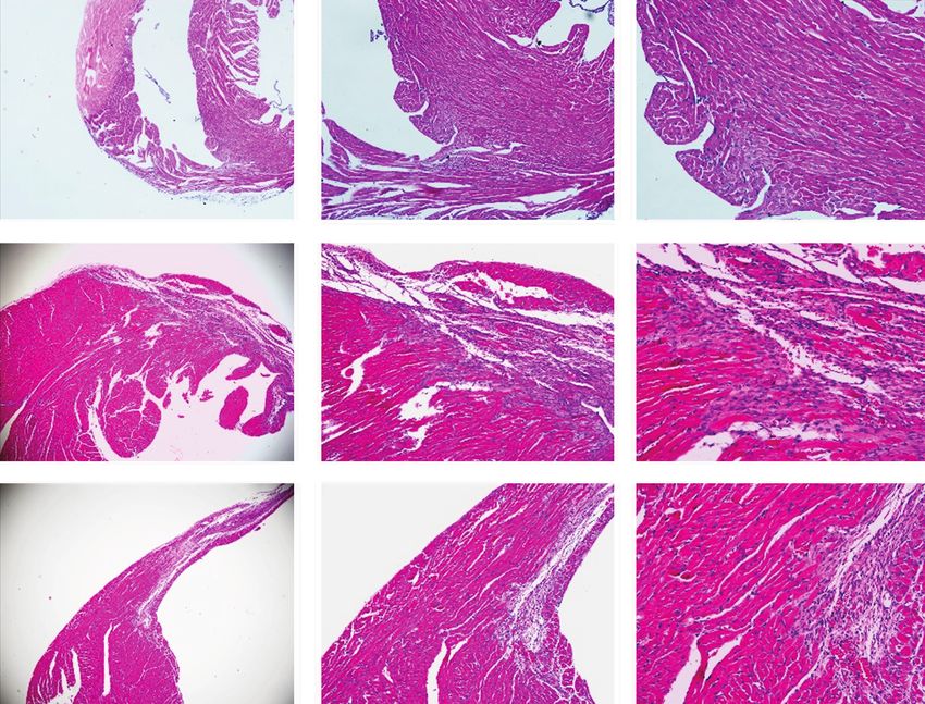
2                                                                                    Oxidative Medicine and Cellular Longevity

[3]. Reduction in the mortality rate of AMI is one big success     angiogenesis through increasing the expression of VEGF,
story of modern medicine [3]. However, the process of              VEGFR2, Flk-1, bFGF, and angiopoietin-1 (Ang-1) in ische-
restoring coronary blood flow to the ischemic myocardium            mic stroke models both in vivo and in vitro [14, 29–33]. The
may lead to myocardial ischemia/reperfusion (I/R) injury           regulation of BHD on the vascular endothelial growth factor
such as myocardial stunning, no-reflow phenomenon, and              (VEGF) signaling pathway according to the pathway enrich-
reperfusion arrhythmias. Several strategies such as pharma-        ment analysis deserves to be studied in order to fully appre-
cological treatment and mechanical therapies could reduce          hend its latent capacity on treatment and its correlation
I/R injury in animal studies or small-scale clinical trials, but   with angiogenesis [34].
the results are inconclusive [4]. Thus, there is still a need to        Caveolin-1 (Cav-1), the signature protein of endothelial
develop a novel cardioprotective strategy for AMI patients.        cell caveolae, is involved in many physiological and patholog-
     Angiogenesis is defined as the growth and proliferation of     ical processes such as antifibrosis [35], inflammation [36],
new blood vessels from preexisting vascular structures [5].        and oxidative stress [37]. Recent studies [38, 39] have dem-
Therapeutic angiogenesis refers to utilizing angiogenic            onstrated that Cav-1 is highly expressed in the vasculature
growth factors to increase the growth of collateral blood ves-     in the process of blood vessel growth and could regulate the
sels and promote new vascularization, so as to improve blood       angiogenic activity of endothelial cells. Loss of Cav-1 would
flow and tissue perfusion [6]. Promoting angiogenesis in            lead to the inhibition of vessel development and vascular
ischemic myocardium that lack sufficient perfusion remains           remodeling [40]. Furthermore, Cav-1 plays a pivotal role in
a promising strategy for MI patients [7, 8]. Although there        the signaling pathway of VEGF/VEGFR2-stimulated angio-
will be a spontaneous angiogenic response in AMI which             genesis and is associated with angiogenic biological activities
could partly reestablish blood flow in myocardium, this             [41]. Resveratrol, a Cav-1 agonist, significantly elevated
protective response is usually insufficient to restore the phys-     eNOS and VEGF protein levels in hypercholesterolemic rats
iological level of tissue perfusion [7]. However, limited          with focal myocardial ischemic injury [42]. These evidences
medical therapies have yet been proved to be able to success-      suggested that the Cav-1/VEGF pathway might play a critical
fully promote angiogenesis in AMI patients [9].                    role in angiogenesis after myocardial ischemic injury. Thus,
     Growing evidences have shown that Chinese herbal med-         in the present study, we aim to investigate the effects of
icines (CHM) could provide therapeutic effect on AMI by             BHD on angiogenesis through the Cav-1/VEGF pathway
targeting angiogenesis [10–12]. Buyang Huanwu decoction            on the MI model of mice.
(BHD), originally recorded in Yilin Gaicuo (Correction on
Errors in Medical Classics) written by Wang in 1830, is a          2. Materials and Methods
famous Chinese herbal prescription, has been used for the
treatment of various vascular diseases in China for hundreds       2.1. Animals. Thirty adult C57BL/6 male mice at 6-8 weeks of
of years [13], and now is still being used in China and other      age and 20-25 g weight were obtained from Shanghai Slack
countries around the world. BHD is composed of seven kinds         Laboratory Animal Research Center and housed in the labo-
of Chinese herbs (Table 1): (a) huang qi (radix astragali, the     ratory animal center of Wenzhou Medical University. All the
dried roots of Astragalus membranaceus (Fisch.) Bunge),            mice were kept under 12 h light/dark cycles, temperature 22
(b) dang gui (radix angelicae sinensis, the dried lateral roots     ± 1° C, and provided with food and water ad libitum. The
of Angelica sinensis (Oliv.) Diels), (c) chi shao (radix paeo-     animals used were treated in accordance with the Guide for
niae rubra, the dried roots of Paeonia lactiflora Pall), (d)        the Care and Use of Laboratory Animals, published by the
chuan xiong (rhizoma chuanxiong the dried rhizomes of              National Institutes of Health (NIH). The study instructions
Ligusticum striatum DC), (e) hong hua (flos carthami, the           were approved by the Animal Ethics Committee of the labo-
dried flowers of Carthamus tinctorius L), (f) tao ren (peach        ratory animal center of Wenzhou Medical University (num-
kernel, the dried seeds of Prunus persica (L.) Batsch), and        ber wydw2014-0058). All efforts were made to minimize the
(g) di long (Lumbricus, the dried bodies of Pheretima asper-       suffering of animals used.
gillum (E. Perrier)), all of which are recorded in http://www
.theplantlist.org and Chinese Pharmacopoeia. Based on tradi-       2.2. Drugs and Reagents. BHD which consists of huang qi
tional Chinese medicine theory, BHD has the function of            (radix astragali seuhedysari), dang gui (radix angelica sinen-
invigorating the body, enhancing blood circulation, and acti-      sis), chi shao (radix paeoniae rubra), chuan xiong (rhizoma
vating Qi flow through energy meridians. Growing evidence           ligustici chuanxiong), hong hua (flos carthami), tao ren
has suggested the cardio-/cerebral protective functions of         (semen persicae), and di long (Lumbricus) with a dispensing
BHD in humans and animal models [14–22]. Recent studies            ratio of 120 : 6 : 4.5 : 3 : 3 : 3 : 3 was purchased from Sanjiu
on pharmacology and biochemistry also have shown that the          Medical & Pharmaceutical Co. Ltd., Shenzhen, China
protective functions of BHD on cardiocerebrovascular               (granule preparations, approval number: country medicine
disease at least in part through the following mechanisms:         accurate character Z44020711); CD31 antibody (ab28364),
antioxidant [18, 23, 24], antiapoptosis [25, 26], anti-            Cav-1 polyclonal antibody (ab2910), and VEGF polyclonal
inflammatory [19, 27], and improving hemorheological dis-           antibody (ab46154) were purchased from Abcam (UK);
orders [19]. An overview of systematic reviews indicated that      extracellular regulated protein kinases (ERK1/2) monoclonal
BHD could treat a wide range of vascular disorders such as         antibody (4695), phosphorylated extracellular regulated pro-
acute ischemic stroke and angina pectoris through targeting        tein kinases (p-ERK1/2) monoclonal antibody (4370), glycer-
vascularity [28]. Studies showed that BHD could promote            aldehyde phosphate dehydrogenase (GAPDH) monoclonal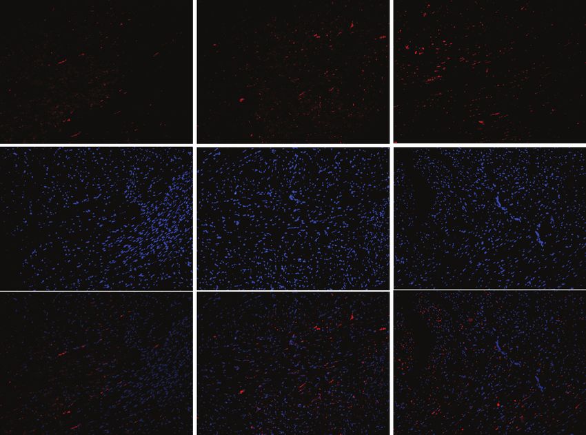
Oxidative Medicine and Cellular Longevity                                                                                            3

                                          Table 1: Overview of Buyang Huanwu decoction.

Chinese name           Common name                                Latin name/family/medicinal parts                      Amount (%)
Huang qi               Radix astragali          Astragalus membranaceus (Fisch.) Bunge/Leguminosae/dried roots           120 g (84.2%)
Dang gui           Radix angelicae sinensis         Angelica sinensis (Oliv.) Diels/Apiaceae/dried lateral roots           6 g (4.2%)
Chi shao            Radix paeoniae rubra                  Paeonia lactiflora Pall/Paeoniaceae/dried roots                  4.5 g (3.2%)
Chuan xiong         Rhizoma chuanxiong                  Ligusticum striatum DC/Apiaceae/dried rhizomes                     3 g (2.1%)
Hong hua               Flos carthami                    Carthamus tinctorius L/Compositae/dried flowers                     3 g (2.1%)
Tao ren                 Peach kernel                     Prunus persica (L.) Batsch/Rosaceae/dried seeds                   3 g (2.1%)
Di long                  Lumbricus                       Pheretima aspergillum (E. Perrier)/dried bodies                   3 g (2.1%)

antibody (5174), and vascular endothelial growth factor               for 30 minutes and then cut into 5 slices (1 mm thick) in a
receptor 2 (VEGFR2) monoclonal antibody (2479) were pur-              perpendicular way to the long axis. The slices were incubated
chased from Cell Signaling Technology (USA).                          in 1% 2,3,5-triphenyltetrazolium chloride solution (TTC) at
                                                                      37°C for 15 minutes. After carefully evaluating the whole sur-
2.3. AMI Model Establishment. Establishment of the AMI                face area, segments with brick red staining were identified as
model referred to the previous publication [43]. Briefly, mice         viable (noninfarcted area) and those without staining were
were anesthetized by isoflurane, and respiration was assisted          identified as nonviable (infarcted area). Finally, the 3rd slice
with a ventilator (Inspira, Harvard Apparatus, Holliston,             of each heart was chosen to calculate the infarct size
MA) in a volume-controlled mode at 80 strokes per minute.             infarcted area/ noninfarcted area + infarcted area     by the
After fixation, thoracotomy was done at the 3rd intercostal            Image-Pro Plus 6.0 software (Media Cybernetics, Silver
space via the left lateral chest wall to expose the pericardium       Spring, USA).
and heart. The AMI model was established by perpetually
ligating the left anterior descending coronary artery (LAD)           2.6. Myocardial Histopathology. The left ventricle including
in 2 mm from its origin (near the main pulmonary artery)              the region of MI was embedded in paraffin. The samples were
with a 7-0 silk suture, resulting in the development of pale          then sectioned into 5 μm thick slices. Masson staining and
color in the distal part of ligation.                                 hematoxylin-eosin (HE) staining were applied separately.
     All C57BL/6 mice (n = 55) were randomly divided into 3           Morphological changes of nuclei and cytoplasm around the
groups by the table of random number: (1) sham-operated               marginal zone of MI in HE staining were observed by an
group (sham, n = 15), the LAD was encircled by a 7-0 silk             optical microscope (Olympus, Japan). Image-Pro Plus 6.0
suture without ligation; (2) AMI group (AMI+sham, n = 20              software (Media Cybernetics, Silver Spring, USA) was used
), gavage with 0.2 ml 0.9% normal saline (once a day) 3 days          to calculate the percentage of collagen deposition around
before modeling until 14 days after modeling; and (3) BHD-            the marginal zone of MI to assess the degree of fibrosis in
treated group (AMI+BHD, n = 20), gavage with 0.2 ml BHD               the infarcted myocardium.
(20 g/kg, once a day) 3 days before modeling until 14 days
after modeling.                                                       2.7. Western Blot Analysis. Total protein isolated from the
                                                                      myocardium was separated by SDS-PAGE and transferred to
2.4. Doppler Echocardiography Study. Fourteen days after              a polyvinylidene difluoride (PVDF) membrane. The mem-
modeling, mice undergo transthoracic echocardiography by              branes were then blocked with 5% fat-free milk and incubated
the M-mode transducer (Acuson Sequoia 512, Sonos, Ger-                overnight at 4°C with primary antibodies including Cav-1
many) after induction of anesthesia. At the papillary muscle          (1 : 1000), VEGF (1 : 1000), VEGFR2 (1 : 1000), GADPH
level, M-mode tracings through short-axis view were                   (1 : 10000), ERK1/2 (1 : 1000), and p-ERK1/2 (1 : 2000). After
recorded through the anterior and posterior left ventricle            washing with TBST for three times, the membranes were incu-
(LV) walls to measure LV end-diastolic dimension (LVEDd),             bated with secondary antibodies (1 : 10000) for 2 h at room
LV end-systolic dimension (LVESd), LV fraction shortening             temperature. ChemiDoc™ XRS+ Imaging System was used
(LVFS), and LV ejective fraction (LVEF) with the Simpson              to visualize the signals. Javas freely available NIH ImageJ soft-
approach. All measurements were done by an experienced                ware (NIH, Bethesda, MD, USA) was used to quantify the
doctor who was blinded to the experimental design.                    intensity of immune reactivity.

2.5. Determination of Myocardial Infarct Size. Euthanasia             2.8. Immunofluorescence Staining. After routine dewaxing
was done on the mice at 14 days after modeling through                and hydration, the antigens in myocardial tissue sections
intraperitoneal injection of excessive pentobarbital sodium.          were repaired by sodium citrate buffer at 100°C. After wash-
The heart of the mice was separated from the aortic arch,             ing thrice with PBS, tissues were treated with 3% hydrogen
major blood vessels, and extracardiac connective tissue and           peroxide for 30 min. 1% bovine serum albumin (BSA) was
rinsed in phosphate-buffered saline to wash away the blood-            used to block the antigen. The tissues were then incubated
stain. The heart tissues were semifreezed in a −20°C freezer          with CD31 antibody (1 : 300), Cav-1 antibody (1 : 500),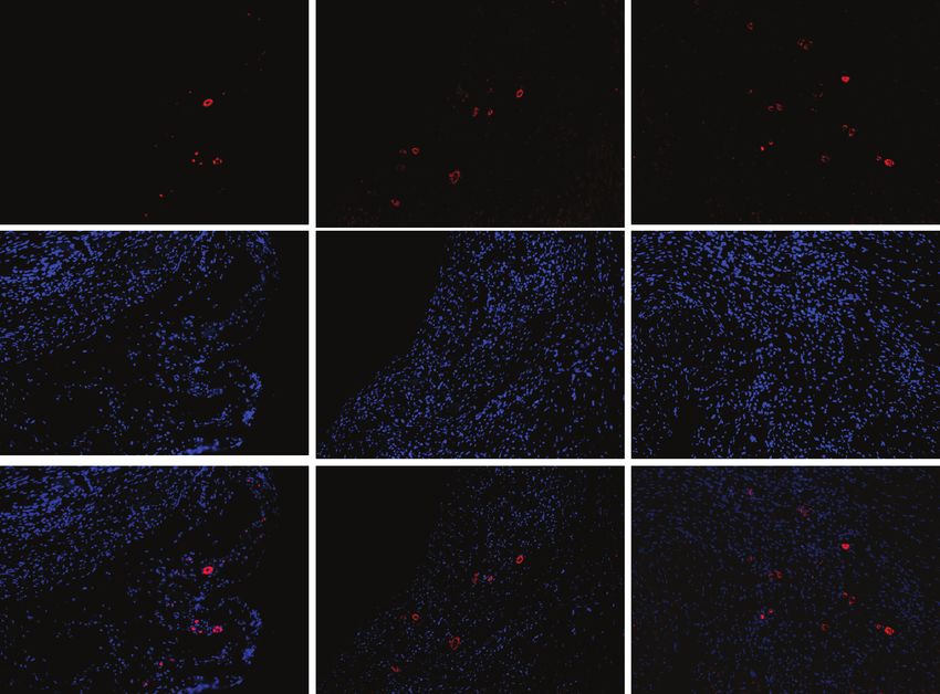
4                                                                                                                      Oxidative Medicine and Cellular Longevity

                                                                   100

                                                                         80

                                  Percent survival
                                                                         60

                                                                         40
                                                                              0             5                  10                 15
                                                                                                      Days
                                                                                  Sham
                                                                                  AMI
                                                                                  AMI+BHD
                                                                                                (a)
                                                                          0.8                           ⁎
                                      Heart weight/body weight (×10−2)

                                                                                                                         #
                                                                          0.6

                                                                          0.4

                                                                          0.2
                                                                                    Sham              AMI            AMI+BHD
                                                                                                (b)

Figure 1: Survival rate and heart weight/body weight ratio at 14 days after AMI in mice (sham: n = 15, AMI: n = 15, and BHD+AMI: n = 18).
(a) The survival rate of mice in the BHD-treated group compared with the AMI group (log-rank: P = 0 0829); (b) the heart weight/body
weight ratio of mice (mean ± SD). ∗ P < 0 05, compared with the sham group; # P < 0 05, compared with the AMI group.

VEGF antibody (1 : 400), VEGFR2 antibody (1 : 200), and p-                                            Gene-specific primers were as follows: Cav-1: forward, 5 ′
ERK antibody (1 : 200) at 4°C and then followed by 60 min of                                          -GACCTAATCCAACCATCAT-3 ′ and reverse, 5 ′ -AGCA
incubation with Alexa Flour 647- or 488-conjugated anti-                                              AGAACATTACCTCAA-3 ′ ; VEGF: forward, 5 ′ -GACTAT
body (1 : 400) at 37°C. To visualize the nuclei, the cells were                                       TCAGCGGACTCA-3 ′ and reverse, 5 ′ -AAGAACCAACC
counterstained with 4 ′ ,6-diamidino-2-phenylindole (DAPI)                                            TCCTCAA-3 ′ ; VEGFR2: forward, 5 ′ -AATGATTGTTG
for 5 min in the dark. The images were captured using a
                                                                                                      GCGATGAA-3 ′ and reverse, 5 ′ -GTGAGGATGACCGT
fluorescence microscope and then analyzed with the
Image-Pro Plus 6.0 software (Media Cybernetics, Silver                                                GTAG-3 ′ ; and β-actin: forward, 5 ′ -ACCTGCCCTTTAGA
Spring, USA).                                                                                         ACTT-3 ′ and reverse, 5 ′ -GCTCCAGGGACTATCTTT-3 ′ .

2.9. Real-Time Quantitative Reverse Transcription                                                     2.10. Statistical Analysis. All data were expressed as mean ±
Polymerase Chain Reaction (RT-qPCR). Total RNA was iso-                                               standard deviation (SD). Difference between two groups
lated using the TRIzol reagent (Invitrogen, USA). RNA sam-                                            was analyzed using two-tailed Student’s t-test. Multiple
ples from each group were reverse transcribed into cDNA                                               groups were compared using one-way analysis of variance
using the PrimeScript™ RT reagent Kit (TAKARA, Japan).                                                (ANOVA) and followed by LSD post hoc comparisons when
Quantitative RT-qPCR was performed on a LightCycler                                                   appropriate. All statistical analyses were performed using the
thermal cycler system (Bio-Rad, USA) using SYBR® Premix                                               SPSS 20.0 software and GraphPad Prism 6.05. Values of
Ex Taq™ II (TAKARA, Japan) and gene-specific primers.                                                  P < 0 05 were considered as statistically significant.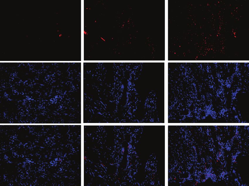
Oxidative Medicine and Cellular Longevity                                                                                     5

                                                                          80

     Sham
                                                                                                                      #
                                                                          60

                                                               LVEF (%)
     AMI                                                                                                       ⁎

                                                                          40

     BHD

                                                                          20
                                                                                           Sham               AMI   AMI+BHD
                         (a)                                                                            (b)
                                                  50

                                                  40
                                       LVFS (%)

                                                                                                    #
                                                  30

                                                  20                                 ⁎

                                                  10
                                                        Sham                         AMI          AMI+BHD
                                                                               (c)
                                             6
                                                                                     ⁎

                                             5
                                                                                                     #
                               LVID d (mm)

                                             4

                                             3

                                             2
                                                       Sham                      AMI              AMI+BHD
                                                                               (d)

                                                               Figure 2: Continued.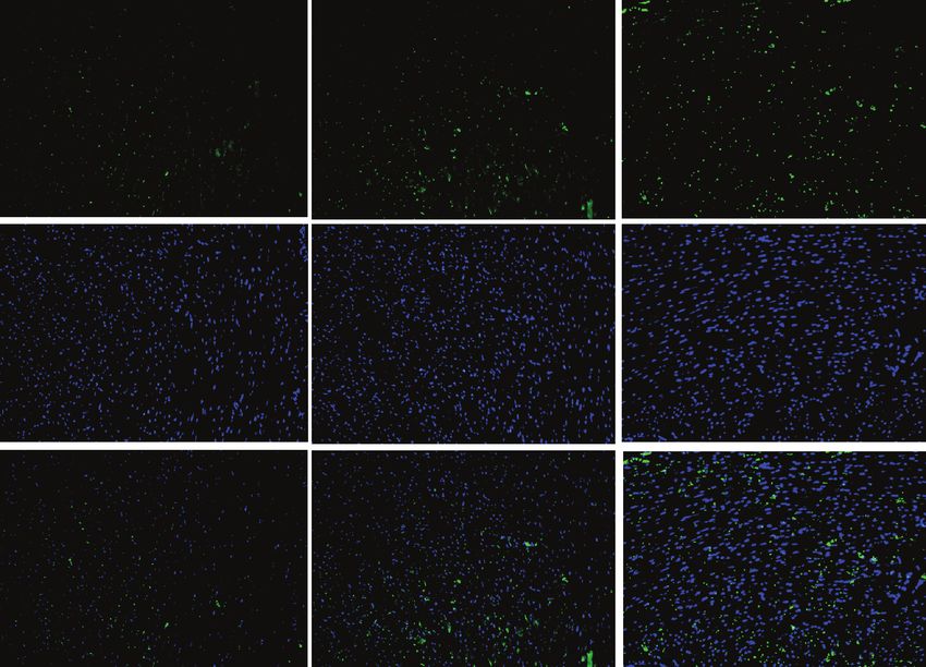
6                                                                                                     Oxidative Medicine and Cellular Longevity

                                                           5
                                                                                      ⁎

                                                           4                                              #

                                  LVIDs (mm)
                                                           3

                                                           2

                                                           1
                                                                       Sham          AMI            AMI+BHD
                                                                               (e)

                                 Sham

                                  AMI

                                 BHD

                                                                               (f)
                                                                  80

                                                                                      ⁎
                                                                  60
                                               Infarct size (%)

                                                                                                      #
                                                                  40

                                                                  20

                                                                  0
                                                                        Sham         AMI          AMI+BHD
                                                                               (g)

Figure 2: Cardiac function and infarct size at 14 days after AMI in mice. (a) M-mode echocardiographic images of the mice in each group. (b)
The analysis of LVEF (n = 6). (c) The analysis of LVFS (n = 6). (d) The analysis of LVIDd (n = 6). (e) The analysis of LVIDs (n = 6). (f)
Representative image of infarct size by cardiac 2,3,5-triphenyltetrazolium chloride (TTC) staining. (g) The analysis of the infarcted size
(sham: n = 5, AMI: n = 5, and BHD+AMI: n = 6). ∗ P < 0 05, compared with the sham group; # P < 0 05, compared with the AMI group.

3. Result                                                                            statistical significance (log-rank: P = 0 0829, Figure 1(a)).
                                                                                     The heart weight/body weight ratio was significantly
3.1. Effect of BHD on the Survival Rate and the Heart                                 decreased in the BHD-treated group compared with the
Weight/Body Weight Ratio after AMI. After 14 days, all mice                          AMI group (P < 0 05, Figure 1(b)).
in the sham group survived, while the BHD-treated group
exhibited a trend towards an improved overall survival rate                          3.2. Effect of BHD on Cardiac Function and Infarct Size after
after the induction of AMI, but differences did not reach                             AMI. As shown by echocardiography images (Figure 2(a)),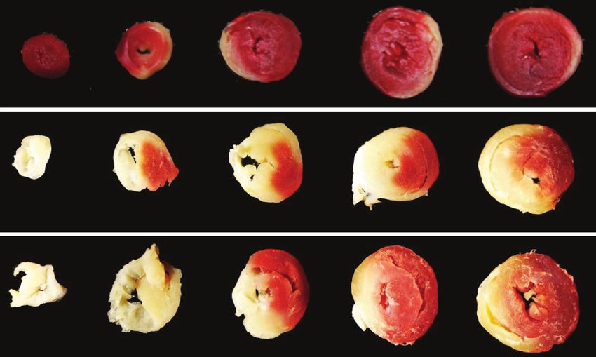
Oxidative Medicine and Cellular Longevity 7
40x 100x 200x
Sham
AMI
AMI+BHD
Figure 3: Histological changes in myocardial tissue at 14 days after AMI in mice (sham: n = 4, AMI: n = 4, and BHD+AMI: n = 6).
there was significant improvement of LVEF in the BHD- AMI group (P < 0 05, Figures 6(c) and 6(d)). Immunofluores-
treated group (75 65 ± 0 64%) compared with the AMI cence indicated that the integrated optical density of Cav-1
group (39 40 ± 2 21%) at 14 days after AMI (Figure 2(b)). (Figures 7(a) and 7(e)), VEGF (Figures 7(b) and 7(f)),
Significant improvements in cardiac function were also VEGFR2 (Figures 7(c) and 7(g)), and p-ERK (Figures 7(d)
observed in LVFS, LVIDd, and LVIDs (P < 0 05). The infarct and 7(h)) was significantly increased in the BHD-treated
size in the AMI group was 56 20 ± 2 26% (Figures 2(g) and group compared with the AMI group (P < 0 05). RT-PCR
2(h)). Compared with the AMI group, the infarct size showed that the mRNA level of Cav-1 (Figure 8(a)), VEGF
(36 74 ± 1 22%) was markedly reduced in the BHD-treated (Figure 8(b)), and VEGFR2 (Figure 8(c)) in the BHD-treated
group (P < 0 05). group was significantly increased compared with the AMI
group (P < 0 05).
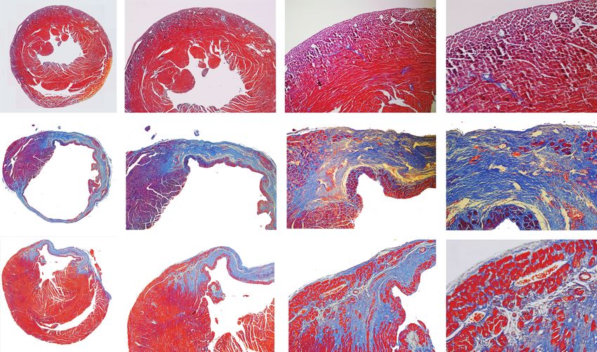
3.3. Effect of BHD on Histological Changes and Fibrosis in
Myocardial Tissue after AMI. By HE staining, the AMI group 4. Discussion
showed marked necrotic changes in myofibrils with severe
infiltration of inflammation and interstitial edema (Figure 3). During AMI, the damage inflicted on the myocardium
BHD-treated group exhibited only focal tissue necrosis, results in two processes: ischemia and the following reperfu-
mild inflammatory infiltration, and interstitial edema sion (I/R) [44]. The edema/sarcolemma rupture, calcium
(Figure 3). Compared with the AMI group, Mason staining overload/hypercontracture, mitochondrial dysfunction, pro-
of the collagen deposition area on myocardial fibrosis was teolysis (caspase, calpain), and apoptosis lead to a large
significantly decreased in the BHD-treated group (P < 0 05 amount of reduction of cardiomyocytes. And, the embolism,
, Figures 4(a) and 4(b)). vasomotor disorder, leukocyte adherence/infiltration, stasis,
and capillary rupture/hemorrhage appeared in coronary
3.4. Effect of BHD on Angiogenesis after AMI. To verify vascular caused severe myocardial injury [45]. Thus, cardio-
whether myocardial protection of BHD is associated with vascular protection drugs generally work through one or
angiogenesis in the infarction border zone, the immunohis- combined aspects of the above targets. In the present study,
tochemical analysis was performed by CD31 staining. As BHD reduced the myocardial fibrosis and inflammation, pro-
shown in Figure 5(b), the density of microvessel in the moted angiogenesis in the infarction border zone via Cav-
BHD-treated group was much higher than that in the AMI 1/VEGF signaling pathway, then reduced the MI size, and
group (P < 0 05). improved the cardiac function. It may be because of these
improvements that we finally observed a trend towards an
3.5. Effect of BHD on Expression of Cav-1, VEGF, VEGFR2, improved overall survival rate of the BHD-treated group.
and p-ERK in the Infarction Border Zone after AMI. Expres- Cav-1 is a major component of the caveola membrane
sion of Cav-1, VEGF, and VEGFR2 was elevated in the AMI that is expressed in the majority of differentiated cells [46]
group compared with the sham group (P < 0 05). Further- and plays an important role in regulating the cellular signal
more, the expression of Cav-1, VEGF, and VEGFR2 was fur- transduction, endocytosis, transcytosis, and molecular trans-
ther increased in the BHD-treated group compared with the port [47]. The cardioprotective effects of Cav-1 in ischemic
AMI group (P < 0 05, Figures 6(a) and 6(b)). BHD treatment heart disease have been well reported [48] in both mouse
promoted the phosphorylation of ERK compared with the and human specimens; an increase of Cav-1 in an infarcted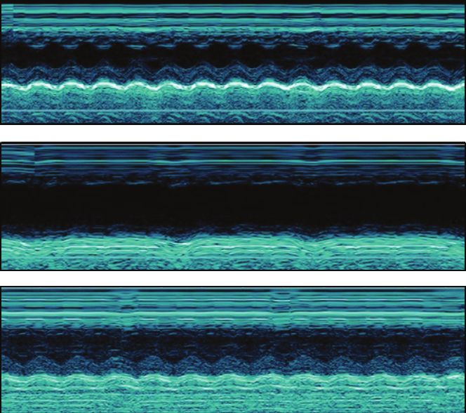
8                                                                                           Oxidative Medicine and Cellular Longevity

                          Holistic diagram              40x                      100x                     200x

                SHAM

                  AMI

                  BHD

                                                                   (a)
                                                   80

                                                   60
                                    Fibrosis (%)

                                                                                        #

                                                   40

                                                   20
                                                        AMI                        AMI+BHD
                                                                   (b)

Figure 4: Fibrosis in myocardial tissue at 14 days after AMI in mice (mean ± SD; sham: n = 4, AMI: n = 4, and BHD+AMI: n = 6). (a)
Representative images of Masson’s trichrome staining. (b) Quantitative analysis of the collagen deposition area. # P < 0 05, compared with
the AMI group.

area was detected in the early stage of MI [38]. Several studies             Previous studies also indicated that Cav-1 could reduce
have shown that the activation or preservation of Cav-1                  infarct volume and promote angiogenesis through the VEGF
played a protective role in myocardial I/R injury [49–51].               signaling pathway [57, 58]. Recent studies showed that the
Subsequently, compared with the wild-type mice, Cav-1-/-                 expression of Cav-1 and VEGF was significantly decreased
mice showed a more severe cardiac dysfunction and a lower                after the use of the caveolin-1 inhibitor, resulted in increase
survival rate after MI [52]. In Cav-1-/- mice, a low-intensity           in neurological deficit and infarction volume [59–61]. Other
pulsed ultrasound, which is a potential cardiac protection               studies also confirmed this phenomenon at the genetic level.
strategy, presented absent cardioprotective effects after myo-            The ablation of Cav-1 gene in mice could result in an impair-
cardial ischemic injury [38]. Cav-1 is also a vital regulator of         ment in angiogenesis and reduction of VEGF expression [56,
vascular endothelial homeostasis which controls angiogene-               62]. VEGF is a pivotal regulator of blood vessel formation
sis and vessel function [53]. The adverse influence on angio-             during embryogenesis and angiogenesis [63]. Lots of evi-
genesis after Cav-1 knockout has been confirmed in multiple               dences have shown that VEGF, through combining with its
disease models, including hindlimb ischemia [54], sclero-                receptor VEGFR2, could trigger multiple downstream signals
derma fibroblasts [55], colitis [39], AMI [38], and cerebral              such as p-ERK, thereby promoting angiogenesis [64–66].
ischemia [56]. In the present study, BHD increased angio-                Taken together, these results indicate that Cav-1 could pro-
genesis and the expression of Cav-1 in the infarction border             mote angiogenesis by upregulating the VEGF signaling path-
zone, suggesting that the cardioprotective effect of BHD tar-             way. The present study indicated that BHD increased the
geted angiogenesis by Cav-1.                                             CAV-1, VEGF, VEGFR2, and p-ERK in the infarction border
Oxidative Medicine and Cellular Longevity                                                                                                                                                  9

                Sham               AMI              AMI+BHD                                                                 30

                                                                                                                                                                                    #
    CD31

                                                                                               The density of microvessel
                                                                                                                            20

    DAPI

                                                                                                                            10

   Merge

                                                                                                                             0
                                                                                                                                        Sham                     AMI             AMI+BHD
                                 (a)                                                                                                                       (b)

Figure 5: Density of microvessel in the infarction border zone at 14 days after AMI in mice (mean ± SD; sham: n = 4, AMI: n = 4, and BHD
+AMI: n = 6). (a) Representative images of CD31 staining. (b) Quantitative analysis of the density of microvessel. # P < 0 05, compared with
the AMI group.

                                                                                         400
                                                                                                                                                                   #
                                                       The level of protein expression

                                                                                                                                                             #          #
                                                                                         300                                                     ⁎
                                                                                                                                           ⁎          ⁎
                               Sham     AMI   BHD
                                                               (% of control)

                       Cav-1
                                                                                         200
                   VEGFA

                  VEGFR2                                                                 100

                   GAPDH
                                                                                           0
                                                                                                                             Sham               AMI          AMI+BHD

                                                                                                                            Cav-1
                                                                                                                            VEGFA
                                                                                                                            VEGFR2
                                  (a)                                                                                                    (b)
                                                                                         400

                                                                                                                                                                            #
                                                       The level of protein expression

                                                                                         300
                                                                                                                                                      ⁎
                                                                (% of control)

                                                                                         200

                          Sham         AMI    BHD

                p-ERK                                                                    100

                  ERK
                                                                                          0
                                                                                                                                 Sham                AMI               AMI+BHD
                                 (c)                                                                                                           (d)

Figure 6: Western blot analysis of Cav-1, VEGF, VEGFR2, and p-ERK1/2 expression in the infarction border zone at 14 days after AMI in
mice (mean ± SD, n = 6). (a) Western blot analysis of the expression of Cav-1, VEGF, and VEGFR2. (b) Quantitative analysis for the western
blot results of Cav-1, VEGF, and VEGFR2. (c) Western blot analysis of the expression of p-ERK1/2. (d) Quantitative analysis for the western
blot results of p-ERK1/2. ∗ P < 0 05, compared with the sham group; # P < 0 05, compared with the AMI group.
10                                              Oxidative Medicine and Cellular Longevity

              Sham             AMI           AMI+BHD

      Cav-1

      DAPI

     Merge

                              (a)
              Sham                  AMI        AMI+BHD

     VEGFA

      DAPI

      Merge

                              (b)
               Sham                 AMI        AMI+BHD

     VEGFR2

       DAPI

      Merge

                              (c)

                      Figure 7: Continued.
Oxidative Medicine and Cellular Longevity 11
Sham AMI AMI+BHD
p ERK
DAPI
Merge
(d)
40 #
Integrated optical density of Cav-1s
30 ⁎
(% of DAPI)
20
10
0
Sham AMI AMI+BHD
(e)
40 10
#
#
Integrated optical density of VEGFAs
Integrated optical density of VEGFR2
8
30
⁎ ⁎
(% of DAPI)
(% of DAPI)
6
20
4
10
2
0 0
Sham AMI AMI+BHD Sham AMI AMI+BHD
(f) (g)
Figure 7: Continued.12 Oxidative Medicine and Cellular Longevity
40 #
Integrated optical density of p ERKs
30
(% of DAPI)
⁎
20
10
0
Sham AMI AMI+BHD
(h)
Figure 7: Immunofluorescence staining of Cav-1, VEGF, VEGFR2, and p-ERK1/2 in the infarction border zone at 14 days after AMI in mice
(mean ± SD; sham: n = 4, AMI: n = 4, and BHD+AMI: n = 6). (a) Immunofluorescence staining of Cav-1. (b) Immunofluorescence staining of
VEGF. (c) Immunofluorescence staining of VEGFR2. (d) Immunofluorescence staining of p-ERK1/2. (e) Quantitative analysis of Cav-1. (f)
Quantitative analysis of VEGF. (g) Quantitative analysis of VEGFR2. (h) Quantitative analysis of p-ERK1/2. ∗ P < 0 05, compared with the
sham group; # P < 0 05, compared with the AMI group.
3 # 3
#
⁎
⁎
The relative expression
The relative expression
of VEGFA (fold)
2 2
of CAV-1 (fold)
1
1
0
0
AMI Sham AMI AMI+BHD
Sham AMI+BHD
(a) (b)
3
#
⁎
The relative expression
of VEGFR2 (fold)
2
1
0
Sham AMI AMI+BHD
(c)
Figure 8: The mRNA expression of Cav-1, VEGF, and VEGFR2 at 14 days after AMI in mice (mean ± SD, n = 6). (a) The mRNA expression
of Cav-1. (b) The mRNA expression of VEGF. (c) The mRNA expression of VEGFR2. ∗ P < 0 05, compared with the sham group; # P < 0 05,
compared with the AMI group.Oxidative Medicine and Cellular Longevity 13
zone, suggesting that BHD could promote angiogenesis [4] B. Ibanez, S. James, S. Agewall et al., “ESC guidelines for the
through the Cav-1/VEGF pathway. management of acute myocardial infarction in patients
Herbal formulae, with multicomponents and multitar- presenting with ST-segment elevation: the task force for the
gets, may potentially satisfy the demands of complex disease management of acute myocardial infarction in patients pre-
treatment in an integrated manner. Furthermore, investiga- senting with ST-segment elevation of the European Society
tion on new molecular targets and principles indicated that of Cardiology (ESC),” European Heart Journal, vol. 39, no. 2,
pp. 119–177, 2018.
a single angiogenic substance might be insufficient for induc-
[5] A. Birbrair, T. Zhang, Z. M. Wang et al., “Type-2 pericytes
ing therapeutic angiogenesis [67]. Hundreds of constituents
participate in normal and tumoral angiogenesis,” American
have been identified in BHD such as polysaccharides, astra- Journal of Physiology-Cell Physiology, vol. 307, no. 1,
galosides, and isoflavonoids in radix astragali seuhedysari pp. C25–C38, 2014.
[68], as well as phthalides and phenolic acids in radix angel- [6] Q. Lu, Y. Yao, Z. Hu et al., “Angiogenic factor AGGF1 activates
icae sinensis and rhizoma ligustici chuanxiong, etc. [69, 70]. autophagy with an essential role in therapeutic angiogenesis
Network pharmacology can forecast multiple targets and for heart disease,” PLOS Biology, vol. 14, no. 8, article
pathways affected by the active components in TCM formu- e1002529, 2016.
lae. Among them, key targets/signaling pathways might be [7] K. Albrecht-Schgoer, W. Schgoer, J. Holfeld et al., “The angio-
selected and should be experimentally validated. genic factor secretoneurin induces coronary angiogenesis in a
model of myocardial infarction by stimulation of vascular
5. Conclusion endothelial growth factor signaling in endothelial cells,” Circu-
lation, vol. 126, no. 21, pp. 2491–2501, 2012.
The present study demonstrated that BHD could exert cardi- [8] S. Araki, Y. Izumiya, S. Hanatani et al., “Akt1-mediated
oprotective effects on the mouse model with AMI through skeletal muscle growth attenuates cardiac dysfunction and
targeting angiogenesis via Cav-1/VEGF signaling pathway. remodeling after experimental myocardial infarction,” Circu-
lation Heart Failure, vol. 5, no. 1, pp. 116–125, 2012.
[9] R. Kornowski, “Therapeutic angiogenesis revisited,” Catheter-
Data Availability ization and Cardiovascular Interventions, vol. 82, no. 6,
pp. 907-908, 2013.
The data used to support the findings of this study are
[10] D. Guo, C. E. Murdoch, T. Liu et al., “Therapeutic angiogenesis
available from the corresponding author upon request.
of Chinese herbal medicines in ischemic heart disease: a
review,” Frontiers in Pharmacology, vol. 9, p. 428, 2018.
Conflicts of Interest [11] L. J. Yu, K. J. Zhang, J. Z. Zhu et al., “Salvianolic acid exerts
cardioprotection through promoting angiogenesis in animal
The authors declare that they have no conflicts of interest. models of acute myocardial infarction: preclinical evidence,”
Oxidative Medicine and Cellular Longevity, vol. 2017, Article
Authors’ Contributions ID 8192383, 11 pages, 2017.
[12] K. J. Zhang, J. Z. Zhu, X. Y. Bao, Q. Zheng, G. Q. Zheng, and
JZZ and XYB contributed equally to this work. JZZ, XYB, Y. Wang, “Shexiang baoxin pills for coronary heart disease in
QZ, QT, PCZ, ZZ, and YW designed the study; JZZ and animal models: preclinical evidence and promoting angiogen-
XYB performed the experiments. QZ, QT, PCZ, and ZZ esis mechanism,” Front Pharmacol, vol. 8, p. 404, 2017.
analyzed the data; JZZ, XYB, and QZ wrote the manu- [13] Q. R. Wang, Yilin Gaicuo (Correction on Errors in Medical
script. Jia-Zhen Zhu and Xiao-Yi Bao contributed equally Classics), People’s Medical Publishing House, Beijing, China,
to this work. 2005.
[14] G. Cai, B. Liu, W. Liu et al., “Buyang Huanwu Decoction can
improve recovery of neurological function, reduce infarction
Acknowledgments volume, stimulate neural proliferation and modulate VEGF
and Flk1 expressions in transient focal cerebral ischaemic rat
This work was supported by the grant of the National brains,” Journal of Ethnopharmacology, vol. 113, no. 2,
Natural Science Foundation of China (81473491/81573750/ pp. 292–299, 2007.
81173395/H2902). [15] L. S. Chu, Y. J. Yin, Q. Ke, W. Chen, and F. Chen, “Effect of
buyanghuanwu decoction on angiogenesis and Ang-1/Tie-2
References expression after focal cerebral ischemia in mice,” Chinese Jour-
nal of Behavioral Medicine and Brain Science, vol. 20, no. 3,
[1] K. Thygesen, J. S. Alpert, A. S. Jaffe et al., “Fourth universal pp. 202–204, 2011.
definition of myocardial infarction,” Journal of the American [16] G. Cai and B. Liu, “Buyang Huanwu Decoction increases vas-
College of Cardiology, vol. S0735-1097, no. 18, pp. 36941– cular endothelial growth factor expression and promotes
36949, 2018. angiogenesis in a rat model of local cerebral ischemia,” Neural
[2] E. J. Benjamin, M. J. Blaha, S. E. Chiuve et al., “Heart disease Regeneration Research, vol. 5, no. 22, pp. 1733–1738, 2010.
and stroke statistics-2017 update: a report from the American [17] C. Z. Hao, F. Wu, J. Shen et al., “Clinical efficacy and safety of
Heart Association,” Circulation, vol. 135, no. 10, pp. e146– buyang huanwu decoction for acute ischemic stroke: a system-
e603, 2017. atic review and meta-analysis of 19 randomized controlled tri-
[3] G. W. Reed, J. E. Rossi, and C. P. Cannon, “Acute myocardial als,” Evidence-based Complementary and Alternative Medicine,
infarction,” Lancet, vol. 389, no. 10065, pp. 197–210, 2017. vol. 2012, Article ID 630124, 10 pages, 2012.14 Oxidative Medicine and Cellular Longevity
[18] Y. Liu, R. Lin, X. Shi et al., “The roles of buyang huanwu decoc- focal cerebral ischemia,” Journal of Molecular Neuroscience,
tion in anti-inflammation, antioxidation and regulation of vol. 56, no. 4, pp. 898–906, 2015.
lipid metabolism in rats with myocardial ischemia,” [31] H. J. Cui, A. L. Yang, H. J. Zhou et al., “Buyang huanwu decoc-
Evidence-based Complementary and Alternative Medicine, tion promotes angiogenesis via vascular endothelial growth
vol. 2011, Article ID 561396, 8 pages, 2011. factor receptor-2 activation through the PI3K/Akt pathway
[19] W. R. Wang, R. Lin, H. Zhang et al., “The effects of Buyang in a mouse model of intracerebral hemorrhage,” BMC Comple-
Huanwu Decoction on hemorheological disorders and energy mentary and Alternative Medicine, vol. 15, no. 1, p. 91, 2015.
metabolism in rats with coronary heart disease,” Journal of [32] F. Liao, Y. Meng, H. Zheng et al., “Biospecific isolation and char-
Ethnopharmacology, vol. 137, no. 1, pp. 214–220, 2011. acterization of angiogenesis-promoting ingredients in Buyang
[20] G. Yang, Z. Fang, Y. Liu et al., “Protective effects of Chinese Huanwu decoction using affinity chromatography on rat brain
traditional medicine buyang huanwu decoction on myocardial microvascular endothelial cells combined with solid-phase
injury,” Evidence-based Complementary and Alternative Med- extraction, and HPLC-MS/MS,” Talanta, vol. 179, pp. 490–
icine, vol. 2011, Article ID 930324, 7 pages, 2011. 500, 2018.
[21] H. Zhang, W. R. Wang, R. Lin et al., “Buyang Huanwu decoc- [33] R. L. Wei, H. J. Teng, B. Yin et al., “A systematic review and
tion ameliorates coronary heart disease with Qi deficiency and meta-analysis of buyang huanwu decoction in animal model
blood stasis syndrome by reducing CRP and CD40 in rats,” of focal cerebral ischemia,” Evidence-based Complementary
Journal of Ethnopharmacology, vol. 130, no. 1, pp. 98–102, and Alternative Medicine, vol. 2013, Article ID 138484, 13
2010. pages, 2013.
[22] Y. C. Zhou, B. Liu, Y. J. Li et al., “Effects of buyang huanwu [34] Q. Guo, M. Zhong, H. Xu, X. Mao, Y. Zhang, and N. Lin, “A
decoction on ventricular remodeling and differential protein systems biology perspective on the molecular mechanisms
profile in a rat model of myocardial infarction,” Evidence- underlying the therapeutic effects of buyang huanwu decoc-
based Complementary and Alternative Medicine, vol. 2012, tion on ischemic stroke,” Rejuvenation Research, vol. 18,
Article ID 385247, 11 pages, 2012. no. 4, pp. 313–325, 2015.
[23] H. Cui, T. Liu, P. Li et al., “An intersectional study of LncRNAs [35] D. Gvaramia, M. E. Blaauboer, R. Hanemaaijer, and V. Everts,
and mRNAs reveals the potential therapeutic targets of buyang “Role of caveolin-1 in fibrotic diseases,” Matrix Biology,
huanwu decoction in experimental intracerebral hemorrhage,” vol. 32, no. 6, pp. 307–315, 2013.
Cellular Physiology and Biochemistry, vol. 46, no. 5, pp. 2173–
[36] S. G. Royce and C. J. Le Saux, “Role of caveolin-1 in asthma
2186, 2018.
and chronic inflammatory respiratory diseases,” Expert Review
[24] J. Shen, Y. Zhu, K. Huang et al., “Buyang Huanwu Decoc- of Respiratory Medicine, vol. 8, no. 3, pp. 339–347, 2014.
tion attenuates H2O2-induced apoptosis by inhibiting reac-
[37] Z. C. Nwosu, M. P. Ebert, S. Dooley, and C. Meyer, “Caveolin-
tive oxygen species-mediated mitochondrial dysfunction
1 in the regulation of cell metabolism: a cancer perspective,”
pathway in human umbilical vein endothelial cells,” BMC
Molecular Cancer, vol. 15, no. 1, p. 71, 2016.
Complementary and Alternative Medicine, vol. 16, no. 1,
p. 154, 2016. [38] T. Shindo, K. Ito, T. Ogata et al., “Low-intensity pulsed ultra-
sound enhances angiogenesis and ameliorates left ventricular
[25] H. W. Wang, K. T. Liou, Y. H. Wang et al., “Deciphering the
dysfunction in a mouse model of acute myocardial infarction,”
neuroprotective mechanisms of Bu-yang Huan-wu decoction
Arteriosclerosis, Thrombosis, and Vascular Biology, vol. 36,
by an integrative neurofunctional and genomic approach in
no. 6, pp. 1220–1229, 2016.
ischemic stroke mice,” Journal of Ethnopharmacology,
vol. 138, no. 1, pp. 22–33, 2011. [39] J. H. Chidlow Jr, J. J. M. Greer, C. Anthoni et al., “Endothelial
[26] L. Fan, K. Wang, and B. Cheng, “Effects of buyang huanwu caveolin-1 regulates pathologic angiogenesis in a mouse model
decoction on apoptosis of nervous cells and expressions of of colitis,” Gastroenterology, vol. 136, no. 2, pp. 575–84.e2,
Bcl-2 and bax in the spinal cord of ischemia-reperfusion injury 2009.
in rabbits,” Journal of Traditional Chinese Medicine, vol. 26, [40] S. E. Woodman, A. W. Ashton, W. Schubert et al., “Caveolin-1
no. 2, pp. 153–156, 2006. knockout mice show an impaired angiogenic response to exog-
[27] B. Dou, W. Zhou, S. Li et al., “Buyang huanwu decoction atten- enous stimuli,” The American Journal of Pathology, vol. 162,
uates infiltration of natural killer cells and protects against no. 6, pp. 2059–2068, 2003.
ischemic brain injury,” Cellular Physiology and Biochemistry, [41] S. A. Tahir, S. Park, and T. C. Thompson, “Caveolin-1 regu-
vol. 50, no. 4, pp. 1286–1300, 2018. lates VEGF-stimulated angiogenic activities in prostate cancer
[28] J. H. Li, A. J. Liu, H. Q. Li, Y. Wang, H. C. Shang, and G. Q. and endothelial cells,” Cancer Biology & Therapy, vol. 8, no. 23,
Zheng, “Buyang huanwu decoction for healthcare: evidence- pp. 2286–2296, 2009.
based theoretical interpretations of treating different diseases [42] S. V. Penumathsa, S. Koneru, S. M. Samuel et al., “Strategic tar-
with the same method and target of vascularity,” Evidence- gets to induce neovascularization by resveratrol in hypercho-
based Complementary and Alternative Medicine, vol. 2014, lesterolemic rat myocardium: role of caveolin-1, endothelial
Article ID 506783, 17 pages, 2014. nitric oxide synthase, hemeoxygenase-1, and vascular endo-
[29] Z. Q. Zhang, T. Tang, J. K. Luo et al., “Effect of qi-tonifying thelial growth factor,” Free Radical Biology & Medicine,
and stasis-eliminating therapy on expression of vascular vol. 45, no. 7, pp. 1027–1034, 2008.
endothelial growth factor and its receptors Flt-1, Flk-1 in [43] E. Gao, Y. H. Lei, X. Shang et al., “A novel and efficient model
the brain of intracerebral hemorrhagic rats,” Chinese Journal of coronary artery ligation and myocardial infarction in the
of Integrative Medicine, vol. 13, no. 4, pp. 285–290, 2007. mouse,” Circulation Research, vol. 107, no. 12, pp. 1445–
[30] J. Yang, F. Gao, Y. Zhang, Y. Liu, and D. Zhang, “Buyang 1453, 2010.
huanwu decoction (BYHWD) enhances angiogenic effect of [44] B. Ibáñez, G. Heusch, M. Ovize, and F. van de Werf, “Evolving
mesenchymal stem cell by upregulating VEGF expression after therapies for myocardial ischemia/reperfusion injury,” JournalOxidative Medicine and Cellular Longevity 15
of the American College of Cardiology, vol. 65, no. 14, pp. 1454– upregulating the caveolin-1/VEGF signaling pathway,” Jour-
1471, 2015. nal of Molecular Neuroscience, vol. 64, no. 2, pp. 211–223,
[45] G. Heusch and B. J. Gersh, “The pathophysiology of acute 2018.
myocardial infarction and strategies of protection beyond [60] Q. Xie, J. Cheng, G. Pan et al., “Treadmill exercise ameliorates
reperfusion: a continual challenge,” European Heart Journal, focal cerebral ischemia/reperfusion-induced neurological defi-
vol. 38, no. 11, pp. 774–784, 2017. cit by promoting dendritic modification and synaptic plasticity
[46] C. M. Thomas and E. J. Smart, “Caveolae structure and func- via upregulating caveolin-1/VEGF signaling pathways,” Exper-
tion,” Journal of Cellular and Molecular Medicine, vol. 12, imental Neurology, vol. 313, pp. 60–78, 2019.
no. 3, pp. 796–809, 2008. [61] Z. Chen, Q. Hu, Q. Xie et al., “Effects of treadmill exercise on
[47] P. W. Shaul and R. G. W. Anderson, “Role of plasmalemmal motor and cognitive function recovery of MCAO mice
caveolae in signal transduction,” American Journal of through the caveolin-1/VEGF signaling pathway in ischemic
Physiology-Lung Cellular and Molecular Physiology, vol. 275, penumbra,” Neurochemical Research, vol. 44, no. 4, pp. 930–
no. 5, pp. L843–L851, 1998. 946, 2019.
[48] Y. Yang, Z. Ma, W. Hu et al., “Caveolin-1/-3: therapeutic tar- [62] J. H. Chidlow Jr and W. C. Sessa, “Caveolae, caveolins, and
gets for myocardial ischemia/reperfusion injury,” Basic cavins: complex control of cellular signalling and inflamma-
Research in Cardiology, vol. 111, no. 4, p. 45, 2016. tion,” Cardiovascular Research, vol. 86, no. 2, pp. 219–225,
[49] K. R. Chaudhary, W. J. Cho, F. Yang et al., “Effect of ischemia 2010.
reperfusion injury and epoxyeicosatrienoic acids on caveolin [63] S. Dehghani, R. Nosrati, M. Yousefi et al., “Aptamer-based bio-
expression in mouse myocardium,” Journal of Cardiovascular sensors and nanosensors for the detection of vascular endothe-
Pharmacology, vol. 61, no. 3, pp. 258–263, 2013. lial growth factor (VEGF): a review,” Biosensors and
[50] L. H. Young, Y. Ikeda, and A. M. Lefer, “Caveolin-1 peptide Bioelectronics, vol. 110, pp. 23–37, 2018.
exerts cardioprotective effects in myocardial ischemia- [64] S. Ikeda, M. Ushio-Fukai, L. Zuo et al., “Novel role of ARF6 in
reperfusion via nitric oxide mechanism,” American Journal vascular endothelial growth factor-induced signaling and
of Physiology-Heart and Circulatory Physiology, vol. 280, angiogenesis,” Circulation Research, vol. 96, no. 4, pp. 467–
no. 6, pp. H2489–H2495, 2001. 475, 2005.
[51] T. Murata, M. I. Lin, Y. Huang et al., “Reexpression of caveolin-1 [65] J. Oshikawa, S. J. Kim, E. Furuta et al., “Novel role of p66Shc in
in endothelium rescues the vascular, cardiac, and pulmonary ROS-dependent VEGF signaling and angiogenesis in endothe-
defects in global caveolin-1 knockout mice,” The Journal of lial cells,” American Journal of Physiology. Heart and Circula-
Experimental Medicine, vol. 204, no. 10, pp. 2373–2382, 2007. tory Physiology, vol. 302, no. 3, pp. H724–H732, 2012.
[52] J. F. Jasmin, G. Rengo, A. Lymperopoulos et al., “Caveolin-1 [66] M. Simons, E. Gordon, and L. Claesson-Welsh, “Mechanisms
deficiency exacerbates cardiac dysfunction and reduces sur- and regulation of endothelial VEGF receptor signalling,”
vival in mice with myocardial infarction,” American Journal Nature Reviews Molecular Cell Biology, vol. 17, no. 10,
of Physiology-Heart and Circulatory Physiology, vol. 300, pp. 611–625, 2016.
no. 4, pp. H1274–H1281, 2011. [67] P. Carmeliet and R. K. Jain, “Molecular mechanisms and clin-
[53] F. Braet, “Rac1, caveolin-1 and vascular endothelial growth ical applications of angiogenesis,” Nature, vol. 473, no. 7347,
factor-mediated liver sinusoidal endothelial cell angiogenesis,” pp. 298–307, 2011.
Liver International, vol. 29, no. 2, pp. 143-144, 2009. [68] C. Chu, L. W. Qi, E. H. Liu, B. Li, W. Gao, and P. Li, “Radix
[54] P. Sonveaux, P. Martinive, J. DeWever et al., “Caveolin-1 Astragali (Astragalus): latest advancements and trends in
expression is critical for vascular endothelial growth factor- chemistry, analysis, pharmacology and pharmacokinetics,”
induced ischemic hindlimb collateralization and nitric oxide- Current Organic Chemistry, vol. 14, no. 16, pp. 1792–1807,
mediated angiogenesis,” Circulation Research, vol. 95, no. 2, 2010.
pp. 154–161, 2004. [69] X. Ran, L. Ma, C. Peng, H. Zhang, and L. P. Qin, “Ligusticum
[55] V. Liakouli, J. Elies, Y. M. el-Sherbiny et al., “Scleroderma fibro- chuanxiong Hort: a review of chemistry and pharmacology,”
blasts suppress angiogenesis via TGF-β/caveolin-1 dependent Pharmaceutical Biology, vol. 49, no. 11, pp. 1180–1189, 2011.
secretion of pigment epithelium-derived factor,” Annals of the
[70] X. D. Liu, W. D. Li, and B. C. Cai, “Advances in research of
Rheumatic Diseases, vol. 77, no. 3, pp. 431–440, 2018.
chemical constituents and the pharmacological activities on
[56] J.-F. Jasmin, S. Malhotra, M. Singh Dhallu, I. Mercier, D. M. cardio- and cerebro-vascular systems of Angelicae Sinensis
Rosenbaum, and M. P. Lisanti, “Caveolin-1 deficiency Radix,” Journal of Nanjing Traditional Chinese Medicine
increases cerebral ischemic injury,” Circulation Research, University, vol. 26, pp. 155–157, 2010.
vol. 100, no. 5, pp. 721–729, 2007.
[57] Y. Gao, Y. Zhao, J. Pan et al., “Treadmill exercise promotes
angiogenesis in the ischemic penumbra of rat brains through
caveolin-1/VEGF signaling pathways,” Brain Research,
vol. 1585, pp. 83–90, 2014.
[58] Q. Pang, H. Zhang, Z. Chen et al., “Role of caveolin-1/vascular
endothelial growth factor pathway in basic fibroblast growth
factor-induced angiogenesis and neurogenesis after treadmill
training following focal cerebral ischemia in rats,” Brain
Research, vol. 1663, pp. 9–19, 2017.
[59] M. Liu, Y. Wu, Y. Liu et al., “Basic fibroblast growth factor pro-
tects astrocytes against ischemia/reperfusion injury byMEDIATORS of
INFLAMMATION
The Scientific Gastroenterology Journal of
World Journal
Hindawi Publishing Corporation
Research and Practice
Hindawi
Hindawi
Diabetes Research
Hindawi
Disease Markers
Hindawi
www.hindawi.com Volume 2018
http://www.hindawi.com
www.hindawi.com Volume 2018
2013 www.hindawi.com Volume 2018 www.hindawi.com Volume 2018 www.hindawi.com Volume 2018
Journal of International Journal of
Immunology Research
Hindawi
Endocrinology
Hindawi
www.hindawi.com Volume 2018 www.hindawi.com Volume 2018
Submit your manuscripts at
www.hindawi.com
BioMed
PPAR Research
Hindawi
Research International
Hindawi
www.hindawi.com Volume 2018 www.hindawi.com Volume 2018
Journal of
Obesity
Evidence-Based
Journal of Stem Cells Complementary and Journal of
Ophthalmology
Hindawi
International
Hindawi
Alternative Medicine
Hindawi Hindawi
Oncology
Hindawi
www.hindawi.com Volume 2018 www.hindawi.com Volume 2018 www.hindawi.com Volume 2018 www.hindawi.com Volume 2018 www.hindawi.com Volume 2013
Parkinson’s
Disease
Computational and
Mathematical Methods
in Medicine
Behavioural
Neurology
AIDS
Research and Treatment
Oxidative Medicine and
Cellular Longevity
Hindawi Hindawi Hindawi Hindawi Hindawi
www.hindawi.com Volume 2018 www.hindawi.com Volume 2018 www.hindawi.com Volume 2018 www.hindawi.com Volume 2018 www.hindawi.com Volume 2018You can also read