Elevated activity in the dorsal dentate gyrus reduces expression of fear memory after fear extinction training
←
→
Page content transcription
If your browser does not render page correctly, please read the page content below
Research Paper
Elevated activity in the dorsal dentate gyrus
reduces expression of fear memory after
fear extinction training
Yujie Zhang, PhD; Zongliang Wang, BS; Jun Ju, PhD;
Jianxiang Liao, MD, PhD; Qiang Zhou, PhD
Background: Effectively reducing the expression of certain aversive memories (fear or trauma memories) with extinction training is gen-
erally viewed to be therapeutically important. A deeper understanding of the biological basis for a more effective extinction process is
also of high scientific importance. Methods: Our study involved intraventricular injection or local injection into the dorsal dentate gyrus of
anti-neuregulin 1 antibodies (anti-NRG1) before fear extinction training, followed by testing the expression of fear memory 24 hours after-
ward or 9 days later. We used local injection of chemogenetic or optogenetic viruses into the dorsal dentate gyrus to manipulate the ac-
tivity of the dorsal dentate gyrus and test the expression of fear memory. We also examined the effect of deep brain stimulation in the
dorsal dentate gyrus on the expression of fear memory. Results: Mice that received intraventricular injection with anti-NRG1 antibodies
exhibited lower expression of fear memory and increased density of activated excitatory neurons in the dorsal dentate gyrus. Injection of
anti-NRG1 antibodies directly into the dorsal dentate gyrus also led to lower expression of fear memory and more activated neurons in
the dorsal dentate gyrus. Inhibiting the activity of dorsal dentate gyrus excitatory neurons using an inhibitory designer receptor exclu-
sively activated by designer drugs (DREADD) eliminated the effects of the anti-NRG1 antibodies. Enhancing the activity of the dorsal
dentate gyrus with an excitatory DREADD or optogenetic stimulation resulted in lower expression of fear memory in mice that did not re-
ceive infusion of anti-NRG1 antibodies. Deep brain stimulation in the dorsal dentate gyrus effectively suppressed expression of fear
memory, both during and after fear extinction training. Limitations: The mechanism for the contribution of the dorsal dentate gyrus to
the expression of fear memory needs further exploration. Conclusion: Activation of the dorsal dentate gyrus may play an important role
in modulating the expression of fear memory; its potential use in fear memory extinction is worthy of further exploration.
Introduction shown that neutralizing endogenous NRG1 inhibits tone-
cued fear conditioning,6 and that NRG1 expression is sig-
A Pavlovian conditioning paradigm has been widely used to nificantly increased in the medial prefrontal cortex after
investigate the biological basis of fear memory and fear ex- tone-cued fear conditioning.7 In previous studies, elevated
tinction.1 Fear extinction in laboratory animals has also been NRG1 signalling was found after anti-NRG1 injection and
used to model exposure therapy in humans — a treatment induced schizophrenia-like phenotypes independent of
commonly used for anxiety disorder.2,3 Extinction of re- ErbB4 signalling. 8,9 During phenotype analysis of these
sponses to fearful stimuli is important for the effective treat- mice, we found reduced fear expression during subsequent
ment of these disorders, especially in the context of psychiat- recall. These results provided a potentially interesting scen
ric disorders such as posttraumatic stress disorder (PTSD) ario in which fear expression could be modulated, provid-
and specific phobias. It has been postulated that people with ing an opportunity to better understand the key brain re-
these disorders may exhibit stronger fear memory or an im- gions involved in fear expression and identify possible
paired ability to inhibit fear responses to conditioned stimuli, mechanisms for more efficacious treatment.
leading to poor extinction efficacy. The dentate gyrus plays an important role in learning and
Neuregulin 1 (NRG1), a trophic factor, belongs to a fam- memory,10–13 but its effect on auditory fear conditioning is
ily of growth factors and is expressed in both developing poorly understood. In a previous study, we found that the
nervous systems and adult brains.4,5 Previous studies have dorsal dentate gyrus (dDG) participated in auditory fear
Correspondence to: Qiang Zhou, Peking University Shenzhen Graduate School, Shenzhen University Town, Lishui Road, Xili Town,
Nanshan District, Shenzhen, China; zhouqiang.sz@pku.edu.cn
Submitted Aug. 7, 2020; Revised Nov. 2, 2020; Revised Dec. 18, 2020; Accepted Jan. 23, 2021
DOI: 10.1503/jpn.200151
© 2021 CMA Joule Inc. or its licensors
E390 J Psychiatry Neurosci 2021;46(3)
Elevated activity in the dorsal dentate gyrus reduces fear memory expression
extinction, regulating fear renewal.14 After fear extinction and the control group was injected with rAAV-CaMKIIα-
training, the return of fear responses to a cue in a context that GFP; in the second, a treatment group received rAAV-
is different from the context used in extinction (i.e., fear re- CaMKIIα-hChR2-eYFP injected in the dDG with optofibre im-
newal) is widely believed to represent limited efficacy of ex- planted in the dDG to stimulate dDG neurons, and a control
posure therapy. We found that mice without significant fear group was injected with rAAV-CaMKIIα-GFP.
renewal showed elevated levels of Dnmt3a in the dDG, and Intraventricular injections were administered to the left lat-
that overexpression of Dnmt3a resulted in a higher density of eral ventricle (−0.35 anteroposterior, +1.0 mediolateral, −2.25
c-Fos neurons in the dDG. We also found that overexpression dorsoventral) at 1 µL/min for a total volume of 5 µL. Local
and knockdown of Dnmt3a in the dDG could regulate fear re- injections were administered to the dDG (−2.1 anteropos
newal in a bidirectional manner. The findings of that study terior, ±1.4 mediolateral, −2.35 dorsoventral) at 200 nL/min
suggested that elevated activity in the dDG may be associ- for a total volume of 1 µL. All brain region coordinates were
ated with reduced fear expression. relative to Bregma (in mm). After injection, we used a 5 min-
In the present study, we used a combination of behav- ute rest period to allow virus diffusion.
ioural analysis, immunocytochemistry and chemogenetic To ensure adequate viral expression, all behavioural tests
manipulations to show that elevated activation of dDG were performed at least 4 weeks after viral injections.
excitatory neurons after infusion of anti-NRG1 antibodies Mice that received rAAV-CaMKIIα-hM3D, -hM4D or -GFP
into the brain led to reduced levels of freezing. To confirm injections were given clozapine-N-oxide (CNO, 3 mg/kg
this result, we selectively activated dDG excitatory neurons i.p.), which potently activates hM3D or hM4D.16 Fear extinc-
by expressing calcium/calmodulin-dependent protein tion training was performed 30 minutes after CNO injection.
kinase II α (CaMKIIα)−human muscarinic-3 receptor de-
signer receptor exclusively activated by designer drugs Behavioural tests
(DREADD)-mutation (hM3D) or CaMKIIα–H134R mutation
in channelrhodopsin-2 (hChR2) in the dDG and evaluated Fear conditioning and testing
changes in fear expression. We conducted fear conditioning and testing following previ-
ous methods.8
Methods For fear conditioning, mice underwent 5 trials of a condi-
tioned stimulus (tone; 80 dB, 6000 Hz) co-terminated with a
Animals foot shock (0.8 mA, 2 s). We used an intensity of 1 mA in the
chemogenetic experiments. Each tone lasted for 30 s, with a
We purchased C57BL/6J wild-type mice from Guangdong 90 s intertrial interval.
Medical Laboratory Animal Centre. We obtained GAD67- For recall tests (1, 2 and 3), mice were placed in a different
GFP mice from Dr. Shengxi Wu.15 We used male mice aged cage with a different shape (35 × 20 × 20 cm) from the fear
9 to 16 weeks for all experiments. Mice were maintained in a conditioning context, and 3 tones were presented with a 90 s
pathogen-free, temperature-controlled (22 ± 1°C) mouse facil- intertrial interval. Recall 1 took place 24 hours after fear con-
ity and housed on a reversed 12-hour light–dark cycle (lights ditioning to determine the formation of fear memory. To test
on at 8 am) with 5 to 6 mice per cage; mice had access to food fear memory expression recalls 2 and 3 took place 24 hours
and water ad libitum. All behavioural experiments were per- and 10 days after fear extinction, respectively.
formed between 9 am and 6 pm. For fear extinction training, 15 tones were presented. Each
tone lasted for 30 s, with a 60 s intertrial interval. Mice were
Antibodies and viral injections placed in the same context as for recall tests.
The primary outcome measured was freezing time. Freez-
We obtained anti-NRG1 and anti-RAG antibodies from Genen ing was defined as the complete absence of movement except
tech and prepared them following a previously published for normal respiration. Freezing time was defined as the per-
method.8 Briefly, antibodies were dissolved in a saline solution centage of total time in a state of freezing during tone presen-
(2 mg/mL) and administered as an intraventricular injection tation and was calculated automatically using FreezeFrame
(5 µL) or as a local injection into the dDG (1 µL per side). software (Coulbourn Instruments).8
Mice were anesthetized with isoflurane (RWD Life Science
Co.) using a 5 μL microsyringe (Hamilton) connected to a mi- Open field test
crosyringe pump (KD Scientific, World Precision Instruments). To test the effect of anti-NRG1, hM3D and deep brain stimu-
Then, we injected 400 nL of recombinant adeno-associated lation (DBS) on movement ability, we performed an open
viruses (rAAVs; rAAV-CaMKIIα-hM4D[Gi]-mCherry; rAAV- field test after recall testing. For the experiments using anti-
CaMKIIα-hM3D[Gq]-mCherry; rAAV-CaMKIIα-GFP; rAAV- NRG1 and hM3D, we performed an open field test after re-
CaMKIIα-hChR2-eYFP) bilaterally into the dDG, at 80 nL/ call 2. In the DBS experiment, we performed an open field
min. To inhibit dDG neurons, a treatment group was injected test after recall 1.
with rAAV-CaMKIIα-hM4D[Gi]-mCherry and a control group The test was performed in a 50 × 50 × 50 cm chamber,
injected with rAAV-CaMKIIα-GFP. To enhance dDG neuronal which was cleaned with 75% alcohol after each test. Each
activity, we used 2 strategies: in the first, a treatment group mouse started in the centre of the chamber and was allowed
was injected with rAAV-CaMKIIα-hM3D[Gq]-mCherry to move freely for 15 minutes. Total distance travelled in
J Psychiatry Neurosci 2021;46(3) E391
Zhang et al.
15 minutes was recorded and analyzed using ANY-maze Immunostaining protocols were taken from previous studies.14
software (Global Biotech Inc.).8 Briefly, sections were treated with 3% H2O2 in phosphate-
buffered saline (PBS) for 30 minutes at room temperature,
Single prolonged stress blocked with mouse immunoglobulin G blocking reagent
To establish a disease model for PTSD, we used a modified from a Mouse on Mouse Basic Kit (BMK-2202; Vector Labora-
procedure for single prolonged stress. 17 Mice were re- tories) for 1.5 hours at room temperature and then incubated
strained for 2 hours in a 50 mL plastic tube with several air with mouse anti-c-Fos primary antibody (ab208942; 1:1000;
holes. After 3 minutes of rest, the mice underwent a Abcam) overnight at 4°C. Sections were incubated with
15 minute forced swim test in a glass beaker (15 cm diam biotinylated goat anti-mouse secondary antibody (Vectastain
eter, 30 cm height, 25°C). Mice were dried under a warm ABC Kit; Vector Laboratories) for 1.5 hours at room tempera-
lamp, exposed to rat bedding scent for 20 minutes and then ture. Sections were washed with PBS and then incubated
exposed to diethyl ether until loss of consciousness. They with ABC liquid for 2 hours at room temperature. Sections
were then left undisturbed in their home cages with new were soaked in DAB colour reagent (SK-4105; Vector Labora-
bedding for 1 week. tories) until they turned brown, and the reaction was stopped
All mice underwent fear conditioning training and then using double-distilled water. Sections were washed in PBS
were divided into 2 groups with the same freezing levels. and cover-slipped for imaging.
The 2 groups received intraventricular injection of anti-NRG1 For double immunostaining of c-Fos and neuronal nuclei
or anti-RAG and then underwent fear extinction training. We (NeuN) or glial fibrillary acidic protein (GFAP), sections
compared freezing levels between the anti-NRG1 and anti- were blocked with 10% normal goat serum in PBS with
RAG groups during fear extinction training, recall 2 and 0.5% Triton-100 at room temperature and incubated with
recall 3 to confirm the effect of anti-NRG1 on fear memory rabbit anti-c-Fos antibody (ab190289; 1:3000; Abcam), mouse
expression in a disease model. anti-NeuN antibody (MAB377; 1:5000; Millipore) or mouse anti-
GFAP antibody (MAB360; 1:500; Millipore) overnight at 4°C.
Optogenetic stimulation Secondary antibodies were Alexa Fluor 488 and 546 conju-
gated secondary antibodies (goat anti-mouse 488 and goat
To activate the dDG, a treatment group received rAAV- anti-rabbit 546; 1:400; Invitrogen).
CaMKIIα-hChR2-eYFP injected into the dDG; a control To visualize c-Fos and glutamic acid decarboxylase 67
group was injected with rAAV-CaMKIIα-GFP. After injec- (GAD67), brain sections from GAD67-GFP mice were blocked
tion, both groups of mice were implanted with optic fibre with 10% normal goat serum in PBS with 0.5% Triton-100 at
bilaterally in the dDG (200 μm diameter; 0.37 NA; Nanjing room temperature for 1 hour, incubated with rabbit anti-c-Fos
Thinker Tech) and neurons in the dDG were stimulated with antibody overnight at 4°C and then incubated with secondary
a 473 nm laser (blue light; 30 s, 8 mW, 10 ms, 20 Hz; from the goat anti-rabbit immunoglobulin G antibody conjugated to
end of the optic fibers). After fear conditioning, fear extinc- Alexa Fluor 546 at room temperature for 1 hour. Sections
tion training with 15 conditioned stimuli was paired with were then washed in PBS and cover-slipped for imaging.
blue light stimulation. We tested fear memory expression
during and after fear extinction training. Data analysis
Deep brain stimulation Statistical analysis was performed using unpaired t tests and
2-way repeated-measures analyses of variance, followed by
We made twisted bipolar electrodes from insulated silver Bonferroni post-tests (GraphPad Prism software).19 All results
(0.1 mm diameter) and implanted them bilaterally into the are shown as mean ± standard error of the mean, and p < 0.05
dDG. Stimulating electrodes were implanted bilaterally in was considered statistically significant.
the dDG of a control group but never stimulated. After sur-
gery, mice were allowed to recover for 7 days. Then, they Results
underwent fear conditioning with 4 pairings of conditioned
stimuli/unconditioned stimuli on day 1. On day 2, mice Reduced fear expression and altered fear extinction in mice
underwent fear extinction training with 20 conditioned injected with anti-NRG1 antibodies
stimuli paired with dDG DBS, consisting of a 300 ms train of
square pulses (200 μs pulse width, 100 µA, 100 Hz).18 The We first tested the effect of anti-NRG1 antibodies on fear condi-
DBS was delivered 100 ms after the onset of the conditioned tioning and expression of fear memory. Mice underwent fear
stimulus. We compared freezing levels between the DBS and conditioning 24 hours after intraventricular injection of anti-
sham groups in fear extinction and recall tests. NRG1 or anti-RAG (control) antibodies (Appendix 1, Fig-
ure S1A, available at jpn.ca/200151-a1). Freezing levels in anti-
Immunohistochemistry NRG1 mice were significantly lower than in anti-RAG mice on
the fourth and fifth trials (Appendix 1, Figure S1B; fourth trial
Mice were killed and brain tissues obtained 90 minutes after p < 0.001; fifth trial p < 0.05). Recall 10 days after conditioning re-
the behavioural tests were completed. Sections (30 μm thick- vealed no difference in freezing levels between the anti-NRG1
ness) were cut on a freezing microtome (CM1860 UV; Leica). and anti-RAG groups (Appendix 1, Figure S1C; recall test 2, t24 =
E392 J Psychiatry Neurosci 2021;46(3)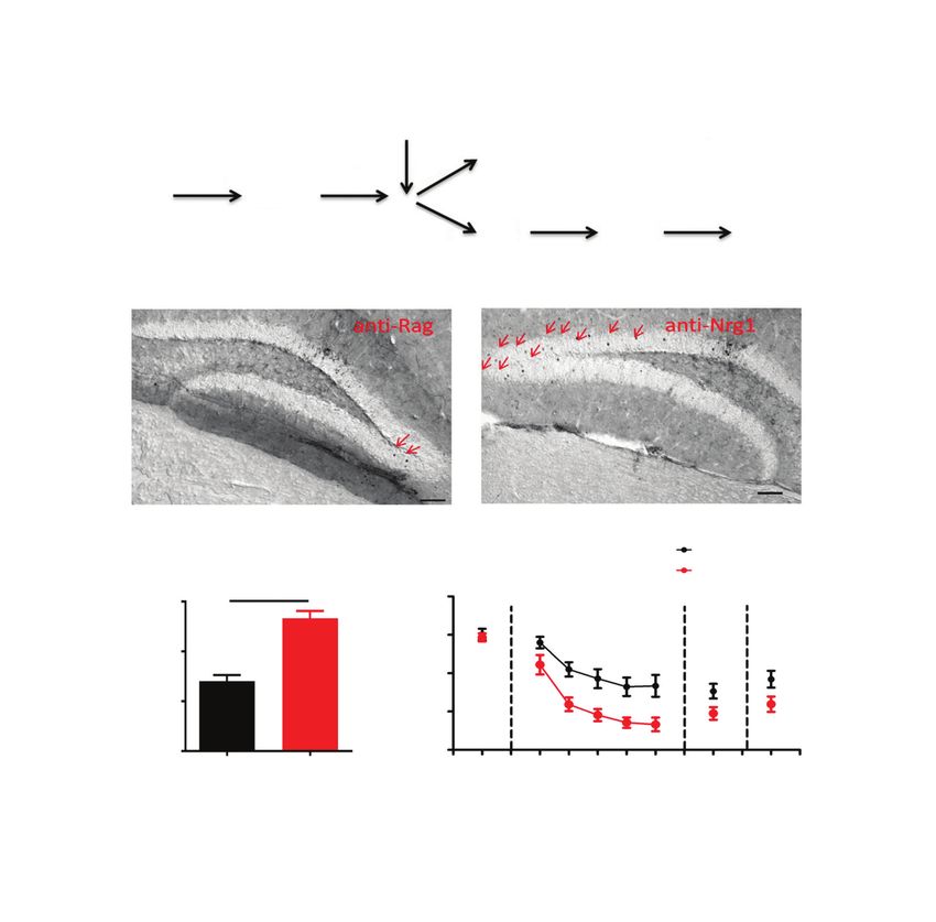
Elevated activity in the dorsal dentate gyrus reduces fear memory expression
1.03, p > 0.05), suggesting that fear memory is not altered by formed after fear conditioning. Freezing levels did not differ
anti-NRG1 antibodies: fear expression is altered instead. between groups injected with anti-RAG and anti-NRG1 anti-
Because anti-NRG1 antibodies also engage peripheral tar- bodies 24 hours after fear conditioning (recall test 1; Figure 1B,
gets to induce behavioural alterations that may have interfered t14 = 0.38, p > 0.05). In contrast, at the start of extinction training,
with our examination of fear responses,9 we used intraven freezing levels were significantly lower in the anti-NRG1
tricular injection to directly introduce antibodies to mouse group (extinction; Figure 1B, F1,56 = 7.49, p < 0.05; Appendix 1,
brains (Figure 1A). To avoid interference, injection was per- Figure S2A, F1,28 = 18.03, p < 0.001). This difference in freezing
A Intraventricular injection (anti-RAG/anti-NRG1)
24 h
Group 1: FC Rec-1
24 h 20 d 24 h 24 h 9d
Group 2: FC Rec-1 Ext Rec-2 Rec-3
7d 24 h 24 h
Group 3: SPS FC Rec-1
B Group 1 C
100 Rec-1 Ext Rec-2 Rec-3 40
75 30
Freezing (%)
*
Distance (m)
50
** 20
25 10
0 0
1 2 3 4 5 Anti-RAG Anti-NRG1
Anti-RAG
Anti-NRG1
D Group 2 E Group 3
100 Rec-1 Ext Rec-2 Rec-3 100 Rec-1 Ext Rec-2 Rec-3
** **
75
Freezing (%)
75
Freezing (%)
**
*
50 50
25 25
0 0
1 2 3 4 5 1 2 3 4 5
Fig. 1: Anti-NRG1 antibody injection led to reduced fear expression. (A) Experimental groups and procedures. (B) Injection of anti-NRG1 anti-
bodies led to lower freezing levels during fear extinction training (anti-NRG1, n = 8 mice; anti-RAG, n = 8 mice). The difference between
groups persisted at recall 24 hours after fear extinction training (Rec-2), but not 9 days later (Rec-3). (C) Injection of anti-NRG1 antibodies did
not affect locomotion in the open field test (anti-NRG1, n = 8 mice; anti-RAG, n = 9 mice). (D) For the remote fear memory group, injection of
anti-NRG1 antibodies led to reduced fear expression (anti-NRG1, n = 6 mice; anti-RAG, n = 6 mice). We observed reduced fear expression at
24 hours after fear extinction training (Rec-2), but not 9 days later (Rec-3). (E) In a single prolonged stress model of post-traumatic stress dis-
order (group 3), injection of anti-NRG1 antibodies led to reduced fear expression (anti-NRG1, n = 6 mice; anti-RAG, n = 6 mice). We observed
reduced fear expression at 24 hours after fear extinction training (Rec-2), but not 9 days later (Rec-3). Freezing score was the average of re-
sponses during 3 conditioned stimuli. *p < 0.05; **p < 0.01. Ext = fear extinction training; FC = fear conditioning; Rec-1 = recall 24 hours after
fear conditioning; Rec-2 = recall 24 hours after fear extinction training; Rec-3 = recall 9 days after Rec-2; SPS = single prolonged stress.
J Psychiatry Neurosci 2021;46(3) E393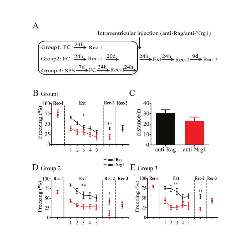
Zhang et al.
was absent when the same groups were tested 9 days later (re- ally no colocalization with these markers, indicating that c-Fos
call test 3; Figure 1B, t14 = 1.58, p > 0.05), mostly because of in- neurons were not G ABAergic neurons or astrocytes. We also
creased freezing levels in the anti-NRG1 group, suggesting that stained brain sections with anti-NeuN and anti-c-Fos simulta-
fear memory was unlikely to be altered, but that its expression neously (Figure 2I) and confirmed that c-Fos-positive cells were
was suppressed for a period after fear extinction training. To neurons (Figure 2J). The above results indicated that excitatory
address whether increased locomotion contributed to this neurons in the dDG were the main target for activation in mice
effect, we measured total distance travelled in an open field test injected with anti-NRG1 antibodies.
and found no significant difference between the anti-NRG1 and
anti-RAG groups (Figure 1C, t15 = 1.47, p > 0.05). Reduced fear expression with elevated neural activity in
It is well known that extinction of remote fear memory is the dDG
much more difficult to achieve.20,21 Thus, we tested whether
anti-NRG1 antibodies were more effective in suppressing fear If the dDG is a key brain region regulating expression of fear
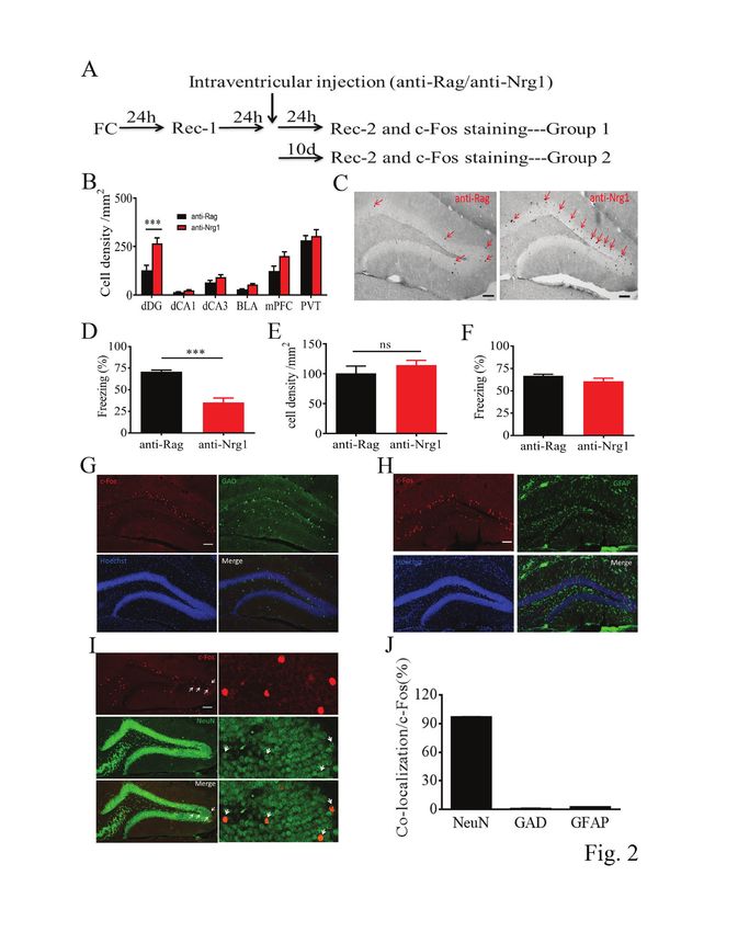
memory after extinction training (Figure 1A, group 2). Similar memory, we would expect that local injection of anti-NRG1 an-
to our findings for recent fear memory, we found reduced ex- tibodies into the dDG would have a similar effect. We injected
pression of fear memory at both the start of fear extinction anti-NRG1 antibodies into the dDG bilaterally after fear condi-
training (Appendix 1, Figure S2B, F1,20 = 64.67, p < 0.001) and tioning and the first recall test (Figure 3A; 1 µL/side). We first
24 hours later. Freezing levels were comparable to that of the quantified c-Fos-positive neurons in the dDG (Figure 3B) and
control group 9 days later (recall test 3; Figure 1D, t10 = 1.41, confirmed a significantly higher density of c-Fos-positive neur
p > 0.05). We also tested whether anti-NRG1 antibodies could ons in the dDG of mice injected with anti-NRG1 antibodies
affect expression of fear memory in a well-established model (Figure 3C, t20 = 6.38, p < 0.001). Expression of fear memory was
of PTSD (single prolonged stress).17 We found lower freezing lower compared to the anti-RAG group (extinction; Figure 3D,
levels and lower expression of fear memory in the anti-NRG1 F1,80 = 12.06, p < 0.01). Expression of fear memory was also sig-
group at the start of extinction training (Appendix 1, nificantly lower at 24 hours (recall test 2; Figure 3D, t20 = 2.82,
Figure S2C, F1,20 = 11. 35, p < 0.01) and 24 hours later (recall test p < 0.05) and 9 days later (recall test 3; Figure 3D, t20 = 2.21, p <
2; Figure 1E, t10 = 4.47, p < 0.01). However, we observed no dif- 0.05) after extinction training in the anti-NRG1 group. Closer
ference in freezing levels between the anti-NRG1 and anti- examination revealed that the difference between these
RAG groups 10 days after extinction training (recall test 3; 2 groups was due in particular to increased freezing levels in
Figure 1E, t10 = 0.30, p > 0.05). Taken together, the above re- the anti-RAG group, which was absent in the other groups
sults suggested that the intraventricular injection of anti- (e.g., Figure 1B, D and E). The effect on fear expression of direct
NRG1 antibodies were associated with lower expression of injection of anti-NRG1 antibodies into the dDG may have been
fear memory for a period of time after extinction training, but because of the density of activated neurons in the dDG. Hence,
fear memory itself appeared to be unaltered. local injection of anti-NRG1 antibodies into the dDG mimicked
intraventricular antibody injection in terms of expression of fear
Selective elevation in c-Fos expression in the dDG with memory and dDG activation.
injection of anti-NRG1 antibodies
Anti-NRG1 antibody effect blocked by DREADD inhibition
To understand which brain regions might contribute to lower and mimicked by DREADD excitation of dDG neurons
expression of fear memory in mice injected with anti-NRG1 an-
tibodies, we examined activated brain regions after fear recall If elevated activity in the dDG is required for lower expression
using c-Fos staining (Figure 2A). Mice were killed 24 hours of fear memory in mice injected with anti-NRG1 antibodies,
(group 1) or 10 days (group 2) after anti-NRG1/anti-RAG injec- then inhibiting neural activity in the dDG should eliminate the
tion. In group 1, we observed a significant increase in the den- antibody effect. To test this, we injected an inhibitory hM4D
sity of c-Fos-positive neurons in the dDG among the few key receptor-mCherry fusion virus (rAAV-CaMKIIα-hM4D-
regions that participate in fear expression and extinction mCherry) into the dDG of anti-NRG1 and anti-RAG mice, and
(Figure 2B and C, p < 0.001), and freezing levels were signifi- GFP reporter virus (rAAV-CaMKIIα-GFP) into the dDG of a
cantly lower in the anti-NRG1 group (Figure 2D, t15 = 7.95, control group (Figure 4A). Twenty-four hours after injection of
p < 0.001). In group 2, we found no difference between the anti- the anti-NRG1 antibodies, CNO was administered (i.p.,
NRG1 and anti-RAG groups in terms of c-Fos density 3 mg/kg) to inhibit dDG neurons, followed by extinction
(Figure 2E, t12 = 0.93, p > 0.05) or freezing levels (Figure 2F, t12 = training in 30 minutes. We found no difference in freezing lev-
1.59, p > 0.05). These results suggested that the density of acti- els or their rate of reduction in the anti-NRG1 and anti-RAG
vated dDG cells may be related to freezing levels. We also groups expressing the hM4D virus (Figure 4B; extinction F1,80 =
found that the density of activated dDG cells was significantly 3.08, p > 0.05; recall test 2 t20 = 0.43, p > 0.05). In mice injected
higher in mice injected with anti-NRG1 than in those injected with the GFP virus, the anti-NRG1 group showed lower freez-
with anti-RAG in a novel experiment, but no difference be- ing levels than the anti-RAG group (Figure 4C; extinction
tween the 2 groups in the home cage (Appendix 1, Figure S3). F1, 72 = 18.51, p < 0.001; recall test 2 t18 = 2.17, p < 0.05). These re-
To identify the cell types activated, we used c-Fos staining in sults demonstrated that hM4D-mediated inhibition of dDG
GAD 67-GFP mice (Figure 2G) or double staining of c-Fos with excitatory cells abolished the effect of anti-NRG1 antibody in-
GFAP (a marker of astrocytes; Figure 2H), but we found virtu- jection on the expression of fear memory.
E394 J Psychiatry Neurosci 2021;46(3)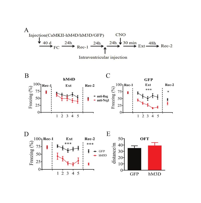
Elevated activity in the dorsal dentate gyrus reduces fear memory expression

                  A                                             Intraventricular injection (anti-RAG/anti-NRG1)

                                            24 h                      24 h         24 h
                      FC                                   Rec-1                                            Rec-2 and c-Fos staining, group 1
                                                                                   10 d
                                                                                                            Rec-2 and c-Fos staining, group 2

                  B                                                                                         C
                                           500                                                                                                                     Anti-RAG                                Anti-NRG1
                        Cell density/mm2

                                                               Anti-RAG
                                                    ***        Anti-NRG1

                                           250

                                                0
                                                    dDG dCA1 dCA3          BLA mPFC PVT

                  D                                                            E                                                                                      F
                                       100                                                            150              NS                                                                100
                                                                                   Cell density/mm2

                                                               ***
                      Freezing (%)

                                                                                                                                                                          Freezing (%)
                                           75                                                                                                                                            75
                                                                                                      100
                                           50                                                                                                                                            50
                                                                                                      50
                                           25                                                                                                                                            25

                                            0                                                          0                                                                                  0
                                                    Anti-RAG     Anti-NRG1                                  Anti-RAG       Anti-NRG1                                                           Anti-RAG   Anti-NRG1

                  G                                                                                                    H

                  I                                                                                                    J
                                                                                                                             Colocalization with c-Fos (%)

                                                                                                                                                             120

                                                                                                                                                             90

                                                                                                                                                             60

                                                                                                                                                             30

                                                                                                                                                              0
                                                                                                                                                                    NeuN                       GAD        GFAP

Fig. 2: Intraventricular injection of anti-NRG1 antibodies led to reduced fear expression and activated excitatory neurons in the dentate gyrus. (A) Ex-
perimental procedures. (B) Quantification of c-Fos-positive cells in a few key brain regions. We found a significant difference in the dDG (anti-NRG1,
n = 9 mice; anti-RAG, n = 8 mice). (C) Sample images of c-Fos immunostaining in mice injected with anti-RAG (left) or anti-NRG1 (right) antibodies.
Scale bar, 100 μm. (D) Injection of anti-NRG1 antibodies suppressed fear expression at 24 hours after injection (anti-NRG1, n = 9 mice; anti-RAG,
n = 8 mice). (E) Injection of anti-NRG1 antibodies had no effect on the density of c-Fos-positive neurons 10 days after injection (anti-NRG1, n = 7 mice;
anti-RAG, n = 7 mice). (F) Injection of anti-NRG1 antibodies had no effect on fear expression 10 days after injection (anti-NRG1, n = 7 mice; anti-RAG,
n = 7 mice). (G) Sample images for c-Fos and GAD67 staining. Scale 100 μm. (H) Sample images for c-Fos and GFAP staining. Scale 100 μm.
(I) Sample images for c-Fos and NeuN staining. Scale 100 μm. (J) Colocalization between c-Fos and NeuN, GAD67 or GFAP. ***p < 0.001. BLA = ba-
solateral amygdala; dCA1 = dorsal hippocampus subfield 1; dCA3 = dorsal hippocampus subfield 3; dDG = dorsal dentate gyrus; FC = fear condition-
ing; GAD67 = glutamic acid decarboxylase 67; GFAP = glial fibrillary acidic protein; mPFC = medial prefrontal cortex; NeuN = neuronal nuclei; NS = not
significant; PVT = paraventricular thalamus; Rec-1 = recall 24 hours after fear conditioning; Rec-2 = recall 10 days after fear extinction training.

                                                                               J Psychiatry Neurosci 2021;46(3)                                                                                                        E395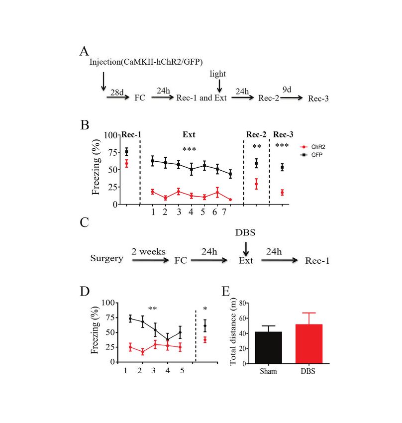
Zhang et al.
A dDG injection (anti-RAG/anti-NRG1)
h Rec-2 and killed
24 h 24 h 24
FC Rec-1
24 24 h 9d
h Ext Rec-2 Rec-3
B
Anti-RAG Anti-NRG1
Anti-RAG
C D Anti-NRG1
150 *** 100 Rec-1 Ext Rec-2 Rec-3
** * *
Cell density/mm2
75
Freezing (%)
100
50
50
25
0 0 0
Anti-RAG Anti-NRG1 1 2 3 4 5
Fig. 3: Injection of anti-NRG1 antibodies led to reduced fear expression and elevated c-Fos expression in the dentate gyrus. (A) Experimental pro
cedures. (B) Sample immunostaining images of c-Fos in mice injected with anti-RAG (left) or anti-NRG1 (right) antibodies. (C) Quantification of the
density of c-Fos-positive neurons in the dDG in mice injected with anti-NRG1 or anti-RAG antibodies (anti-NRG1, n = 12 mice; anti-RAG, n =
10 mice). (D) Injection of anti-NRG1 antibodies in the dDG led to lower fear expression without affecting initial freezing levels. Freezing levels were
significantly lower in the anti-NRG1 group 24 hours after extinction training (Rec-2) and 9 days later (Rec-3; anti-NRG1, n = 11 mice; anti-RAG, n =
11 mice). Freezing score was the average of responses during 3 conditioned stimuli. *p < 0.05; **p < 0.01; ***p < 0.001. dDG = dorsal dentate gyrus;
Ext = fear extinction training; FC = fear conditioning; Rec-1 = recall 24 hours after fear conditioning; Rec-2 = recall 24 hours after fear extinction train-
ing; Rec-3 = recall 9 days after Rec-2.
Next, we conducted a contrasting experiment by expressing terms of total distance travelled (Figure 4E, t14 = 0.66, p > 0.05).
excitatory hM3D receptor-mCherry fusion protein (rAAV- We found c-Fos-expressing cells in the dDG after CNO in-
CaMKIIα-hM3D-mCherry) into the dDG of wild-type mice, jection in the hM3D group, but not in the hM4D group (Ap-
and a GFP reporter virus (rAAV- CaMKIIα-GFP) into the pendix 1, Figure S4). Thus, hM3D-mediated activation of dDG
dDG of control mice (Figure 4A). The rationale was that en- excitatory cells suppressed expression of fear memory.
hancing dDG neuronal activity using hM3D should mimic the
effect of anti-NRG1 injection in wild-type mice. Compared to Suppressed expression of fear memory with stimulation of
the GFP group, we found significantly lower freezing levels in dDG neurons
the h3MD group (Figure 4D; extinction F1,68 = 30.19, p < 0.001;
recall test 2 t17 = 6.37, p < 0.001), resembling the effects in mice To further confirm the effect of activation of the dDG on
injected with anti-NRG1 antibodies. We found that CNO in- fear expression and fear extinction, mice injected with
jection did not alter locomotion in an open field test, and we adeno-associated viruses (rAAV-CaMKIIα-hChR2-eYFP or
found no difference between the hM3D and GFP groups in rAAV-CaMKIIα-GFP) underwent auditory fear conditioning
E396 J Psychiatry Neurosci 2021;46(3)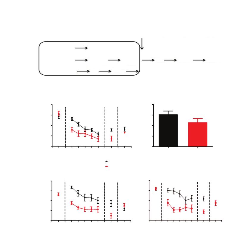
Elevated activity in the dorsal dentate gyrus reduces fear memory expression
A
Injection (CaMKIIα-hM4D, -hM3D or -GFP) CNO
40 d 24 h 24 h 24 h 30 min 48 h
FC Rec-1 Ext Rec-2
Intraventricular injection
B C
hM4D GFP
100 100
Rec-1 Ext Rec-2 Rec-1 Ext Rec-2
***
Freezing (%)
Freezing (%)
75 75
*
Anti-RAG
50 50
Anti-NRG1
25 25
0 0
1 2 3 4 5 1 2 3 4 5
D E
100 Rec-1 Ext Rec-2 50 Open field test
*** *** 40
75
Freezing (%)
Distance/m
GFP 30
50
hM3D
20
25
10
0 0
1 2 3 4 5
GFP hM3D
Fig. 4: DREADD modulation of neuronal activity in the dDG affected fear expression. (A) Experimental procedures. (B) Inhibition of dDG activ-
ity with an inhibitory CaMKIIα-hM4D-mCherry fusion protein abolished the reduction in fear expression induced by anti-NRG1. Freezing levels
were not significantly different between the 2 groups at 24 hours after fear extinction training (anti-NRG1, n = 10 mice; anti-RAG, n = 12 mice).
(C) Expression of CaMKIIα-GFP in the dDG had no effect on the reduction of fear expression induced by anti-NRG1. Freezing levels were sig-
nificantly lower in the anti-NRG1 group than in the anti-RAG group (anti-NRG1, n = 9 mice; anti-RAG, n = 11 mice). (D) Excitation of the dDG
led to reduced fear expression. We also observed reduced fear expression 48 hours after fear extinction training (hM3D, n = 8 mice; GFP,
n = 11 mice); ***p < 0.001. (E) Increased neuronal activity in the dDG with an excitatory CaMKIIα-hM3D-mCherry fusion protein did not affect
locomotion in the open field test (hM3D, n = 8 mice; GFP, n = 8 mice). Freezing score was the average of responses during 3 conditioned
stimuli. *p < 0.05; ***p < 0.001. CaMKIIα-GFP = calcium/calmodulin-dependent protein kinase II α green fluorescent protein; CaMKIIα-hM3D =
calcium/calmodulin-dependent protein kinase II α human muscarinic-3 receptor DREADD-mutation; CaMKIIα-hM4D = calcium/calmodulin-
dependent protein kinase II α human muscarinic-4 receptor DREADD-mutation; dDG = dorsal dentate gyrus; DREADD = designer receptor
exclusively activated by designer drugs; Ext = fear extinction training; FC = fear conditioning; Rec-1 = recall 24 hours after fear conditioning;
Rec-2 = recall 48 hours after fear extinction training.
(Figure 5A). After fear conditioning, the 2 groups of mice 10 days after extinction training in the hChR2 group
had similar freezing levels during recall test 1. However, (Figure 5B; extinction F1,90 = 57.85, p < 0.001; recall test 2 t15 =
when the mice were exposed to the conditioned stimulus 3.05, p < 0.01; recall test 3 t15 = 5.57, p < 0.001), resembling
with blue light delivered to the bilateral dDG using optical the effect of hM3D-mediated activation of dDG excitatory
fibres, freezing levels in the hChR2 group were significantly cells. We also tested the effect of optostimulation in dDG
lower than levels in the GFP group. As well, expression of and found numerous c-Fos-expressing cells in the dDG after
fear memory was significantly lower at 24 hours and blue light stimulation (Appendix 1, Fig. S4).
J Psychiatry Neurosci 2021;46(3) E397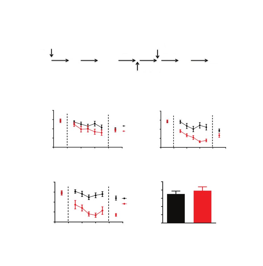
Zhang et al.
A
Injection (CaMKIIα-hChR2 or -GFP)
Light
28 d 24 h 24 h 9d
FC Rec-1 and Ext Rec-2 Rec-3
B
100 Rec-1 Ext Rec-2 Rec-3
75 *** ** *** hChR2
Freezing (%)
GFP
50
25
0
1 2 3 4 5 6 7
C
DBS
2w 24 h 24 h
Surgery FC Ext Rec-1
D E
100 80
** *
Total distance (m)
Freezing (%)
75 60
50 40
25 20
0 0
1 2 3 4 5
Sham DBS
Fig. 5: Stimulation of dDG neurons resulted in reduced fear expression. (A) Experimental procedures. (B) Optogenetic activation of dDG neur
ons led to reduced fear expression. We also observed reduced fear expression 24 hours after extinction training (hChR2, n = 8 mice; GFP, n =
9 mice). (C) Experimental procedures. (D) Fear expression was suppressed in the DBS group during both fear extinction training and recall
test 1 (n = 9 mice; sham, n = 6 mice). (E) DBS in the dDG did not affect locomotion measured in the open field test (unpaired t test, t8 = 0.35,
p > 0.05; DBS, n = 9 mice; sham, n = 8 mice). *p < 0.05; **p < 0.01; ***p < 0.001. Freezing score was the average of responses during 2 con-
ditioned stimuli in B and during 3 conditioned stimuli in D. CaMKIIα-GFP = CaMKIIα-GFP = calcium/calmodulin-dependent protein kinase II α
green fluorescent protein; CaMKIIα-hChR2 = calcium/calmodulin-dependent protein kinase II α H134R mutation in channelrhodopsin-2;
DBS = deep brain stimulation; dDG = dorsal dentate gyrus; Ext = fear extinction training; FC = fear conditioning; Rec-1 = recall 24 hours after
fear conditioning; Rec-2 = recall 48 hours after fear extinction training; Rec-3 = recall 9 days after Rec-2.
We also conducted a DBS experiment to modulate the ac- ure S4). Taken together, the above results showed an effective
tivity of the dDG (Figure 5C). Two weeks after surgery, mice reduction in fear expression using DBS in the dDG.
were fear-conditioned and tested. Freezing levels were sig
nificantly lower during both extinction and recall in the DBS Discussion
group than in the sham group (Figure 5D; F1,52 = 10.46, p <
0.01; recall test 1 t13 = 2.39, p < 0.05), but locomotion in the open In this study we examined the contribution of the dDG to
field test was not different between the 2 groups (Figure 5E; fear expression. We found the following: intraventricular in-
t15 = 0.56, p > 0.05). After DBS in the dDG, we also found jection of anti-NRG1 antibodies before fear extinction was as-
abundant c-Fos-expressing cells in the dDG (Appendix 1, Fig- sociated with lower freezing levels in fear-conditioned naive
E398 J Psychiatry Neurosci 2021;46(3)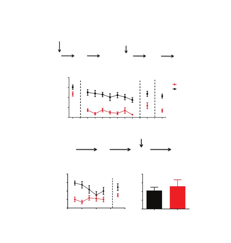
Elevated activity in the dorsal dentate gyrus reduces fear memory expression
mice, PTSD model mice, and testing of remote fear memory; The dDG also receives inputs from the locus coeruleus and
intraventricular injection of anti-NRG1 antibodies selectively diffuse projections from the ventral tegmental area, which
activated excitatory neurons in the dDG; local injection of participate in regulating novelty detection.34–36 It has been
anti-NRG1 antibodies in the dDG activated dDG neurons demonstrated that the dorsal hippocampus also participates
and reduced fear expression; hM4D-mediated inhibition of in novelty-related exploration.37–40 Moncada and colleagues39
the dDG blocked anti-NRG1 effects on fear expression; found that previous exposure to a novel environment pro-
hM3D or optogenetic activation of the dDG suppressed fear moted long-term memory formation. The dDG also plays a
expression; and DBS in the dDG reduced fear expression. role in novelty detection,41 and activating parvalbumin neur
These findings support the idea that elevated activity in the ons in the dDG appears to impair social interaction induced
dDG suppress fear expression. by novelty.23 Some studies have shown that novelty exposure
The major finding of the current study is that activation of before fear extinction training facilitated and strengthened
the dDG can reduce the expression of auditory fear memory. fear extinction.42–45 Rats exposed to a novel environment for
This finding was consistent with those of our previous work: 5 minutes before fear extinction training showed enhanced
that Dnmt3a overexpression in the dDG prevents fear re- fear extinction, which was fully blocked by anisomycin in the
newal and elevates the activity of dDG, and that knockdown dorsal CA1.46 Whether elevated activation of the dDG by
of Dnmt3a in the dDG promotes fear renewal.14 Overexpres- novelty exploration can modulate the expression of fear
sion of Dnmt3a in the dDG resulted in higher density of c-Fos memory requires further investigation.
neurons in the dDG.14 This result means that the activity of Fear memory did not appear to be altered by anti-NRG1
the dDG is associated with expression levels of Dnmt3a in the antibody injection. The effect of dDG activation may thus be
dDG. Mice with Dnmt3a overexpression in the dDG showed limited to modulating the expression of conditioned fear. If
no significant fear renewal, supporting our finding in the this is the case, one advantage of targeting the underlying
present study about a correlation between fear expression mechanism or system is to suppress or modulate fear ex-
and the activity of the dDG. pression while preserving the fear memory. To explore the
The exact contribution of the dorsal hippocampus to audi- possibility of manipulating the dDG dynamically with meth-
tory fear conditioning is poorly understood, especially with ods transferable to human treatment, we tested DBS. We
respect to the contribution of the dDG. Although some stud- found that freezing levels in mice that received DBS in the
ies have suggested that modulating the activity of the dorsal dDG during fear extinction were significantly lower than in
hippocampus did not affect auditory fear conditioning,11,22,23 mice from the sham group; DBS may help to suppress fear
Quinn and colleagues24 and Bast and colleagues25 both found expression during fear extinction training. Although DBS
that freezing levels during tone presentation were signifi- has been used routinely to treat depression47 and obsessive–
cantly reduced after neurotoxic lesion of the dorsal hippo- compulsive disorder48 and preclinical DBS studies in fear
campus. Activating dDG neurons that were tagged during conditioning animal models have suggested its potential use
auditory fear conditioning was sufficient to trigger expres- for treatment-resistant PTSD,49–51 very few studies have ex-
sion of that memory.26 Before fear extinction training, inacti- plored the clinical effect of DBS in PTSD.52–54 About 20% to
vation of the dorsal hippocampus reduced the rate of extinc- 30% of PTSD patients do not respond to medications and
tion. 27 Thus, the dorsal hippocampus is implicated in conventional psychotherapy. 49,51 A recent clinical trial
auditory fear conditioning and extinction. Our finding that showed that 6 patients with treatment-resistant PTSD ex
dDG activation suppressed fear expression during memory perienced a 30% decrease in clinician-administered PTSD
recall provides the most direct evidence for the contribution scale scores after bilateral basolateral amygdala high-
of the dDG to cued expression of fear memory. frequency stimulation.52,54 Our results supported DBS as a
The dDG granule cells receive their major inputs from the potential therapeutic tool for refractory PTSD.
entorhinal cortex (EC) and then project to the hippocampal
subfield 3 (CA3). In turn, the CA3 pyramidal cells project to Limitations
the hippocampal subfield 1 (CA1), which projects back to the
EC, creating a synaptic loop (EC–DG–CA3–CA1–EC). The We found that elevating dDG activity suppressed the expres-
CA3 region also projects to the lateral septum,28 which is acti- sion of fear memory, but we did not investigate the neural
vated after auditory fear conditioning,29 and pretraining inacti- circuits underlying this phenomenon. We propose that this
vation of the lateral septum abolishes learned fear response.30 suppression occurs via CA3 projection to the lateral septum
The lateral septum has strong bidirectional interaction with the and then the amygdala. We also did not address whether fear
amygdala and hypothalamus, and both structures play an im- memory was still intact with dDG activation.
portant role in auditory fear learning.29,31,32 Taking these find-
ings together, it is possible that the dDG suppresses fear re- Conclusion
sponse by directly regulating the expression of fear memory,
via CA3 projection to the lateral septum and subsequently to We have provided evidence that the dDG may modulate
the amygdala. Electrical stimulation of the lateral septum had auditory fear conditioning, in that dDG activation sup-
a predominantly inhibitory effect on neurons in the amyg- pressed the expression of conditioned fear. Because reduced
dala,33 but whether this effect is mediated or can be mimicked fear expression is seen after DBS in the dDG, we suggest
by CA3 inputs to the lateral septum is unknown. that modulating dDG activity may have therapeutic potential
J Psychiatry Neurosci 2021;46(3) E399Zhang et al.
in treating diseases that involve aberrant emotional responses, 15. Chen L, McKenna JT, Leonard MZ et al. GAD67-GFP knock-in
mice have normal sleep-wake patterns and sleep homeostasis.
such as PTSD. Neuroreport 2010;21:216-20.
16. Sternson SM, Roth BL. Chemogenetic tools to interrogate brain
Acknowledgements: We thank T. Li for the virus injection and functions. Annu Rev Neurosci 2014;37387-407.
Dr. Z. Gong for suggestions on immunohistochemistry methods. This 17. Yan R, Wang T, Zhou Q. Elevated dopamine signaling from ven-
work is supported by grants (2019SHIBS0004, SZBL2019062801003, tral tegmental area to prefrontal cortical parvalbumin neurons
KQTD2015032709315529, SZSM201812005). drives conditioned inhibition. Proc Natl Acad Sci U S A 2019;
116:13077-86.
Affiliations: From the Peking University, Shenzhen Graduate 18. Milad MR, Quirk GJ. Neurons in medial prefrontal cortex signal
School, School of Chemical Biology and Biotechnology, State Key memory for fear extinction. Nature 2002;420:70-4.
Laboratory of Chemical Oncogenomics, Key Laboratory of Chemical 19. Zhang Y Ouyang K. Lipina TV, et al. Conditioned stimulus pre-
Genomics, Shenzhen 518055, Peoples R China (Zhang, Wang, Zhou); sentations alter anxiety level in fear-conditioned mice. Mol Brain
the Precision Medicine Centre, the Seventh Affiliated Hospital, Sun 2019;12:28.
Yat-sen University, Shenzhen, 518107, Guangdong, China (Ju); and 20. Graff J, Joseph NF, Horn ME et al. Epigenetic priming of memory
the Pediatric Neurology, Shenzhen Children’s Hospital, Shenzhen, updating during reconsolidation to attenuate remote fear mem
518038, China (Zhang, Liao). ories. Cell 2014;156:261-76.
21. Tsai LH, Graff J. On the resilience of remote traumatic memories
Competing interests: None declared. against exposure therapy-mediated attenuation. EMBO Rep
2014;15:853-61.
Contributors: Y. Zhang and Q. Zhou designed the study. Y. Zhang, 22. Cho HY, Kim M, Han JH. Specific disruption of contextual mem-
Z. Wang, J. Ju and J. Liao acquired the data, which Y. Zhang, ory recall by sparse additional activity in the dentate gyrus.
Z. Wang, J. Ju and Q. Zhou analyzed. Y. Zhang and Q. Zhou Neurobiol Learn Mem 2017;145:190-8.
wrote the article, which all authors reviewed. All authors ap- 23. Zou D, Chen L, Deng D et al. DREADD in parvalbumin interneur
proved the final version to be published and can certify that no ons of the dentate gyrus modulates anxiety, social interaction and
other individuals not listed as authors have made substantial con- memory extinction. Curr Mol Med 2016;16:91-102.
tributions to the paper. 24. Quinn JJ, Wied HM, Ma QD et al. Dorsal hippocampus involve-
ment in delay fear conditioning depends upon the strength of the
Content licence: This is an Open Access article distributed in ac- tone-footshock association. Hippocampus 2008;18:640-54.
cordance with the terms of the Creative Commons Attribution 25. Bast T, Zhang WN, Feldon J. Dorsal hippocampus and classical
(CC BY-NC-ND 4.0) licence, which permits use, distribution and fear conditioning to tone and context in rats: effects of local
reproduction in any medium, provided that the original publica- NMDA-receptor blockade and stimulation. Hippocampus 2003;
tion is properly cited, the use is noncommercial (i.e., research or 13:657-75.
educational use), and no modifications or adaptations are made. 26. Liu X, Ramirez S, Pang PT et al. Optogenetic stimulation of a hip-
See: https://creativecommons.org/licenses/by-nc-nd/4.0/ pocampal engram activates fear memory recall. Nature 2012;
484:381-5.
27. Corcoran KA, Desmond TJ, Frey KA et al. Hippocampal inactiva-
References tion disrupts the acquisition and contextual encoding of fear ex-
1. Morrison FG, Ressler KJ. From the neurobiology of extinction to tinction. J Neurosci 2005;25:8978-87.
improved clinical treatments. Depress Anxiety 2014;31:279-90. 28. Fanselow MS, Dong HW. Are the dorsal and ventral hippocampus
2. Hofmann SG. Cognitive processes during fear acquisition and ex- functionally distinct structures? Neuron 2010;65:7-19.
tinction in animals and humans: implications for exposure therapy 29. Butler CW, Wilson YM, Gunnersen JM et al. Tracking the fear
of anxiety disorders. Clin Psychol Rev 2008;28:199-210. memory engram: discrete populations of neurons within amyg-
3. Hofmann SG, Smits JAJ, Asnaani A et al. Cognitive enhancers for dala, hypothalamus, and lateral septum are specifically activated
anxiety disorders. Pharmacol Biochem Behav 2011;99:275-84. by auditory fear conditioning. Learn Mem 2015;22:370-84.
4. Mei L, Xiong WC. Neuregulin 1 in neural development, synaptic 30. Calandreau L, Jaffard R, Desmedt A. Dissociated roles for the lat-
plasticity and schizophrenia. Nat Rev Neurosci 2008;9:437-52. eral and medial septum in elemental and contextual fear condi-
5. Huang YZ, Won S, Ali DW et al. Regulation of neuregulin signal- tioning. Learn Mem 2007;14:422-9.
ing by PSD-95 interacting with ErbB4 at CNS synapses. Neuron 31. Sheehan TP, Chambers RA, Russell DS. Regulation of affect by the
2000;26:443-55. lateral septum: implications for neuropsychiatry. Brain Res Brain
6. Lu Y, Sun XD, Hou FQ et al. Maintenance of GABAergic activity Res Rev 2004;46:71-117.
by neuregulin 1-ErbB4 in amygdala for fear memory. Neuron 32. Deng K, Yang L, Xie J et al. Whole-brain mapping of projection
2014;84:835-46. from mouse lateral septal nucleus. Biol Open 2019;8;bio043554.
7. Chen YH, Chen YH, Lan YJ et al. ErbB4 signaling in the prelimbic 33. Thomas E, Dewolfe M, Sancar F et al. Electrophysiological analysis
cortex regulates fear expression. Transl Psychiatry 2017;7:e1168. of the interaction between the lateral septum and the central nu-
8. Ju J, Liu L, Zhang Y et al. Effect of age onset on schizophrenia-like cleus of the amygdala. Neurosci Lett 2012;524:79-83.
phenotypes and underlying mechanisms in model mice. Prog 34. Takeuchi T, Duszkiewicz AJ, Sonneborn A, et al. Locus coeruleus
Neuropsychopharmacol Biol Psychiatry 2019;89:465-74. and dopaminergic consolidation of everyday memory. Nature
9. Dominguez SL, Hegde GV, Hanson JE et al. Antibody-mediated 2016;537:357-62.
stabilization of NRG1 induces behavioral and electrophysiological 35. Hansen N, Manahan-Vaughan D. Locus coeruleus stimulation fa-
alterations in adult mice. Sci Rep 2018;8:8239. cilitates long-term depression in the dentate gyrus that requires ac-
10. Pierson JL, Pullins SE, Quinn JJ. Dorsal hippocampus infusions of tivation of beta-adrenergic receptors. Cereb Cortex 2015;25:1889-96.
CNQX into the dentate gyrus disrupt expression of trace fear con- 36. Kempadoo KA, Mosharov EV, Choi SJ et al. Dopamine release
ditioning. Hippocampus 2015;25:779-85. from the locus coeruleus to the dorsal hippocampus promotes
11. Kheirbek MA, Drew LJ, Burghardt NS, et al. Differential control of spatial learning and memory. Proc Natl Acad Sci U S A 2016;113:
learning and anxiety along the dorsoventral axis of the dentate 14835-40.
gyrus. Neuron 2013;77:955-68. 37. Mendez M, Arias N, Uceda S et al. c-Fos expression correlates with
12. Bernier BE, Lacagnina AF, Ayoub A et al. Dentate gyrus contrib- performance on novel object and novel place recognition tests.
utes to retrieval as well as encoding: evidence from context fear Brain Res Bull 2015;117:16-23.
conditioning, recall, and extinction. J Neurosci 2017;37:6359-71. 38. Arias N, Mendez M, Arias JL. The recognition of a novel-object in
13. Denny CA, Kheirbek MA, Alba EL et al. Hippocampal memory a novel context leads to hippocampal and parahippocampal c-Fos
traces are differentially modulated by experience, time, and adult involvement. Behav Brain Res 2015;292:44-9.
neurogenesis. Neuron 2014;83:189-201. 39. Moncada D, Viola H. Induction of long-term memory by exposure
14. Gong Z, Zhou Q. Dnmt3a in the dorsal dentate gyrus is a key to novelty requires protein synthesis: evidence for a behavioral
regulator of fear renewal. Sci Rep 2018;8:5093. tagging. J Neurosci 2007;27:7476-81.
E400 J Psychiatry Neurosci 2021;46(3)Elevated activity in the dorsal dentate gyrus reduces fear memory expression
40. Lee I, Hunsaker MR, Kesner RP. The role of hippocampal subre- 48. Holland MT, Trapp NT, McCormick LM et al. Deep brain stimu-
gions in detecting spatial novelty. Behav Neurosci 2005;119:145-53. lation for obsessive-compulsive disorder: a long term naturalistic
41. Morellini F, Sivukhina E, Stoenica L et al. Improved reversal learn- follow up study in a single institution. Front Psychiatry 2020;
ing and working memory and enhanced reactivity to novelty in 11:55.
mice with enhanced GABAergic innervation in the dentate gyrus. 49. Reznikov R, Binko M, Nobrega JN et al. Deep brain stimulation in
Cereb Cortex 2010;20:2712-27. animal models of fear, anxiety, and posttraumatic stress disorder.
42. Dunsmoor JE, Campese VD, Ceceli AO et al. Novelty-facilitated Neuropsychopharmacology 2016;41:2810-7.
extinction: providing a novel outcome in place of an expected 50. Hamani C, Temel Y. Deep brain stimulation for psychiatric dis-
threat diminishes recovery of defensive responses. Biol Psychiatry ease: contributions and validity of animal models. Sci Transl Med
2015;78:203-9. 2012;4:142rv8.
43. Menezes J, Alves N, Borges S et al. Facilitation of fear extinction by 51. Reznikov R, Hamani C. Posttraumatic stress disorder: perspectives
novelty depends on dopamine acting on D1-subtype dopamine re- for the use of deep brain stimulation. Neuromodulation 2017;20:7-14.
ceptors in hippocampus. Proc Natl Acad Sci U S A 2015;112:E1652-8. 52. Koek RJ, Langevin JP, Krahl SE, et al. Deep brain stimulation of
44. de Carvalho Myskiw J, Furini CRG, Benetti F et al. Hippocampal the basolateral amygdala for treatment-refractory combat post-
molecular mechanisms involved in the enhancement of fear extinc- traumatic stress disorder (PTSD): study protocol for a pilot ran-
tion caused by exposure to novelty. Proc Natl Acad Sci U S A 2014; domized controlled trial with blinded, staggered onset of stimula-
111:4572-7. tion. Trials 2014;15:356.
45. Lucas K, Luck CC, Lipp OV. Novelty-facilitated extinction and the 53. Gouveia FV, Davidson B, Meng Y et al. Treating post-traumatic
reinstatement of conditional human fear. Behav Res Ther 2018;109: stress disorder with neuromodulation therapies: transcranial mag-
68-74. netic stimulation, transcranial direct current stimulation, and deep
46. de Carvalho Myskiw J, Benetti F, Izquierdo I. Behavioral tagging brain stimulation. Neurotherapeutics 2020.
of extinction learning. Proc Natl Acad Sci U S A 2013;110:1071-6. 54. Langevin JP, Koek RJ, Schwartz HN et al. Deep brain stimulation
47. Mayberg HS, Lozano AM, Voon V et al. Deep brain stimulation for of the basolateral amygdala for treatment-refractory posttraumatic
treatment-resistant depression. Neuron 2005;45:651-60. stress disorder. Biol Psychiatry 2016;79:e82-4.
J Psychiatry Neurosci 2021;46(3) E401You can also read