Potential hazardous effects of printing room PM2.5 exposure include promotion of lung inflammation and subsequent injury
←
→
Page content transcription
If your browser does not render page correctly, please read the page content below
Molecular Medicine REPORTS 22: 3213-3224, 2020

     Potential hazardous effects of printing room PM2.5 exposure
   include promotion of lung inflammation and subsequent injury
     CHANGWEI ZOU1, HONG YANG1, LANYUE CUI2, XINYI CAO2, HONG HUANG1 and TINGTAO CHEN3

                      1
                      School of Resources Environmental and Chemical Engineering, Key Laboratory of
    Poyang Lake Environment and Resource Utilization, Ministry of Education, 2Nanchang University Queen Mary School;
 3
   National Engineering Research Center for Bioengineering Drugs and The Technologies, Institute of Translational Medicine,
                                Nanchang University, Nanchang, Jiangxi 330031, P.R. China

                                        Received March 8, 2020; Accepted June 26, 2020

                                                  DOI: 10.3892/mmr.2020.11399

Abstract. There have been few studies investigating the poten-      exposure is potential threat to individuals who spend a signifi-
tial effects of indoor sources of particulate matter on human       cant amount of time in printing rooms.
health. In this study, the effect of different concentrations of
fine particulate matter (PM2.5) collected from a printing room      Introduction
on lung health was examined using cultured cells and a mouse
model. Further, the mechanism of lung injury was exam-              Previous epidemiological studies have shown that air pollution
ined. The results indicated that PM2.5 significantly enhanced       is associated with increased morbidity and mortality world-
malondialdehyde activity (P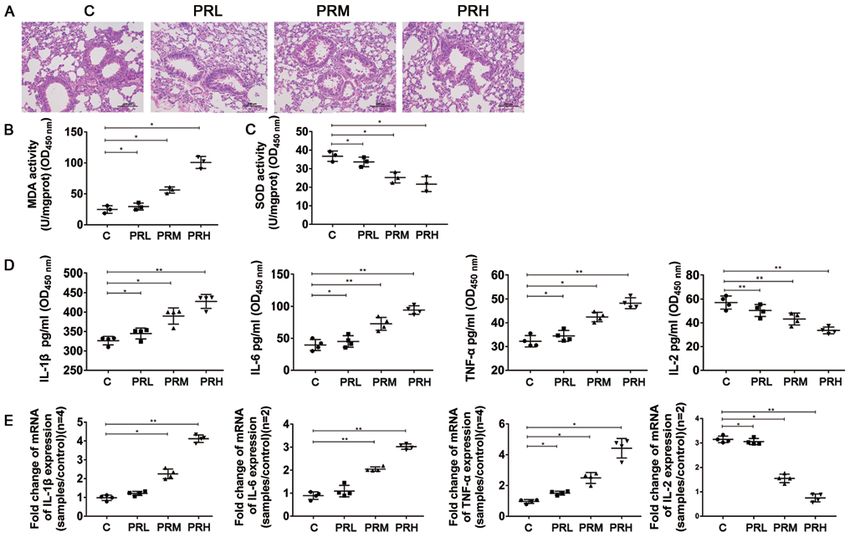
3214                      ZOU et al: POTENTIAL HAZARDOUS EFFECTS OF PM2.5 FROM THE PRINTING ROOM

     PM2.5, which are atmospheric PM with a diameter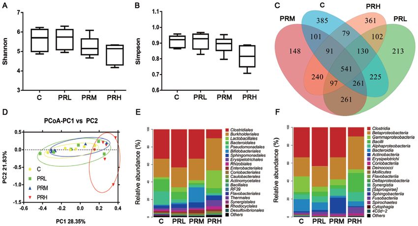
Molecular Medicine REPORTS 22: 3213-3224, 2020 3215
(p)‑ERK (1:1,000; cat. no. AF1015; Affinity Biosciences), cyclo- TCTACTG GTGT‑3', reverse primer 5'‑GTCATCATAC TT
oxygenase‑2 (COX2; 1:1,000; cat. no. 12375‑1‑AP; ProteinTech GGCAGGTT‑3') were analyzed using the 2‑ΔΔCq method (24).
Group, Inc.), NF‑κ B (p65; 1:2,000; cat. no. 10745‑1‑AP;
ProteinTech Group, Inc.) and p‑NF‑ κ B (p‑p65; 1:1,000; Hematoxylin and eosin (HE) staining and immunohistochem‑
cat. no. bs0982R; BIOSS). istry (IHC). Samples containing tissues from each group were
fixed in 10% buffered formalin for 24 h at 4˚C, embedded
Animals and treatments. In total, 48 male C57BL/6 mice (age, into paraffin and sliced into 4‑5 µm‑thick sections, The wax
8 weeks; weight, 25‑30 g) were purchased from Hunan SJA sheets were smoothed on polylysine‑treated slides in the
Laboratory Animal Co., Ltd. (http://hnslk.mfqyw.com/) and spreading machine tank (45˚C). The surrounding water was
housed in cages in the Institute of Translational Medicine of drained with absorbent paper and tissues were place in an oven
Nanchang University using standard conditions (humidity at 60˚C for 4 h. Subsequently, H&E staining was performed
51±13%, temperature 23±3˚C, 12/12‑h light‑dark cycle). Each (hematoxylin staining for 5‑10 min; 0.6% ammonia reflux
mouse was distinguished via ear piercing (a loophole in the blue; eosin staining for 3‑5 min; treatment with 70% ethanol
front, middle and rear of the margin of the left ear represented and 80% ethanol for 10‑20 sec, 95% ethanol for 3‑5 min,
numbers 1, 2 and 3, respectively, and a loophole in the front, 100% ethanol for 3‑5 min, and transparent xylene for 3‑5 min.
middle and rear of the margin of the right ear represented All the operations were performed at room temperature) and
numbers 4, 5 and 6, respectively). Subsequently, mice were tissues were observed using a light microscope (magnifica-
randomly divided into four groups as follows: The first group, tion, x200; Olympus Corporation) to compare the pathological
mice treated with PBS (C group, n=12); The second group, features of samples. IHC was performed using caspase‑3
mice given a 5 µg/g PM 2.5 suspension on days 1 and 3 via (1:250; cat. no. 19677‑1‑AP; ProteinTech Group, Inc.),
tracheal perfusion (PRL group, n=12); The third group, mice caspase‑8 (1:250; cat. no. 13426‑1‑P; ProteinTech Group, Inc.),
given a 10 µg/g PM2.5 suspension on days 1 and 3 via tracheal Bax (1:2,500; cat. no. 50599‑2‑lg; ProteinTech Group, Inc.)
perfusion (PRM group, n=12) and the fourth group, mice given and Bcl‑2 (1:1,000; cat. no. 12789‑1‑AP; ProteinTech Group,
a 15 µg/g PM2.5 suspension on days 1 and 3 via tracheal perfu- Inc.) antibodies (incubated at 4˚C overnight; reheated at room
sion (PRH group, n=12). On day 4, 100 µl serum was collected temperature for 45 min), and secondary antibody (1:1,000;
via tail vein blood collection after mice were anesthetized with Servicebio, Inc.; cat. no. GB24303; 37˚C, 1 h) antibodies.
1% pentobarbital sodium solution (40 mg/kg), then mice were
sacrificed and the lung tissue were collected and stored at ‑80˚C High‑throughput sequencing analyses. Lung tissues from
for use in subsequent experiments. The concentrations of PM2.5 groups C (n=6), PRL (n=6), PRM (n=6) and PRH (n=6)
used in the present study were determined via referencing were collected, and total genomic DNA was extracted from
previous studies (21,22) and the present pre‑experimental samples using a genomic DNA extraction kit (cat. no. 69506;
results (date not shown). The present study was approved by Qiagen GmbH) and a bead beating method. Subsequently,
the Ethical Committee of the Second Affiliated Hospital of 515F (5'‑GTGC CAG CMG CCG CGG TAA‑3') and 806R
Nanchang University, and all experiments were performed (5'‑GGAC TAC VSG GGTATC TAAT‑3') primers were
in accordance with approved guidelines (The Guide for Care used to amplify the V4 region of the 16S ribosomal DNA
and Use of Laboratory Animals; National Institutes of Health gene for high‑throughput sequencing, and PCR products
publication no. 85‑23) (23). were sequenced using an IlluminaHiSeq 2000 platform
(Illumina, Inc.; sequencing read archive (SRA) accession
Reverse transcription‑quantitative PCR (RT‑qPCR). Total no. SRP217605]. The extraction quality of DNA was deter-
RNA was extracted from lung tissue using TRIzol® reagent mined using 0.8% agar‑gel electrophoresis, and the DNA
(Invitrogen; Thermo Fisher Scientific, Inc.), and RNA purity was quantified using Uv‑spectrophotometer (Thermo Fisher
was determined using a NanoDrop™ 2000 spectrophotometer Scientific, Inc.). The Quant‑iT PicoGreen dsDNA Assay kit
(Thermo Fisher Scientific, Inc.). cDNA was reversed tran- (cat. no. P11496; Thermo Fisher Scientific, Inc.) was used
scribed from RNA (cat. no. 4374967; Applied Biosystems; to quantify DNA in the Promega QuantiFluor fluorescence
Thermo Fisher Scientific, Inc.) and qPCR was performed using quantitative system. The cut adapt and UCHIME algorithm,
a 7900HT Fast Real‑Time PCR system (Applied Biosystems; from the UPARSE software package (UPARSE; http://drive5.
Thermo Fisher Scientific, Inc.) and 2X SYBRGreen master com/uparse/ and Supplementary Software; version 7.0.100),
mix (Bio‑Rad Laboratories, Inc.). Samples with appropriate Qiime software (Quantitative insights Into Microbial Ecology;
primers were incubated at 95˚C for 30 sec, and 40 cycles of versions 1.8.0 and 1.9.1; http://qiime.org/index.html; used to
60˚C for 30 sec, and 72˚C for 30 sec were performed after compare the richness and diversity indexes, such as Shannon
a 1 min incubation step at 95˚C to activate the reaction. index and Simpson index), and SIMCA‑P software (Umetrics;
Expression levels of IL‑6 (forward primer 5'‑GAAATCGTG version 11.5) were used to evaluate α (intra‑sample) and β
GAAvATGAG‑3', reverse primer 5'‑GCTTAGGCATAACGC (inter‑sample) diversity.
ACT‑3'), TNF‑α (forward primer 5'‑GTGGAACTGGCAGAA The Kyoto Encyclopedia of Genes and Genomes (KEGG)
GAGG CA‑3', reverse primer 5'‑AGAG GGAGGC CAT TT database (http://www.genome.jp/kegg/pathway.html) was used
GGGA AC‑3'), IL‑1β (forward primer 5'‑GTGTCTT TCCCG to predict the correlation between microbiota changes with
TGGACCTTC‑3', reverse primer 5'‑TCATCTCGGAGCCTG environmental adaptation, immune responses and energy
TAGTGC‑3'), IL‑2 (forward primer 5'‑GCCA AGAGCTGA metabolism (25). The Operational taxonomic units (OTUs) and
CCAACTTC‑3', reverse primer 5'‑ATCG CCCACACTA AG species classification were analyzed based on effective data
AGCAT‑3') and GAPDH (forward primer 5'‑CTCGTGGAG (excluded errors and questionable data), and then the abundance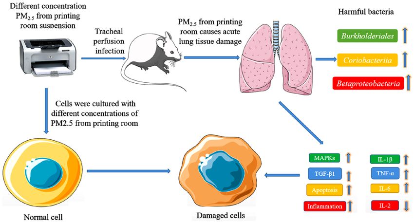
3216                         ZOU et al: POTENTIAL HAZARDOUS EFFECTS OF PM2.5 FROM THE PRINTING ROOM

Figure 1. Oxidative damage resulting from printing room‑derived PM 2.5 exposure in human HBE and HUVEC cells. (A) Determination of HBE and
HUVEC cell viability. (B) MDA and SOD activity in HBE and HUVEC cells. Levels of IL‑1β, IL‑6, TNF‑ α and IL‑2 expression in (C) HBE cells and
(D) HUVECs. Data are presented as the mean ± standard deviation. n=12/group. *P
Molecular Medicine REPORTS 22: 3213-3224, 2020                                                        3217

Figure 2. Effects of printing room‑derived PM2.5 exposure on protein expression in human HBE and HUVEC cells. Levels of (A) COX2, p‑p65 and p65,
(B) TGF‑β1 and (C) Bcl‑2 and Bax protein in HBE cells. Levels of (D) COX2, p‑p65, p65, (E) TGF‑β1 and (F) Bcl‑2 and Bax protein in HUVECs. Data are
presented as the mean ± standard deviation. n=12/group. *P
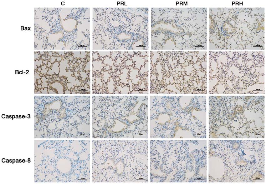
3218                          ZOU et al: POTENTIAL HAZARDOUS EFFECTS OF PM2.5 FROM THE PRINTING ROOM

Figure 3. Inflammatory damage in lung tissues occurs post‑PM2.5 exposure in male C57BL/6 mice. (A) Hematoxylin and eosin staining images (magnification,
x200). (B) MDA and (C) SOD activity. (D) Levels of IL‑1β, IL‑6, TNF‑α and IL‑2. (E) Gene expression of IL‑1β, IL‑6, TNF‑α and IL‑2. Data are presented as
the mean ± standard deviation. n=12/group. *P
Molecular Medicine REPORTS 22: 3213-3224, 2020                                                            3219

Figure 4. Effects of printing room‑derived PM2.5 exposure on signaling pathways in vivo. Protein levels of (A) COX2 and p‑p65/p65 (B) p‑ERK/ERK (C) TGF‑βand
(D) Bcl‑2 and Bax in mouse lungs are shown. Data are presented as the mean ± standard deviation. n=12/group. *P
3220                          ZOU et al: POTENTIAL HAZARDOUS EFFECTS OF PM2.5 FROM THE PRINTING ROOM

Figure 6. Effects of printing room‑derived PM2.5 on the pulmonary microflora of mice. (A) Shannon index and (B) Simpson index values. (C) The composition
and relative abundance of microbial diversity at the genera level in mouse lung of C, PRL, PRM and PRH groups are compared. (D) PCoA associated with
β diversity index. Microbial composition at the (E) order and (F) microbial class levels. PM2.5, fine particulate matter; C, cells treated with PBS; PRL, cells
treated with 5 µg/ml PM2.5; PRM, cells treated with 10 µg/m PM2.5; PRH, cells treated with 15 µg/ml PM2.5.

the environment (0.0011 vs. 0.0015 for PRH and group C,                          pro‑inflammatory factors IL‑1β, IL‑6 and TNF‑α, inhibited
respectively), immune system activity (0.00055 vs. 0.00087                       the production of inflammatory factor IL‑2, activated the
for PRH and group C, respectively), and altered energy                           inflammatory response and promoted lung injury and fibrosis.
metabolism (0.05 vs. 0.055 for PRH and group C, respectively).                   These present results are consistent with previously published
PM2.5 exposure notably enhanced the appearance of biodeg-                        studies (29,30). Therefore, PM2.5 activated oxidative stress and
radation and metabolism of xenobiotics (0.057 and 0.036 for                      inflammatory responses in lung cells, which has the potential
PRH and group C, respectively), endocrine system activity                        to cause cellular injury and even death.
(0.0061 and 0.0041 for PRH and group C, respectively) and                            To further study the potential mechanisms by which PM2.5
infectious diseases (0.0041 and 0.0032 for PRH and group C,                      alters lung cell function, key proteins of inflammatory, fibrosis
respectively) that occurred in PRH compared with control                         and apoptosis pathways were analyzed. PM 2.5 significantly
cells (Fig. 7). A graphical summary of the study is presented                    increased the levels of COX2 and p‑p65/p65. COX2 is an
in Fig. 8.                                                                       important pro‑inflammatory gene, and studies have shown
                                                                                 that overexpression of COX2 promotes inflammation and
Discussion                                                                       adversely affects fibrosis (31). p65 belongs to the NF‑κ B
                                                                                 family of proteins, and these proteins exist either as homolo-
Exposure to carbon dust and volatile organic compounds                           gous or heterologous dimers formed between family members
affects the respiratory, immune and nervous systems, and staff                   under normal conditions (31). When cells were stimulated by
working in offices may have symptoms of cough and throat                         different external factors, such as stress, lipopolysaccharide,
discomfort that are caused by PM present in the air (26).                        viruses and free oxygen radicals, NF‑κ B immediately disasso-
Since the importance of indoor particulate matter on health                      ciates and translocates to the nucleus to enhance transcription
has been neglected, the effects of PM2.5 (collected from the                     of inflammatory genes (32). p65 has also been shown to be a
printing room) exposure on lung injury, as well as its potential                 transcription factor involved in the NF‑KB pathway via regu-
mechanisms, was studied both in vitro and in vivo.                               lation of pro‑inflammatory cytokines TNF‑α and IL‑6 (33).
    First, the effects of exposure to different concentrations                   As PM 2.5 concentration increased, the expression of COX2
of PM 2.5 on HBE and HUVEC cells was evaluated. The                              and p‑p65/p65 were enhanced in a dose‑dependent manner.
results indicated that PM 2.5 exposure significantly reduced                     This finding indicated that as PM2.5 concentrations increase,
cell viability in a dose‑dependent manner, enhanced the lipid                    the degree of pulmonary inflammation and fibrosis become
oxidation index of MDA and decreased the anti‑oxidative                          increasingly severe as a result of enhanced expression of
stress index of SOD, which was in line with previously                           COX2 and p‑p65/p65.
published reports (27,28). Moreover, PM2.5 addition to cell                          Overexpression of COX2 and p‑p65/p65 greatly enhanced
culture media significantly increased the accumulation of                        pulmonary fibrosis via enhancing the expression of pro‑fibrotic
Molecular Medicine REPORTS 22: 3213-3224, 2020 3221 Figure 7. Effects of printing room‑derived PM2.5 exposure on metabolic pathways. The relative abundance of (A) Burkholderiales, (B) Coriobacteriia and (C) Betaproteobacteria were assessed. Furthermore, (D) environmental adaptations, (E) immunity, (F) energy metabolism, (G) xenobiotic biodegradation and metabolism, (H) endocrine system and (I) infectious disease of groups were compared. *P
3222                     ZOU et al: POTENTIAL HAZARDOUS EFFECTS OF PM2.5 FROM THE PRINTING ROOM

cytokine TGF‑ β1 (34) and apoptosis markers, including              Coriobacteriia (at the order level). Both Betaproteobacteria
Bax/Bcl‑2 (35). TGF‑β1 is critical for regulatory T cell func-      and Burkholderiales can cause lung inflammation associated
tion and T‑helper (Th) 9 and Th17 differentiation, which play       with lung disease, and as abundance of Betaproteobacteria
important roles in the regulation of tissue repair, embryogen-      and Burkholderiales increase, the degree of lung damage is
esis, cartilage homeostasis, cellular growth and proliferation      expected to correspondingly increase (45). Therefore, PM2.5
and cancer, and are associated with the development of autoim-      exposure has the ability to damage human health through
mune disorders, chronic inflammatory conditions and allergic        increasing pathogen abundance in the lung. This can lead to
diseases (36). Bcl‑2 is an anti‑apoptotic marker, and downreg-      increased lung inflammation and damage. Environmental PM2.5
ulation of Bcl‑2 may promote the production of pro‑apoptotic        is also capable of causing lung damage and energy metabolism
proteins (37). For example, increased Bax levels can lead to        disorders, such as diarrhea (46). Multiple lines of evidence
apoptotic cell death (38). Of TGF‑β1 and Bax expression levels      have shown that PM2.5 exposure is associated with metabolic
were upregulated in groups exposed to PM2.5. Simultaneously,        disorders, especially in children and the elderly or genetically
Bcl‑2 expression was downregulated. Taken together, these           susceptible experimental animal models (47). Environmental
results indicated that exposure to increased concentrations         adaptation, immune response and energy metabolism in PRH
of PM2.5 enhanced pulmonary injury as a result of enhanced          groups markedly decreased compared group C. Furthermore,
pulmonary inflammation and fibrosis via downregulation of           infectious disease occurrence, endocrine activity and the xeno-
Bcl‑2 and upregulation of TGF‑β1.                                   biotic biodegradation and metabolism may have an association
     A mouse model was established and the results indicated        with PM2.5 exposure.
that PM 2.5 exposure lead to inflammatory cell infiltration,            In the present study, the effect of printing room PM 2.5
airway epithelial cell shedding and necrosis. Consistent with       exposure on the lungs was assessed. The results indicated that
cell culture findings, exposure to PM2.5 significantly increased    PM2.5 caused cellular and tissue injury as a result of increased
the accumulation of pro‑inflammatory factors IL‑1β, IL‑6 and        oxidative stress, inflammation, fibrosis and apoptosis.
TNF‑α, and reduced production of the inflammatory factor,           Additionally, PM2.5 exposure enhanced pathogen abundance
IL‑2. This finding indicated that PM2.5 induced inflammation        in lung tissues. Therefore, printing room PM 2.5 may nega-
in the lung via increasing the production of pro‑inflammatory       tively affect individuals exposed for long periods. However,
factors in lung tissue. Western blotting results indicated that     considering limitations of the present acute lung injury model,
protein expression of COX2, p‑p65/p65, TGF‑β1 and Bax/Bcl‑2         more work will be needed to clarify the health risks associated
were enhanced post‑exposure to PM2.5, which indicated that          with printing room PM2.5 exposure. In conclusion, the present
PM2.5 induced fibrosis and apoptosis in the lung via increasing     study evaluated mechanisms of lung injury caused by printing
the production of pro‑fibrotic cytokines and apoptosis markers.     room‑derived PM2.5, and found that they are closely related the
In addition, the expression of p‑ERK/ERK was evaluated, and         inflammation, apoptosis and fibrosis caused, and provided a
it was found that increased concentrations of PM2.5 resulted        theoretical foundation for further research.
in MAPK pathway activation, whereby increased levels of
p‑ERK relative to ERK were observed. The MAPK pathway               Acknowledgements
involves p38, JNK and ERK1/2, which play key roles in acute
lung injury, and the ERK pathway exerts a critical function         Not applicable.
in the management of levels of various cytokines, including
TNF‑α, IL‑1β and IL‑6 (39). Furthermore, ERK is involved in         Funding
the regulation of numerous cellular processes, including stress
response, inflammatory pathways, proliferation, differentia-        This study was supported by the National Natural Science
tion, apoptosis and survival (34). Elevated p‑ERK/ERK ratios        Foundation of China (grant no. 41765009), the Excellent
in the MAPK pathway indicated that increased concentrations         Youth Foundation of the Jiangxi Scientific Committee (grant
of PM 2.5 can lead to lung inflammation. Additionally, IHC          no. 20171BCB23028), the Science and Technology Plan of
results indicated that PM2.5 upregulated the expression of the      the Jiangxi Health Planning Committee (grant no. 20175526)
pro‑apoptotic protein, Bax, and downregulated the accumula-         and the Science and Technology Project of Jiangxi (grant
tion of the anti‑apoptotic protein, Bcl‑2, which eventually lead    nos. 20181BBG70028 and 20181BCB24003).
to enhanced expression of apoptosis proteins caspase‑8 and
caspase‑3 (40).                                                     Availability of data and materials
     The lungs have previously been considered a sterile
environment, and assessment of pulmonary disease has                The datasets used and/or analyzed during the current study are
been examined from a bacterial pathology perspective (41).          available from the corresponding author on reasonable request.
Recently, 16s ribosomal RNA studies and metagenomics
revealed the presence of a flexible microbiota in the upper and     Authors' contributions
lower respiratory tract, blood, placenta and amniotic fluid (42).
Increasing evidence has shown that bacteria are present in the      TC and HH made substantial contributions to conception
lungs and serve key roles in fatal pneumonia, which can be both     and design. CZ, HY, LC and XC performed the analysis and
beneficial and harmful (43,44). The present high‑throughput         interpretation of data. CZ, TC, HY, LC and XC drafted the
sequencing results showed that PM2.5 altered microbial diver-       manuscript and revised the important intellectual content. All
sity in the lung by increasing abundance of Burkholderiales         authors read and approved the final manuscript, and TC gave
(at the class level), Betaproteobacteria (at the order level) and   the final approval of the version to be published.
Molecular Medicine REPORTS 22: 3213-3224, 2020 3223
Ethics approval and consent to participate 15. Ya P, Xu H, Ma Y, Fang M, Yan X, Zhou J and Li F: Liver injury
induced in Balb/c mice by PM2.5 exposure and its alleviation by
compound essential oils. Biomed Pharmacother 105: 590‑598,
The present study was approved by the Ethical Committee of 2018.
the Second Affiliated Hospital of Nanchang University, and 16. Tavera Busso I, Vera A, Mateos AC, Amarillo AC and
Carreras H: Histological changes in lung tissues related with
all experiments were performed in accordance with approved sub‑chronic exposure to ambient urban levels of PM2.5 in
guidelines (The Guide for Care and Use of Laboratory Córdoba, Argentina. Atmos Environ 167: 616‑624, 2017.
Animals; National Institutes of Health publication 85‑23). 17. Yang B, Guo J and Xiao C: Effect of PM2.5 environmental
pollution on rat lung. Environ Sci Pollut Res Int 25: 36136‑36146,
2018.
Patient consent for publication 18. Li H, Zhao Q, Liu R, Yang L, Chen H and Cui X: Protective effect
and potential mechanism of simvastatin on myocardial injury
Not applicable. induced by diabetes with hypoglycemia. Exp Clin Endocrinol
Diabetes 126: 148‑161, 2018.
19. Huang H, Zou C, Cao J and Tsang P: Carbonaceous aerosol
Competing interests characteristics in outdoor and indoor environments of Nanchang,
China, during summer 2009. J Air Waste Manag Assoc 61:
1262‑1272, 2011.
The authors declare that they have no competing interests. 20. Huang H, Zou C, Cao J, Tsang P, Zhu F, Yu C and Xue S:
Water‑soluble Ions in PM2.5 on the Qianhu Campus of
Nanchang University, Nanchang City: Indoor‑outdoor distri-
References bution and source implications. Aerosol Air Qual Res 12:
435‑443, 2012.
1. Holzer M, Bihari P, Praetner M, Uhl B, Reichel C, Fent J, 21. Wang H, Song L, Ju W, Wang X, Dong L, Zhang Y, Ya P, Yang C
Vippola M, Lakatos S and Krombach F: Carbon‑based nanoma- and Li F: The acute airway inflammation induced by PM2.5
terials accelerate arteriolar thrombus formation in the murine exposure and the treatment of essential oils in Balb/c mice. Sci
microcirculation independently of their shape. J Appl Toxicol 34: Rep 7: 44256, 2017.
1167‑1176, 2014. 22. Zhang X, Zhong W, Meng Q, Lin Q, Fang C, Huang X, Li C,
2. Cohen AJ, Brauer M, Burnett R, Anderson HR, Frostad J, Estep K, Huang Y and Tan J: Ambient PM2.5 exposure exacerbates
Balakrishnan K, Brunekreef B, Dandona L, Dandona R, et al: severity of allergic asthma in previously sensitized mice.
Estimates and 25‑year trends of the global burden of disease J Asthma 52: 785‑794, 2015.
attributable to ambient air pollution: An analysis of data from 23. Tian P, Xu D, Huang Z, Meng F, Fu J, Wei H and Chen T:
the global burden of diseases study 2015. Lancet 389: 1907‑1918, Evaluation of truncated G protein delivered by live attenuated
2017. Salmonella as a vaccine against respiratory syncytial virus.
3. Urlaub S, Grün G, Foldbjerg P and Sedlbauer K: Ventilation and Microb Pathog 115: 299‑303, 2018.
health ‑ a review. Proc AVIC Conf, 2015. 24. Livak KJ and Schmittgen TD: Analysis of relative gene expression
4. Chen Y, Du W, Shen G, Zhuo S, Zhu X, Shen H, Huang Y, Su S, data using real‑time quantitative PCR and the 2(‑Delta Delta
Lin N, Pei L, et al: Household air pollution and personal exposure C(T)) method. Methods 25: 402‑408, 2001.
to nitrated and oxygenated polycyclic aromatics (PAHs) in rural 25. Hammer y, Harper DA and Ryan PD: PAST: Paleontological
households: Influence of household cooking energies. Indoor statistics software package for education and data analysis.
Air 27: 169‑178, 2017. Palaeontol Electron 4: 1‑9, 2001.
5. Du B, Gao J, Chen J, Stevanovic S, Ristovski Z, Wang L and 26. Tang T, Hurraß J, Gminski R and Mersch‑Sundermann V: Fine
Wang L: Particle exposure level and potential health risks of and ultrafine particles emitted from laser printers as indoor air
domestic Chinese cooking. Build Environ 123: 564‑574, 2017. contaminants in German offices. Environ Sci Pollut Res Int 19:
6. Liu T, Liu Q, Li Z, Huo L, Chan M, Li X, Zhou Z and Chan CK: 3840‑3849, 2012.
Emission of volatile organic compounds and production of 27. Kohl H, Orth R, Riebartsch O, Galeitzke M and Cap JP: Support
secondary organic aerosol from stir‑frying spices. Sci Total of innovation networks in manufacturing industries through iden-
Environ 599‑600: 1614‑1621, 2017. tification of sustainable collaboration potential and best‑practice
7. Liu Y, Chen YY, Cao JY, Tao FB, Zhu XX, Yao CJ, Chen DJ, transfer. Procedia CIRP 26: 185‑189, 2015.
Che Z, Zhao QH and Wen LP: Oxidative stress, apoptosis, and 28. Zhang ZQ, Zhang CZ, Shao B, Pang DH, Han GZ and Lin L:
cell cycle arrest are induced in primary fetal alveolar type II Effects of abnormal expression of fusion and fission genes on the
epithelial cells exposed to fine particulate matter from cooking morphology and function of lung macrophage mitochondria in
oil fumes. Environ Sci Pollut Res Int 22: 9728‑9741, 2015. SiO2‑induced silicosis fibrosis in rats in vivo. Toxicol Lett 312:
8. Khatri M, Bello D, Pal AK, Woskie S, Gassert TH, Demokritou P 181‑187, 2019.
and Gaines P: Toxicological effects of PM0.25‑2.0 particles 29. Smith RE, Strieter RM, Phan SH, Lukacs N and Kunkel SL: TNF
collected from a photocopy center in three human cell lines. and IL‑6 mediate MIP‑1alpha expression in bleomycin‑induced
Inhal Toxicol 25: 621‑632, 2013. lung injury. J Leukoc Biol 64: 528‑536, 1998.
9. Pirela SV, Bhattacharya K, Wang Y, Zhanga Y, Wanga G, 30. Cavarra E, Carraro F, Fineschi S, Naldini A, Bartalesi B, Pucci A
Christophic CA, Godleskia J, Thomasd T, Qiane Y, and Lungarella G: Early response to bleomycin is characterized
Orandle MS, et al: A 21‑day sub‑acute, whole‑body inhalation by different cytokine and cytokine receptor profiles in lungs. Am
exposure to printer‑emitted engineered nanoparticles in rats: J Physiol Lung Cell Mol Physiol 287: L1186‑L1192, 2004.
Exploring pulmonary and systemic effects. NanoImpact 15: 31. Dai P, Shen D, Shen J, Tang Q, Xi M, Li Y and Li C: The roles
100176, 2019. of Nrf2 and autophagy in modulating inflammation mediated
10. Pirela S, Molina R, Watson C, Cohen JM, Bello D, Demokritou P by TLR4‑NFκ B in A549 cell exposed to layer house particulate
and Brain J: Effects of copy center particles on the lungs: A matter 2.5 (PM2.5). Chemosphere 235: 1134‑1145, 2019.
toxicological characterization using a Balb/c mouse model. Inhal 32. Hayden MS and Ghosh S: NF‑κ B in immunobiology. Cell Res 21:
Toxicol 25: 498‑508, 2013. 223‑244, 2011.
11. Jensen M and Rold‑Petersen J: Itching erythema among post 33. Barker HE, Paget JT, Khan AA and Harrington KJ: The tumour
office workers caused by a photocopying illachine with wet microenvironment after radiotherapy: Mechanisms of resistance
toner. Contact Dermatitis 5: 389‑391, 1979. and recurrence. Nat Rev Cancer 15: 409‑425, 2015.
12. Ga l ia rdo M, Romero P, Sá nchez‑ Quevedo MC a nd 34. Lazzara F, Fidilio A, Platania CBM, Giurdanella G, Salomone S,
López‑Caballero JJ: Siderosilicosis due to photocopier toner Leggio GM, Tarallo V, Cicatiello V, De Falco S, Eandi CM, et al:
dust. Lancet 344: 412‑413, 1994. Aflibercept regulates retinal inflammation elicited by high
13. Grifka J: Use of drugs in renal impairment. Internist (Berl) 49: glucose via the PlGF/ERK pathway. Biochem Pharmacol 168:
126, 2008 (In German). 341‑351, 2019.
14. Theegarten D, Boukercha S, Philippou S and Anhenn O: 35. Kuroki M, Noguchi Y, Shimono M, Tomono K, Tashiro T, Obata Y,
Submesothelial deposition of carbon nanoparticles after toner Nakayama E and Kohno S: Repression of bleomycin‑induced
exposition: Case report. Diagn Pathol 5: 77, 2010. pneumopathy by TNF. J Immunol 170: 567‑574, 2003.3224 ZOU et al: POTENTIAL HAZARDOUS EFFECTS OF PM2.5 FROM THE PRINTING ROOM
36. Ndaw VS, Abebayehu D, Spence AJ, Paez PA, Kolawole EM, 43. Kaimala S, Al‑Sbiei A, Cabral‑Marques O, Fernandez‑Cabezudo MJ
Taruselli MT, Caslin HL, Chumanevich AP, Paranjape A, and Al‑Ramadi BK: Attenuated bacteria as immunotherapeutic
Baker B, et al: TGF‑ β1 suppresses IL‑33‑induced mast cell tools for cancer treatment. Front Oncol 8: 136, 2018.
function. J Immunol 199: 866‑873, 2017. 44. Zhao C, He J, Cheng H, Zhu Z and Xu H: Enhanced therapeutic
37. Ashkenazi A, Fairbrother WJ, Leverson JD and Souers AJ: From effect of an antiangiogenesis peptide on lung cancer in vivo
basic apoptosis discoveries to advanced selective BCL‑2 family combined with salmonella VNP20009 carrying a Sox2 shRNA
inhibitors. Nat Rev Drug Discov 16: 273‑284, 2017. construct. J Exp Clin Cancer Res 35: 107, 2016.
38. Hii LW, Lim SE, Leong CO, Chin SY, Tan NP, Lai KS and 45. Pacello F, D'Orazio M and Battistoni A: An ERp57‑mediated
Mai CW: The synergism of Clinacanthus nutans Lindau extracts disulphide exchange promotes the interaction between
with gemcitabine: Downregulation of anti‑apoptotic markers in Burkholderia cenocepacia and epithelial respiratory cells. Sci
squamous pancreatic ductal adenocarcinoma. BMC Complement Rep 6: 21140, 2016.
Altern Med 19: 257, 2019. 46. Ning X, Ji X, Li G and Sang N: Ambient PM2.5 causes lung
39. Jiang C, Zhong R, Zhang J, Wang X, Ding G, Xiao W and Ma S: injuries and coupled energy metabolic disorder. Ecotoxicol
Reduning injection ameliorates paraquat‑induced acute lung Environ Saf 170: 620‑626, 2019.
injury by regulating AMPK/MAPK/NF‑κ B signaling. J Cell 47. Pan K, Jiang S, Du X, Zeng X, Zhang J, Song L, Zhou J, Kan H,
Biochem 120: 12713‑12723, 2019. Sun Q, Xie Y and Zhao J: AMPK activation attenuates inflam-
40. Che Z, Liu Y, Chen Y, Cao J, Liang C, Wang L and Ding R: matory response to reduce ambient PM2.5‑induced metabolic
The apoptotic pathways effect of fine particulate from cooking disorders in healthy and diabetic mice. Ecotoxicol Environ
oil fumes in primary fetal alveolar type II epithelial cells. Mutat Saf 179: 290‑300, 2019.
Res Genet Toxicol Environ Mutagen 761: 35‑43, 2014.
41. Wang Y, Chen J, Tang B, Zhang X and Hua ZC: Systemic
administration of attenuated salmonella typhimurium in combi-
nation with interleukin‑21 for cancer therapy. Mol Clin Oncol 1: This work is licensed under a Creative Commons
461‑465, 2013. Attribution-NonCommercial-NoDerivatives 4.0
42. Yu K, Rodriguez MD, Paul Z, Gordon E, Rice K, Triplett EW, International (CC BY-NC-ND 4.0) License.
Keller‑Wood M and Wood CE: Proof of principle: Physiological
transfer of small numbers of bacteria from mother to fetus in
late‑gestation pregnant sheep. PLoS One 14: e0217211, 2019.You can also read