ATM/NEMO signaling modulates the expression of PD- L1 following docetaxel chemotherapy in prostate cancer
←
→
Page content transcription
If your browser does not render page correctly, please read the page content below
Open access Original research
ATM/NEMO signaling modulates the
J Immunother Cancer: first published as 10.1136/jitc-2020-001758 on 22 July 2021. Downloaded from http://jitc.bmj.com/ on October 17, 2021 by guest. Protected by copyright.
expression of PD-L1 following docetaxel
chemotherapy in prostate cancer
Zongren Wang,1,2 Xueling Zhang,1 Wuguo Li,3 Qiao Su,3 Zhaoyang Huang,1
Xinyao Zhang,1,2 Haiqi Chen,1 Chengqiang Mo,2 Bin Huang,2 Wei Ou,2
Junxing Chen,2 Guangyin Zhao,3 Lingwu Chen,2 Lan Shao 1
To cite: Wang Z, Zhang X, ABSTRACT programmed cell death ligand-1 (PD- L1).
Li W, et al. ATM/NEMO Background The efficacy of docetaxel-based High levels of PD- L1 in tumor cells are
signaling modulates the chemotherapy is limited by the development of drug
expression of PD-L1 following
important for the induction of immunosup-
resistance. Recent studies demonstrated the efficacy of pression, immune tolerance, and immune
docetaxel chemotherapy in
anti-programmed death-1 (PD-1)/programmed cell death evasion in the tumor microenvironment.1–3
prostate cancer. Journal for
ImmunoTherapy of Cancer ligand-1 (PD-L1) immunotherapies in metastatic prostate
Among men, the most common type of inva-
2021;9:e001758. doi:10.1136/ cancer. The ataxia telangiectasia mutation (ATM) protein
plays a crucial role in maintaining genome stability and sive cancer is that of the prostate cancer, and
jitc-2020-001758
function of mitosis. Here, we aimed to determine whether it is the second leading cause of death. Recent
►► Additional online PD-1/PD-L1 signaling contributes to the resistance to DTX studies have reported that PD-1 is expressed
supplemental material is and to elucidate the mechanism underlying DTX-induced on the tumor-infiltrating T cells in patients
published online only. To view, PD-L1 expression. with prostate cancer and responsible for
please visit the journal online Methods In this retrospective study, PD-L1 expression rapid tumor progression.4–7 However, reports
(http://dx.doi.org/10.1136/jitc- was analyzed in 33 tumor tissue samples from prostate on the expression of PD-L1 in prostate cancer
2020-001758). cancer patients. Prostate cell lines were used to perform cells are conflicting. Although several studies
functional assays and examine underlying mechanisms
ZW, XZ and WL contributed support the involvement of PD-L1 expression
in vitro. A fully mouse prostate cancer model and a
equally. and the PD-L1/PD-1 pathway in the progres-
humanized chimeric mouse bearing human prostate
Accepted 25 May 2021 tumors and peripheral blood mononuclear cells were used sion of prostate tumors,5 8 9 some reports
for in vivo assays. suggested that PD- L1 is downregulated in
Results We have shown that DTX, a chemotherapeutic prostate cancer.10 Therefore, a compre-
drug which causing microtubule interference, could hensive analysis of PD-L1 expression under
significantly induce the expression of PD-L1 in prostate different medical and therapeutic conditions
cancer cells. This effect is blocked by the inhibition of is necessary for the development of check-
ATM, suggesting that it plays an essential role in PD-L1 point inhibitors in prostate cancer.
© Author(s) (or their expression upregulated by DTX. Mechanistic studies
employer(s)) 2021. Re-use Docetaxel (DTX)- based chemotherapy is
have shown that ATM activity in cancer cells enhances
permitted under CC BY-NC. No
the stability of the NF-κB essential modulator (NEMO),
the first-line treatment of metastatic castrate-
commercial re-use. See rights resistant prostate cancer and recent reports
and permissions. Published by which leading to an increase in the NF-κB activity and
BMJ. PD-L1 expression. Using the mouse model, it was further also suggest the improving outcomes of DTX
1
The Center for Translational demonstrated that a combination of ATM and NEMO in metastatic hormone sensitive prostate
Medicine, The First Affiliated inhibitors along with DTX augmented the antitumor cancer.11–13 DTX inhibits microtubule depo-
Hospital, Sun Yat-sen University, efficacy of chemotherapy, which are comparable to that of lymerization, thereby activating the M-phase
Guangzhou, China PD-L1 antibody. cycle checkpoint.14–17 An important issue
2
Department of Urology, The Conclusions Our findings have revealed that a previously limiting DTX chemotherapy is the develop-
First Affiliated Hospital, Sun unrecognized ATM-NEMO signaling which induced by DTX
Yat-sen University, Guangzhou,
ment of drug resistance, which is the primary
is capable of suppressing tumor immunity by activating the
China expression of PD-L1, suggesting that the ATM-NEMO-NF-
cause of disease aggravation.18 Several studies
3
Animal Experiment Center, The
κB axis can be exploited to restore the immune balance have reported that anti-PD-1/PD-L1 immu-
First Affiliated Hospital, Sun notherapies are effective for the treatment
and overcome cancer resistance triggered by DTX.
Yat-sen University, Guangzhou,
Graphic Abstract: supplementary file 1 of metastatic prostate cancer (mPCa),15 19
China
suggesting that the PD-1/PD-L1 signaling may
Correspondence to contribute to the chemoresistance to DTX.
Dr. Lan Shao; INTRODUCTION In this study, we showed that DTX chemo-
shaolan@mail.sysu.edu.c n
Programmed death-1 (PD-1) is an important therapeutic treatment upregulated the PD-L1
Dr. Lingwu Chen; immune checkpoint molecule that medi- expression in prostate cancer cells, and iden-
c henlwu@mail.sysu.edu.c n ates immunosuppression by binding to the tified that ATM-NEMO as the major pathway
Wang Z, et al. J Immunother Cancer 2021;9:e001758. doi:10.1136/jitc-2020-001758 1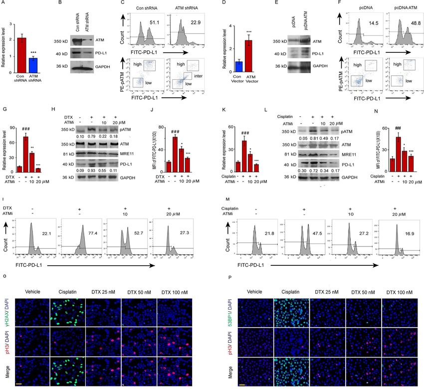
Open access
activating PD- L1 expression. Our research provides a
J Immunother Cancer: first published as 10.1136/jitc-2020-001758 on 22 July 2021. Downloaded from http://jitc.bmj.com/ on October 17, 2021 by guest. Protected by copyright.
Table 1 Baseline demographics and clinical characteristics
new understanding of the mechanism underlying PD-L1
expression in cancer cells, would open a new strategy to No of patients 55
overcoming DTX resistance and demonstrates a new way Age, median, years 66.3
to improve the efficacy of DTX chemotherapy by thera- Range, years 52–80
peutically blocking NEMO or ATM signaling. Characteristics No of patients (%)
Ataxia telangiectasia mutation (ATM) is the main
TPSA (µg/L)
kinase that regulates the G2/M cell cycle checkpoint
and activated instinctively in mitosis.20 21 Recent studies 20 16 (29.1)
(NF-κB) is a strong inducer of the PD-L1 gene expression
FPSA (µg/L)
in multiple types of cancers.26 The activation of NF-κB in
response to multiply cell stresses via an ATM-dependent 0–1 31 (56.4)
mechanism. Activated ATM phosphorylates NF-κB essen- >1 24 (43.6)
tial modulator (NEMO), which results in its modification F/T
by SUMOylation and monoubiquitination. Ubiquitinated 0–0.25 44 (80.0)
NEMO is then exported to the cytoplasm, where it phos-
0.25–1 9 (16.4)
phorylates IκB kinase-β (IKK β) in association with ATM
and ELKS (glutamate-rich, leucine-rich, lysine-rich, and >1 2 (3.6)
serine-
rich) proteins, leading to degradation of IκBα. Gleason sum score
Thus, p65/RelA is liberated and translocated from the 6 6 (10.9)
cytoplasm to the cell nucleus.27–29 However, whether DTX 7 21 (38.2)
affects PD-L1 expression and the role of ATM and NF-κB
8 10 (18.2)
signaling in this process remain to be elucidated.
9 17 (30.9)
10 1 (1.82)
METHODS Disease status
Study design cT
This study determined whether PD-1/PD- L1 signaling
T1 6 (10.9)
contributes to the resistance to DTX, and elucidated the
mechanism underlying DTX-induced PD-L1 expression. T2a 1 (1.82)
PD-L1 expression was analyzed in 33 tumor tissue samples T2b 17 (30.9)
from prostate cancer patients. Prostate cell lines were T2c 8 (14.5)
used to perform functional assays in vitro. Two mouse T3a 6 (10.9)
models with RM-1 and PC-3 bearing prostate tumors
T3b 15 (27.3)
were used for in vivo assays. All the surgically resected
prostate cancer tissues and peripheral blood mononu- T4 2 (3.64)
clear cells (PBMCs) were obtained from 55 patients at N
the Department of Urology, the First Affiliated Hospital, N0 38 (69.1)
Sun Yat-sen University (Guangzhou, China). Table 1 pres- N1 17 (30.9)
ents the demographic characteristics of 55 patients. The
M
clinical tumor node metastasis (TNM) stage of cancer
was assessed by the eighth edition of the American Joint M0 39 (70.9)
Committee on Cancer (AJCC). The cancer stage was M1 16 (29.1)
defined according to AJCC prognostic staging system of Stage
prostate cancer. I 2 (3.64)
IIA 3 (5.45)
Cell culture and transfection
PC-3, DU-145 and RM-1 prostate cancer cells were obtained IIB 5 (9.09)
from the American Type Culture Collection. The cancer IIC 1 (1.82)
cells were cultured in RPMI 1640 supplemented with IIIA 6 (10.9)
10% FBS. DTX (MedChemExpress, Monmouth Junction,
IIIB 7 (12.7)
New Jersey, USA), which promotes microtubule stabili-
zation and inhibits the mitosis of cancer cells; cisplatin IVA 3 (5.45)
(MedChemExpress), a replicative polymerase inhibitor, IVB 28 (50.9)
were used at concentrations of 0, 25, 50, 100 nM and 0, Continued
2 Wang Z, et al. J Immunother Cancer 2021;9:e001758. doi:10.1136/jitc-2020-001758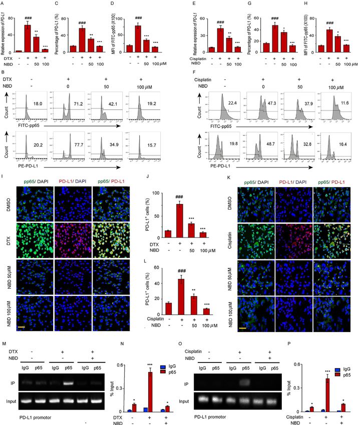
Open access
A/G PLUS-Agarose, and then incubated with anti-ATM or
J Immunother Cancer: first published as 10.1136/jitc-2020-001758 on 22 July 2021. Downloaded from http://jitc.bmj.com/ on October 17, 2021 by guest. Protected by copyright.
Table 1 Continued
anti-NEMO with 20 µL of protein A/G PLUS-Agarose at
No of patients 55 4°C for 12 hours. Immune complexes were collected after
Surgical margin status each immunoprecipitation by centrifugation at 13 000 g
Positive 20 (36.4) for 10 min. The immune complexes were subjected to
sodium dodecyl sulfate- polyacrylamide gel electropho-
Negative 35 (63.6)
resis (SDS-PAGE), followed by immunoblotting with ATM
cT, clinical T; FPSA, free prostate-specific antigen; TPSA, total and NEMO specific antibodies.
prostate-specific antigen. Previously reported methods were applied for western
blotting.30 In brief, whole-cell lysates were prepared
in radioimmunoprecipitation buffer. Equal amounts
10, 20, 30 µM, respectively. Paclitaxel (PTX, MedChemEx- of total protein from each sample were loaded into an
press), was used at concentration of 200 nM. Additionally, SDS-PAGE gel and transferred to a polyvinylidene diflu-
indicated concentrations of the ATM inhibitor KU55933 oride membrane. Antibodies specific for PD-L1 (Abcam,
(Sigma-Aldrich, St. Louis, Missouri, USA) or the NEMO Cambridge, Massachusetts, USA), ATM, phospho-ATM,
binding peptide (NBD) (Enzo Biochem, New York, USA) MRE11, p65, phospho- p65 and GAPDH (Santa Cruz
were added at 60 min before the induction of PD-L1. Biotechnology, Santa Cruz, California, USA) were added
For inhibitor screening, S3I-201(100 µM), Fludarabine 12 hours at 4°C. The membranes were subsequently
(10 µM), UO126- EtOH (10 µM), Bay11-7082 (10 µM), incubated with secondary antibodies and detected with
JSH-23 (5 µM), SD-208 (2 µM) which obtained from a chemiluminescent detection system (Beyotime Biotech-
Sigma-Aldrich were added and the cells were collected nology, Shanghai, China).
after 6 hours.
Chromatin immunoprecipitation assay
Flow cytometry
Chromatin immunoprecipitation (chIP) assay was
For extracellular staining, cells were surface stained with
preformed to identify the binding site of NF-κB p65 in
FITC-PDL1, PE-PDL1, PE-PDL2, PE-CD8 antibodies
PD-L1 promoter region. PC-3 cells were crosslinked with
(Biolegend, San Diego, California, USA). For intracellular
1% formaldehyde at 37°C for 10 min, and neutralized
staining, cells were first surface stained with FITC-PDL1,
with 0.2 M glycine. Then the chromatin was obtained and
PE-CD8, APC-CD4, PE-CD25, APC-EGFR (Biolegend).
fractured according to the instructions of chIP assay kit
In addition, the intracellular staining was performed
(Beyotime Biotechnology), followed by precipitating with
with a BD permeabilization/Fixation kit. 107 cells/mL
p65 antibody or IgG isotype 12 hours at 4°C. After puri-
were stained with PE-phospho-ATM, FITC-FOXP3, phos-
fication, the precipitated DNA and input were de-cross-
pho-p65, and KI67 antibodies (Cell signaling Technology,
linked at 68°C. The purified DNA was amplified by PCR
Danvers, Massachusetts, USA), followed by an Alexa Fluro
and analyzed by 2% agarose gel, and the capacity of p65
488-conjugated Donkey anti-mouse IgG (Jackson Immu-
binding to PD-L1 promoter was quantified by qPCR.
noResearch, West Grove, Pennsylvania, USA), and then
Primer sequences used in chIP- qPCR were as follows:
detected by LSRII flow cytometer. Data were analyzed by
sense 5’-CTTCCGCAGCCTTAATCCTTA-3’ and antisense
FlowJo software.
5’-ATCGTGGATTCTGTGACTTCCTC-3’.
For prepared single-cell suspensions from the tumor
tissue, The Tumor Dissociation Kit (Mitenyi Biotec,
ATM shRNA-mediated knockdown and overexpression
Teterow, Germany) was used.
To knockdown ATM expression, shRNA ATM and control
RNA isolation and quantitative PCR vector were obtained from addgene, 6 µg shRNA were
Total RNA was extracted from 1.0×10⁵ cells, and cDNA was transfected into cells by Oligofectamine (Life Technol-
synthesized with AMV-reverse transcriptase and random ogies, Invitrogen), following protocols provided by the
hexamer primers (Roche Diagnostic, Indianapolis, manufacturer. At 24 hours post transfection, cells were
Indiana, USA). Quantitative PCR (qPCR) was performed treated with 50 nM DTX for 6 hours, and then collected
using StepOneplus (Applied Biosystems, Carlsbad, Cali- for analysis.
fornia, USA), the gene expression was normalized to To overexpress ATM plasmids, 2.5 µg pcDNA3.1- His
GAPDH and the relative amount of mRNA were calculated and pcDNA3.1-Flag-His-ATM (Addgene) were transfected
using the 2−ΔΔCt method. The primer sequences used for into tumor cells by Lipofectamine 2000. At 24 hours post
PD-L1: 5′-GGAGATTAGATCCTGAGGAAAACCA-3′ and transfection, cells were collected for analysis.
5′-AACGGAAGATGAATGTCAGTGCTA-3′; GAPDH: 5′-
CTCCTCTGACTTCAACAGCGA-3′ and 5′-CCAAATTC Immunofluorescence and immunohistochemistry
GTTGTCATACCAGGA-3′. PC-3 cells were cultured in the cell culture chamber. The
cells were fixed in 4% paraformaldehyde for 15 min,
Immunoprecipitation and western blotting permeabilized using 0.2% triton X-100 for 10 min,
Cancer cells were lysed in 1.5 mL of cold lysis buffer. The followed by staining with rabbit anti-human γH2AX, rabbit
supernatants of the cell lysates were precleared by protein anti-human 53BP1, mouse anti-human phospho-histone
Wang Z, et al. J Immunother Cancer 2021;9:e001758. doi:10.1136/jitc-2020-001758 3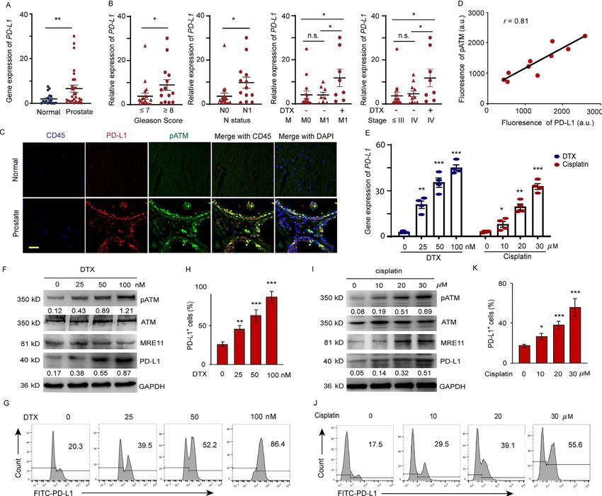
Open access
H3, rabbit anti-human phospho- p65 (Cell signal tech- RESULTS
J Immunother Cancer: first published as 10.1136/jitc-2020-001758 on 22 July 2021. Downloaded from http://jitc.bmj.com/ on October 17, 2021 by guest. Protected by copyright.
nology) and mouse anti-human PD-L1 antibody (Abcam) Phosphorylated ATM is correlated with the upregulation of
or normal control IgG 6–8 hours at 4°C. Then, Alexa PD-L1 induced by DTX
Flour 488- conjugated goat anti- rabbit IgG and Alexa PD-L1 expressions in 18 normal and 33 prostate tumor
Flour 594-conjugated goat anti-mouse IgG were added tissues were detected by qPCR (online supplemental table
and incubated for 1 hour at 37°C. The nucleus was 1). As shown in figure 1A, higher expression of PD-L1 was
stained with 4'6-diamidino-2-phenylindole and mounted observed in the prostate tumor tissues compared with that
with ProLong Gold Antifade Reagent (Life Technolo- in normal tissues. There is a positive correlation between
gies). The images were analyzed using a Zeiss LSM 880 PD-L1 expression and Gleason score (score ≤ 7 vs. score ≥
Confocal Imaging System (Zeiss, Jena, Germany).31 8, p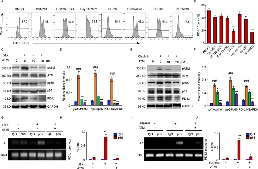
Open access
J Immunother Cancer: first published as 10.1136/jitc-2020-001758 on 22 July 2021. Downloaded from http://jitc.bmj.com/ on October 17, 2021 by guest. Protected by copyright.
Figure 1 DTX induces PD-L1 expression in patients with prostate cancer. (A–D) Expression of PD-L1 in prostate cancer tissue
from prostate cancer patients. (A) PD-L1 gene expression was quantified by qPCR in normal and prostate cancer tissues. The
results from 33 patients are represented as scatter plots and bar graphs displaying the mean±SEM of the normal and prostate
cancer tissues. (B) Relative PD-L1 expression in 33 prostate patients was assessed for correlation with Gleason sum score,
TNM status, cancer stage, and docetaxel (DTX) treatment. symbols represent individual subjects; bars show the mean±SEM.
(C, D) Immunofluorescence staining of PD-L1 (green), pATM (red) and CD45 (blue) in the sections of normal and prostate tumor
tissues from 10 patients with prostate cancer. (C) A representation image was shown. (D) Correlation of PD-L1 fluorescence
intensity with pATM. (E–K) Cell stresses upregulate PD-L1 expression due to DTX and cisplatin in a dose dependent manner.
PC-3 cells were examined at 6 hours after treating with DTX at concentrations of 0, 25, 50, 100 nM; at 24 hours after treating with
cisplatin at concentrations of 0, 10, 20, and 30 µM. (E) PD-L1 gene expression after DTX and cisplatin treatment was quantified
by qPCR. Results from four experiments. (F–H) DTX treatment. (F) Representative blotting for ATM activation, MRE11 and PD-
L1 expression after DTX treatment. results from four experiments. (G, H) PD-L1 expression was detected by surface staining
with FITC-PD-L1 antibody with isotype IgG as a control. Representative FACS results from four experiments are shown in (G)
and with quantifications in (H). (I–K) Cisplatin treatment. (I) Representative blotting for ATM activation and PD-L1 expression
after cisplatin treatment. Results from three experiments. (J, K) PD-L1 expression was detected by FITC-PD-L1 antibody with
isotype IgG as a control. Representative FACS results from three experiments are shown in (J) and with quantifications in (K). All
data are shown as mean±SEM. *POpen access
J Immunother Cancer: first published as 10.1136/jitc-2020-001758 on 22 July 2021. Downloaded from http://jitc.bmj.com/ on October 17, 2021 by guest. Protected by copyright.
Figure 2 ATM regulates PD-L1 expression induced by DTX. (A–C) PC-3 cells were exposed to ATM shRNA. Forty-eight hours
later, the cells were treated with 50 nM DTX for 6 hours and then collected. (A) Knockdown efficiency was detected by qPCR.
(B) ATM and PD-L1 expression was determined by Western blotting. (C) The effect of ATM knockdown on the PD-L1 expression
was detected by FACS. (D–F) PC-3 cells were transfected with the control and ATM plasmid without DTX treatment. Forty-
eight hours later, the cells were collected. (D) Transfection efficiency was detected by qPCR. (E) PD-L1 expression was detected
by Western blotting. (F) The effect of ATM transfection on PD-L1 expression was detected by FACS. (G–J) ATM inhibitor
(KU55933) can block the PD-L1 expression induced by DTX. PC-3 cells were treated with 50 nM DTX following treatment
with the KU55933. (G) PD-L1 gene expression was quantified by qPCR. (H) ATM, pATM, MRE11 and PD-L1 expression
was determined by Western blotting. (I, J) PD-L1 expression was detected by FACS. (K–N) ATM inhibitor blocks the PD-L1
expression induced by cisplatin. PC-3 cells were treated with 20 µM cisplatin following treatment with the KU55933. (K) PD-
L1 gene expression was quantified by qPCR. (L) ATM, pATM and PD-L1 expression was determined by Western blotting. (M,
N) PD-L1 expression was detected by FACS. (O, P) DTX induces PD-L1 expression is DDR independent. PC-3 cells were
examined after treating with DTX and cisplatin. (O) Co-immunostaining of γH2AX (green) and phospho-histone H3 (red) in the
PC-3 cells. Scale bar, 20 µm. (P) Co-immunostaining of 53BP1 (green) and phospho-histone H3 (red). Scale bar, 20 µm. All
data are shown as mean±SEM. ###POpen access
ATM knockdown markedly reduced the protein concen- suppressed cisplatin but not DTX-induced PD-L1 expres-
J Immunother Cancer: first published as 10.1136/jitc-2020-001758 on 22 July 2021. Downloaded from http://jitc.bmj.com/ on October 17, 2021 by guest. Protected by copyright.
tration of PD-L1 (figure 2B). To link increased PD-L1 sion, suggesting DTX- induced ATM and PD- L1 activa-
expression with the activation of ATM kinase activity, tion is not trigged by DDR-induced G2/M checkpoint
extracellular PD- L1 and intracellular pATM expres- (online supplemental figure 6). To further verify the
sion levels were compared by FACS in individual cells association of observed responses with the interruption
(figure 2C). We subsequently found that the higher the in microtubule, PTX, a microtubule inhibitor, was added
mean fluorescence intensity (MFI) of pATM, the more to PC-3 cells with the reported treatment which did not
intense the expression of PD-L1 in the cell membrane. induce DNA damage.37 PTX activated ATM signal and
To test the hypothesis that ectopic activation of ATM PD-L1 expression was similar to DTX, which confirmed
could upregulate PD- L1 expression, His- ATM or
Flag- that disruption of microtubule-induced ATM-dependent
control constructs were transfected into PC-3 cells. It PD-L1 expression (online supplemental figure 7).
was observed that the PD-L1 protein levels had substan-
tially increased in the cells transfected with ATM plasmid PD-L1 upregulation is mediated via the NF-κB pathway
(figure 2D–F), indicating that ATM kinase activation is To elucidate the mechanism underlying PD-L1 activation
required for DTX-induced PD-L1 expression. by DTX, a panel of signal pathway inhibitors (STAT3i:
To further investigate whether the PD-L1 expression S3I-201; STAT1i: Fludarabine; MEK1/2i: UO126-EtOH;
is associated with ATM activation under DTX chemo- Bay11-7082: TNFα-induced NF-κBi; JSH-23: NF-κBi for
therapy, cancer cells were treated with DTX in combina- p65 nuclear translocation; TGF-βRIi: SD-208; ATMi:
tion with different doses of the ATM inhibitor. Cisplatin, KU55933) were screened to identify the exact pathways
which activates ATM through DDR signaling, was used as regulating the transcription of PD- L1. DTX- induced
a positive control (figure 2G–N). Inhibition of the enzy- PD-L1 upregulation in cancer cells was substantially
matic activity of ATM attenuated the DTX-induced upreg- suppressed by JSH-23, which normalized the PD- L1
ulation of PD- L1 expression (figure 2G–J), as well as expression as effectively as ATM inhibitor (figure 3A,B),
cisplatin-induced PD-L1 expression (figure 2K–N). Treat- indicating that p65- dependent NF-κB activation was
ment with 20 µM of KU55933 was sufficient to suppress involved in the modulation of PD-L1 expression.
PD-L1 expression to the level observed in the untreated Further, we confirmed the link between the induction
cells. Taken together, these results suggested that the of the ATM activity, PD- L1 expression and increased
activation of ATM kinase was directly responsible for the activation of NF-κB signaling in the cancer cells after
enhanced expression of PD-L1 in cancer cells after treat- DTX or cisplatin treatment (figure 3C–J). The activity
ment with DTX. of ATM kinase induced by both treatments was inhib-
ited by KU55933 in tumor cells. Moreover, we observed
DTX-induced PD-L1 expression is not dependent on DDR increased phosphorylation of the NF-κB subunit p65 in
activated G2/M checkpoint the cancer cells treated with either DTX (figure 3C,D)
Several proteins involved in DNA damage signaling such or cisplatin (figure 3E,F). The results of a dose depen-
as γH2AX and 53BP1 produce discrete DNA damage dent experiment indicated that the inhibition of ATM
foci.34 To further compare the mechanisms of DTX and activity was accompanied by a marked loss of the NF-κB
cisplatin-induced PD-L1 expression, the expressions of activity and followed by a reduction in the PD-L1 expres-
γH2AX and 53BP1 after DTX and cisplatin treatment were sion . In addition, we further performed chIP assays in
checked. Accumulation of DNA breaks was confirmed by DTX treated PC-3 cells with and without ATM inhibitor.
immunostaining for γH2AX and 53BP1 in the nuclei of Increased p65 bindings to the PD-L1 promotor were
cisplatin treated cells. However, γH2AX and 53BP1 foci observed in response to DTX or cisplatin, and the ATM
in the nuclei of DTX treated cells were not significantly inhibitor effectively blocked the binding of p65 to the PD-
higher (figure 2O,P). Moreover, ATM undergoes dimer L1 promoter (figure 3G–J), suggesting that NF-κB p65
dissociation in response to DSBs.35 No significant dimer/ acts downstream of the ATM to block PD-L1 expression
monomer transmission was observed after DTX treatment on both DTX and cisplatin chemotherapy.
(online supplemental figure 4), suggesting that DTX-
induced ATM activation might not show DDR response. NEMO is required for PD-L1 upregulation induced by DTX
DTX-induced cell accumulation in G2/M phase was indi- Multiple cell stress can activate ATM, which phosphor-
cated by phospho-histone H3 staining (figure 2O,P) and ylates NEMO and enhances its SUMOylation. This step
cell cycle analysis via propidium iodide (online supple- is necessary for the p65 nuclear translocation and NF-κB
mental figure 5). However, there were no distinct γH2AX activation.38–40 NBD is a cell-permeable synthetic peptide
and 53BP1 signals in both phospho-H3 positive and nega- capable of selectively inhibiting the activation of NF-κB
tive cells after DTX treatment (figure 2O,P). Generally, by disrupting the formation of NEMO and the IKKβ
ATM functions upstream of ATR in DDR-induced G2/M complex, thus, decreasing the NF-κB p65 dependent
checkpoint arrest.36 To further confirm DTX- induced gene expression by inhibiting its nuclear translocation.
ATM activation being DDR-induced G2/M checkpoint To determine whether ATM activated NF-κB and upreg-
independent, PC-3 cells were treated with ATR inhib- ulated PD-L1 expression through NEMO, PC-3 cells were
itor following DTX or cisplatin. ATR inhibitor effectively treated with the NBD followed by a treatment with either
Wang Z, et al. J Immunother Cancer 2021;9:e001758. doi:10.1136/jitc-2020-001758 7Open access
J Immunother Cancer: first published as 10.1136/jitc-2020-001758 on 22 July 2021. Downloaded from http://jitc.bmj.com/ on October 17, 2021 by guest. Protected by copyright.
Figure 3 NF-κB activity is required for the upregulation of PD-L1 in PC-3 cells after DTX treatment. (A, B) PC-3 cells were
treated with 50 nM DTX following treatment with different pathway inhibitors S31-201, U0126-EtOH, Bay11-7082, JSH-23,
SD-208, fludarabine, KU55933 or a vehicle. (A) PD-L1 expression was detected by surface staining with FITC-PD-L1 antibody,
followed by FACS with isotype IgG as the control. representative results of FACS from three experiments are shown in (A) and
with quantifications in (B), data are shown as mean±SEM. *POpen access
J Immunother Cancer: first published as 10.1136/jitc-2020-001758 on 22 July 2021. Downloaded from http://jitc.bmj.com/ on October 17, 2021 by guest. Protected by copyright.
Figure 4 The small NEMO blocking peptide (NBD) blocks PD-L1 expression induced by DTX. (A–D) PC-3 cells were exposed
to 50 nM DTX following treatment with indicated doses of NBD. (A) PD-L1 gene expression after DTX treatment was quantified
by qPCR. (B) PD-L1 and pp65 expressions were detected by FACS. (C) Percentage of PD-L1 positive cells were quantified
from three experiments. (D) MFI of FITC-pp65 was quantified from three experiments. (E–H) PC-3 cells were exposed to 20 µM
cisplatin with indicated doses of NBD. (E) PD-L1 gene expression was quantified by qPCR. (F) PD-L1 and pp65 expressions
were detected by FACS. (G) Percentage of PD-L1 positive cells were quantified from three experiments. (H) MFI of FITC-pp65
was quantified from three experiments. (I, J) Co-immunostaining of pp65 (green) and PD-L1 (red) in the PC-3 cells after DTX
and NBD treatment. (I) Representative images are from one of three sections from indicate treated groups. Scale bar, 20 µm. (J)
Quantification of PD-L1 positive cells from three experiments. (K, L) Co-immunostaining of pp65 (green) and PD-L1 (red) in the
PC-3 cells after cisplatin and NBD treatment. (K) Representative images are from one of three sections from indicate treated
groups. Scale bar, 20 µm. (L) Quantification of PD-L1 positive cells from three experiments. ###POpen access
ATM regulated PD-L1 expression through association with by immunoblotting with anti-NEMO or co-IP with anti-
J Immunother Cancer: first published as 10.1136/jitc-2020-001758 on 22 July 2021. Downloaded from http://jitc.bmj.com/ on October 17, 2021 by guest. Protected by copyright.
NEMO NEMO and immunoblotting with anti- ATM, showed
It has been reported that DNA damage inducing stress enhanced stress- induced ATM- NEMO colocalization
responses increased the ATM activation and binding to in the tumor cells after DTX (figure 5A,B) or cisplatin
NEMO in the nuclei and a further study indicated that treatment (figure 5C,D). To confirm that NEMO and
this mechanism also works with other cell toxic stress ATM interaction is dependent on the phosphorylation
even without DNA damage.40 To further confirm that of ATM, PC-3 cells were treated with DTX plus KU55933
the interaction between ATM and NEMO is a critical (figure 5E,F)or cisplatin plus KU55933 (figure 5G,H).
event involved in the activation of PD-L1 in response to The results of reciprocal co-IP showed that the binding of
DTX, we performed co- immunoprecipitation (co- IP) ATM to NEMO had decreased substantially in response to
experiment. Reciprocal co- IP, with anti-
ATM, followed increase concentration of the ATM inhibitor, suggesting
Figure 5 ATM regulates PD-L1 expression by interacting with NEMO. (A, B) PC-3 cells were treated with DTX (0 nM, 25 nM,
50 nM) for 6 hours. (A) Association between DTX-induced ATM and NEMO was determined by the coimmunoprecipitation assay
with anti-ATM (a mouse monoclonal IgG) followed by immunoblotting with anti-NEMO (a goat polyclonal antibody), and (B)
anti-NEMO (a goat polyclonal IgG) followed by immunoblotting with anti-ATM (a mouse monoclonal antibody). Representative
blots from three experiments are shown. (C, D) PC-3 cells were treated with cisplatin (0 µM, 10 µM, 20 µM) for 24 hours. (C)
Association between ATM and NEMO was determined by the coimmunoprecipitation assay with anti-ATM, followed by
immunoblotting with anti-NEMO and (D) anti-NEMO, followed by immunoblotting with anti-ATM. Representative blots from
three experiments are shown. (E, F) PC-3 cells were treated with 50 nM DTX followed by ATM inhibitors or vehicle for 6 hours.
(E) Association between ATM and NEMO was determined by the coimmunoprecipitation assay with anti-ATM, followed by
immunoblotting with anti-NEMO, and (F) anti-NEMO followed by immunoblotting with anti-ATM. Representative blots from three
experiments are shown. (G, H) PC-3 cells were treated with 20 µM cisplatin, followed by ATM inhibitors or vehicle for 24 hours.
(G) Association between ATM and NEMO was determined by the co-immunoprecipitation assay with anti-ATM followed by
immunoblotting with anti-NEMO, and (H) anti-NEMO followed by immunoblotting with anti-ATM. Representative blots from
three experiments are shown. ATM, ataxia telangiectasia mutation; DTX, docetaxel; NEMO, NF-κB essential modulator; PD-L1,
programmed cell death ligand-1.
10 Wang Z, et al. J Immunother Cancer 2021;9:e001758. doi:10.1136/jitc-2020-001758Open access
that the interaction between NEMO and ATM depends antitumor effect of NEMO and ATM inhibitors depends
J Immunother Cancer: first published as 10.1136/jitc-2020-001758 on 22 July 2021. Downloaded from http://jitc.bmj.com/ on October 17, 2021 by guest. Protected by copyright.
on ATM kinase activity. on T cells.
Moreover, we found that oxidative, ethanol and elec- To further confirm that pharmacological inhibition
tric treatments which enhanced NF-κB signaling through of NEMO and ATM enhances the antitumor efficacy of
NEMO SUMOylation also upregulated PD-L1 expression DTX, a chimeric mouse was created using PC-3 cells and
in PC-3 cells, suggesting the key role of SUMOylation PBMCs of patients (online supplemental figure 9A–H).
of NEMO in regulating PD-L1 signaling (online supple- Similar to the aforementioned results, the combination
mental figure 8). of NBD or KU55933 with DTX substantially increased
the tumor inhibitory rate, while no therapeutic benefit
DTX with either NEMO or ATM inhibitor restricted tumor of the combination treatment was observed in the T cell
growth in vivo depletion groups (Online supplemental figure 9I–L).
To determine whether inhibition of the ATM or NEMO Furthermore, KU55933 and NBD attenuated the DTX-
could enhance the antitumor efficacy of DTX in vivo, induced PD-L1 upregulation (online supplemental figure
RM-1 prostate cancer cells were injected into mice, which 9M–P). An immunohistochemically analysis of the CD3+
were subsequently treated with DTX, DTX plus KU55933, T cells in the tumor sections indicated that the inhibitors
and DTX plus NBD separately, while DTX plus an anti- could dramatically increase the T cell tumor-infiltration
PD-L1 blockade was used as the therapeutic control. and percentage of CD3+IFNγ+ effector T cells compared
Although DTX alone attenuated the tumor growth, its with that of DTX (online supplemental figure 9Q,R).
antitumor efficacy was dramatically enhanced in combi- Notably, the proliferation capacity of CD8+ T cells was also
increased in the combination treatment groups (online
nation with NBD or KU55933, which was as effective
supplemental figure 9S,T), confirming that the inhibition
as anti-PD-
L1 antibody, as indicated by the decreased
of NEMO and ATM plus DTX resulted in a significantly
tumor inhibitory rate (40.3% DTX vs. 62.8% NBD+DTX,
greater antitumor response than that by DTX alone.
74.9% KU55933+DTX, 58.2% anti-PD-L1+DTX, pOpen access
J Immunother Cancer: first published as 10.1136/jitc-2020-001758 on 22 July 2021. Downloaded from http://jitc.bmj.com/ on October 17, 2021 by guest. Protected by copyright.
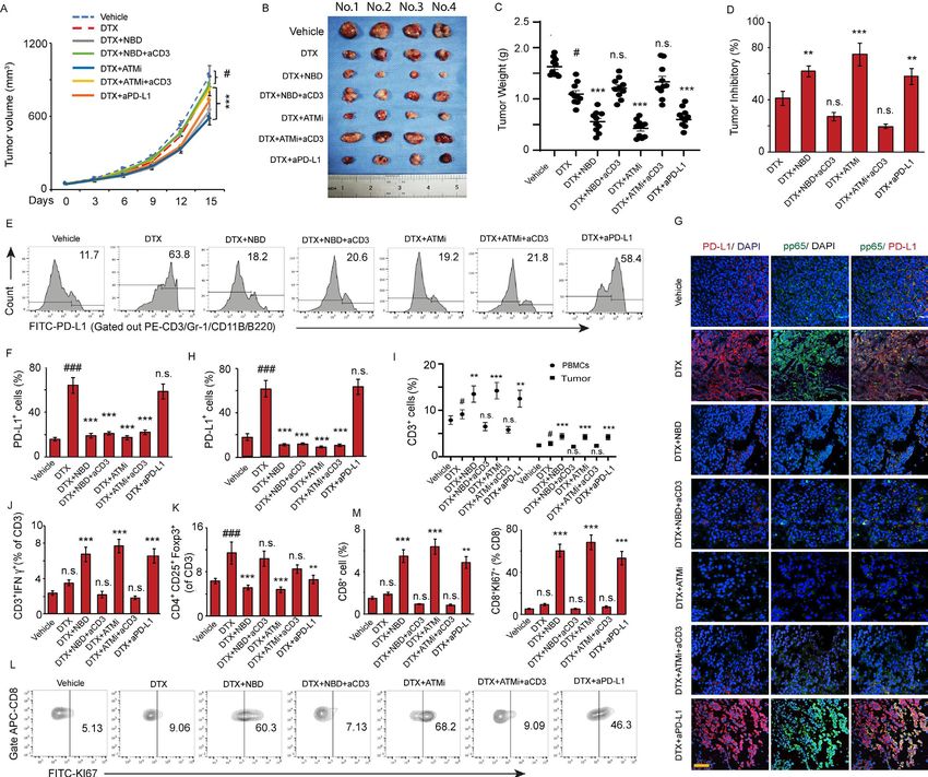
Figure 6 ATM and NEMO inhibitors promote the antitumor effect of DTX through downregulation of PD-L1. C57BL/6 mice
implanted with RM-1 prostate tumors were divided to 7 groups with 10 mice per group and treated with (1) vehicle, (2) DTX,
(3) NBD plus DTX, (4) NBD plus DTX and anti-CD3, (5) KU55933 plus DTX, (6) KU55933 plus DTX and anti-CD3, and (7)
anti-PD-L1 plus DTX as a therapeutic control group. (A–D) ATM and NEMO inhibitors promote the antitumor effect of DTX.
(A) Tumor volumes were measured at the indicated day after treatment. ANOVA, #pOpen access
One of the key mechanistic questions was whether the CONCLUSION
J Immunother Cancer: first published as 10.1136/jitc-2020-001758 on 22 July 2021. Downloaded from http://jitc.bmj.com/ on October 17, 2021 by guest. Protected by copyright.
dual signal initiation model of ATM-NEMO for the PD-L1 Our study identified a previously uncharacterized type
upregulation was relevant to the PD- L1 upregulation of ATM- NEMO signaling induced by DTX capable of
following DTX chemotherapy in prostate cancer, which suppressing the tumor immunity by activating PD- L1
does not induce obvious DNA damage signaling. Indeed, expression. The present findings indicated that combina-
we found that chemotherapeutic treatment with DTX tion treatment with DTX and NEMO or ATM inhibitors
caused a 2-fold higher of PD-L1 induction than cisplatin can be exploited to develop a new therapeutic strategy to
with a considerably lower dose in prostate cancer cells. overcome cancer immune tolerance associated with the
Consistently, this activation involved both ATM activa- use of DTX, providing an alternative to PD-L1 antibodies
as a means of restoring cancer immunity. This might be
tion and ATM-NEMO binding. Moreover, both ATM and
of great benefit for patients resistant to antibody therapy.
NEMO function inhibition can blunt the PD-L1 in pros-
tate cancer cells induced by DTX. These results revealed Acknowledgements We thank all patients and medical staff who generously
a novel mechanism of DTX resistance by the upregula- contributed to this study. All authors have read and approved the final submitted
tion of PD-L1. version of the manuscript.
Our findings are clinically significant and highly rele- Contributors All authors were involved in drafting the article or revising it critically
for important intellectual content. Study conception and design: ZW, XZ, WL, LC
vant for the treatment of cancers that lack PD-L1 expres- and LS. Acquisition of data: ZW, XZ, WL, QS, ZYH, XZ, HC, CM, BH, WO, JC and GZ.
sion, such as prostate cancer. Recent reports on the Analysis and interpretation of data: ZW, XZ, WL, LC and LS.
PD-1/PD-L1 immunotherapy for prostate cancer are rela- Funding This work was supported by the National Natural Science Foundation
tively disappointing.44 45 However, we have observed an of China (grant no. 81901849 and 82071818), the Guangdong Natural Science
Foundation (grant no. 2018A030310328 and 2021A1515011030), Medical
increased PD-L1 expression after DTX treatment, which
Scientific Research Foundation of Guangdong Province, China (grant no. A2018344).
correlated with tumor progression in the mouse bearing
Competing interests No, there are no competing interests.
the prostate tumor. The combination of DTX with the
Patient consent for publication Not required.
pharmacological inhibition of ATM or NBD attenuated
Ethics approval The ethical approval was authorized by the Institutional Review
DTX-induced PD-L1 upregulation, which had synergisti-
Board of the First Affiliated Hospital of Sun Yat-sen University, and written consent
cally enhanced the anti-tumor efficacy of DTX.46 Mech- was obtained from all patients included.
anistically, DTX dramatically promoted the nuclear Provenance and peer review Not commissioned; externally peer reviewed.
translocation of NF-κB p65 and induced PD-L1 expres- Data availability statement Data are available on reasonable request. The
sion. ATM phosphorylates NEMO, which results in NEMO datasets used and/or analyzed during the current study are available from the
SUMOylation, is a key step for NF-κB p65 nuclear trans- corresponding author on reasonable request.
location. We observed that a dose-dependent increasing Supplemental material This content has been supplied by the author(s). It has
of ATM-NEMO association after DTX treatment. There- not been vetted by BMJ Publishing Group Limited (BMJ) and may not have been
peer-reviewed. Any opinions or recommendations discussed are solely those
fore, the NF-κB-PD-L1 axis was activated by DTX through of the author(s) and are not endorsed by BMJ. BMJ disclaims all liability and
promoting ATM-NEMO binding. ATM inhibition effec- responsibility arising from any reliance placed on the content. Where the content
tively interrupted ATM interaction with NEMO and NBD includes any translated material, BMJ does not warrant the accuracy and reliability
of the translations (including but not limited to local regulations, clinical guidelines,
specifically blocked NEMO activation. Thus, the inhibi- terminology, drug names and drug dosages), and is not responsible for any error
tion of NEMO and ATM along with DTX resulted in a and/or omissions arising from translation and adaptation or otherwise.
significantly greater antitumor response than that of DTX Open access This is an open access article distributed in accordance with the
alone. Creative Commons Attribution Non Commercial (CC BY-NC 4.0) license, which
permits others to distribute, remix, adapt, build upon this work non-commercially,
The question was raised how ATM-NEMO mediates and license their derivative works on different terms, provided the original work is
PD-L1 expression, which can be related to the enhanced properly cited, appropriate credit is given, any changes made indicated, and the use
immunosuppression following DTX chemotherapy. It is non-commercial. See http://creativecommons.org/licenses/by-nc/4.0/.
has been reported that PD-1 is upregulated on the tumor- ORCID iD
infiltrating T cells in the tumor microenvironment of Lan Shao http://orcid.org/0000-0001-6725-4433
prostate cancer,47 which characterized by lacking the
expression of perforin and IFN-γ.48–50 We found that
suppressing the PD-L1 expression by inhibiting NEMO REFERENCES
and ATM increased the number of circulating and 1 Weber JS, Kudchadkar RR, Yu B, et al. Safety, efficacy, and
biomarkers of nivolumab with vaccine in ipilimumab-refractory or
tumor-infiltrating T cells in both RM-1 and PC-3 tumor- -naive melanoma. J Clin Oncol 2013;31:4311–8.
bearing mice. Specifically, an increase percentage of the 2 Garon EB, Rizvi NA, Hui R, et al. Pembrolizumab for the treatment of
non-small-cell lung cancer. N Engl J Med 2015;372:2018–28.
IFN-γ positive cytotoxic T cells and decrease of the regu- 3 Zou W, Wolchok JD, Chen L. Pd-L1 (B7-H1) and PD-1 pathway
latory T cells contribute to the reverse of the immuno- blockade for cancer therapy: mechanisms, response biomarkers,
and combinations. Sci Transl Med 2016;8:328rv4.
suppressive state of tumor. Furthermore, an increase in 4 Thoma C. Prostate cancer: PD-L1 expression is common and
the proliferation capacity of CD8 T cells may also play an indicates poor prognosis. Nat Rev Urol 2016;13:5.
5 Gevensleben H, Dietrich D, Golletz C, et al. The immune checkpoint
important role in the enhancing of the antitumor effect regulator PD-L1 is highly expressed in aggressive primary prostate
of DTX. cancer. Clin Cancer Res 2016;22:1969–77.
Wang Z, et al. J Immunother Cancer 2021;9:e001758. doi:10.1136/jitc-2020-001758 13Open access
6 Massari F, Ciccarese C, Caliò A, et al. Magnitude of PD-1, PD-L1 28 Wu Z-H, Wong ET, Shi Y, et al. Atm- and NEMO-dependent ELKS
J Immunother Cancer: first published as 10.1136/jitc-2020-001758 on 22 July 2021. Downloaded from http://jitc.bmj.com/ on October 17, 2021 by guest. Protected by copyright.
and T lymphocyte expression on tissue from castration-resistant ubiquitination coordinates TAK1-mediated IKK activation in response
prostate adenocarcinoma: an exploratory analysis. Target Oncol to genotoxic stress. Mol Cell 2010;40:75–86.
2016;11:345–51. 29 McCool KW, Miyamoto S. Dna damage-dependent NF-κB
7 Schepisi G, Farolfi A, Conteduca V, et al. Immunotherapy for prostate activation: NEMO turns nuclear signaling inside out. Immunol Rev
cancer: where we are headed. Int J Mol Sci 2017;18:2627. 2012;246:311–26.
8 Papanicolau-Sengos A, Yang Y, Pabla S, et al. Identification 30 Shao L, Fujii H, Colmegna I, et al. Deficiency of the DNA repair
of targets for prostate cancer immunotherapy. Prostate enzyme ATM in rheumatoid arthritis. J Exp Med 2009;206:1435–49.
2019;79:498–505. 31 Shao L, Zhou HJ, Zhang H, et al. SENP1-mediated NEMO
9 Barata P, Agarwal N, Nussenzveig R, Gerendash B, et al. Clinical deSUMOylation in adipocytes limits inflammatory responses and
activity of pembrolizumab in metastatic prostate cancer with type-1 diabetes progression. Nat Commun 2015;6:8917.
microsatellite instability high (MSI-H) detected by circulating tumor 32 Vendetti FP, Karukonda P, Clump DA, et al. Atr kinase inhibitor
DNA. J Immunother Cancer 2020;8:e001065. AZD6738 potentiates CD8+ T cell-dependent antitumor activity
10 Martin AM, Nirschl TR, Nirschl CJ, et al. Paucity of PD-L1 expression following radiation. J Clin Invest 2018;128:3926–40.
in prostate cancer: innate and adaptive immune resistance. Prostate 33 Gao Y, Nihira NT, Bu X, et al. Acetylation-Dependent regulation
Cancer Prostatic Dis 2015;18:325–32. of PD-L1 nuclear translocation dictates the efficacy of anti-PD-1
11 Messina C, Messina M, Boccardo F. Abiraterone or docetaxel for immunotherapy. Nat Cell Biol 2020;22:1064–75.
Castration-sensitive metastatic prostate cancer? that is the question! 34 Fernandez-Capetillo O, Chen H-T, Celeste A, et al. DNA damage-
Eur Urol 2018;73:147–8. induced G2-M checkpoint activation by histone H2AX and 53BP1.
12 Tucci M, Bertaglia V, Vignani F, et al. Addition of docetaxel to Nat Cell Biol 2002;4:993–7.
androgen deprivation therapy for patients with hormone-sensitive 35 Bakkenist CJ, Kastan MB. DNA damage activates ATM through
metastatic prostate cancer: a systematic review and meta-analysis. intermolecular autophosphorylation and dimer dissociation. Nature
Eur Urol 2016;69:563–73. 2003;421:499–506.
13 Tsao C-K, Galsky MD, Oh WK. Docetaxel for metastatic hormone- 36 Jazayeri A, Falck J, Lukas C, et al. ATM- and cell cycle-dependent
sensitive prostate cancer: urgent need to minimize the risk of regulation of ATR in response to DNA double-strand breaks. Nat Cell
neutropenic fever. Eur Urol 2016;70:707–8. Biol 2006;8:37–45.
14 Tannock IF, de Wit R, Berry WR, et al. Docetaxel plus prednisone or 37 Poruchynsky MS, Komlodi-Pasztor E, Trostel S, et al. Microtubule-
mitoxantrone plus prednisone for advanced prostate cancer. N Engl Targeting agents augment the toxicity of DNA-damaging agents by
J Med 2004;351:1502–12. disrupting intracellular trafficking of DNA repair proteins. Proc Natl
15 Nuhn P, De Bono JS, Fizazi K, et al. Update on systemic prostate Acad Sci U S A 2015;112:1571–6.
cancer therapies: management of metastatic castration-resistant 38 Wu Z-H, Shi Y, Tibbetts RS, et al. Molecular linkage between the
prostate cancer in the era of precision oncology. Eur Urol kinase ATM and NF-kappaB signaling in response to genotoxic
2019;75:88–99. stimuli. Science 2006;311:1141–6.
16 Leduc C, Adam J, Louvet E, et al. Tpf induction chemotherapy 39 Mabb AM, Wuerzberger-Davis SM, Miyamoto S. Piasy mediates
increases PD-L1 expression in tumour cells and immune cells NEMO sumoylation and NF-kappaB activation in response to
in head and neck squamous cell carcinoma. ESMO Open genotoxic stress. Nat Cell Biol 2006;8:986–93.
2018;3:e000257. 40 Wuerzberger-Davis SM, Nakamura Y, Seufzer BJ, et al. Nf-kappaB
17 Francini E, Sweeney CJ. Docetaxel activity in the era of Life- activation by combinations of NEMO sumoylation and ATM
prolonging hormonal therapies for metastatic castration-resistant activation stresses in the absence of DNA damage. Oncogene
prostate cancer. Eur Urol 2016;70:410–2. 2007;26:641–51.
18 Conteduca V, Jayaram A, Romero-Laorden N, et al. Plasma androgen 41 Wang Z, Zhao J, Wang G, et al. Comutations in DNA damage
receptor and docetaxel for metastatic castration-resistant prostate response pathways serve as potential biomarkers for immune
cancer. Eur Urol 2019;75:368–73. checkpoint blockade. Cancer Res 2018;78:6486–96.
19 Fernández-García EM, Vera-Badillo FE, Perez-Valderrama B, et al. 42 Herbst RS, Soria J-C, Kowanetz M, et al. Predictive correlates of
Immunotherapy in prostate cancer: review of the current evidence. response to the anti-PD-L1 antibody MPDL3280A in cancer patients.
Clin Transl Oncol 2015;17:339–57. Nature 2014;515:563–7.
20 Yang Z, Shen Y, Oishi H, et al. Restoring oxidant signaling 43 Huang TT, Wuerzberger-Davis SM, Wu Z-H, et al. Sequential
suppresses proarthritogenic T cell effector functions in rheumatoid modification of NEMO/IKKgamma by SUMO-1 and ubiquitin
arthritis. Sci Transl Med 2016;8:331ra38. mediates NF-kappaB activation by genotoxic stress. Cell
21 Oricchio E, Saladino C, Iacovelli S, et al. ATM is activated by default 2003;115:565–76.
in mitosis, localizes at centrosomes and monitors mitotic spindle 44 Comiskey MC, Dallos MC, Drake CG. Immunotherapy in prostate
integrity. Cell Cycle 2006;5:88–92. cancer: teaching an old dog new tricks. Curr Oncol Rep 2018;20:75.
22 Zhu Z, Chen P, Yan Z. DNA damage response signaling as a 45 Haffner MC, Guner G, Taheri D, et al. Comprehensive evaluation of
predictive biomarker and synergistic therapeutic target for anti-PD-1/ programmed Death-Ligand 1 expression in primary and metastatic
PD-L1 immunotherapy in non-small cell lung cancer. Thorac Cancer prostate cancer. Am J Pathol 2018;188:1478–85.
2018;9:901–3. 46 Lacour M, Hiltbrunner S, Lee S-Y, et al. Adjuvant chemotherapy
23 Teo MY, Seier K, Ostrovnaya I, et al. Alterations in DNA damage increases programmed Death-Ligand 1 (PD-L1) expression in non-
response and repair genes as potential marker of clinical benefit from small cell lung cancer recurrence. Clin Lung Cancer 2019;20:391–6.
PD-1/PD-L1 blockade in advanced urothelial cancers. J Clin Oncol 47 Ebelt K, Babaryka G, Frankenberger B, et al. Prostate cancer lesions
2018;36:1685–94. are surrounded by Foxp3+, PD-1+ and B7-H1+ lymphocyte clusters.
24 Sun L-L, Yang R-Y, Li C-W, et al. Inhibition of ATR downregulates PD- Eur J Cancer 2009;45:1664–72.
L1 and sensitizes tumor cells to T cell-mediated killing. Am J Cancer 48 Ebelt K, Babaryka G, Figel AM, et al. Dominance of CD4+
Res 2018;8:1307–16. lymphocytic infiltrates with disturbed effector cell characteristics
25 Sato H, Niimi A, Yasuhara T, et al. DNA double-strand break repair in the tumor microenvironment of prostate carcinoma. Prostate
pathway regulates PD-L1 expression in cancer cells. Nat Commun 2008;68:1–10.
2017;8:1751. 49 Petitprez F, Fossati N, Vano Y, et al. PD-L1 expression and CD8 + T-
26 Jin X, Ding D, Yan Y, et al. Phosphorylated RB promotes cancer cell infiltrate are associated with clinical progression in patients with
immunity by inhibiting NF-κB activation and PD-L1 expression. Mol node-positive prostate cancer. Eur Urol Focus 2019;5:192-196.
Cell 2019;73:22–35. 50 Barach YS, Lee JS, Zang X. T cell coinhibition in prostate cancer:
27 Miyamoto S. Nuclear initiated NF-κB signaling: NEMO and ATM take new immune evasion pathways and emerging therapeutics. Trends
center stage. Cell Res 2011;21:116–30. Mol Med 2011;17:47–55.
14 Wang Z, et al. J Immunother Cancer 2021;9:e001758. doi:10.1136/jitc-2020-001758You can also read