Infusion of Plasma from Exercised Mice Ameliorates Cognitive Dysfunction by Increasing Hippocampal Neuroplasticity and Mitochondrial Functions in ...
←
→
Page content transcription
If your browser does not render page correctly, please read the page content below
International Journal of
            Molecular Sciences

Article
Infusion of Plasma from Exercised Mice Ameliorates
Cognitive Dysfunction by Increasing Hippocampal
Neuroplasticity and Mitochondrial Functions in
3xTg-AD Mice
Tae-Woon Kim 1,2 , Sang-Seo Park 2 , Joon-Young Park 3 and Hye-Sang Park 3, *
 1    Exercise Rehabilitation Research Institute, Department of Exercise & Health Science, Sangmyung University,
      Seoul 03016, Korea; twkim0806@naver.com
 2    Department of Physiology, College of Medicine, KyungHee University, Seoul 02447, Korea; sps07@naver.com
 3    Department of Kinesiology, College of Public Health and Cardiovascular Research Center, Lewis Katz School
      of Medicine, Temple University, Philadelphia, PA 19122, USA; parkjy@temple.edu
 *    Correspondence: hspark.happy@gmail.com

 Received: 26 March 2020; Accepted: 30 April 2020; Published: 6 May 2020

 Abstract: Alzheimer’s disease is the most common neurodegenerative brain disease causing dementia.
 It is characterized by slow onset and gradual worsening of memory and other cognitive functions.
 Recently, parabiosis and infusion of plasma from young mice have been proposed to have positive
 effects in aging and Alzheimer’s disease. Therefore, this study examined whether infusion of plasma
 from exercised mice improved cognitive functions related to the hippocampus in a 3xTg-Alzheimer’s
 disease (AD) model. We collected plasma from young mice that had exercised for 3 months and
 injected 100 µL of plasma into the tail vein of 12-month-old 3xTg-AD mice 10 times at 3-day intervals.
 We then analyzed spatial learning and memory, long-term memory, hippocampal GSK3β/tau proteins,
 synaptic proteins, mitochondrial function, apoptosis, and neurogenesis. In the hippocampus of
 3xTg-AD mice, infusion of plasma from exercised mice improved neuroplasticity and mitochondrial
 function and suppressed apoptosis, ultimately improving cognitive function. However, there was
 no improvement in tau hyperphosphorylation. This study showed that plasma from exercised mice
 could have a protective effect on cognitive dysfunction and neural circuits associated with AD via a
 tau-independent mechanism involving elevated brain-derived neurotrophic factor due to exercise.

 Keywords: Alzheimer’s disease; cognitive function; hippocampus; neuroplasticity; mitochondria;
 young plasma; exercise

1. Introduction
     Alzheimer’s disease (AD) is the most common cause of dementia, characterized by slow onset
and progressive decline of memory and cognitive functions. AD is associated with various cellular
changes in the brain, including synaptic injury, alterations in mitochondrial structure and function,
abnormal inflammatory response, extracellular accumulation of amyloid beta (Aβ), and intracellular
neurofibrillary tangles [1–3]. AD is caused by atrophy, senile plaques, and hyperphosphorylated tau
protein aggregates in the hippocampus, one of the neuroanatomical areas responsible for memory and
learning [4,5]. Cognitive dysfunction, including that involving memory and learning, is associated
with decreased neurogenesis in the hippocampus, which could result from decreased expression of
immature neuron factors, such as DCX (doublecortin), that signal the birth of new neurons [6].
     Tau overexpression and hyperphosphorylation have been found to impair axonal movement of cell
organelles, including mitochondria [7–9]. There has been much evidence demonstrating mitochondrial

Int. J. Mol. Sci. 2020, 21, 3291; doi:10.3390/ijms21093291                            www.mdpi.com/journal/ijms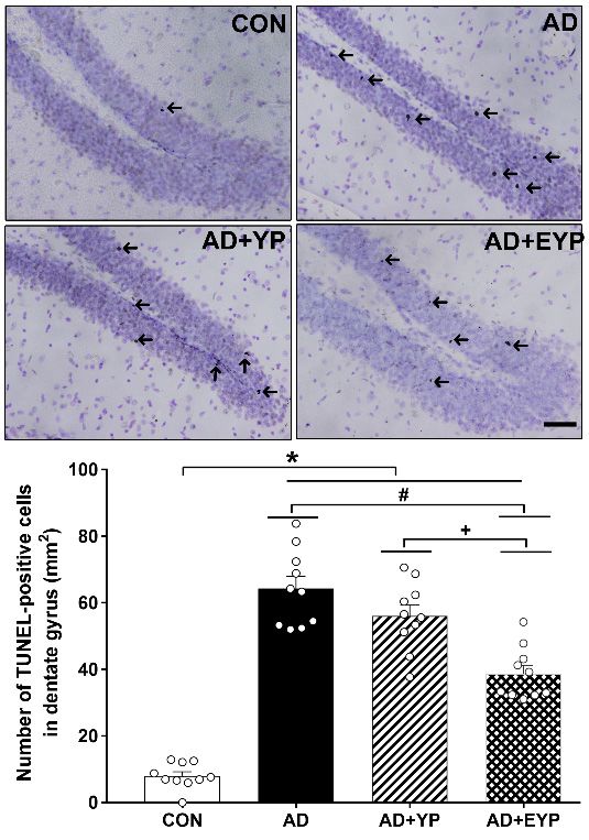
Int. J. Mol. Sci. 2020, 21, 3291                                                                                             2 of 16

dysfunction accompanied by aging and aging-related neurodegenerative disease [10]. In particular,
mitochondrial dysfunction has been observed in fibroblasts and hematopoietic cells from the brains
of AD transgenic mouse models and human patients with AD [11–14]. It has been suggested that
mitochondrial dysfunction could develop when the course of AD worsens or in all stages of the disease,
and that this process may occur not only in the brain, but systemically [13].
     In recent research on aging-related treatments, injection of young mouse blood into aged mice
has shown positive effects, which may be applied as a new experimental approach for the treatment
of aging-related diseases. According to previous studies, parabiosis between 18-month-old and
5-week-old mice resulted in increased expression of genes related to brain activity than in mice
that did not receive blood transfusions, especially immediate-early genes in hippocampal cells [15].
This indicated that parabiosis increased brain activity and improved memory. With increasing age,
the synapses, which form the network for communication between neurons, begin to regress, leading
to degeneration of neurons, atrophy of the brain, and a sudden increase in neurodegenerative disease.
Blood links the diverse tissues of the body. Blood is not only a transport medium for oxygen and cells,
but also helps to fight infectious disease and conveys information in the form of hormones and other
molecules. In other words, blood plays an important role in conveying information between cells and
tissues, including the brain.
     Meanwhile, exercise not only has a positive effect in patients with neurodegenerative diseases such
as AD and Parkinson’s disease, but also has positive effects on plasma. Exercise-induced neuroplasticity,
characterized by inhibited apoptosis and increased neurogenesis and brain-derived neurotrophic factor
(BDNF), facilitates recovery from brain damage after traumatic brain injury, ischemia, and stroke [16–19].
The plasma concentration of BDNF increases after aerobic exercise [20], and exercise training has also
been reported to increase resting plasma BDNF [21]. As AD worsens, it eventually leads to loss of
motor function. Although exercise has various positive effects on the brain, the drawback is that these
effects are only possible when the individual is capable of physical activity. Thus, in this study, we
aimed to examine whether transfusion of plasma from exercised mice could have similar effects to
exercise on cognitive function, hippocampal neuroplasticity, and mitochondrial function in AD.

2. Results

2.1. Effect of Exercise on Plasma BDNF in Donation Mice
     Enzyme-linked immunosorbent assay (ELISA) was performed to investigate changes in plasma
BDNF concentration. The group treated with exercised young plasma (EYP) showed significantly
increased plasma BDNF concentrations compared with the group treated with young plasma (YP)
(p < 0.001)
        Int. J.(Figure     1). 21, x
                Mol. Sci. 2020,                                                   3 of 17

      Figure 1.  Effect
               Figure     of exercise
                       1. Effect        on plasma
                                 of exercise on plasma brain-derived   neurotrophic
                                                         brain-derived neurotrophic    factor
                                                                                    factor    (BDNF)
                                                                                           (BDNF)         in donation
                                                                                                    in donation  plasma plasma
      mice. YP:mice. YP: young
                  young           plasma,
                            plasma,    EYP: EYP:  exercisedyoung
                                              exercised     young plasma.
                                                                   plasma. Data
                                                                            Dataareare
                                                                                    expressed  as the
                                                                                       expressed    asmean   ± standard
                                                                                                        the mean    ± standard
      error of error of the mean
               the mean     (SEM). (SEM).
                                      * p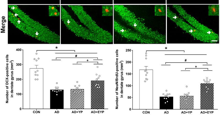
Int. J. Mol. Sci. 2020, 21, 3291 3 of 16
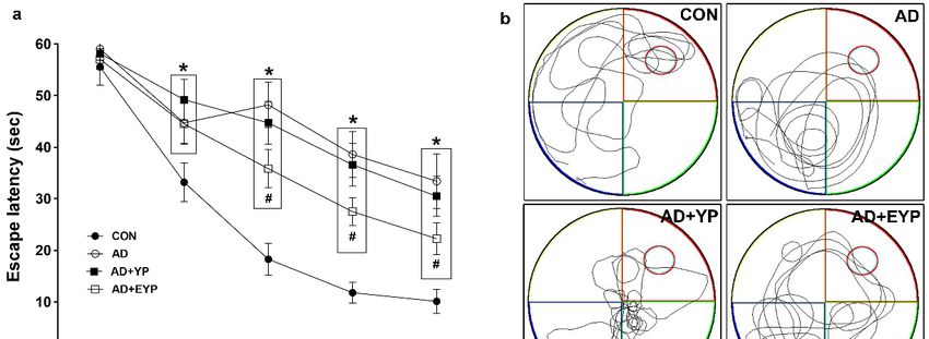
2.2. Effect of Plasma from Young Exercised Mice on Cognitive Functions in 3xTg-AD Mice
To assess spatial learning and long-term memory, the Morris water maze and step-through
avoidance tasks were performed. Spatial learning was assessed by measuring the time on the platform.
In spatial learning ability, the AD group took longer to find the platform from day 2 compared to the
control (CON) group (p < 0.001). The group treated with EYP took less time to find the platform than
the AD group starting from day 3 (Day 3: p = 0.039, Day 4: p = 0.017, Day 5: p < 0.001) (Figure 2a).
The AD group showed reduced spatial memory (p < 0.001) and long-term memory (p < 0.001) compared
to the CON group. The group treated with YP infusion did not demonstrate statistically significant
differences in these tests; however, infusion of plasma from young exercised mice improved spatial
memory and long-term memory compared with the AD group (p = 0.008 and p = 0.030, respectively).
Treatment comparisons revealed significant differences in spatial and long-term memory between AD
+ YP and AD + EYP combined groups (p < 0.001, p = 0.028, respectively) (Figure 2b,c). Infusion of
plasma from exercised young mice conferred positive effects in improving spatial memory, learning
ability, and
Int. J. Mol. Sci. long-term
2020, 21, x memory. 4 of 17
Figure Effectofofplasma
2. Effect
Figure 2. plasmafrom from young
young exercised
exercised mice
mice on on cognitive
cognitive functions
functions in 3xTg-AD
in 3xTg-AD mice.mice.
The
The Morris water maze task for spatial learning (a) and memory (b) and step through task
Morris water maze task for spatial learning (a) and memory (b) and step through task for long-term for long-term
memory
memory (c).
(c). CON:
CON:wild-type,
wild-type,AD: AD:3xTg-AD,
3xTg-AD, ADAD+ YP: 3xTg-AD
+ YP: 3xTg-ADandand
young plasma
young injection,
plasma AD +AD
injection, EYP:
+
3xTg-AD and exercised young plasma injection group. Data are expressed as
EYP: 3xTg-AD and exercised young plasma injection group. Data are expressed as the mean ± the mean ± standard
error of the
standard mean
error (SEM).
of the mean < 0.05 *compared
* p(SEM). with the with
p < 0.05 compared CONthe
group. p < 0.05# compared
CON# group. with thewith
p < 0.05 compared AD
group. + p < 0.05 between AD + YP and AD + EYP
the AD group. + p < 0.05 between AD + YP and AD + EYP groups.groups.
2.3. Effect of Plasma from Young Exercised Mice on GSK3β and Tau Protein Expression in Hippocampus
2.3. Effect of Plasma from Young Exercised Mice on GSK3β and Tau Protein Expression in Hippocampus
Western blot analysis was performed to investigate changes in GSK3β/tau protein in the
Western blot analysis was performed to investigate changes in GSK3β/tau protein in the
hippocampus. For group comparisons, results were normalized relative to the CON group. AD, AD
hippocampus. For group comparisons, results were normalized relative to the CON group. AD, AD
+ YP, and AD + EYP groups showed significantly decreased p-GSK3β/GSK3β ratios (p < 0.001) and
+ YP, and AD + EYP groups showed significantly decreased p-GSK3β/GSK3β ratios (p < 0.001) and
significantly increased p-tau (Ser262, Thr205)/tau ratios (p < 0.001) compared with the CON group.
significantly increased p-tau (Ser262, Thr205)/tau ratios (p < 0.001) compared with the CON group.
Differences between groups not including the CON group were not statistically significant (Figure 3).
Differences between groups not including the CON group were not statistically significant (Figure
3).
Int. J. Mol. Sci. 2020, 21, 3291                                                                                       4 of 16
Int. J. Mol. Sci. 2020, 21, x                                                                                          5 of 17

      Figure 3. Effect of plasma from young exercised mice on GSK3β and tau protein expression in the
      Figure 3. Effect CON:
      hippocampus.     of plasma  from young
                              wild-type,       exercised AD
                                         AD: 3xTg-AD,    mice+onYP:GSK3β  and tau
                                                                     3xTg-AD    andprotein
                                                                                    young expression   in the
                                                                                           plasma injection,
      hippocampus.   CON:   wild-type, AD:  3xTg-AD,  AD  + YP: 3xTg-AD   and   young plasma injection,
      AD + EYP: 3xTg-AD and exercised young plasma injection group. Data are expressed as the mean      AD ±+
      EYP: 3xTg-AD
      standard error ofand
                        the exercised young
                            mean (SEM).  * p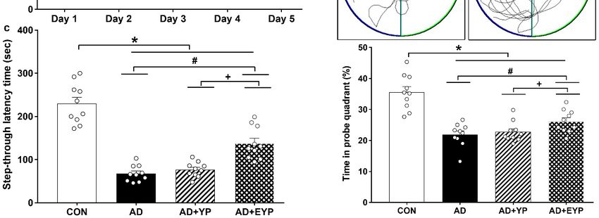
Int. J. Mol. Sci. 2020, 21, 3291 5 of 16
Int. J. Mol. Sci. 2020, 21, x 6 of 17
Int. J. Mol. Sci. 2020, 21, x 6 of 17
Figure 4. Effect of plasma from young exercised mice on mitochondrial Ca2+ retention and H2 O2
Figure 4. Effect of plasma from young exercised mice on mitochondrial Ca2+ retention and H2O2
emission in hippocampus. CON: wild-type, AD: 3xTg-AD, AD + YP: 3xTg-AD and young plasma
emission in hippocampus. CON: wild-type, AD: 3xTg-AD, AD + YP: 3xTg-AD and young plasma
injection, AD + EYP: 3xTg-AD and exercised young plasma injection group. Data are expressed as the
injection, AD + EYP: 3xTg-AD and exercised young plasma injection group. Data are expressed as the
mean ± standard error of the mean (SEM). * p < 0.05 compared to the CON group. # p < 0.05 compared
meanFigure
± standard error
4. Effect of the from
of plasma meanyoung
(SEM). * p < 0.05
exercised compared
mice to the CON
on mitochondrial group. #and
Ca2+ retention p < 0.05
H2O2 compared
to the AD group. + p < 0.05 between the AD + YP and AD + EYP groups.
to theemission in hippocampus.
AD group. CON: wild-type,
+ p < 0.05 between the ADAD:+ YP3xTg-AD,
and ADAD + YP:groups.
+ EYP 3xTg-AD and young plasma
2.5. Effectinjection,
of Plasma AD + EYP: 3xTg-AD and exercised young plasma injection group. Data are expressed as the
from Young Exercised Mice on Apoptosis in the Hippocampus
mean ± standard error of the mean (SEM). * p < 0.05 compared to the CON group. # p < 0.05 compared
2.5. Effect of Plasma from Young Exercised Mice on Apoptosis in the Hippocampus
to the AD group.
To investigate + p < 0.05
changes inbetween
apoptoticthe AD + YP and AD
proteins + EYP
in the groups.
hippocampus, we analyzed the expression
To investigate changes in apoptotic proteins
levels of Bax, Bcl-2, cytochrome c, apaf-1, and cleaved caspase-3 and -9. in the hippocampus, weTo analyzed
investigatethe expression
cell death,
2.5. Effect of Plasma from Young Exercised Mice on Apoptosis in the Hippocampus
levels
we of Bax,the
analyzed Bcl-2, cytochrome
number c, apaf-1, andcells.
of TUNEL-positive cleaved caspase-3cell
To compare anddeath
-9. Tobetween
investigate cell death,
groups, the CONwe
To investigate
analyzed
group the
results number
were setchanges
as andin apoptotic proteins
of 1,TUNEL-positive cells.inTo
the other groups’ thecompare
resultshippocampus,
cell we
were normalized analyzed
death the expression
between
relative groups,
to the CON thegroup.
CON
levels of Bax, Bcl-2, cytochrome c, apaf-1, and cleaved caspase-3 and -9. To investigate cell death, we
group
AD, AD + YP,were
results and set
ADas + 1,
EYPandgroups
the other groups’
showed results were
significant normalized
increased relativeoftoBax,
expressions the CON group.
cytochrome
analyzed the number of TUNEL-positive cells. To compare cell death between groups, the CON
AD,
c, and AD +
cleavedYP, and AD
caspase-3 + EYP
and groups
-9, and showed
significant significant
decreased increased
expression expressions
of Bcl-2
group results were set as 1, and the other groups’ results were normalized relative to the CON group. of Bax,
compared cytochrome
to the CON c,
and
groupAD, (p <
cleavedAD caspase-3
0.001). TheAD
+ YP, and ADand + EYP
+ EYP-9, and significant
combination
groups decreased expression
group demonstrated
showed significant of Bcl-2
significantly
increased expressions compared to the
decreasedc,expression
of Bax, cytochrome CON
group
of BaxandInt. J. Mol. Sci. 2020, 21, 3291 6 of 16
Int. J. Mol. Sci. 2020, 21, x 7 of 17
2.6. Effect of Plasma from Young Exercised Mice on Cell Death in the Hippocampal Dentate Gyrus
2.6. Effect of Plasma from Young Exercised Mice on Cell Death in the Hippocampal Dentate Gyrus
To investigate cell death in the hippocampal dentate gyrus (DG) we utilized a TUNEL assay.
To investigate cell death in the hippocampal dentate gyrus (DG) we utilized a TUNEL assay.
AD, AD + YP, and AD + EYP groups showed a significantly increased numbers of hippocampal
AD, AD + YP, and AD + EYP groups showed a significantly increased numbers of hippocampal
TUNEL-positive cells compared to the CON group (p < 0.001). The AD + EYP group demonstrated a
TUNEL-positive cells compared to the CON group (p < 0.001). The AD + EYP group demonstrated a
significantly reduced number of TUNEL-positive cells compared to the AD group (p = 0.012) and the
significantly reduced number of TUNEL-positive cells compared to the AD group (p = 0.012) and the
AD + YP group (p = 0.011), whereas there was no significant difference between the AD and AD + YP
AD + YP group (p = 0.011), whereas there was no significant difference between the AD and AD + YP
groups (Figure 6). Thus, plasma from young exercised mice inhibited cell death in the hippocampus of
groups (Figure 6). Thus, plasma from young exercised mice inhibited cell death in the hippocampus
AD mice.
of AD mice.
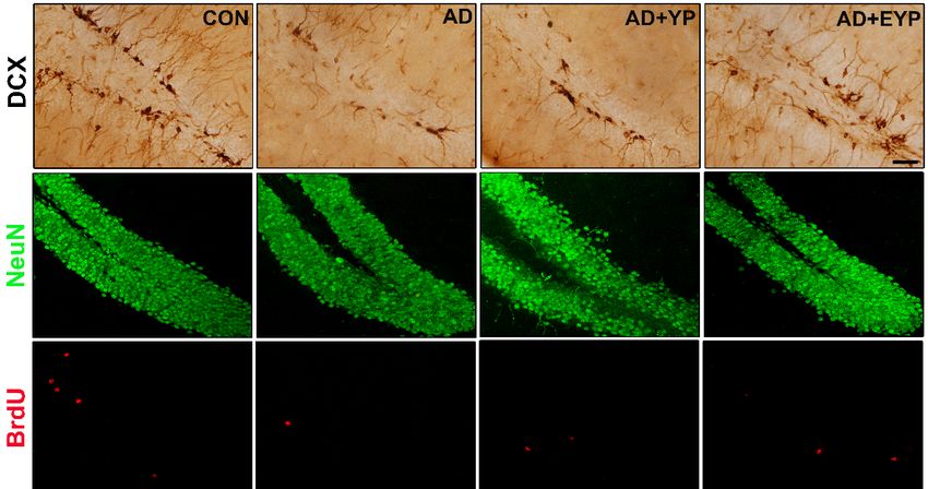
Figure 6. Effect of plasma from young exercised mice on cell death in the hippocampal dentate gyrus
FigurePhotomicrographs
(DG). 6. Effect of plasmaand
from young
data exercised mice on
of TUNEL-positive cell(arrow).
cells death inThe
the hippocampal dentate50gyrus
scale bar represents µm.
(DG). wild-type,
CON: Photomicrographs and data
AD: 3xTg-AD, AD of TUNEL-positive
+ YP: 3xTg-AD andcells
young(arrow).
plasmaThe scale bar
injection, ADrepresents 50 µm.
+ EYP: 3xTg-AD
CON:
and wild-type,
exercised AD:plasma
young 3xTg-AD, AD + group.
injection YP: 3xTg-AD and
Data are young plasma
expressed as the injection, AD + EYP:
mean ± standard 3xTg-AD
error of the
mean (SEM). * p < 0.05 compared to the CON group. # p < 0.05 compared to the AD group. + pInt. J. Mol. Sci. 2020, 21, 3291 7 of 16
Int. J. Mol. Sci. 2020, 21, x 8 of 17
Figure 7. Effect of plasma from young exercised mice on the expressions of BDNF, PSD 95,
Figure 7. Effect of plasma from young exercised mice on the expressions of BDNF, PSD 95, and
and synaptophysin in the hippocampus. CON: wild-type, AD: 3xTg-AD, AD + YP: 3xTg-AD and
synaptophysin
young in the hippocampus.
plasma injection, CON: wild-type,
AD + EYP: 3xTg-AD AD: 3xTg-AD,
and exercised AD +injection
young plasma YP: 3xTg-AD and
group. young
Data are
expressed as the mean ± standard error of the mean (SEM). * p < 0.05 compared to the CON group. #are
plasma injection, AD + EYP: 3xTg-AD and exercised young plasma injection group. Data pInt. J. Mol. Sci. 2020, 21, 3291 8 of 16
Int. J. Mol. Sci. 2020, 21, x 9 of 17
Figure
Figure 8. Effect of
8. Effect of plasma
plasma from
from young
young exercised
exercised mice
mice on
on cell
cell proliferation
proliferation and
and neurogenesis
neurogenesis in in the
the
hippocampus. CON: wild-type, AD: 3xTg-AD, AD + YP: 3xTg-AD and Young
hippocampus. CON: wild-type, AD: 3xTg-AD, AD + YP: 3xTg-AD and Young plasma injection, AD plasma injection, AD+
+EYP:
EYP: 3xTg-AD
3xTg-AD andand exercised
exercised young
young plasma
plasma injection
injection group.
group. Photomicrographs
Photomicrographs and and
datadata of DCX-
of DCX- and
and NeuN/BrdU-positive cells (white arrows). The scale bar represents 50 µm. Data
NeuN/BrdU-positive cells (white arrows). The scale bar represents 50 µm. Data are expressed as the are expressed
as the ±mean
mean ± standard
standard error oferror of the(SEM).
the mean mean *,(SEM).
p < 0.05 p < 0.05 compared
*, compared to the
to the CON CON
group. #, group.
p < 0.05 #, p < 0.05
compared
compared to the AD group. +, p < 0.05 between AD +
to the AD group. +, p < 0.05 between AD + YP and AD + EYP groups.YP and AD + EYP groups.
3. Discussion
3. Discussion
Increasing evidence suggests that AD pathogenesis is not restricted to the neuronal compartment.
Increasing evidence suggests that AD pathogenesis is not restricted to the neuronal
Glial cells, particularly reactive astrocytes and activated microglia, appear to play critical and interactive
compartment. Glial cells, particularly reactive astrocytes and activated microglia, appear to play
roles in neurodegeneration [22,23]. Accumulating evidence of their role in brain energy metabolism
critical and interactive roles in neurodegeneration [22,23]. Accumulating evidence of their role in
and reduced oxygen supply to the brain clearly point to their critical involvement in the prevention,
brain energy metabolism and reduced oxygen supply to the brain clearly point to their critical
initiation, and progression of neurodegenerative diseases, including AD [10,24]. However, AD is
involvement in the prevention, initiation, and progression of neurodegenerative diseases, including
an age-related neurodegenerative disease caused by accumulation of Aβ, neurofibrillary tangles,
AD [10,24]. However, AD is an age-related neurodegenerative disease caused by accumulation of Aβ,
gradual nerve injury, and cognitive deficits. In 3xTg mice, we observed pathological elements
neurofibrillary tangles, gradual nerve injury, and cognitive deficits. In 3xTg mice, we observed
similar to those of AD, including tau hyperphosphorylation and impaired spatial memory, which are
pathological elements similar to those of AD, including tau hyperphosphorylation and impaired
consistent with previous studies [25]. These symptoms have been reported to gradually worsen over
spatial memory, which are consistent with previous studies [25]. These symptoms have been reported
time [26]. Among the pathological elements of AD, overexpression and phosphorylation of tau is
to gradually worsen over time [26]. Among the pathological elements of AD, overexpression and
correlated with mitochondrial dysfunction including ROS production, reduced ATP, fragmenting of
phosphorylation of tau is correlated with mitochondrial dysfunction including ROS production,
mitochondrial membrane potential, and ultimately, neuronal damage [27,28]. Specifically, elevated
reduced ATP, fragmenting of mitochondrial membrane potential, and ultimately, neuronal damage
[27,28]. Specifically, elevated levels of ROS, including H2O2, resulting from impaired regulation of
mitochondrial Ca2+ homeostasis have been shown to increase the sensitivity of mitochondrial PTPInt. J. Mol. Sci. 2020, 21, 3291 9 of 16
levels of ROS, including H2 O2 , resulting from impaired regulation of mitochondrial Ca2+ homeostasis
have been shown to increase the sensitivity of mitochondrial PTP opening [29], which in turn promotes
apoptosis [30]. In the present study, the AD group showed tau hyperphosphorylation, reduced levels
of synaptic plasticity markers such as BDNF, PSD95, and synaptophysin, reduced cell proliferation
and neurogenesis, reduced mitochondrial Ca2+ retention, and elevated H2 O2 . There were increased
levels of Bax, apaf-1, cytochrome c, cleaved caspase-3 and -9, decreased Bcl-2 levels, and increased cell
death in the AD group. In the hippocampus, tau hyperphosphorylation impairs cognitive function
by not only inhibiting cell proliferation and neurogenesis, but also by increasing the number of cells
undergoing apoptosis via mitochondrial dysfunction.
Various studies have demonstrated that exercise has positive effects on brain health, and exercise
is especially known to play a beneficial role in AD treatment. However, the obvious but essential
precondition for exercise is that the individual must physically able. AD is known to start as mild
cognitive impairment and eventually progress to loss of motor function, eliminating the possibility
of exercise. However, recent research on aging has suggested that parabiosis could have positive
effects on cognitive function in the elderly. When plasma from young mice was infused into aged
mice, behavioral tests, including those for contextual fear conditioning, spatial learning, and memory
showed improvements in aging-related cognitive dysfunction [15]. Conversely, when plasma from
elderly mice was infused into young mice, neurogenesis, learning, and memory decreased [31].
Yuan et al. [32] reported that treatment with plasma from young mice reduced acute brain injury
induced by aging-related hemorrhagic stroke. In one recent study, parabiosis and intravenous infusion
of plasma from young mice resulted in near-complete recovery of synaptic and neural protein levels in
an animal model of AD [33]. These results suggest that factors carried in the blood might partially
contribute to synaptic plasticity, neurogenesis, and cognitive function. In the present study, the young
plasma group showed partial, trend-level improvements in hippocampal GSKβ/Tau expression,
neuroplasticity, and mitochondrial function, but these were not statistically significant. Conversely,
the group infused with plasma from mice that had exercised for 12 weeks showed overall improvements
accompanied by positive effects on cognitive function. The effects of plasma from exercised mice may
be due to elevated levels of BDNF in the blood resulting from exercise. Among the young plasma
donor mice in this study, those that had exercised showed higher levels of plasma BDNF than those
that had not exercised. Previous animal and clinical studies have also shown that blood BDNF levels
increase after exercise [34,35]. Qin et al. [36] reported that reduced peripheral blood BDNF is important
clinical evidence of AD or mild cognitive impairment, supporting a relationship between decreasing
BDNF level and AD progression. Specifically, plasma BDNF level can reflect hippocampal BDNF.
Therefore, plasma BDNF level is an important and stable blood biomarker of AD [37,38].
From a neuroprotective stance, BDNF secreted at the synaptic cleft modulates neuroplasticity to
confer protection from neural cell death caused by Aβ aggregates and tau protein, which are involved
in the pathology of AD [39]. In a study by Jiao et al. [40], delivery of the BDNF gene was proposed to
be a good treatment method for tau-related neural degeneration in AD and other tauopathy-related
neurodegenerative diseases. BDNF is involved in the control of neurogenesis; normal BDNF-TrkB
signaling is essential for the long-term survival of new neurons in the hippocampal dentate gyrus [41]
and can also inhibit caspase-3 activity as a result of apoptotic stimuli [42]. Mitochondrial function
in the brain is increased by BDNF in a dose-dependent manner, and BDNF counteracts damage
caused by mitochondrial Ca2+ overload [43]. However, in the present study, plasma from young
control and exercised mice failed to improve tau protein levels. In previous studies, heterochronic
parabiosis and infusion of plasma from young mice improved synaptic and neuronal proteins as
well as cognitive function in mutant mice for amyloid precursor protein without a decrease in Aβ
burden [33]. Meanwhile, BDNF supplementation relieved behavior deficits, protected against neuronal
loss, and alleviated synaptic degeneration and neuronal abnormality in P301L mutant tau transgene
mice, but had no effect on tau hyperphosphorylation [40]. Consistent with previous studies on plasma
infusion and BDNF, the present study found that infusion of plasma from exercised mice in an ADInt. J. Mol. Sci. 2020, 21, 3291 10 of 16
model did not reduce tau hyperphosphorylation but did enhance cognitive function via improvements
in hippocampal synaptic proteins, neuroplasticity, including apoptosis, and mitochondrial function.
4. Material and Methods
4.1. Animals
All animal experiments were conducted according to the National Institutes of Health and National
Institutes of Health and Korean Academy of Medical Sciences guidelines. The study protocol was
approved by the Kyung Hee University Animal Care and Use Committee (Approval No. KHUASP
[SE] -17-103, 14 August 2017). The mice were kept under constant temperature (25 ± 1 ◦ C) and light
(7 AM to 7 PM) conditions, and food and water were provided arbitrarily. The 12-month-old male
mice were randomly divided into the following groups: wild type (CON), 3xTG-AD (AD), 3xTg-AD,
young plasma injection (AD + YP), and 3xTg-AD and exercised young plasma injection (AD + EYP)
groups (n = 10 in each group). The plasma used for the donation was obtained from 4-month-old
C57BL/6 mice from the same mouse line that had undergone a 12-week exercise period. 3xTg-AD
mice were obtained commercially from the Jackson Laboratory (MMRRC stock number 008880) and
were maintained by breeding in our facilities. Genotypes were determined by PCR analysis of DNA
collected from tail biopsies. BrdU (Sigma, St. Louis, MO, USA) was administered intraperitoneally
(i.p.) at 100 mg/kg/day for 7 days, and the mice were sacrificed 4 weeks after the first day of BrdU
injection to observe neurogenesis.
4.2. Plasma Donation Mice EXERCISE Protocol
The 4-week-old mice (C57BL/6) that would eventually be used for blood donation exercised on a
treadmill for animals at an inclination of 0◦ . For the first 4 weeks, the animals performed 5 min of
warm-up exercise at a speed of 3 m/min, followed by 30 min of the main exercise at a speed of 10
m/min, and finally 5 min of a cool-down exercise at a speed of 3 m/min. Thereafter, the exercise load of
the main exercise was gradually increased to a speed of 11 m/min in weeks 5–6, 12 m/min in weeks 7–8,
13 m/min in weeks 9–10, and eventually 50 min of 14 m/min in weeks 11–12. Exercise was performed
five times per week, for a total of 12 weeks.
4.3. Infusion of Young Plasma and Young Plasma from Exercised Mice
For young plasma infusion, we performed plasma infusion immediately after sacrificing 20,
4-month-old male donor mice each from the exercise and non-exercise groups, two mice every 3 days.
As described previously [15], the plasma was isolated by centrifugation and injected into the tail veins
of 3xTg-AD mice in 100-µL doses, one dose every 3 days, for a total of 10 doses.
4.4. Behavioral Analysis
4.4.1. Morris Water Maze
Spatial learning and memory were analyzed using the Morris water maze task. One day before
starting training, for acclimation, the animals were made to swim freely in the swimming pool for
60 s without an escape platform. Training (learning) consisted of the animals trying to find an escape
platform, and was performed four times a day for 5 days. Animals that could not find the escape
platform within 60 s were guided to the platform, while swimming, by the experimenter. The animals
were allowed to rest for 30 s on the platform. A probe trial was performed 24 h after the last training
session, in which animals were allowed to swim freely for 60 s with no escape platform. Video tracking
was used to automatically measure how well the animals remembered the previous location of the
escape platform.Int. J. Mol. Sci. 2020, 21, 3291 11 of 16
4.4.2. Step-Through Avoidance Test
Long-term memory was measured using the step-through avoidance test. On the first day of
training, the animals were placed on a platform brightly lit by a halogen lamp, and the door to a box
was left open. Once the animal entered the box, the door was closed and the animal was allowed to
remain inside the box for 20 s. This method was repeated twice. Finally, on the third trial, as soon as
the door was closed, the animal was given a single 0.3 mA electrical shock for 2 s through the floor.
After 24 h, the animal was placed on the brightly lit platform, and after the door to the box was opened,
the time was measured until the animal entered the box (latency). The latency was measured up to
300 s, and animals that did not enter the box within this time were scored a latency of 300 s.
4.5. Preparation of Tissue
The animals were euthanized immediately after the water maze test. To prepare the
brain slices, the mice were fully anesthetized with ethyl ether, perfused transcardially with
50 mM phosphate-buffered saline (PBS), and then fixed with a freshly prepared solution of 4%
paraformaldehyde in 100 mM phosphate buffer (pH 7.4). The brains were then removed, post-fixed in
the same fixative overnight, and transferred into a 30% sucrose solution for cryoprotection. Coronal
sections with a thickness of 40 µm were created using a freezing microtome (Leica, Nussloch, Germany).
From each group of 10 animals, five were used for immunohistochemistry and five for Western blot and
mitochondrial function analysis. The hippocampal tissue for Western blot analysis was immediately
stored at −70 ◦ C until use. For immunohistochemistry, two sections from each group were analyzed,
resulting in a total of 10 slices. Western blot was performed to analyze all five samples in each group
and then was re-quantified for each protein and reanalyzed in the five samples. Therefore, the density
value was calculated and analyzed a total of 10 times in each group.
4.6. Immunohistochemistry
To visualize cell proliferation expression, immunohistochemistry for doublecortin (DCX) in the
dentate gyrus was performed. The sections were incubated in PBS for 10 min, and then washed three
times for 5 min in the PBS. The sections were then incubated in 1% H2 O2 for 30 min. The sections were
selected from each brain and incubated overnight with goat anti-DCX antibody (1:500; Santa cruz,
Dallas, TX, USA) and then with biotinylated secondary antibody (rabbit) (1:250; Vector Laboratories,
Burlingame, CA, USA) for another 1.5 h. Signal from the secondary antibody was amplified with the
Vector Elite ABC kit® (1:100; Vector Laboratories). Antibody-biotin-avidin-peroxidase complexes were
visualized using a DAB substrate kit (Vector Laboratories). The slides were air-dried overnight at
room temperature, and the coverslips were mounted using Permount ® (Fisher scientific, Fair lawn,
NJ, USA).
4.7. Immunofluorescence
BrdU/NeuN-positive cells in the dentate gyrus were tested for immunofluorescence. In brief,
the brain sections were permeabilized by incubation in 0.5% Triton X-100 in PBS for 20 min, then
incubated in 50% formamide-2× standard saline citrate at 65 ◦ C for 2 h, denaturated in 2 N HCl at
37 ◦ C for 30 min, and rinsed twice in 100 mM sodium borate (pH 8.5). The sections were incubated
overnight with a rat anti-BrdU antibody (1:500; Abcam, Cambridge, UK) and mouse anti-NeuN
antibody (1:500; Millipore, Temecula, CA). The brain sections were then washed in PBS and incubated
with the appropriate secondary antibodies for 1.5 h. The secondary antibodies used were anti-mouse
IgG Alexa Fluor-488 and anti-rat IgG Alexa Fluor-550. Images were captured using an FV3000 confocal
microscope (Olympus, Tokyo, Japan).Int. J. Mol. Sci. 2020, 21, 3291 12 of 16
4.8. TUNEL Staining
To visualize DNA fragmentation, we performed TUNEL staining using an In Situ Cell Death
Detection Kit (Roche Diagnostics, Risch-Rotkreuz, Switzerland) according to the manufacturer’s
protocol. The sections were post-fixed in ethanol-acetic acid (2:1), rinsed, incubated with proteinase K
(100 mg/mL), and then rinsed again. Next, the sections were incubated in 3% H2 O2 , permeabilized
with 0.5% Triton X-100, rinsed again, and incubated in the TUNEL reaction mixture. The sections were
rinsed and visualized using Converter-POD with 0.03% DAB, counterstained with Nissl, and mounted
onto gelatin-coated slides. The slides were air-dried overnight at room temperature and cover-slipped
using Permount mounting medium.
4.9. Western Blotting
The hippocampal tissues were homogenized on ice and lysed in a lysis buffer containing
50 mM Tris–HCl (pH 7.5), 150 mM NaCl, 0.5% deoxycholic acid, 1% Nonidet P40, 0.1% sodium
dodecyl sulfate, 1 mM PMSF, and leupeptin 100 mg/mL. The protein content was measured using
a colorimetric protein assay kit (Bio-Rad, Hercules, CA, USA). Thirty micrograms of protein were
separated on sodium dodecyl sulfate-polyacrylamide gels and transferred onto a nitrocellulose
membrane, which was incubated with antibodies against β-actin (1:2000; Santa Cruz Biotechnology),
GAPDH (1:2000; Santa Cruz Biotechnology), t-GSK3β, p-GSK3β (ser 9) (1:1000; Cell Signaling), t-Tau,
p-Tau (ser262) (1:1000; Thermo Fisher), p-tau (thr 205) (1:1000; Thermo Fisher), Bcl-2 (1;1000; Santa
Cruz Biotechnology), cytochrome c (1:1000; Santa Cruz Biotechnology), Bax (1:1000; Cell Signaling),
cleaved caspase-3,-9 (1:700; Cell Signaling), BDNF (1:1000; Alomone), PSD95 (1:1000; Cell Signaling),
and synaptophysin (1:1000; Abcam). Horseradish peroxidase-conjugated anti-mouse antibodies for
Bcl-2, p-Tau, cytochrome c, β-actin and GAPDH, and anti-rabbit antibodies for t-tau, t-GSK3β, p-GSK3β,
Bax, cleaved caspase-3, BDNF, PSD95, and synaptophysin were used as secondary antibodies.
4.10. Mitochondrial Ca2+ Retention Capacity
The mitochondrial calcium retention capacity was tested to assess the susceptibility of the
permeability transition pore (PTP) to opening. Briefly, after grinding the hippocampal tissue, overlaid
traces of changes in fluorescence induced by Calcium Green-5 N were measured continuously (∆F/min)
at 37 ◦ C during state 4 respiration using a Spex FluoroMax 4 spectrofluorometer (Horiba Scientific,
Edison, NJ, USA). After establishing the background ∆F (hippocampal tissue in the presence of 1 µM
Calcium Green-5 N, 1 U/mL hexokinase, 0.04 mM EGTA, 1.5 nM thapsigargin, 5 mM 2-deoxyglucose,
5 mM glutamate, 5 mM succinate, and 2 mM malate), the reaction was initiated by addition of Ca2+
pulses (12.5 nM), with excitation and emission wavelengths set at 506 and 532 nm, respectively.
The total mitochondrial calcium retention capacity prior to PTP opening (i.e., release of Ca2+ ) was
expressed as pmol/mg tissue weight.
4.11. Mitochondrial H2 O2 Emission
The mitochondrial H2 O2 emission was measured at 37 ◦ C (∆F/min) during state 4 respiration
(10 µg/mL oligomycin) by continuously monitoring oxidation of Amplex Red (excitation/emission
λ = 563/587 nm) using a Spex FluoroMax 4 spectrofluorometer with the following protocol: 10 µM
Amplex Red, 1 U/mL horseradish peroxidase, and 10 µg/mL oligomycin; followed by 1 mM malate
+ 2 mM glutamate (complex I substrates); 3 mM succinate (complex II substrate); and 10 mM
glycerol-3-phosphate (lipid substrate). The mitochondrial H2 O2 emission rate after removing the
background value from each of the standard values (standard curve) was calculated from the ∆F/min
gradient values and expressed as pmol/min/mg tissue weight.Int. J. Mol. Sci. 2020, 21, 3291 13 of 16
4.12. Plasma BDNF Analysis in Donation Mice
The plasma BDNF concentration was measured using ELISA. The ELISA kit was purchased from
R & D Biology Inc. (Total BDNF Quantikine ELISA kit DBNT00, R & D Systems), and the experiment
was performed strictly according to the manufacturer’s instructions. The specific protocol was as
follows: first, the reagents, samples, and standard products were prepared. The samples and standard
products were added and reacted for 120 min at 37 ◦ C. After washing the plate three times, antibody
working solution was added and the mixture was left to react for 60 min at 37 ◦ C. The plate was
washed three times again, horseradish peroxidase (HRP) was added, and the mixture was left to react
for 30 min at 37 ◦ C. After washing the plate three times, substrate working solution was added, and the
mixture was left to react in a dark place for 5–10 min. After adding stop buffer, a BioTek ELx800
full-automatic enzyme labeling system ( Winooski, VT, USA) was used to detect the optical density
(OD) at 450 nm within 30 min.
4.13. Statistical Analysis
Cell counting and optical density quantification for TUNEL, DCX, and BrdU/NeuN-positive cells
were performed using Image-Pro® Plus (Media Cyberbetics Inc. Rockville, MD, USA) attached to a
light microscope (Olympus, Tokyo, Japan). The data were analyzed with one-way ANOVA, followed
by Tukey post-hoc tests. Plasma BDNF was analyzed using t-tests. All values are expressed as the
mean ± standard error of the mean (S.E.M.), and p-values < 0.05 were considered significant.
5. Conclusions
Treatment with plasma from young control or exercised mice showed positive effects on cognitive
function in a 3xTg-AD animal model, with partial improvements in hippocampal neuroplasticity and
mitochondrial function. In particular, in plasma from young exercised mice, elevated plasma BDNF
levels, as a result of exercise, might have a protective effect against cognitive dysfunction and on
important AD-related neural pathways, acting via tau-independent mechanisms. Further studies will
be needed to investigate potential mechanisms mediating these interactions.
Author Contributions: Conceptualization, T.-W.K., S.-S.P., H.-S.P.; Analysis, T.-W.K., S.-S.P.; Writing—original
draft preparation, T.-W.K., H.-S.P.; Writing—review, J.-Y.P.; Funding Acquisition, H.-S.P. All authors have read and
agreed to the published version of the manuscript.
Funding: This work was supported by the Ministry of Education of the Republic of Korea and the National
Research Foundation of Korea (NRF-2017S1A5A2A01024208).
Conflicts of Interest: The authors declare no conflicts of interest.
Abbreviations
AD Alzheimer’s disease
Aβ Amyloid beta
BDNF Brain-derived neurotrophic factor
BrdU 5-Bromo-2’-Deoxyuridine
DAB 3,30 -Diaminobenzidine
DCX Doublecortin
EYP Exercised young plasma
GM Glutamate + malate
GM+S GM + succinate
GMS+G3P GMS + glycerol-3-phosphate
GSK-3β Glycogen synthase kinase-3 beta
NeuN Neuronal nuclei
PBS phosphate-buffered salineInt. J. Mol. Sci. 2020, 21, 3291 14 of 16
PSD95 postsynaptic density protein 95
ROS Reactive oxygen species
TUNEL Terminal deoxynucleotidyl transferase dUTP nick end labeling
YP Young plasma
3xTg-AD Triple-transgenic mouse model-AD
References
1. Mattson, M.P. Pathways towards and away from Alzheimer’s disease. Nature 2004, 430, 63–69. [CrossRef]
2. Swerdlow, R.H.; Khan, S.M. The Alzheimer’s disease mitochondrial cascade hypothesis: An update.
Exp. Neurol. 2009, 218, 308–315. [CrossRef] [PubMed]
3. Kumar, A.; Singh, A.; Ekavali. A review on Alzheimer’s disease pathophysiology and its management:
An update. Pharmacol. Rep. 2015, 67, 195–203. [CrossRef]
4. Blennow, K.; de Leon, M.J.; Zetterberg, H. Alzheimer’s disease. Lancet 2006, 368, 387–403. [CrossRef]
5. Arendt, T. Synaptic degeneration in Alzheimer’s disease. Acta Neuropathol. 2009, 118, 167–179. [CrossRef]
[PubMed]
6. Siwak-Tapp, C.T.; Head, E.; Muggenburg, B.A.; Milgram, N.W.; Cotman, C.W. Neurogenesis decreases with
age in the canine hippocampus and correlates with cognitive function. Neurobiol. Learn. Mem. 2007, 88,
249–259. [CrossRef]
7. Stamer, K.; Vogel, R.; Thies, E.; Mandelkow, E.; Mandelkow, E.M. Taublockstraffic of organelles,
neurofilaments, and APPvesicles in neurons and enhance soxidativestress. Cell Biol. 2002, 156, 1051–1063.
8. Mandelkow, E.M.; Stamer, K.; Vogel, R.; Thies, E.; Mandelkow, E. Clogging of axons by tau, inhibition of
axonaltraffic and starvation of synapses. Neurobiol. Aging 2003, 24, 1079–1085. [CrossRef] [PubMed]
9. Dubey, M.; Chaudhury, P.; Kabiru, H.; Shea, T.B. Tau inhibits anterograde axonal transport and perturbs
stability in growing axonal neurites in part by displacing kinesin cargo: Neurofilaments attenuate
tau-mediated neurite instability. Cell Motil. Cytoskelet. 2008, 65, 89–99. [CrossRef]
10. Bordone, M.P.; Salman, M.M.; Titus, H.E.; Amini, E.; Andersen, J.V.; Chakraborti, B.; Diuba, A.V.;
Dubouskaya, T.G.; Ehrke, E.; Espindola de Freitas, A.; et al. The energetic brain—A review from students to
students. J. Neurochem. 2019, 151, 139–165. [CrossRef]
11. Gibson, G.E.; Sheu, K.F.; Blass, J.P. Abnormalities of mitochondrial enzymes in Alzheimer disease. J. Neural
Transm. (Vienna) 1998, 105, 855–870. [CrossRef]
12. Devi, L.; Prabhu, B.M.; Galati, D.F.; Avadhani, N.G.; Anandatheerthavarada, H.K. Accumulation of amyloid
precursor protein in the mitochondrial import channels of human Alzheimer’s disease brain is associated
with mitochondrial dysfunction. J. Neurosci. 2006, 26, 9057–9068. [CrossRef]
13. Parker, W.D., Jr.; Filley, C.M.; Parks, J.K. Cytochrome oxidase deficiency in Alzheimer’s disease. Neurology
1990, 40, 1302–1303. [CrossRef] [PubMed]
14. Wang, X.; Su, B.; Siedlak, S.L.; Moreira, P.I.; Fujioka, H.; Wang, Y.; Casadesus, G.; Zhu, X. Amyloid-beta
overproduction causes abnormal mitochondrial dynamics via differential modulation of mitochondrial
fission/fusion proteins. Proc. Natl. Acad. Sci. USA 2008, 105, 19318–19323. [CrossRef]
15. Villeda, S.A.; Plambeck, K.E.; Middeldorp, J.; Castellano, J.M.; Mosher, K.I.; Luo, J.; Smith, L.K.; Bieri, G.;
Lin, K.; Berdnik, D.; et al. Young blood reverses age-related impairments in cognitive function and synaptic
plasticity in mice. Nat. Med. 2014, 20, 659–663. [CrossRef] [PubMed]
16. Kim, D.H.; Ko, I.G.; Kim, B.K.; Kim, T.W.; Kim, S.E.; Shin, M.S.; Kim, C.J.; Kim, H.; Kim, K.M.; Baek, S.S.
Treadmill exercise inhibits traumatic brain injury-induced hippocampal apoptosis. Physiol. Behav. 2010, 101,
660–665. [CrossRef]
17. Luo, C.X.; Jiang, J.; Zhou, Q.G.; Zhu, X.J.; Wang, W.; Zhang, Z.J.; Han, X.; Zhu, D.Y. Voluntary exercise-induced
neurogenesis in the postischemic dentate gyrus is associated with spatial memory recovery from stroke.
J. Neurosci. Res. 2007, 85, 1637–1646.
18. Ke, Z.; Yip, S.P.; Li, L.; Zheng, X.X.; Tong, K.Y. The effects of voluntary, involuntary, and forced exercises on
brain-derived neurotrophic factor and motor function recovery: A rat brain ischemia model. PLoS ONE
2011, 6, e16643. [CrossRef] [PubMed]
19. Lee, S.U.; Kim, D.Y.; Park, S.H.; Choi, D.H.; Park, H.W.; Han, T.R. Mild to moderate early exercise promotes
recovery from cerebral ischemia in rats. Can. J. Neurol. Sci. 2009, 36, 443–449. [CrossRef]Int. J. Mol. Sci. 2020, 21, 3291 15 of 16
20. Zoladz, J.A.; Pilc, A.; Majerczak, J.; Grandys, M.; Zapart-Bukowska, J.; Duda, K. Endurance training increases
plasma brain-derived neurotrophic factor concentration in young healthy men. J. Physiol. Pharmacol. 2008, 7,
119–132.
21. Seifert, T.; Brassard, P.; Wissenberg, M.; Rasmussen, P.; Nordby, P.; Stallknecht, B.; Adser, H.; Jakobsen, A.H.;
Piegaard, H.; Nielsen, H.B.; et al. Endurance training enhances BDNF release from the human brain. Am. J.
Physiol.-Regul. Integr. Comp. Physiol. 2010, 298, R372–R377. [CrossRef]
22. Salman, M.M.; Kitchen, P.; Woodroofe, M.N.; Bill, R.M.; Conner, A.C.; Heath, P.R.; Conner, M.T. Transcriptome
Analysis of Gene Expression Provides New Insights into the Effect of Mild Therapeutic Hypothermia on
Primary Human Cortical Astrocytes Cultured under Hypoxia. Front. Cell. Neurosci. 2017, 11, 386. [CrossRef]
[PubMed]
23. Chun, H.; Marriott, I.; Lee, C.J.; Cho, H. Elucidating the Interactive Roles of Glia in Alzheimer’s Disease
Using Established and Newly Developed Experimental Models. Front. Neurol. 2018, 9, 797. [CrossRef]
[PubMed]
24. Heneka, M.T.; Carson, M.J.; El Khoury, J.; Landreth, G.E.; Brosseron, F.; Feinstein, D.L.; Jacobs, A.H.;
Wyss-Coray, T.; Vitorica, J.; Ransohoff, R.M.; et al. Neuroinflammation in Alzheimer’s disease. Lancet Neurol.
2015, 14, 388–405. [CrossRef]
25. Oddo, S.; Caccamo, A.; Shepherd, J.D.; Murphy, M.P.; Golde, T.E.; Kayed, R.; Metherate, R.; Mattson, M.P.;
Akbari, Y.; LaFerla, F.M. Triple-transgenic model of Alzheimer’s disease with plaques and tangles: Intracellular
Abeta and synaptic dysfunction. Neuron 2003, 39, 409–421. [CrossRef]
26. Carroll, J.C.; Rosario, E.R.; Chang, L.; Stanczyk, F.Z.; Oddo, S.; LaFerla, F.M.; Pike, C.J. Progesterone and
estrogen regulate Alzheimer-like neuropathology in female 3xTg-AD mice. J. Neurosci. 2007, 27, 13357–13365.
[CrossRef]
27. Reddy, P.H. Abnormal tau, mitochondrial dysfunction, impaired axonal transport of mitochondria,
and synaptic deprivation in Alzheimer’s disease. Brain Res. 2011, 1415, 136–148. [CrossRef]
28. Atlante, A.; Amadoro, G.; Bobba, A.; de Bari, L.; Corsetti, V.; Pappalardo, G.; Marra, E.; Calissano, P.;
Passarella, S. A peptide containing residues 26–44 of tau protein impairs mitochondrial oxidative
phosphorylation acting at the level of the adenine nucleotide translocator. Biochim. Biophys. Acta 2008, 1777,
1289–1300. [CrossRef]
29. Kristian, T.; Pivovarova, N.B.; Fiskum, G.; Andrews, S.B. Calcium-induced precipitate formation in brain
mitochondria: Composition, calcium capacity, and retention. J. Neurochem. 2007, 102, 1346–1356. [CrossRef]
30. Tsujimoto, Y.; Shimizu, S. Role of the mitochondrial membrane permeability transition in cell death. Apoptosis
2007, 12, 835–840. [CrossRef]
31. Villeda, S.A.; Luo, J.; Mosher, K.I.; Zou, B.; Britschgi, M.; Bieri, G.; Stan, T.M.; Fainberg, N.; Ding, Z.; Eggel, A.;
et al. The ageing systemic milieu negatively regulates neurogenesis and cognitive function. Nature 2011, 477,
90–94. [CrossRef] [PubMed]
32. Yuan, J.J.; Zhang, Q.; Gong, C.X.; Wang, F.X.; Huang, J.C.; Yang, G.Q.; Liu, L.; Zhou, K.; Xu, R.; Chen, Q.; et al.
Young plasma ameliorates aging-related acute brain injury after intracerebral hemorrhage. Biosci. Rep. 2019,
17, 39. [CrossRef] [PubMed]
33. Middeldorp, J.; Lehallier, B.; Villeda, S.A.; Miedema, S.S.; Evans, E.; Czirr, E.; Zhang, H.; Luo, J.; Stan, T.;
Mosher, K.I.; et al. Preclinical Assessment of Young Blood Plasma for Alzheimer Disease. JAMA Neurol.
2016, 73, 1325–1333. [CrossRef]
34. Belviranlı, M.; Okudan, N. Exercise training increases cardiac, hepatic and circulating levels of brain-derived
neurotrophic factor and irisin in young and aged rats. Horm. Mol. Biol. Clin. Investig. 2018, 36, 1–8.
[CrossRef] [PubMed]
35. Gilder, M.; Ramsbottom, R.; Currie, J.; Sheridan, B.; Nevill, A.M. Effect of fat free mass on serum and plasma
BDNF concentrations during exercise and recovery in healthy young men. Neurosci. Lett. 2014, 560, 137–141.
[CrossRef]
36. Qin, X.Y.; Cao, C.; Cawley, N.X.; Liu, T.T.; Yuan, J.; Loh, Y.P.; Cheng, Y. Decreased peripheral brain-derived
neurotrophic factor levels in Alzheimer’s disease: A meta-analysis study (N = 7277). Mol. Psychiatry 2017,
22, 312–320. [CrossRef]
37. Nagata, T.; Kobayashi, N.; Shinagawa, S.; Yamada, H.; Kondo, K.; Nakayama, K. Plasma BDNF levels are
correlated with aggressiveness in patients with amnestic mild cognitive impairment or Alzheimer disease.
J. Neural. Transm. (Vienna) 2014, 121, 433–441. [CrossRef]Int. J. Mol. Sci. 2020, 21, 3291 16 of 16
38. Klein, A.B.; Williamson, R.; Santini, M.A.; Clemmensen, C.; Ettrup, A.; Rios, M.; Knudsen, G.M.; Aznar, S.
Blood BDNF concentrations reflect brain-tissue BDNF levels across species. Int. J. Neuropsychopharmacol.
2011, 14, 347–353. [CrossRef]
39. Nagahara, A.H.; Merrill, D.A.; Coppola, G.; Tsukada, S.; Schroeder, B.E.; Shaked, G.M.; Wang, L.; Blesch, A.;
Kim, A.; Conner, J.M.; et al. Neuroprotective effects of brain-derived neurotrophic factor in rodent and
primate models of Alzheimer’s disease. Nat. Med. 2009, 15, 331–337. [CrossRef]
40. Jiao, S.S.; Shen, L.L.; Zhu, C.; Bu, X.L.; Liu, Y.H.; Liu, C.H.; Yao, X.Q.; Zhang, L.L.; Zhou, , H.D.; Walker, D.G.;
et al. Brain-derived neurotrophic factor protects against tau-related neurodegeneration of Alzheimer’s
disease. Transl. Psychiatry 2016, 6, e907. [CrossRef]
41. Sairanen, M.; Lucas, G.; Ernfors, P.; Castrén, M.; Castrén, E. Brain-derived neurotrophic factor and
antidepressant drugs have different but coordinated effects on neuronal turnover, proliferation, and survival
in the adult dentate gyrus. J. Neurosci. 2005, 25, 1089–1094. [CrossRef] [PubMed]
42. Han, B.H.; D’Costa, A.; Back, S.A.; Parsadanian, M.; Patel, S.; Shah, A.R.; Giddy, J.M.; Srinivasan, A.;
Deshmukh, M.; Holtzman, D.M. BDNF blocks caspase-3 activation in neonatal hypoxia-ischemia. Neurobiol.
Dis. 2000, 7, 38–53. [CrossRef] [PubMed]
43. Markham, A.; Cameron, I.; Franklin, P.; Spedding, M. BDNF increases rat brain mitochondrial respiratory
coupling at complex I, but not complex II. Eur. J. Neurosci. 2004, 20, 1189–1196. [CrossRef] [PubMed]
© 2020 by the authors. Licensee MDPI, Basel, Switzerland. This article is an open access
article distributed under the terms and conditions of the Creative Commons Attribution
(CC BY) license (http://creativecommons.org/licenses/by/4.0/).You can also read