Immunohistochemistry in Prostate Pathology
←
→
Page content transcription
If your browser does not render page correctly, please read the page content below
ED U C AT I O N AL

IHC        Prostate Pathology

                                Immunohistochemistry
                                 in Prostate Pathology
                                                     Glen Kristiansen1, MD; Jonathan I. Epstein2, MD

                                          Institute of Pathology, University Hospital Bonn, Germany1
                                   The Departments of Pathology, Urology, and Oncology, The Johns
                                                Hopkins Hospital Medical Institutions, Baltimore, MD2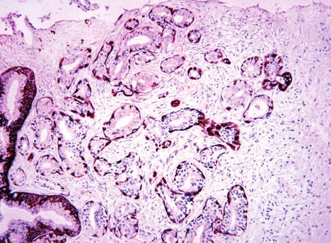
Glen Kristiansen, MD                                            Jonathan I. Epstein, MD

Glen Kristiansen, MD, studied medicine in Freiburg i.Br.,     Jonathan I. Epstein, MD, obtained a combined BA-MD
Berlin and London. He started residency at the Depart-        degree from Boston University’s 6-Year Medical Program
ment of Pathology of the Free University Berlin in 1996       (1975–1981). Following his residency in anatomic pathol-
and moved on to the Institute of Pathology of the Charité     ogy at The Johns Hopkins Hospital in Baltimore, Maryland
in 1998, when his career as a prostate cancer researcher      and a fellowship in oncologic pathology at Memorial Sloan
began. After board certification in anatomical and surgi-     Kettering Cancer Center in New York, he joined the staff at
cal pathology (2003), his post-doctoral thesis (2004) on      The Johns Hopkins Hospital and has been there his entire
differential gene expression of prostate cancer, he took      career. At The Johns Hopkins Medical Institutions, he is
up a professorship for molecular tumor pathology at the       Professor of Pathology, Urology, and Oncology; the recip-
University of Zurich in 2007. Since 2011 he is full profes-   ient of the Reinhard Chair of Urological Pathology; and
sor of pathology and chairman of the Institute of Patholo-    Director of Surgical Pathology. He is the past President of
gy of the University Bonn, Germany.                           the International Society of Urological Pathology.

Dr. Kristiansens research field covers diagnostic and         Dr. Epstein has over 700 publications in the peer-re-
prognostic biomarkers of solid tumors, with a predomi-        viewed literature and has authored 49 book chapters. His
nant focus on prostate cancer. He has published more          most-frequently cited first or last authored publications is
than 270 peer reviewed papers and is a frequent speak-        ‘‘Pathological and Clinical Findings to Predict Tumor Ex-
er in national and international conferences. In Bonn, he     tent of Nonpalpable (stage T1c) Prostate Cancer,’’ pub-
has established next to his translational research working    lished in JAMA, which establishes the criteria for active
group a GU pathology consult service, is active in post-      surveillance. He was also the leading author to develop
graduate teaching of pathologists and urologists and is       the WHO Consensus Conference on Classification of
centrally involved in several prostate cancer studies (in-    Urothelial Neoplasia (1998) and the consensus on updat-
cluding the german PREFERE-trial, funded by the Ger-          ing the Gleason grading system (2005).
man Cancer Aid). He is also part of the steering commit-
tee of the European Network of Uropathology (ENUP).           He is the author or co-author of five books including “Inter-
                                                              pretation of Prostate Biopsies” which is in its 4th edition, and
                                                              “Bladder Biopsy Interpretation” which is in its 2nd edition.
                                                              He is a co-editor of the “WHO Pathology and Genetics of Tu-
                                                              mours of the Urinary System and Male Genital Organs”, and
                                                              a co-author of the 2011 AFIP Fascicle, 4th Series on “Tumors
                                                              of the Prostate Gland, Seminal Vesicles, Male Urethra, and
                                                              Penis”. He has one of the largest surgical pathology consult-
                                                              ing services in the world with approximately 12,000 cases
                                                              per year, covering the full range of urologic pathology. Dr.
                                                              Epstein uses these consultations to train four genitourinary
                                                              pathology fellows each year, with 42 fellows trained to date.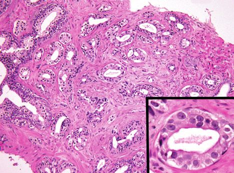
Prostate Pathology | IHC

Table of Contents

Prostate Cancer  . . . . . . . . . . . . . . . . . . . . . . . . . . . . . . . . . . . . . . . . . . . . . . . . . . . . . . . . . . . . . . . . . . . . . . . . . . . . . . . . . . . . . . . . . . . . . . . . . . . . . . . . . . . . . . . 4

Adenocarcinoma of the Prostate (Limited)  . . . . . . . . . . . . . . . . . . . . . . . . . . . . . . . . . . . . . . . . . . . . . . . . . . . . . . . . . . . . . . . 4
Negative Markers of Malignancy – Basal Cell Markers . . . . . . . . . . . . . . . . . . . . . . . . . . . . . . . . . . . . . . . . . . . . . 4
CK HMW, CK 5/6 and p63                                                              . . . . . . . . . . . . . . . . . . . . . . . . . . . . . . . . . . . . . . . . . . . . . . . . . . . . . . . . . . . . . . . . . . . . . . . . . . . . . . . . . . . . . . . . . . . . .   4

Positive Markers of Malignancy . . . . . . . . . . . . . . . . . . . . . . . . . . . . . . . . . . . . . . . . . . . . . . . . . . . . . . . . . . . . . . . . . . . . . . . . . . . . . . . . . . . 6
AMACR (Alpha-methylacyl-CoA racemace) . . . . . . . . . . . . . . . . . . . . . . . . . . . . . . . . . . . . . . . . . . . . . . . . . . . . . . . . . . . . . . . 6
ERG (Ets-related gene product)  . . . . . . . . . . . . . . . . . . . . . . . . . . . . . . . . . . . . . . . . . . . . . . . . . . . . . . . . . . . . . . . . . . . . . . . . . . . . . . . . . . . . 8
FASN (Fatty acid synthase) . . . . . . . . . . . . . . . . . . . . . . . . . . . . . . . . . . . . . . . . . . . . . . . . . . . . . . . . . . . . . . . . . . . . . . . . . . . . . . . . . . . . . . . . . . 10
GOLPH2 (Golgi phosphoprotein 2) . . . . . . . . . . . . . . . . . . . . . . . . . . . . . . . . . . . . . . . . . . . . . . . . . . . . . . . . . . . . . . . . . . . . . . . . . . . .  10
CYCS, ICK and IKBKB . . . . . . . . . . . . . . . . . . . . . . . . . . . . . . . . . . . . . . . . . . . . . . . . . . . . . . . . . . . . . . . . . . . . . . . . . . . . . . . . . . . . . . . . . . . . . . . . . .  10

Primary Adenocarcinoma of the Prostate from Secondary Tumors  . . . . . . . . . . . . . . . .  10
PSA (Prostate-specific antigen)  . . . . . . . . . . . . . . . . . . . . . . . . . . . . . . . . . . . . . . . . . . . . . . . . . . . . . . . . . . . . . . . . . . . . . . . . . . . . . . . . . .  10
PSMA (Prostate-specific membrane antigen) . . . . . . . . . . . . . . . . . . . . . . . . . . . . . . . . . . . . . . . . . . . . . . . . . . . . . . . . . .  11
Prostein (P501S)  . . . . . . . . . . . . . . . . . . . . . . . . . . . . . . . . . . . . . . . . . . . . . . . . . . . . . . . . . . . . . . . . . . . . . . . . . . . . . . . . . . . . . . . . . . . . . . . . . . . . . . . . . . . .  11
AR (Androgen receptor) . . . . . . . . . . . . . . . . . . . . . . . . . . . . . . . . . . . . . . . . . . . . . . . . . . . . . . . . . . . . . . . . . . . . . . . . . . . . . . . . . . . . . . . . . . . . . . .  11
ERG (Ets-related gene product)  . . . . . . . . . . . . . . . . . . . . . . . . . . . . . . . . . . . . . . . . . . . . . . . . . . . . . . . . . . . . . . . . . . . . . . . . . . . . . . . . .  11
NKX3.1 (Homeobox protein NKX3.1) . . . . . . . . . . . . . . . . . . . . . . . . . . . . . . . . . . . . . . . . . . . . . . . . . . . . . . . . . . . . . . . . . . . . . . . . .  12
AMACR (Alpha-methylacyl-CoA racemase) . . . . . . . . . . . . . . . . . . . . . . . . . . . . . . . . . . . . . . . . . . . . . . . . . . . . . . . . . . . . .  12

Specific Differential Diagnoses . . . . . . . . . . . . . . . . . . . . . . . . . . . . . . . . . . . . . . . . . . . . . . . . . . . . . . . . . . . . . . . . . . . . . . . . . . . . . . . . .  12
Prostate Cancer (PCa) vs. Urothelial Cancer (UC)  . . . . . . . . . . . . . . . . . . . . . . . . . . . . . . . . . . . . . . . . . . . . . . . . .  12
Prostate Cancer (PCa) vs. Colorectal Cancer (CRC)  . . . . . . . . . . . . . . . . . . . . . . . . . . . . . . . . . . . . . . . . . . . .  12
Diagnosis of Pretreated Prostate Carcinomas  . . . . . . . . . . . . . . . . . . . . . . . . . . . . . . . . . . . . . . . . . . . . . . . . . . . . . . . . .  12

Pitfalls in the Use of Prostatic Markers . . . . . . . . . . . . . . . . . . . . . . . . . . . . . . . . . . . . . . . . . . . . . . . . . . . . . . . . . . . . . . . . . . .  13

Concluding Remarks . . . . . . . . . . . . . . . . . . . . . . . . . . . . . . . . . . . . . . . . . . . . . . . . . . . . . . . . . . . . . . . . . . . . . . . . . . . . . . . . . . . . . . . . . . . . . . . . . . .  14

Dako Antibodies for Prostate Tissue Antigens . . . . . . . . . . . . . . . . . . . . . . . . . . . . . . . . . . . . . . . . . . . . . . . . . . . . . .  14

Stains using Dako Antibodies  . . . . . . . . . . . . . . . . . . . . . . . . . . . . . . . . . . . . . . . . . . . . . . . . . . . . . . . . . . . . . . . . . . . . . . . . . . . . . . . . . . .  15

References  . . . . . . . . . . . . . . . . . . . . . . . . . . . . . . . . . . . . . . . . . . . . . . . . . . . . . . . . . . . . . . . . . . . . . . . . . . . . . . . . . . . . . . . . . . . . . . . . . . . . . . . . . . . . . . . . . . . .  17
IHC | Prostate Pathology
Prostate Cancer
Prostate cancer is the second most common cancer
in men in the United States. It is estimated that about
240,000 new cases of prostate cancer will be diag-
nosed annually. This accumulates to 16% of all men will
be diagnosed with prostate cancer during his lifetime
with an average age at the time of diagnosis about
67 years old. Almost 30,000 men will die of prostate
cancer in 2013 in the US making it the second leading
cause of cancer death in American men, behind only
lung cancer (statistics from www.cancer.org).
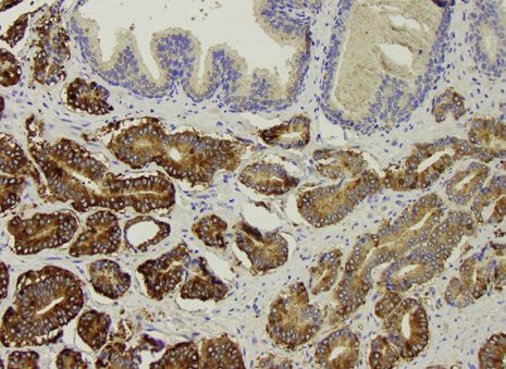
Immunohistochemical (IHC) markers are often used as Figure 1: Cocktail labeling with brown chromogen labeling
both basal cell nuclei (p63) and cytoplasm in benign glands
an aid in the diagnosis of prostatic adenocarcinoma,
(right side). Prostate adenocarcinoma (left side) with absence
especially in the diagnosis of limited primary prostate of basal cell staining.
carcinoma on needle biopsy. The diagnosis of prostate
adenocarcinoma is aided by IHC staining for basal cell
layer markers, such as p63, cytokeratin 5/6 (CK 5/6), A lack of basal cell staining may also be seen in several
and high molecular weight cytokeratin (CK HMW) as well benign mimickers of prostatic adenocarcinoma. In aden-
as prostate-‘specific’ markers. osis (atypical adenomatous hyperplasia (AAH)), usually
>50% of the glands label with basal cells markers, yet
This document will discuss the potentials and pitfalls of the as few as 10% may be positive (2). However, the stain-
individual markers used in the diagnosis of prostate cancer. ing is patchy within individual glands and sometimes only
one or two basal cells are identified (Figure 2). If specific
staining occurs in the negative control tissue, patient spec-
Adenocarcinoma of the Prostate (Limited) imen’s results must be considered invalid.
Negative Markers of Malignancy
– Basal Cell Markers On needle biopsy, if a small glandular focus is atypical,
The loss of basal cells in prostate carcinomas is the most yet has features suggestive of adenosis, despite being
important diagnostic hallmark of malignancy, and basal entirely negative for basal cells, an appropriate diagno-
cell markers has been the immunohistochemical cor- sis is “Atypical glandular proliferation. Adenosis cannot
nerstone of prostate diagnostics for more than 15 years be excluded”. Partial atrophy and high grade prostatic
(1,2). Malignancy is strongly supported by the absolute intraepithelial neoplasia (HGPIN) show similar staining
absence of basal cell staining by IHC in a morphological- to adenosis. There is often focal and patchy basal cells
ly suspicious lesion. The lack of basal cell layer staining staining with occasional glands being totally negative for
should be supported by the simultaneous demonstration basal cells (Figures 3-4) (7).
of a positive basal cell layer in adjacent unequivocally
benign glands (that serve as an internal quality control). CK HMW, CK 5/6 and p63
Basal cell cytokeratins (CK HMW, CK 5/6, CK 14) and A pitfall in the use of immunohistochemistry for the di-
p63 are both equally eligible for staining of basal cells agnosis of prostate adenocarcinoma is false positive
and yield similar results (Figure 1) (3,4). The sensitivity staining for basal cell markers. This can occur in several
to detect basal cells can even be increased by a combi- patterns. A type of false positive staining with basal cell
nation of both (5,6). markers are uncommon cases of acinar adenocarcinoma
4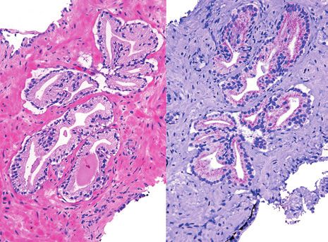
Prostate Pathology | IHC
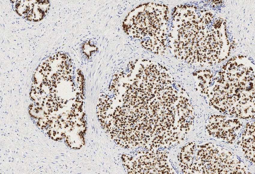
A B C
Figure 2: A) Low magnification of crowded glands of adenosis mimicking carcinoma. B) Higher magnification showing small glands
with pale cytoplasm and benign cytology. C) CK HMW stain showing patchy basal cell staining of scattered adenosis glands. Although
some glands are negative, these glands are identical morphologically to glands with basal cells and the entire lesion should be consid-
ered benign.
A A
B B
Figure 3: A) Partial atrophy. B) Patchy basal cell staining with Figure 4: A) High grade prostatic intraepithelial neoplasia (HG-
CK HMW analogous to the staining seen in adenosis. PIN). B) Patchy basal cell staining with p63 in HGPIN glands.
5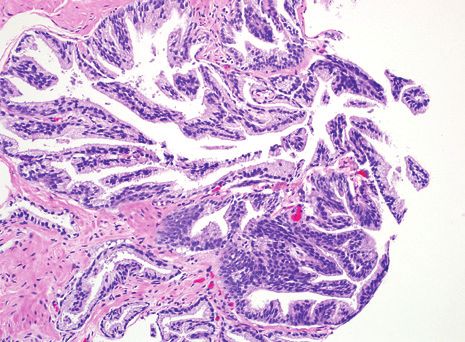
IHC | Prostate Pathology

          that label focally with CK HMW and less so with p63 in a             3) other basal cell markers such as CK HMW, and CK 5/6
          non-basal cell distribution (Figure 5). This phenomenon                 are totally negative. The other differential diagnosis for
          can be seen in all grades of prostate cancer, although                  a malignant lesion with p63 positivity is basal cell
          more commonly encountered in Gleason scores 8-10 (9).                   carcinoma. Not in favor of the diagnosis of basal cell
          Retention of basal cells in early adenocarcinoma is an                  carcinoma is the total negativity for other basal cell
          extremely rare phenomenon even in highly selected con-                  markers such as CK HMW and CK 5/6 in p63-
          sultation material (Figure 6) (8). Therefore this diagnosis             positive prostate cancers along with its positivity for
          should be made with great caution only when there is un-                prostatic secretory cell markers such as prostate-
          equivocal cancer on the hematoxylin and eosin slide, and                specific antigen (PSA).
          preferentially after consulting with an expert pathologist.
                                                                               Apart from high molecular weight cytokeratins and p63, a
          Non-specific staining seems to depend on the antigen re-             range of other markers that label basal cells in the prostate
          trieval method used, with the hot plate method showing               has been suggested (e.g. P-cadherin, podoplanin (D2-40),
          more non-specific reaction than the pepsin predigestion              CD109 or BCL2) (13-16). Since the experience with these
          and microwave retrieval methods (5,9-11). p63 has great-             experimental markers is limited, these are not recommend-
          er specificity for basal cells compared with CK HMW,                 ed in a routine setting.
          showing less non-specific reactions with cancer cells. A
          unique problem with p63 is aberrant diffuse expression of
          p63 in acinar adenocarcinoma (Figure 7) (12).                        Positive Markers of Malignancy
                                                                               AMACR (Alpha-methylacyl-CoA racemace)
          These cases differ from those showing the non-specific               It has long been a desire of surgical pathologists to com-
          staining of basal cell markers in adenocarcinoma described           plement basal cell markers, which stain negative in car-
          above in three major aspects:                                        cinoma, with an affirmative positive marker of malignan-
          1) the staining for p63 is strong and diffuse within the             cy. AMACR was the first such candidate positive marker.
             malignant glands;                                                 AMACR is a mitochondrial and peroxisomal enzyme that
          2) the majority of cases with aberrant p63 show distinc-             is involved in beta-oxidation of branched-chain fatty acids
             tive morphology of infiltrative glands, nests and cords           and in bile acid biosynthesis (17). It is expressed in var-
             with atrophic cytoplasm, hyperchromatic nuclei and                ious normal tissues, e.g. hepatocytes, renal tubular epi-
             visible nucleoli; and                                             thelial cells and gall bladder mucosa, but also in a variety

            A                                           B                                           C

          Figure 5. A) Adenocarcinoma of the prostate (arrows). B) CK HMW labeling several cancer cells. The positivity is not in a basal cell
          distribution as seen in adjacent benign glands (right side). C) Same cancer glands are negative for p63.

6
Prostate Pathology | IHC

                                                                     of dysplastic tissues or malignant tumors including colon
                                                                     cancer and papillary renal cancer (18-20). The highest
                                                                     rates of AMACR overexpression (>95% of cases) have
                                                                     been reported for prostate cancer, which has led to its
                                                                     widespread use as a positive diagnostic biomarker; so
                                                                     far, it is the only one that has gained clinical acceptance.
                                                                     In combination with basal cell markers, AMACR stain-
                                                                     ing can significantly increase the diagnostic accuracy
                                                                     and thus help avoiding unnecessary re-biopsies (21-27).
                                                                     Without AMACR, only "atypical glands" would have been
                                                                     reported in some instances.
 A

                                                                     However, the interpretation of AMACR staining requires
                                                                     experience, since it also introduces new pitfalls. Approx-
                                                                     imately 20% of small foci of adenocarcinoma on needle
                                                                     biopsy are negative for AMACR. Foamy gland, atrophic,
                                                                     pseudohyperplastic, and hormone-treated carcinomas ex-
                                                                     press AMACR to an even lesser extent (28-29). AMACR
                                                                     expression also lacks specificity. It is as frequently over-
                                                                     expressed in HGPIN as in adenocarcinoma, and certain
                                                                     benign mimickers of adenocarcinoma such as adenosis,
                                                                     partial atrophy and post-atrophic hyperplasia may ex-
                                                                     press AMACR (30). Consequently, it is essential to inter-
 B
                                                                     pret AMACR in the context of the entire lesion, using it to
Figure 6. A) Typical case of adenocarcinoma of the prostate          confirm a morphological impression of malignancy in a
with HGPIN gland. B) Carcinoma glands are positive for CK            focus of suspicious glands. A suspicious glandular focus
HMW and p63.
                                                                     that fulfills the histological criteria of carcinoma and that is
                                                                     negative for basal cell markers can still be diagnosed as
                                                                     adenocarcinoma even in the absence of AMACR reactivity.

 A                                            B                                            C

Figure 7. A) Adenocarcinoma on both sides of benign glands (arrow) with atrophic appearance and multilayered nuclei. B) p63/CK
HMW cocktail with carcinoma positive for p63 only labeling nuclei with surrounding benign glands having positivity in both nuclei (p63)
and cytoplasm (CK HMW). C) CK HMW stain with p63 positive carcinoma negative for CK HMW.

                                                                                                                                                 7
IHC | Prostate Pathology

            A                                                                 A

            B                                                                 B

          Figure 8. A) Gleason score 3+3=6 adenocarcinoma. Note ad-          Figure 9. A) Classic partial atrophy. B) Cocktail labeling with
          mixed benign glands (*) with paler cytoplasm and luminal infold-   brown chromogen basal cells (p63 and CK HMW) and red chro-
          ing. B) Cocktail labeling with brown chromogen labeling both ba-   mogen labeling AMACR. The diagnosis is still partial atrophy
          sal cell nuclei (p63) and cytoplasm (CK HMW) in benign glands.     despite the lack of basal cells and positive AMACR.
          Carcinoma lacks basal cells. Red chromogen labels cancer cy-
          toplasm (AMACR) and is negative in the benign glands.

          A common dual stain includes p63 and AMACR antibod-                absent and AMACR is positive (Figures 9-10). In other
          ies, but a potential problem with this stain is that p63 may       cases, where the glands are suspicious for partial atro-
          show background staining in the cytoplasm of benign                phy or HGPIN yet not definitive and the basal stains are
          glands, which may be confused with AMACR immunore-                 negative (+/- AMACR positivity); these lesions should
          activity. Another problem is that it is more difficult to iden-    be reported as: “Atypical glands, suspicious for ade-
          tify sparse brown p63-positive basal cells in the setting of       nocarcinoma.” (Figure 11). Even entirely benign glands
          intense brown AMACR-positive cytoplasm. A triple stain             can occasionally lack basal cells and express AMACR
          with AMACR labeled with a red chromogen and both p63               (Figure 12).
          and CK HMW labeled with a brown chromogen circum-
          vents this problem (Figure 8).                                     ERG (Ets-related gene product)
                                                                             The diagnostic value of ERG IHC is now widely under
          In a classic case of partial atrophy or HGPIN, these               investigation (31,32). A limitation of ERG as an affirm-
          diagnoses can be established even if basal cells are               ative positive cancer marker is the large fraction of

8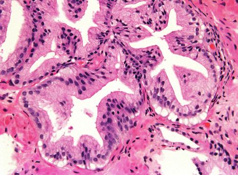
Prostate Pathology | IHC

 A                                                                 A

 B                                                                 B

Figure 10. A) HGPIN. B) HGPIN negative for basal cells (brown)   Figure 11. A) Atypical glands at the edge of the core that are
and positive for AMACR (red).                                    larger than typical cancer glands and could represent HGPIN.
                                                                 B) Despite negative stains for basal cells and positive AMACR
                                                                 (red), the atypical glands represent carcinoma yet HGPIN cannot
                                                                 be excluded.

ERG-negative carcinomas. Earlier studies have report-
ed ERG fusion in 15-72% of cases, depending on co-
hort design, tumor grade, zonal origin and even patient
ethnicity, but the mean prevalence in western countries
appears to level around 50% (33-43). However, on limit-
ed foci of carcinoma on needle biopsy, the positive rate
is more 30-40% (Figure 13). HGPIN is also positive for
ERG in a minority of cases. Even though these cave-
ats limit the diagnostic value of ERG to detect primary
invasive prostate cancer, combined staining of basal
cell markers with ERG may be useful in selected (ERG
positive) cases.
                                                                 Figure 12. Entirely benign prostate glands with negative stains
                                                                 for basal cells (brown) and positive AMACR (red).

                                                                                                                                           9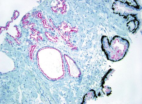
IHC | Prostate Pathology

                                                                               FASN (Fatty acid synthase)
                                                                               FASN overexpression in prostate cancer is well de-
                                                                               scribed (Figure 14) (44-49). A main difference with
                                                                               AMACR is the more prevalent expression of FASN in
                                                                               normal tissues and HGPIN, which makes it necessary
                                                                               to compare the staining of atypical glands with adja-
                                                                               cent clearly benign glands. However, if this compari-
                                                                               son is performed, FASN can be helpful, particularly in
                                                                               AMACR negative cases, which almost always are posi-
                                                                               tive for FASN (50, 51).

                                                                               GOLPH2 (Golgi phosphoprotein 2)
           Figure 13. Adenocarcinoma labeling with ERG. Note internal          GOLPH2 (GOLM1) is a 73kDa Golgi phosphoprotein of
           positive control of endothelial cells.
                                                                               yet unknown function that has been reported in various
                                                                               profiling studies of prostate cancer (52-54). So far, four
                                                                               groups have independently confirmed the strong over-
                                                                               expression of GOLPH2 in prostate cancer at the protein
                                                                               level, which can be used diagnostically in an experimen-
                                                                               tal setting (55-58).

                                                                               CYCS, ICK and IKBKB
                                                                               Other candidate positive markers that have been pro-
                                                                               posed are somatic cytochrome C (CYCS), intestinal cell
                                                                               kinase (ICK) and inhibitor of nuclear factor-kB kinase
                                                                               subunit (IKBKB) in prostate cancer (59). The very limited
                                                                               experience with these markers requires extensive valida-
                                                                               tion, before they can be recommended.
           Figure 14 FASN overexpression in prostate cancer (bottom
           area) relative to benign glands (top area).

                                                                               Primary Adenocarcinoma of the Prostate
                                                                               from Secondary Tumors
                                                                               PSA (Prostate-specific antigen)
                                                                               Prostate-specific antigen (PSA, KLK3) is a 33 kDa serine
                                                                               protease that is widely used to confirm the prostatic origin
                                                                               of metastatic carcinoma (61). PSA is however not entirely
                                                                               specific for prostate since it has also been detected in
                                                                               carcinomas of the ovary and the breast, including male
                                                                               breast cancer and other tissues, but it still is probably the
                                                                               most commonly used prostate marker (62-64). The pan-
                                                                               el of PSA, prostein (P501S), and NKX3.1 minimizes false
             A                     B                    C
                                                                               negative immunoreactivity in a poorly differentiated pros-
           Figure 15. A) Gleason score 10 adenocarcinoma of the prostate. B)   tatic adenocarcinoma (Figure 15).
           Negative PSA. C) Positive for P501S.

10
Prostate Pathology | IHC
PSMA (Prostate-specific membrane antigen) is slightly less indicative since ERG expression is seen
PSMA is a folate dehydrolase that is strongly expressed in vascular tumors, thymomas and gynecological neo-
by most prostate carcinomas and their metastases (65). plasms (80,81). It is also possible, that the sensitivity
In contrast to PSA, PSMA shows increasing expression in prostate cancer metastases exceeds that of primary
levels in high grade tumors and metastases, however it tumors, since TMPRSS2-ERG rearrangement might be
is now acknowledged that it is not prostate specific at more prevalent in metastases (82).
all, but is rather widely expressed in various solid tumors
including renal cancer, gastrointestinal neoplasms and Four studies of independent groups have analyzed ERG
urothelial carcinomas (66-68). rearrangement as a marker for small cell carcinoma of
the prostate, which can be difficult to differentiate from
Prostein (P501S) small cell carcinomas of other sites (83-86). All four stud-
Prostein’s prostate-specificity has been independently ies found ERG rearrangements detected by FISH exclu-
confirmed and several groups have successfully ap- sively in prostatic small cell carcinomas (range 45-86%)
plied prostein IHC to discriminate a prostatic cancer but not in small cell carcinomas of other sites including
origin from tumors of the colon and the bladder (69- bladder and lung. In comparison to other markers pre-
75). Especially, the separation of high grade prostate viously suggested in this respect, ERG clearly outper-
cancer from urothelial carcinoma can be successfully forms these, including PSA and also prostein, which was
achieved with a combination of p63 and prostein (76). found in only 28% of prostatic small cell carcinoma cas-
The biological functions of prostein, which is androgen es (87). The use of ERG to determine a prostatic origin of
regulated and mostly localized to the golgi apparatus small cell carcinomas appears to be the best validated
of the cell, are unclear. However, it is still regarded to contribution of the ERG rearrangement to prostate diag-
be among the best validated immunohistochemical nostics. However, as small cell carcinomas, regardless
markers of prostatic origin. In cases where PSA is neg- of the site of origin, are treated the same it is current-
ative, many will be positive for prostein. An additional ly questionable as to the need to specifically diagnose
advantage is the distinctive granular cytoplasmic stain- small cell carcinoma of the prostate.
ing which distinguishes it from other markers in which
a weak positve cytoplasmic blush can be more difficult
to interpret.
AR (Androgen receptor)
PSA and PSMA are both targets of androgen signal-
ing and the AR itself is also regulated in prostate can-
cer (77,78). Again, the diagnostic use of AR staining is
greatly hampered by the expression of AR in other human
tissues and tumors and it can therefore no longer be rec-
ommended (Figure 16) (79).
ERG (Ets-related gene product)
Although ERG expression clearly lacks sensitivity in
primary prostatic carcinomas (with 50% negatives), it
appears to be quite specific for prostatic origin. More
specifically, the genomic translocation has not been Figure 16. Metastatic prostatic adenocarcinoma to cervical
found in any other carcinoma, whereas the protein level lymph node with positivity for AR.
11IHC | Prostate Pathology
NKX3.1 (Homeobox protein NKX3.1)
Another androgen regulated and mostly prostate-spe-
cifically expressed gene is the homeobox gene NKX3.1,
which is found expressed primarily in secretory prostatic
epithelia of benign and neoplastic cells, but rarely also
in benign testis and invasive lobular carcinomas of the
breast (88-90). Some researchers have described a loss
of NKX3.1 protein in high grade tumors of the prostate
and even a prognostic significance (91, 92). One study
compared several prostate marker candidates including
NKX3.1 and prostein and found both excellent for the dis-
crimination of prostate from urothelial cancer (Figure 17)
(73). More recently, Gurel et al. described NKX3.1 as an Figure 17. Nuclear staining for NKX3.1 in high grade prostate cancer.
excellently sensitive and specific prostate cancer marker,
outperforming PSA in this regard (93). This discrepancy
to earlier studies is explained by a novel, more sensitive Most helpful is to investigate the expression of p63 (posi-
NKX3.1 antibody (sensitivity 98.6%, specificity 99.7%). tive in UC, negative in PCa), prostein (positive in PCa, neg-
ative in UC), NKX3.1 (positive in PCa, negative in UC), and
AMACR (Alpha-methylacyl-CoA racemase) GATA3 (positive in UC, negative in PCa) (Table 1).
Even though AMACR is typically overexpressed in prostate
Table 1. Markers suggested for differential diagnosis of prostate
cancer, it is not restricted to it but is also present in up to 92% cancer vs. urothelial cancer.
of colorectal adenocarcinomas, as well as breast, lung, ovar-
ian, renal cell carcinomas (especially the papillary variant), Marker Prostate Cancer Urothelial Cancer
p63 Neg Pos
as well as bladder urothelial and adenocarcinomas (94-97).
Thus, this marker is not useful in the differential diagnosis of Prostein Pos Neg
prostate cancer from other malignancies. GATA3 Neg Pos
NKX3.1 Pos Neg
Specific Differential Diagnoses Prostate Cancer (PCa) vs. Colorectal Cancer (CRC)
Prostate Cancer (PCa) vs. Urothelial Cancer (UC) The typical immunophenotype of CRC is CK20+/CK7-/
Although the morphology of invasive urothelial carcino- CDX2+. Of these markers CDX2 alone is helpful, since
ma is typically distinct from glandular adenocarcinoma of it is very rarely positive in PCa, however there are ex-
the prostate, the morphological discrimination from high ceptions (98). As stated above, prostein and NKX3.1 are
grade prostate carcinomas can be challenging, particu- helpful to identify PCa. Nuclear staining of beta-catenin is
larly in small biopsies. IHC can be helpful, but beware: more common in CRC, however this lacks sensitivity and
PSA is mostly negative in UC, but may be missing in high specificity and is discouraged as a marker for CRC.
grade prostate cancer. AR is mostly positive in PCa, but
is also seen in UC, its use is therefore discouraged. Ac- Diagnosis of Pretreated Prostate Carcinomas
cumulation and strong nuclear staining of p53 is more The effects of organ sparing therapy, i.e. androgen ablation
prevalent in invasive UC, but may also be positive in high and radiotherapy, on prostatic tissues are well documented
grade PCa. CK7/20 are commonly used markers for UC, (60). Reactive changes in benign tissues and tumor atro-
however both cytokeratins may also be expressed in high phy can markedly obscure the morphology. This introduc-
grade PCa, so they lack discriminatory power. es a risk to over- or underdiagnose prostate cancer and
12Prostate Pathology | IHC
particularly Gleason scores can be markedly altered, e.g. respectively, in contrast to PSA where non-specific diffuse
by an assignment of Gleason pattern 4 to areas that had cytoplasmic staining can be misinterpreted as true positivity
been Gleason 3 prior to therapy. Treated prostate carcino- (Figure 19). PSMA can be found in rare cases of pulmonary
mas tend to show some loss of AMACR expression, limiting small cell carcinoma, hepatocellular carcinoma, papillary renal
its value in a post-treatment situation. In severely regressed cell carcinoma and most importantly in 17% of urothelial car-
cases, stainings for pan-cytokeratin and basal cell markers cinomas (99). The two oldest prostatic markers that exist are
are more helpful to ascertain the presence of residual or re- PSA and PSAP. PSAP is relatively specific although it suffers
current prostate cancer (Figure 18). from relative decreased sensitivity. Situations that can cause
diagnostic difficulty include PSA and PSAP within periurethral
glands, as well as cystitis cystica and cystitis glandularis in
Pitfalls in the Use of Prostatic Markers both men and women (100-102). Other examples of cross-re-
NKX3.1 and prostein (P501S) are the most specific markers active staining include anal glands in men (PSA, PSAP) and
for prostate origin (Figures 15-16). They also have the advan- urachal remnants (PSA) (103,104). Some intestinal carcinoids
tage of nuclear and clumpy granular cytoplasmic staining, and pancreatic islet cell tumors are strongly reactive with anti-
bodies to PSAP, yet are negative with antibodies to PSA (105).
Periurethral gland carcinomas in women and various salivary
gland tumors may also be PSA and PSAP positive (106, 107).
Weak false-positive staining for PSAP has been reported in
several breast and renal cell carcinomas.
Among prostatic markers with the greatest specificity for
prostate, the most sensitive are PSA, P501S, NKX3.1.
There are some situations where the marker is sensitive yet
false negative results can occur. If the positive control slide
shows only weak to moderate staining of benign prostate
glands with a prostatic marker, then poorly differentiated
A
prostatic adenocarcinomas which typically have less an-
B
A B
Figure 18. A) Adenocarcinoma of the prostate with radiation ef-
fect. B) Cocktail stain with benign prostate glands with radiation Figure 19. A) Specific clumpy granular staining of P501S in be-
effect (right side) labeling basal cells brown with CK HMW and nign prostate gland. B) Weak diffuse nonspecific biotin labeling
p63. Carcinoma with treatment effect (left side) lacks basal cells in a case labeled with P501S that can be correctly diagnosed as
and is positive for AMACR (red). being negative. If this was a PSA stain, then it may have been
incorrectly called positive.
13IHC | Prostate Pathology
tigen can be falsely negative. Another pitfall is negative years, immunohistochemistry has become an indispensible
staining with prostate markers in poorly differentiated adeno- tool in surgical pathology and some areas, like lymphoma
carcinomas of the prostate, which is the situation where this classification, even depend strictly on immunophenotyping.
immunohistochemistry is typically performed. PSA immuno- In the evolution of current concepts of prostate pathology, im-
expression is inversely correlated with increasing Gleason munohistochemistry has also become increasingly important.
score, and a minority of Gleason score 10 adenocarcinomas
may be negative for PSA, especially in limited material. P501S In this review, we aimed to illustrate that immunohistochem-
and NKX3.1 expression seems to be unrelated to Gleason istry can in fact be immensely contributive in diagnostic
grade. It is important to note that a small minority (less than prostate pathology, if used with care and experience. No
5%) of poorly differentiated prostatic adenocarcinomas are to- single marker can establish a diagnosis on its own, but
tally negative for all prostatic markers (76). Therefore, the lack has to be used in close conjunction and with a thorough
of immunoreactivity for prostate- specific markers in a poorly assessment of the individual cases’ morphological as well
differentiated tumor, especially if present in limited amount (in as the clinical context, to lead to correct conclusions for
biopsy specimens), does not totally exclude the diagnosis of improved patient care. Every tool has pros and cons. The
a poorly differentiated prostatic adenocarcinoma. generally increased diagnostic certainty achieved with im-
munohistochemistry also opens up the possibility of new
pitfalls that the pathologist must be aware of.
Concluding Remarks
The increasing number of biopsies, time constraints and We have compiled this review to cover the most important
the demands of quality management and legal issues have uses and pitfalls of contemporary immunohistochemistry
prompted pathologists to adapt their workflow accordingly in prostate diagnostics and hope that this may be a help-
and to increase their diagnostic efficiency. Over the past 20 ful companion in daily work.
Dako Antibodies for Prostate Tissue Antigens
Anti- Clone Concentrate Ready-to-Use
AMACR 13H4
AMACR + CK HMW + CK 5/6 13H4 + 34ßE12 + D5/16 B4
Androgen Receptor AR441
Cytokeratin 5/6 D5/16 B4
Cytokeratin HMW 34ßE12
ERG EP111
Ki-67 MIB-1
p53 Protein 318-6-11
p53 Protein DO-7
p63 Protein DAK-p63*
Prostein (P501S) 10E3
Prostate-Specific Antigen (PSA) ER-PR8
Prostate-Specific Antigen (PSA) Poly
Prostate-Specific Membrane Antigen (PSMA) 3E6
Prostatic Acid Phosphatase PASE/4LJ
*Not available in the US.
14Prostate Pathology | IHC
Stains using Dako FLEX RTU antibodies
AMACR AMACR + CK HMW + CK 5/6 Cytokeratin 5/6
Clone 13H4 Clones 13H4 + 34ßE12 Clone D5/16 B4
Prostate adenocarcinoma. The ma- + D5/16 B4 Prostate hyperplasia and prostate
jority of cells show a distinct granular Prostate. Cells labeled by Anti-AMACR carcinoma. The normal and benign
cytoplasmic staining reaction and the antibody display a distinct red cyto- glands show a distinct cytoplasmic
benign glands are mostly negative. plasmic granular staining. Cells labe- staining reaction in the basal cells.
led by Anti-CK HMW and Anti-CK 5/6
antibody display strong brown cyto-
plasmic staining.
Cytokeratin HMW ERG Ki-67
Clone 34ßE12 Clone EP111 Clone MIB-1
Prostate adenocarcinoma. Various Prostate adenocarcinoma. The majori- Tonsil. The germinal center B cells
staining reaction patterns are seen: ty of neoplastic cells show a moderate show a moderate to strong nuclear
Continuous cytoplasmic staining in to strong nuclear staining reaction. staining reaction.
normal gland, discontinuous pattern
in PIN and no staining in invasive
cancer cells.
15IHC | Prostate Pathology
p53 protein p63 protein Prostein (P501S),
Clone DO-7 Clone DAK-p63 Clone 10E3
Breast carcinoma, the neoplastic Benign prostate hyperplasia and nor- Prostate adenocarcinoma. The major-
cells show a moderate to strong nu- mal prostate. The normal and benign ity of neoplastic celles show a mod-
clear staining reaction. glands show a distinct nuclear staining erate to strong granular cytoplasmic
reaction in basal cells, while the secre- staining reaction.
tory and neoplastic cells are negative.
Prostate-Specific Antigen (PSA) Prostate-Specific Membrane
Polyclonal Antigen (PSMA)
Prostate adenocarcinoma. The ne- Clone 3E6
oplastic cells and the hyperplastic Prostate adenocarcinoma. The majori-
glands show a moderate to strong and ty of neoplastic cells show a moderate
diffuse cytoplasmic staining reaction. to strong cytoplasmic and/or membra-
nous staining reaction.
16Prostate Pathology | IHC
References 20. Sonwalkar SA, Rotimi O, Scott N, et al. A study of indefinite for dyspla-
1. Brawer MK, Peehl DM, Stamey TA, Bostwick DG. Keratin immunore sia in Barrett's oesophagus: reproducibility of diagnosis, clinical
activity in the benign and neoplastic human prostate. Cancer Res outcomes and predicting progression with AMACR (alpha-methylacyl-
1985; 45(8):3663-7. CoA-racemase). Histopathol 2010; 56(7):900-7.
2. Hedrick L, Epstein JI. Use of keratin 903 as an adjunct in the diagnosis 21. Carswell BM, Woda BA, Wang X, et al. Detection of prostate cancer
of prostate carcinoma. Am J Surg Pathol 1989; 13(5):389-96. by alpha-methylacyl CoA racemase (P504S) in needle biopsy speci-
3. Signoretti S, Waltregny D, Dilks J, et al. p63 is a prostate basal cell mens previously reported as negative for malignancy. Histopathol
marker and is required for prostate development. Am J Pathol 2000; 2006; 48(6):668-73.
157(6):1769-75. 22. Farinola MA, Epstein JI. Utility of immunohistochemistry for alpha-
4. Weinstein MH, Signoretti S, Loda M. Diagnostic utility of immunohisto- methylacyl-CoA racemase in distinguishing atrophic prostate cancer
chemical staining for p63, a sensitive marker of prostatic basal cells. from benign atrophy. Hum Pathol 2004; 35(10):1272-8.
Mod Pathol 2002; 15(12):1302-8. 23. Herawi M, Epstein JI. Immunohistochemical antibody cocktail staining
5. Shah RB, Zhou M, LeBlanc M, Snyder M, Rubin MA. Comparison of the (p63/HMWCK/AMACR) of ductal adenocarcinoma and Gleason pat-
basal cell-specific markers, 34betaE12 and p63, in the diagnosis tern 4 cribriform and noncribriform acinar adenocarcinomas of the
of prostate cancer. Am J Surg Pathol 2002; 26(9):1161-8. prostate. Am J Surg Pathol 2007; 31(6):889-94.
6. Zhou M, Shah R, Shen R, Rubin MA. Basal cell cocktail (34betaE12 + 24. Jiang Z, Wu CL, Woda BA, et al. Alpha-methylacyl-CoA racemase: a
p63) improves the detection of prostate basal cells. Am J Surg Pathol multi-institutional study of a new prostate cancer marker. Histopathol
2003; 27(3):365-71. 2004; 45(3):218-25.
7. Wang W, Sun X, Epstein JI. Partial atrophy on prostate needle biopsy 25. Zhou M, Aydin H, Kanane H, Epstein JI. How often does alpha-methy-
cores: a morphologic and immunohistochemical study. Am J Surg lacyl-CoA-racemase contribute to resolving an atypical diagnosis on
Pathol 2008; 32(6):851-7. prostate needle biopsy beyond that provided by basal cell markers?
8. Oliai BR, Kahane H, Epstein JI. Can basal cells be seen in adenocarci Am J Surg Pathol 2004; 28(2):239-43.
noma of the prostate?: an immunohistochemical study using high 26. Paner GP, Luthringer DJ, Amin MB. Best practice in diagnostic im
molecular weight cytokeratin (clone 34betaE12) antibody. Am J Surg munohistochemistry: prostate carcinoma and its mimics in needle core
Pathol 2002; 26(9):1151-60. biopsies. Arch Pathol Lab Med 2008; 132(9):1388-96.
9. Ali TZ, Epstein JI. False positive labeling of prostate cancer with high 27. Jiang Z, Iczkowski KA, Woda BA, Tretiakova M, Yang XJ. P504S im
molecular weight cytokeratin: p63 a more specific immunomarker for munostaining boosts diagnostic resolution of "suspicious" foci in pros-
basal cells. Am J Surg Pathol 2008; 32(12):1890-5. tatic needle biopsy specimens. Am J Clin Pathol 2004; 121(1):99-107.
10. Ramnani DM, Bostwick DG. Basal cell-specific anti-keratin antibody 28. Beach R, Gown AM, De Peralta-Venturina MN, et al. P504S immunohisto-
34betaE12: optimizing its use in distinguishing benign prostate and chemical detection in 405 prostatic specimens including 376 18-gauge
cancer. Mod Pathol 1999; 12(5):443-4. needle biopsies. Am J Surg Pathol 2002; 26(12):1588-96.
11. Varma M, Linden MD, Amin MB. Effect of formalin fixation and epitope 29. Zhou M, Jiang Z, Epstein JI. Expression and diagnostic utility of
retrieval techniques on antibody 34betaE12 immunostaining of pros- alpha-methylacyl-CoA-racemase (P504S) in foamy gland and pseudo-
tatic tissues. Mod Pathol 1999; 12(5):472-8. hyperplastic prostate cancer. Am J Surg Pathol 2003; 27(6):772-8.
12. Osunkoya AO, Hansel DE, Sun X, Netto GJ, Epstein JI. Aberrant dif- 30. Herawi M, Parwani AV, Irie J, Epstein JI. Small glandular proliferations
fuse expression of p63 in adenocarcinoma of the prostate on needle on needle biopsies: most common benign mimickers of prostatic
biopsy and radical prostatectomy: report of 21 cases. Am J Surg adenocarcinoma sent in for expert second opinion. Am J Surg Pathol
Pathol 2008; 32(3):461-7. 2005; 29(7):874-80.
13. Kuroda N, Katto K, Tamura M, et al. Immunohistochemical application 31. He H, Magi-Galluzzi C, Li J, et al. The diagnostic utility of novel im
of D2-40 as basal cell marker in evaluating atypical small acinar prolife- munohistochemical marker ERG in the workup of prostate biopsies
ration of initial routine prostatic needle biopsy materials. Medical mo- with "atypical glands suspicious for cancer". Am J Surg Pathol 2011;
lecular morphology 2010; 43(3):165-9. 35(4):608-14.
14. Hasegawa M, Hagiwara S, Sato T, et al. CD109, a new marker for 32. Yaskiv O, Zhang X, Simmerman K, et al. The Utility of ERG/P63 Double
myoepithelial cells of mammary, salivary, and lacrimal glands and Immunohistochemical Staining in the Diagnosis of Limited Cancer
prostate basal cells. Pathol Int 2007; 57(5):245-50. in Prostate Needle Biopsies. Am J Surg Pathol 2011; 35(7):1062-8.
15. Jarrard DF, Paul R, van Bokhoven A, et al. P-Cadherin is a basal 33. Demichelis F, Fall K, Perner S, et al. TMPRSS2:ERG gene fusion asso-
cell-specific epithelial marker that is not expressed in prostate cancer. ciated with lethal prostate cancer in a watchful waiting cohort. Onco-
Clin Cancer Res 1997; 3(11):2121-8. gene 2007; 26(31):4596-9.
16. Ramos Soler D, Mayordomo Aranda E, Calatayud Blas A, et al. [Use- 34. Lapointe J, Li C, Giacomini CP, et al. Genomic profiling reveals
fulness of bcl-2 expression as a new basal cell marker in prostatic alternative genetic pathways of prostate tumorigenesis. Cancer Res
pathology]. Acta Urol Esp 2006; 30(4):345-52. 2007; 67(18):8504-10.
17. Lloyd MD, Darley DJ, Wierzbicki AS, Threadgill MD. Alpha-methylacyl- 35. Braun M, Scheble VJ, Menon R, et al. Relevance of cohort design
CoA racemase--an 'obscure' metabolic enzyme takes centre stage. for studying the frequency of the ERG rearrangement in prostate can-
FEBS 2008; 275(6):1089-102. cer. Histopathol 2011; 58(7):1028-36.
18. Went PT, Sauter G, Oberholzer M, Bubendorf L. Abundant expression of 36. Mosquera JM, Perner S, Demichelis F, et al. Morphological features
AMACR in many distinct tumour types. Pathol 2006; 38(5):426-32. of TMPRSS2-ERG gene fusion prostate cancer. J Pathol 2007;
19. Dorer R, Odze RD. AMACR immunostaining is useful in detecting dys- 212(1):91-101.
plastic epithelium in Barrett's esophagus, ulcerative colitis, and 37. Fine SW, Gopalan A, Leversha MA, et al. TMPRSS2-ERG gene fusion
Crohn's disease. Am J Surg Pathol 2006; 30(7):871-7. is associated with low Gleason scores and not with high-grade morpho-
logical features. Mod Pathol 2010; 23(10):1325-33.
17IHC | Prostate Pathology
38. Guo CC, Zuo G, Cao D, Troncoso P, Czerniak BA. Prostate cancer of 58. Li W, Wang X, Li B, Lu J, Chen G. Diagnostic significance of overex-
transition zone origin lacks TMPRSS2-ERG gene fusion. Mod Pathol pression of Golgi membrane protein 1 in prostate cancer. Urology
2009; 22(7):866-71. 2012; 80(4):952 e1-7.
39. Bismar TA, Trpkov K. TMPRSS2-ERG gene fusion in transition zone 59. Haggarth L, Hagglof C, Jaraj SJ, et al. Diagnostic biomarkers of
prostate cancer. Mod Pathol 2010; 23(7):1040-1; author reply 1-2. prostate cancer. Scan J Urol Nephrol 2011; 45(1):60-7.
40. Falzarano SM, Navas M, Simmerman K, et al. ERG rearrangement is 60. Petraki CD, Sfikas CP. Histopathological changes induced by thera-
present in a subset of transition zone prostatic tumors. Mod Pathol pies in the benign prostate and prostate adenocarcinoma. Histol
2010; 23(11):1499-506. Histopathol 2007; 22(1):107-18.
41. Mao X, Yu Y, Boyd LK, et al. Distinct genomic alterations in prostate can- 61. Bostwick DG. Prostate-specific antigen. Current role in diagnostic pa-
cers in Chinese and Western populations suggest alternative pathways of thology of prostate cancer. Am J Clin Pathol 1994; 102(4 Suppl 1):
prostate carcinogenesis. Cancer Res 2010; 70(13):5207-12. S31-7.
42. Miyagi Y, Sasaki T, Fujinami K, et al. ETS family-associated gene fu- 62. Kraus TS, Cohen C, Siddiqui MT. Prostate-specific antigen and hor-
sions in Japanese prostate cancer: analysis of 194 radical prostatec- mone receptor expression in male and female breast carcinoma.
tomy samples. Mod Pathol 2010; 23(11):1492-8. Diagn Pathol 2010; 5:63.
43. Magi-Galluzzi C, Tsusuki T, Elson P, et al. TMPRSS2-ERG gene fu- 63. Alanen KA, Kuopio T, Koskinen PJ, Nevalainen TJ. Immunohistochem-
sion prevalence and class are significantly different in prostate can- ical labelling for prostate specific antigen in non-prostatic tissues. Pa-
cer of Caucasian, African-American and Japanese patients. Pros- thol, Res Pract 1996; 192(3):233-7.
tate 2011; 71(5):489-97. 64. van Krieken JH. Prostate marker immunoreactivity in salivary gland
44. Prowatke I, Devens F, Benner A, et al. Expression analysis of imbal- neoplasms. A rare pitfall in immunohistochemistry. Am J Surg Pathol
anced genes in prostate carcinoma using tissue microarrays. Br J 1993; 17(4):410-4.
Cancer 2007; 96(1):82-8. 65. Wright GL, Jr., Haley C, Beckett ML, Schellhammer PF. Expression
45. Shurbaji MS, Kalbfleisch JH, Thurmond TS. Immunohistochemical de- of prostate-specific membrane antigen in normal, benign, and malig-
tection of a fatty acid synthase (OA-519) as a predictor of progression nant prostate tissues. Urol Oncol 1995; 1(1):18-28.
of prostate cancer. Hum Pathol 1996; 27(9):917-21. 66. Troyer JK, Beckett ML, Wright GL, Jr. Detection and characterization
46. Baron A, Migita T, Tang D, Loda M. Fatty acid synthase: a metabolic of the prostate-specific membrane antigen (PSMA) in tissue extracts
oncogene in prostate cancer? J Cell Biochem 2004; 91(1):47-53. and body fluids. Int J Cancer 1995; 62(5):552-8.
47. Fiorentino M, Zadra G, Palescandolo E, et al. Overexpression of 67. Kinoshita Y, Kuratsukuri K, Landas S, et al. Expression of prostate-
fatty acid synthase is associated with palmitoylation of Wnt1 and cyto- specific membrane antigen in normal and malignant human tissues.
plasmic stabilization of beta-catenin in prostate cancer. Lab Invest World J Surg 2006; 30(4):628-36.
2008; 88(12):1340-8. 68. Samplaski MK, Heston W, Elson P, Magi-Galluzzi C, Hansel DE. Folate
48. Migita T, Ruiz S, Fornari A, et al. Fatty acid synthase: a metabolic hydrolase (prostate-specific antigen) 1 expression in bladder cancer
enzyme and candidate oncogene in prostate cancer. J Natl Cancer subtypes and associated tumor neovasculature. Mod Pathol 2011;
Inst 2009; 101(7):519-32. 24(11): 1521-9.
49. Rossi S, Graner E, Febbo P, et al. Fatty acid synthase expression de- 69. Xu J, Kalos M, Stolk JA, et al. Identification and characterization of
fines distinct molecular signatures in prostate cancer. Mol Cancer Res prostein, a novel prostate-specific protein. Cancer Res 2001;
2003; 1(10):707-15. 61(4):1563-8.
50. Wu X, Zayzafoon M, Zhang X, Hameed O. Is There a Role for Fatty 70. Kalos M, Askaa J, Hylander BL, et al. Prostein expression is highly
Acid Synthase in the Diagnosis of Prostatic Adenocarcinoma?: A restricted to normal and malignant prostate tissues. Prostate 2004;
Comparison With AMACR. Am J Clin Pathol 2011; 136(2):239-46. 60(3):246-56.
51. Tischler V, Fritzsche FR, Gerhardt J, Jäger C, Stephan C, Jung K, et 71. Sheridan T, Herawi M, Epstein JI, Illei PB. The role of P501S and PSA
al. Comparison of the diagnostic value of fatty acid synthase (FASN) in the diagnosis of metastatic adenocarcinoma of the prostate. Am J
with alpha-methylacyl-CoA racemase (AMACR) as prostatic cancer Surg Pathol 2007; 31(9):1351-5.
tissue marker. Histopathol 2010;56:811-5. 72. Osunkoya AO, Netto GJ, Epstein JI. Colorectal adenocarcinoma involv-
52. Luo JH, Yu YP, Cieply K, et al. Gene expression analysis of prostate ing the prostate: report of 9 cases. Human Pathol 2007; 38(12):1836-41.
cancers. Mol Carcinog 2002; 33(1):25-35. 73. Chuang AY, DeMarzo AM, Veltri RW, et al. Immunohistochemical differ-
53. Lapointe J, Li C, Higgins JP, et al. Gene expression profiling identifies entiation of high-grade prostate carcinoma from urothelial carcinoma.
clinically relevant subtypes of prostate cancer. Proc Natl Acad Sci Am J Surg Pathol 2007; 31(8):1246-55.
U S A 2004; 101(3):811-6. 74. Lane Z, Hansel DE, Epstein JI. Immunohistochemical expression of
54. Kristiansen G, Pilarsky C, Wissmann C, et al. Expression profiling of prostatic antigens in adenocarcinoma and villous adenoma of the
microdissected matched prostate cancer samples reveals CD166/ urinary bladder. Am J Surg Pathol 2008; 32(9):1322-6.
MEMD and CD24 as new prognostic markers for patient survival. J Pathol 75. Lane Z, Epstein JI, Ayub S, Netto GJ. Prostatic adenocarcinoma in
2005; 205(3):359-76. colorectal biopsy: clinical and pathologic features. Human Pathol
55. Wei S, Dunn TA, Isaacs WB, De Marzo AM, Luo J. GOLPH2 and MYO6: 2008; 39(4):543-9.
putative prostate cancer markers localized to the Golgi apparatus. The 76. Srinivasan M, Parwani AV. Diagnostic utility of p63/P501S double
Prostate 2008; 68(13):1387-95. sequential immunohistochemical staining in differentiating urothelial
56. Kristiansen G, Fritzsche FR, Wassermann K, et al. GOLPH2 protein carcinoma from prostate carcinoma. Diagn Pathol 2011;6:67.
expression as a novel tissue biomarker for prostate cancer: impli- 77. Fleischmann A, Rocha C, Schobinger S, et al. Androgen receptors
cations for tissue-based diagnostics. Brit J Can 2008; 99(6):939-48. are differentially expressed in Gleason patterns of prostate cancer and
57. Varambally S, Laxman B, Mehra R, et al. Golgi protein GOLM1 is a tis- down-regulated in matched lymph node metastases. Prostate 2011;
sue and urine biomarker of prostate cancer. Neoplasia 2008; 71(5):453-60.
10(11):1285-94.
18Prostate Pathology | IHC
78. Loda M, Fogt F, French FS, et al. Androgen receptor immunohisto- 97. Suh N, Yang XJ, Tretiakova MS, Humphrey PA, Wang HL. Value of
chemistry on paraffin-embedded tissue. Mod Pathol 1994; 7(3):388-91. CDX2, villin, and alpha-methylacyl coenzyme A racemase immuno-
79. Jaspers HC, Verbist BM, Schoffelen R, et al. Androgen receptor-pos- stains in the distinction between primary adenocarcinoma of the
itive salivary duct carcinoma: a disease entity with promising new bladder and secondary colorectal adenocarcinoma. Mod Pathol 2005;
treatment options. J Clin Oncol 2011; 29(16):e473-6. 18(9):1217-22.
80. Scheble VJ, Braun M, Beroukhim R, et al. ERG rearrangement is spe- 98. Herawi M, De Marzo AM, Kristiansen G, Epstein JI. Expression of
cific to prostate cancer and does not occur in any other common CDX2 in benign tissue and adenocarcinoma of the prostate. Human
tumor. Mod Pathol 2010; 23(8):1061-7. Pathol 2007; 38(1):72-8.
81. Minner S, Luebke AM, Kluth M, et al. High level of Ets-related gene 99. Mhawech-Fauceglia P, Zhang S, Terracciano L, et al. Prostate-specific
expression has high specificity for prostate cancer: a tissue microarray membrane antigen (PSMA) protein expression in normal and neoplas-
study of 11 483 cancers. Histopathol 2012; 61(3):445-53. tic tissues and its sensitivity and specificity in prostate adenocarcino-
82. Perner S, Svensson MA, Hossain RR, et al. ERG rearrangement ma: an immunohistochemical study using mutiple tumour tissue mi-
metastasis patterns in locally advanced prostate cancer. Urology croarray technique. Histopathol 2007; 50(4):472-83.
2010; 75(4):762-7. 100. Nowels K, Kent E, Rinsho K, Oyasu R. Prostate specific antigen and
83. Scheble VJ, Braun M, Wilbertz T, et al. ERG rearrangement in small cell acid phosphatase-reactive cells in cystitis cystica and glandularis.
prostatic and lung cancer. Histopathol 2010; 56(7):937-43. Arch Pathol Lab Med 1988;112:734-7.
84. Guo CC, Dancer JY, Wang Y, et al. TMPRSS2-ERG gene fusion in small 101. Pollen JJ, Dreilinger A. Immunohistochemical identification of pro-
cell carcinoma of the prostate. Hum Pathol 2011; 42(1):11-7. static acid phosphatase and prostate specific antigen in female periu-
85. Williamson SR, Zhang S, Yao JL, et al. ERG-TMPRSS2 rearrangement rethral glands. Urology 1984;23:303-4.
is shared by concurrent prostatic adenocarcinoma and prostatic small 102. Tepper SL, Jagirdar J, Heath D, Geller SA. Homology between the
cell carcinoma and absent in small cell carcinoma of the urinary blad- female paraurethral (Skene's) glands and the prostate. Immunohisto-
der: evidence supporting monoclonal origin. Mod Pathol 2011; chemical demonstration. Arch Pathol Lab Med 1984;108:423-5.
24(8): 1120-7. 103. Kamoshida S, Tsutsumi Y. Extraprostatic localization of prostatic acid
86. Lotan TL, Gupta NS, Wang W, et al. ERG gene rearrangements are phosphatase and prostate-specific antigen: distribution in cloaco-
common in prostatic small cell carcinomas. Mod Pathol 2011; genic glandular epithelium and sex-dependent expression in human
24(6):820-8. anal gland. Hum Pathol 1990;21:1108-11.
87. Wang W, Epstein JI. Small cell carcinoma of the prostate. A morpho- 104. Golz R, Schubert GE. Prostatic specific antigen: immunoreactivity in
logic and immunohistochemical study of 95 cases. Am J Surg Pathol urachal remnants. J Urol 1989;141:1480-2.
2008; 32(1):65-71. 105. Sobin LH, Hjermstad BM, Sesterhenn IA, Helwig EB. Prostatic acid
88. Voeller HJ, Augustus M, Madike V, et al. Coding region of NKX3.1, a phosphatase activity in carcinoid tumors. Cancer 1986;58:136-8.
prostate-specific homeobox gene on 8p21, is not mutated in human 106. van Krieken JH. Prostate marker immunoreactivity in salivary gland
prostate cancers. Cancer Res 1997; 57(20):4455-9. neoplasms. A rare pitfall in immunohistochemistry. Am J Surg Pathol
89. He WW, Sciavolino PJ, Wing J, et al. A novel human prostate-specific, 1993;17:410-4.
androgen-regulated homeobox gene (NKX3.1) that maps to 8p21, a 107. Spencer JR, Brodin AG, Ignatoff JM. Clear cell adenocarcino-
region frequently deleted in prostate cancer. Genomics 1997; 43(1):69- ma of the urethra: evidence for origin within paraurethral ducts. J Urol
90. Gelmann EP, Bowen C, Bubendorf L. Expression of NKX3.1 in normal 1990;143:122-5.
and malignant tissues. Prostate 2003; 55(2):111-7.
91. Bethel CR, Faith D, Li X, et al. Decreased NKX3.1 protein expression
in focal prostatic atrophy, prostatic intraepithelial neoplasia, and ade-
nocarcinoma: association with gleason score and chromosome 8p
deletion. Cancer Res 2006; 66(22):10683-90.
92. Bowen C, Bubendorf L, Voeller HJ, et al. Loss of NKX3.1 expression
in human prostate cancers correlates with tumor progression. Cancer
Res 2000; 60(21):6111-5.
93. Gurel B, Ali TZ, Montgomery EA, et al. NKX3.1 as a marker of prostatic
origin in metastatic tumors. Am J Surg Pathology 2010; 34(8):1097-105.
94. Noske A, Zimmermann AK, Caduff R, Varga Z, Fink D, Moch H, et al.
Alpha-methylacyl-CoA racemase (AMACR) expression in epithelial
ovarian cancer. Virch Arch 2011;459:91-7.
95. Gunia S, May M, Scholmann K, Störkel S, Hoschke B, Koch S, et al.
Expression of alpha-methylacyl-CoA racemase correlates with histo-
pathologic grading in noninvasive bladder cancer. Virch Arch
2008;453:165-70.
96. Zhou M, Chinnaiyan AM, Kleer CG, Lucas PC, Rubin MA. Alpha-Methy-
lacyl-CoA racemase: a novel tumor marker over-expressed in several
human cancers and their precursor lesions. Am J Surg Pathol 2002;
26(7):926-31.
19Relentless in our commitment
to fighting cancer. Together.
Corporate Headquarters Australia Canada France Japan Poland United Kingdom
Denmark +61 3 9357 0892 +1 905 335 3256 +33 1 64 53 61 44 +81 3 5802 7211 +48 58 661 1879 +44 (0)1 353 66 99 11
+45 44 85 95 00
Austria China Germany Korea Spain United States of America
+43 1 408 43 34 0 +86 21 3612 7091 +49 40 69 69 470 +82 2 402 6775 +34 93 499 05 06 +1 805 566 6655
29078 27JAN14
Belgium Denmark Ireland The Netherlands Sweden
www.dako.com +32 (0) 16 38 72 20 +45 44 85 97 56 +353 1 479 0568 +31 20 42 11 100 +46 8 556 20 600
Represented in more Brazil Finland Italy Norway Switzerland
than 100 countries +55 11 50708300 +358 9 348 73 950 +39 02 58 078 1 +47 23 14 05 40 +41 41 760 11 66You can also read