Erythropoietin Intensifies the Proapoptotic Activity of LFM-A13 in Cells and in a Mouse Model of Colorectal Cancer

 
CONTINUE READING
Erythropoietin Intensifies the Proapoptotic Activity of LFM-A13 in Cells and in a Mouse Model of Colorectal Cancer
International Journal of
                                                                                                                         Molecular Sciences

                                                                                                              Article
                                                                                                              Erythropoietin Intensifies the Proapoptotic Activity
                                                                                                              of LFM-A13 in Cells and in a Mouse Model
                                                                                                              of Colorectal Cancer
                                                                                                              Anna Tankiewicz-Kwedlo 1, *, Justyna Magdalena Hermanowicz 2,3 , Krystyna Pawlak 1 ,
                                                                                                              Robert Czarnomysy 4 , Krzysztof Bielawski 4 , Izabela Prokop 5 and Dariusz Pawlak 2 ID
                                                                                                               1    Department of Monitored Pharmacotherapy, Medical University of Bialystok, Mickiewicza 2C,
                                                                                                                    15-222 Bialystok, Poland; krystynapawlak@poczta.onet.pl
                                                                                                               2    Department of Pharmacodynamics, Medical University of Bialystok, Mickiewicza 2C, 15-222 Bialystok,
                                                                                                                    Poland; justyna.hermanowicz@umb.edu.pl (J.M.H.); dariuszpawlak@poczta.onet.pl (D.P.)
                                                                                                               3    Department of Clinical Pharmacy, Medical University of Bialystok, Mickiewicza 2C, 15-222 Bialystok, Poland
                                                                                                               4    Department of Synthesis and Technology of Drugs, Medical University of Bialystok, Mickiewicza 2C,
                                                                                                                    15-222 Bialystok, Poland; robert.czarnomysy@umb.edu.pl (R.C.); kbiel@umb.edu.pl (K.B.)
                                                                                                               5    Department of Medicinal Chemistry, Medical University of Bialystok, Mickiewicza 2C,
                                                                                                                    15-222 Bialystok, Poland; izabela.prokop@umb.edu.pl
                                                                                                               *    Correspondence: aniatan@poczta.onet.pl; Tel.: +48-85-748-5601
                                                                                                                                                                                                                      
Pobrano z https://ppm.umb.edu.pl / Downloaded from Repository of Medical University of Bialystok 2021-11-01

                                                                                                               Received: 16 March 2018; Accepted: 18 April 2018; Published: 23 April 2018                             

                                                                                                               Abstract: The Bruton’s tyrosine kinase (BTK) inhibitor LFM-A13 has been widely employed as
                                                                                                               an antileukemic agent, but applications in solid cancer have been found recently. The compound promotes
                                                                                                               apoptosis, has an antiproliferative effect, and increases cancer cell sensitivity to chemotherapy drugs.
                                                                                                               We decided to assess the impact of the simultaneous use of erythropoietin (Epo) and LFM-A13 on signal
                                                                                                               transduction in colon DLD-1 and HT-29 cells, as well as in tumor xenografts. The induction of apoptosis by
                                                                                                               Epo and LFM-A-13 in the cells was confirmed by phosphatidylserine externalization, loss of mitochondrial
                                                                                                               membrane potential, and modulation of the expression of apoptotic protein BAX and antiapoptotic protein
                                                                                                               BCL-2 in colon adenocarcinoma cells. Nude mice were inoculated with adenocarcinoma cells and treated
                                                                                                               with Epo and LFM-A13 in order to evaluate the degree of tumor regression. The simultaneous use of Epo
                                                                                                               and LFM-A13 severely inhibited cell growth, activated apoptosis, and also inhibited tumor growth in
                                                                                                               xenografts. The addition of Epo to LFM-A13 intensified the antiproliferative effect of LFM-A13, confirmed
                                                                                                               by the loss of mitochondrial membrane potential and the accumulation of apoptotic colon cancer cells
                                                                                                               with externalized phosphatidylserine (PS). These preclinical results suggest that the combination of Epo
                                                                                                               and LFM-A13 has a high proapoptotic activity and should be tested in the clinic for the treatment of solid
                                                                                                               tumors such as colon cancer.

                                                                                                               Keywords: apoptosis; Bruton’s tyrosine kinase; colon cancer; erythropoietin; LFM-A13; xenografts

                                                                                                              1. Introduction
                                                                                                                    A dysfunctional apoptotic process can lead to both pathogenesis of colorectal cancer and its
                                                                                                              resistance to chemotherapy and radiotherapy [1]. Apoptosis, a cellular form of cell death, is currently
                                                                                                              an area interest for clinicians involved in colorectal cancer treatment and management [2].
                                                                                                                    Identifying novel intracellular proteins that play a crucial role in the growth and survival of colon
                                                                                                              cells has provided more effective therapies for cancer patients. Protein tyrosine kinase inhibitors are
                                                                                                              currently among the most promising drug types. Tyrosine kinases which often undergo mutations are
                                                                                                              the most commonly identified dominant oncogenes. Overexpression or activating mutation of these

                                                                                                              Int. J. Mol. Sci. 2018, 19, 1262; doi:10.3390/ijms19041262                           www.mdpi.com/journal/ijms
Erythropoietin Intensifies the Proapoptotic Activity of LFM-A13 in Cells and in a Mouse Model of Colorectal Cancer
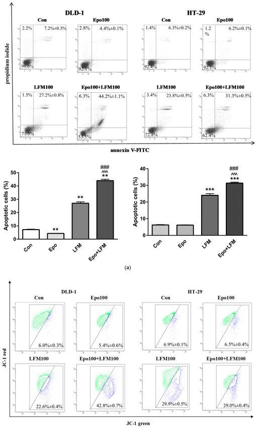
Int. J. Mol. Sci. 2018, 19, 1262                                                                       2 of 17

                                                                                                              enzymes cause increased tumor cell proliferation and apoptosis evasion, while promoting angiogenesis
                                                                                                              and metastasis.
                                                                                                                     LFM-A13 is an active metabolite of leflunomide, designed using computer modeling techniques.
                                                                                                              It is the first inhibitor of Bruton’s tyrosine kinase (BTK), a cytoplasmic protein tyrosine kinase involved
                                                                                                              in signal transduction pathways regulating growth, maturation, differentiation, and cell viability. BTK
                                                                                                              is also an upstream activator of multiple antiapoptotic signaling molecules and networks, including
                                                                                                              the phosphatidylinositol-3-kinase/protein kinase B pathway [3]. Vassilev et al. demonstrated that
                                                                                                              BTK plays a role in the marked resistance of chicken B cell line DT40 as well as human leukemic
                                                                                                              B-cell precursors against Fas-mediated apoptosis. BTK links with Fas receptor via its kinase and
                                                                                                              pleckstrin homology (PH) domains and prevents the FAS–FADD (Fas-associated protein with death
                                                                                                              domain) interaction, which is essential for the recruitment and activation of FLICE (inhibitory proteins:
                                                                                                              regulators of death receptor-mediated apoptosis) by Fas during the apoptotic signal [4]. Recent data
                                                                                                              suggest the implication of BTK in signal transduction pathways affecting gene transcription [5,6].
                                                                                                              Inhibition of BTK, specifically, by the irreversible inhibitor ibrutinib, leads to the upregulation of
                                                                                                              apoptosis-related genes [7]. Bruton tyrosine kinase (BTK) has been demonstrated to be a key element
                                                                                                              of the βcR signaling pathway. βcR crosslinking causes BTK to interact with the inner leaflet of the
                                                                                                              plasma membrane to constitute the βcR signalosome, which subsequently stimulates downstream
                                                                                                              pathways, including nuclear factor (NF)-κB. A strong positive correlation in gene expression between
                                                                                                              BCL-2 and BTK was also proved. In mantle cell lymphoma, BTK activity promotes BCL2 transcription
                                                                                                              via the nuclear factor κ-light-chain-enhancer of activated B cells (NF-κB) pathway [8]. The blockade of
Pobrano z https://ppm.umb.edu.pl / Downloaded from Repository of Medical University of Bialystok 2021-11-01

                                                                                                              BTK activity attenuates βcR signaling and induces apoptosis by decreasing the levels of antiapoptotic
                                                                                                              BCL-2, BCL-XL, and Mcl-1 protein [9].
                                                                                                                     Previous data confirmed that LFM-A13 inhibits phosphoinositide 3-kinase (PI3K) and
                                                                                                              Epo-induced phosphorylation of erythropoietin receptor (EpoR). This compound was proved to block
                                                                                                              Janus kinases (JAK2) binding to EpoR, thus breaking the associated intracellular signal transduction
                                                                                                              pathway [10]. Since a link between BTK and Epo activity exists, we decided to investigate the impact
                                                                                                              of a combined therapy with Epo and LFM-A13 on apoptosis in colon cancer models [11]. It was
                                                                                                              demonstrated that Epo-induced signal transduction is inhibited in cells lacking BTK. Moreover,
                                                                                                              LFM-A13 efficiently inhibits Epo-induced phosphorylation of EpoR and Janus kinase 2 (Jak2),
                                                                                                              thus breaking the related intracellular signaling pathway [10]. It is well known that Epo used to
                                                                                                              treat anemia promotes survival, proliferation, and differentiation of the progenitors of erythropoiesis,
                                                                                                              exerting a proangiogenic and antiapoptotic effect [12,13]. Despite this, the results of our recent study
                                                                                                              indicate that Epo intensifies the antiproliferative activity of LFM-A13 [14].
                                                                                                                     In this study, we examined the effect of these compounds on the viability of colon cancer cells,
                                                                                                              using DLD-1 and HT-29 cell lines. We assessed apoptosis using several biochemical markers, such as
                                                                                                              phosphatidylserine externalization, loss of mitochondrial membrane potential (MMP), expression
                                                                                                              of pro-apoptotic protein BAX and of antiapoptotic protein BCL-2. We also explored the effect of
                                                                                                              the simultaneous use of Epo and LFM-A13 on tumor development in a mouse model of human
                                                                                                              colorectal cancer. The results demonstrated that the simultaneous use of Epo and LFM-A13 can
                                                                                                              strongly induce apoptosis in colon cancer cells in vitro as well as reduce tumor volume in colon cancer
                                                                                                              xenografts in vivo.

                                                                                                              2. Results

                                                                                                              2.1. Erythropoietin in Combination with LFM-A13 Decreases Colon Cancer Viability
                                                                                                                    In the first stage of our study, we decided to choose the effective cytotoxic dose of LFM-A13. The MTT
                                                                                                              test indicated that only the highest dose (100 µM) decreased the viability of colon cancer DLD-1 and
                                                                                                              HT-29 cells (Figure 1a,b). Therefore, we used this dose in further research. Incubation of DLD-1 cells
                                                                                                              with 100 IU/mL of Epo and 30 or 100 µM of LFM-A13 decreased cell viability compared with the control
                                                                                                              (p < 0.001, p < 0.001 respectively) as well as with cells treated with 100 IU/mL of Epo (p < 0.001), 30 µM of
Erythropoietin Intensifies the Proapoptotic Activity of LFM-A13 in Cells and in a Mouse Model of Colorectal Cancer
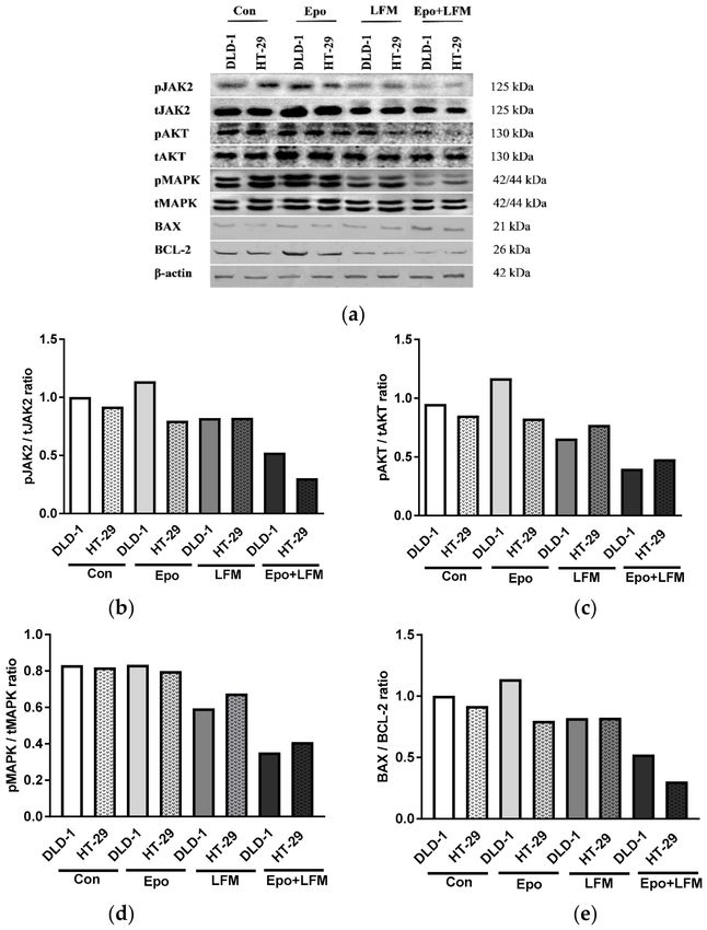
Int. J. Mol. Sci. 2018, 19, 1262                                                                        3 of 17

                                                                                                              LFM-A13 (p < 0.001), and 100 µM of LFM-A13 (p < 0.01) (Figure 1c). In turn, the reduction in cell viability in
                                                                                                              the HT-29 line was also statistically significant, but weaker. The combined treatment significantly reduced
                                                                                                              cell viability only in cells treated with the highest dose (100 µM) of LFM-A13 compared with the control
                                                                                                              (p < 0.001). Adding LFM-A13 (100 µM) to Epo (100 IU/mL) markedly reduced cell viability compared
                                                                                                              with Epo alone (p < 0.001) (Figure 1d). The obtained results indicate that the addition of Epo to LFM-A13
                                                                                                              significantly reduced the viability of colon cancer cells compared with LFM-A13 alone. The quantification of
                                                                                                              the potency of these combinations using the Chou–Talalay method showed synergistic combination indices
                                                                                                              (CI) for Epo and LFM-A13 in a constant ratio, particularly in the DLD-1 line (Figure 1e). In the case of the
                                                                                                              HT-29 line, synergism was observed only at the highest doses; at low doses, the calculated CI was close to 1
                                                                                                              and thus could represent an additive effect (Figure 1f).
Pobrano z https://ppm.umb.edu.pl / Downloaded from Repository of Medical University of Bialystok 2021-11-01

                                                                                                                                                             Figure 1. Cont.
Erythropoietin Intensifies the Proapoptotic Activity of LFM-A13 in Cells and in a Mouse Model of Colorectal Cancer
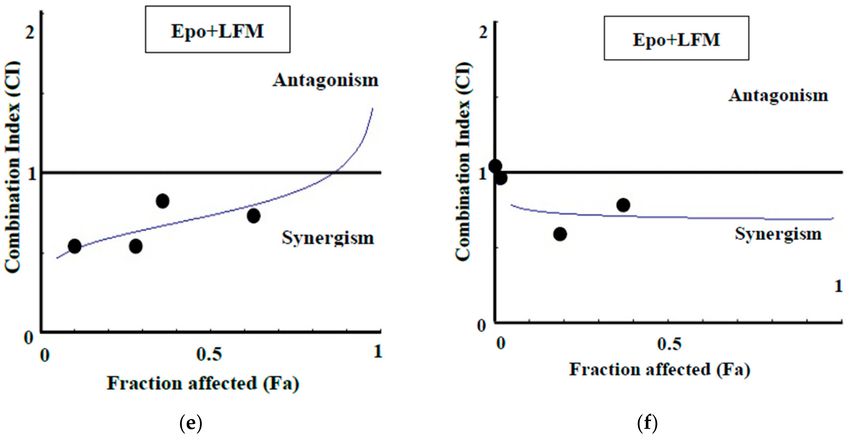
Int. J. Mol. Sci. 2018, 19, 1262                                                                                   4 of 17

                                                                                                                    Figure 1. Effects of erythropoietin β (Epo), LFM-A13 (LFM), and their combinations on colon cancer
                                                                                                                    cells. Impact of LFM-A13 (LFM) on cell viability of DLD-1 cells (a) and HT-29 cells (b); * p < 0.05,
                                                                                                                    ** p < 0.01 (versus control (Con)). Effect of erythropoietin β (Epo), LFM-A13 (LFM), and their combined
Pobrano z https://ppm.umb.edu.pl / Downloaded from Repository of Medical University of Bialystok 2021-11-01

                                                                                                                    activity on cell viability of DLD-1 (c) and HT-29 (d) colon cancer cells; * p < 0.05 (vs. Con), *** p < 0.001
                                                                                                                    (vs. Con); ˆ p < 0.05 (vs. Epo), ˆˆˆ p < 0.001 (vs. Epo); # p < 0.05 (vs. LFM-A13), ## p < 0.01 (vs. LFM-A13),
                                                                                                                    ### p < 0.001 (vs. LFM-A13). Combination index (CI) analysis of erythropoietin (10–100 IU/mL)
                                                                                                                    combined with LFM-A13 (10–100 µM) at a constant ratio in DLD-1 (e) and HT-29 (f) cells. Synergistic
                                                                                                                    effects are defined as CI < 1, additive effects as CI = 1, and antagonistic effects as CI > 1.

                                                                                                              2.2. Erythropoietin in Combination with LFM-A13 Induces Apoptosis through Intracellular Signaling
                                                                                                              Pathway Attenuation
                                                                                                                    To illustrate the mechanism underlying the regulation of intracellular signal by the Epo and
                                                                                                              LFM-A13 combination, we investigated the status of the JAK2, AKT, and mitogen-activated protein
                                                                                                              kinases (MAPK) pathways in colon cancer cells. As shown in Figure 2, simultaneous use of Epo and
                                                                                                              LFM-A13 failed to induce efficient phosphorylation of JAK2, AKT, and MAPK. Moreover, LFM-A13
                                                                                                              with Epo further inhibited the intracellular signaling pathway in comparison with LFM-A13 alone in
                                                                                                              both DLD-1 and HT-29 cells.
                                                                                                                    A 48 h treatment with Epo and LFM-A13 contributed to a marked increase in the expression of
                                                                                                              BAX compared with control cells in both lines. The combination treatment intensified BAX expression
                                                                                                              more than LFM-A13 alone. Simultaneously, we found a decrease in the expression of BCL-2 after
                                                                                                              incubation with Epo and LFM-A13 compared with the control cells. Incubation with Epo led to an
                                                                                                              increase in BCL-2 expression particularly in DLD-1 cells, which could be due to the presence of EpoR
                                                                                                              receptors in these cells. An increase in the BAX/BCL-2 ratio correlated with an increase in the apoptotic
                                                                                                              process. In our study, the ratio of BAX to BCL-2 shifted in favor of BAX compared with the control,
                                                                                                              which confirms the intensification of this process (Figure 2).
                                                                                                                    Apoptosis was detected by staining with annexin V, a phospholipid-binding protein commonly used
                                                                                                              for the detection of early apoptosis. For assessment of the cell death mode induced by Epo+LFM-A13, flow
                                                                                                              cytometric analysis was carried out using annexin V and propidium iodide (PI). Dual-labeling for annexin V
                                                                                                              and PI allows the distinction between live cells (annexin-V−/PI−), early apoptotic cells (annexin-V+/PI−),
                                                                                                              late apoptotic cells (annexin-V+/PI+), and necrotic cells (annexin-V−/PI+) (Figure 3a). As shown in
                                                                                                              Figure 3a, after 48 h of treatment with Epo+LFM-A13, we observed a significant accumulation of annexin
                                                                                                              V-positive cells in both DLD-1 and HT-29 cells. In-term administration of Epo slightly inhibited apoptosis
                                                                                                              in both tested lines after 48 h incubation compared with the control. We found that the apoptotic effect of
Erythropoietin Intensifies the Proapoptotic Activity of LFM-A13 in Cells and in a Mouse Model of Colorectal Cancer
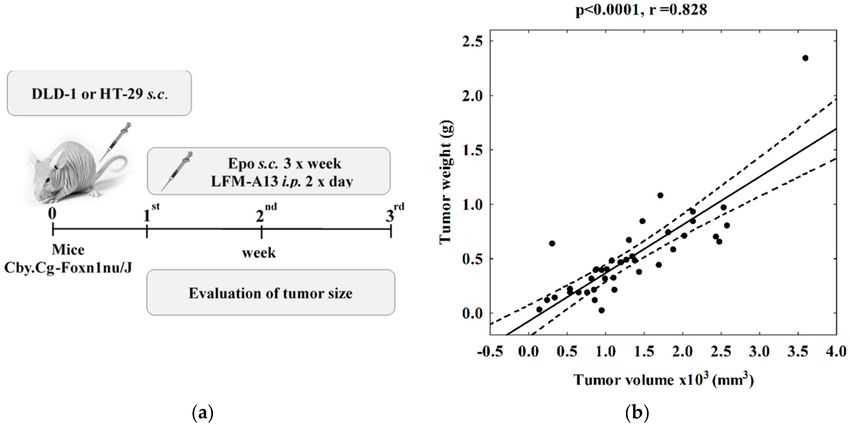
Int. J. Mol. Sci. 2018, 19, 1262                                                                           5 of 17

                                                                                                              Epo+LFM-A13 was stronger than that evoked by LFM-A13 alone, which resulted in fullest induction of
                                                                                                              apoptosis in both DLD-1 and HT-29 cancer cell lines.
                                                                                                                   To examine the cellular mechanism underlying Epo+LFM-A13-induced intrinsic apoptosis in DLD-1
                                                                                                              and HT-29 cells, we evaluated changes in the mitochondrial transmembrane potential using flow cytometry
                                                                                                              analysis. We observed that Epo+LFM-A13 increased the number of cells with decreased levels of MMP
                                                                                                              in both DLD-1 and HT-29 cells at 24 h (Figure 3b). Mitochondria a play key role in the propagation of
                                                                                                              apoptosis [15,16]. It is known that, at an early stage, apoptotic stimuli alter the mitochondrial transmembrane
                                                                                                              potential, which can be measured by determining the fluorescence of the dye JC-1. As shown in Figure 3b,
                                                                                                              Epo+LFM-A13 in DLD-1 and HT-29 cells induced an increase in the proportion of cells with depolarized
                                                                                                              mitochondria. We observed that the effect of Epo+LFM-A13 was stronger than that caused by LFM-A13
                                                                                                              alone. These results are consistent with those obtained in the annexin V and PI test.
Pobrano z https://ppm.umb.edu.pl / Downloaded from Repository of Medical University of Bialystok 2021-11-01

                                                                                                                    Figure 2. Effect of erythropoietin β (Epo), LFM-A13 (LFM), and their combination on intracellular
                                                                                                                    pathway and apoptosis. Immunoblotting analysis for: (a) phospho-JAK2 (pJAK2) and total JAK2 (tJAK2),
                                                                                                                    phospho-AKT (pAKT) and total AKT (tAKT), phospho-MAPK (pMAPK) and total MAPK (tMAPK),
                                                                                                                    BAX and BCL-2 in DLD-1 and HT-29 cells treated with erythropoietin β (Epo 100 IU/mL), LFM-A13
                                                                                                                    (LFM 100 µM), or their combination for 48 h. The samples used for electrophoresis consisted of 20 µg
                                                                                                                    of protein from six pooled cell extracts (n = 6). (b–e) Band staining was quantified by densitometry;
                                                                                                                    (b) pJAK/tJAK, (c) pAKT/tAKT, (d) pMAPK/tMAPK, (e) BAX/BCL-2 ratio the band intensities of the
                                                                                                                    phospho-proteins are normalized with respect to the intensities of the respective total protein bands.
Pobrano z https://ppm.umb.edu.pl / Downloaded from Repository of Medical University of Bialystok 2021-11-01

                                                                                                              Int. J. Mol. Sci. 2018, 19, 1262

                Figure 3. Cont.
                                                                                                              6 of 17
Int. J. Mol. Sci. 2018, 19, 1262                                                                                7 of 17

                                                                                                                    Figure 3. Effect of erythropoietin β (Epo), LFM-A13 (LFM), and their combinations on intracellular
                                                                                                                    pathway and apoptosis. (a) Representative flow cytometry dot plots for Annexin V–FITC (fluorescein
                                                                                                                    isothiocyanate) assay of DLD-1 and HT-29 cells incubated with Epo (Epo100, 100 IU/mL) and LFM-A13
                                                                                                                    (LFM100, 100 µM) for 48 h (mean ± SD; n = 3). The live cells appear at the lower left corner in the
                                                                                                                    plots; the early apoptotic cells appear at the lower right corner; the necrotic cells appear at the upper
                                                                                                                    left corner; the dead cells appear at the upper right corner. Left panel: The percentage of apoptotic
                                                                                                                    DLD-1 cells incubated with Epo and LFM-A13 is shown in the bar diagram as mean ± SD (n = 3).
                                                                                                                    Right panel: The percentage of apoptotic HT-29 cells incubated with Epo and LFM-A13 is shown in
Pobrano z https://ppm.umb.edu.pl / Downloaded from Repository of Medical University of Bialystok 2021-11-01

                                                                                                                    the bar diagram as mean ± SD (n = 3); * p < 0.05 (vs. Con), ** p < 0.01 (vs. Con), *** p < 0.001 (vs. Con);
                                                                                                                    ˆ p < 0.05 (vs. Epo), ˆˆˆ p < 0.001 (vs. Epo); # p < 0.05 (vs. LFM-A13), ### p < 0.001 (vs. LFM-A13).
                                                                                                                    (b) Representative dot plots presenting the loss of mitochondrial membrane potential (MMP) in DLD-1
                                                                                                                    and HT-29 cells incubated with Epo (Epo100, 100 IU/mL) and LFM-A13 (LFM100, 100 µM) for 48 h
                                                                                                                    (mean ± SD; n = 3). Cells with normal MMP are shown on the right side of the plots, cells with
                                                                                                                    decreased MMP on the left side of the plots. The graphs show the percent of cells with decreased
                                                                                                                    mitochondrial membrane potential in DLD-1 (left panel) and HT-29 cells (right panel).

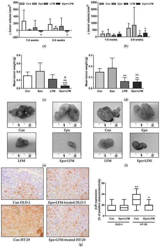
                                                                                                              2.3. Anti-Colon Cancer Activity of the Simultaneous Use of Epo and LFM-A13 in Mouse Xenografts
                                                                                                                   In order to confirm the results of the in vitro experiments, we performed animal studies.
                                                                                                              In DLD-1 xenografts receiving Epo+LFM-A13, differences in tumor growth between the first and the
                                                                                                              zero week as well as the second and the zero week were significantly smaller compared with the
                                                                                                              control (p < 0.05, p < 0.05, respectively, Figure 4a). In HT-29 xenografts treated with Epo+LFM-A13,
                                                                                                              the growth increment was significantly smaller only between the second and the zero week compared
                                                                                                              with the control group (p < 0.05, Figure 4b). Additionally, in HT-29 xenografts, a smaller growth
                                                                                                              increment was obtained after LFM-A13 therapy in the same period compared with the control
                                                                                                              (p < 0.05). As shown in Figure 4c,d, the tumor weights of DLD-1 and HT-29 xenografts treated
                                                                                                              with a combination of Epo+LFM-A13 were significantly lower compared with the control (p < 0.001
                                                                                                              and p < 0.01, respectively). In HT-29 xenografts, LFM-A13 significantly decreased tumor development
                                                                                                              compared with the control animals (p < 0.01). The tumors of mice treated with Epo+LFM-A13 were
                                                                                                              five times smaller compared with the control DLD-1 xenografts, and two-and-a-half times smaller
                                                                                                              compared with the control HT-29 xenografts (Figure 4e,f). Cytoplasmic anti-βcR (β common receptor)
                                                                                                              staining prevailed in all positive slides. In the present study, weak positive immunoreactivity for βcR
                                                                                                              was found in nine control DLD-1 xenografts (90% of all cases); one (10% of all cases) exhibited medium
                                                                                                              staining. We obtained similar results in Epo+LFM-13-treated animals. Interestingly, in control HT-29
                                                                                                              xenografts, medium-positive immunoreactivity for βcR was observed in seven animals (70% of all
                                                                                                              cases); however, one animal (10% of all cases) exhibited strong staining and two (20% of all cases)
                                                                                                              medium staining. In Epo+LFM-A13-treated xenografts, weak immunoreactivity was found in nine
                                                                                                              cases (95% of positive tumors), while medium immunoreactivity was detected in one case (10% of
                                                                                                              positive tumors) (Figure 4g). These results indicate that the combined therapy has potent anticancer
                                                                                                              activity, in which βcR receptors are probably involved.
Int. J. Mol. Sci. 2018, 19, 1262                                                                                   8 of 17
Pobrano z https://ppm.umb.edu.pl / Downloaded from Repository of Medical University of Bialystok 2021-11-01

                                                                                                                    Figure 4. Effect of the combined activity of LFM-A13 and erythropoietin β on a mouse model of colorectal
                                                                                                                    cancer. Impact on tumor volume in DLD-1 (a) and HT-29 (b) xenografts; 0—before substance administration,
                                                                                                                    1—after first week of substance administration, 2—after second week of substance administration. The results
                                                                                                                    are presented as mean values ± SD, n = 5–10. Effect of the combined activity of LFM-A13 (10 mg/kg b.m.)
                                                                                                                    and erythropoietin β (Epo, 600 IU/kg b.m.) on tumor weight in DLD-1 (c) and HT-29 (d) xenografts.
                                                                                                                    The results are presented as mean values ± SD, n = 10. * p < 0.05 (vs. Con), ** p < 0.01 (vs. Con),
                                                                                                                    *** p < 0.001 (vs. Con); ˆ p < 0.05 (vs. Epo), ˆˆ p < 0.01 (vs. Epo), ˆˆˆ p < 0.001 (vs. Epo); # p < 0.05 (vs.
                                                                                                                    LFM-A13). Representative weights of the tumors dissected from the nude mice untreated or treated
                                                                                                                    with Epo, LFM-A13, and Epo+LFM-A13 in DLD-1 (e) and in HT-29 (f). (g) Positive expression of βcR
                                                                                                                    in the cytoplasm of colon cancer xenografts. Left panel: expression in control and Epo-LFM-A13-treated
                                                                                                                    DLD-1 and HT-29 xenografts (H&E staining; magnification, ×200). Right panel: a box-and-whisker plot of
                                                                                                                    percentage βcR expression in DLD-1 and HT-29 tumor xenografts. The results are presented as medians
                                                                                                                    (minimum–maximum), n = 10, * p < 0.05 (vs. Con), ˆ p < 0.05 (Con in DLD-1 vs. Con in HT-29 xenografts).
Int. J. Mol. Sci. 2018, 19, 1262                                                                  9 of 17

                                                                                                              3. Discussion
                                                                                                                    Despite the use of combination therapies in many patients with colon cancer, fully satisfactory
                                                                                                              results have not been achieved. The current work investigates the effect of the simultaneous use
                                                                                                              of erythropoietin (Epo) and LFM-A13 (BTK inhibitor) on the viability of DLD-1 and HT-29 colon
                                                                                                              cancer cells and on their growth in xenografts, as well as its putative mechanism of cytotoxicity.
                                                                                                              The novel drug combination of Epo and LFM-A13 that we used proved to exert a high cytotoxic effect
                                                                                                              in DLD-1 and HT-29 colon cancer cells. The simultaneous use of these agents induced apoptosis in
                                                                                                              the treated cells. We extended these observations in vivo by demonstrating that, in DLD-1 xenografts,
                                                                                                              LFM-A13 alone was able to delay tumor development, while a combination of Epo with LFM-A13 led
                                                                                                              to significant tumor regression.
                                                                                                                    The evaluation of the cytotoxicity of Epo and LFM-A13 used together in both DLD-1 and HT-29
                                                                                                              colon cancer cells demonstrated that this combination had a greater antiproliferative effect than the
                                                                                                              use of the latter drug alone. During the last decade, a plethora of studies has suggested that LFM-A13
                                                                                                              exhibits antiproliferative activity. LFM-A13 revealed mitotic arrest and prevented bipolar spindle
                                                                                                              assembly formation in human cancer cell lines and glioblastoma cells [17]. Uckun et al. also confirmed
                                                                                                              the antiproliferative activity profile of LFM-A13 and reported that this agent blocks cell division
                                                                                                              in a zebra fish embryo model at the 16-cell stage of embryonic development [18]. It has also been
                                                                                                              shown that the inhibition of BTK expression by LFM-A13 decreased the proliferation of prostate cancer
                                                                                                              cells, but not of normal prostate epithelial cells, which express very little BTK [19]. On the basis of
Pobrano z https://ppm.umb.edu.pl / Downloaded from Repository of Medical University of Bialystok 2021-11-01

                                                                                                              these results and the report indicating a relationship between BTK, LFM-A13, and erythropoietin [10],
                                                                                                              we used a combination of these agents. The results of our study indicate that the addition of Epo to
                                                                                                              LFM-A13 enhances the antiproliferative activity of LFM-A13. Similar results were obtained in previous
                                                                                                              studies, in which we observed that the combined use of Epo and cytotoxic agents such as 5-fluorouacil
                                                                                                              and active metabolite of irinotecan (SN-38) enhanced the antitumor effect on DLD-1 human colon
                                                                                                              cancer cells [20].
                                                                                                                    Then, we attempted to clarify the potential mechanism of action. On one hand, the activity
                                                                                                              of BTK, which is involved in the activation of the signaling pathways responsible for maturation,
                                                                                                              viability, and cell differentiation, is inhibited by LFM-A13. In turn, we supposed that Epo may increase
                                                                                                              pBTK expression and enhance the antiproliferative action of LFM-A13 through the elevation of the
                                                                                                              levels of its target proteins. Our experiments, carried out by flow cytometry assessment of annexin V
                                                                                                              binding, revealed that LFM-A13 increased the number of DLD-1 and HT-29 apoptotic cells. Epo with
                                                                                                              LFM-A13 further enhanced these effects and induced a high level of apoptosis in both DLD-1 and
                                                                                                              HT-29 (Figure 2).
                                                                                                                    Mitochondria play a key role in the apoptosis of mammalian cells and undergo characteristic
                                                                                                              functional and structural changes at an early stage of the death process [21]. Almost all apoptotic
                                                                                                              pathways concentrate on the mitochondria, such as the decrease of the mitochondrial transmembrane
                                                                                                              potential. A decrease of MMP characterizes an early and already invariant stage of apoptosis [15,22].
                                                                                                              Incubation with Epo and LFM-A13 led to a stronger decrease of the mitochondrial membrane potential
                                                                                                              in DLD-1 than incubation with LFM-A13 alone.
                                                                                                                    We also examined the mechanism through which the used combination of compounds was able
                                                                                                              to induce apoptosis. By profiling the relevant pathways, we found that simultaneous use of Epo
                                                                                                              and LFM-A13 resulted in the activation of pro-apoptotic BAX and downregulation of antiapoptotic
                                                                                                              BCL-2, which may cause a release of cytochrome C from the mitochondria in DLD-1 and HT-29
                                                                                                              cells. It was previously shown that a high ratio of BAX to BCL-2 can lead to loss of mitochondrial
                                                                                                              membrane potential, causing a release of cytochrome c and consequently inducing apoptosis [23,24].
                                                                                                              It was also demonstrated that another BTK inhibitor—ibrutinib—induced concentration-dependent
                                                                                                              apoptosis in both Mino or Jeko-1 cells and decreased the levels of antiapoptotic BCL-2, BCL-XL,
                                                                                                              and Mcl-1 proteins [9]. Moreover, simultaneous use of ibrutinib and bortezomib synergistically
                                                                                                              increased mitochondrial injury and apoptosis in diffuse large B cell lymphoma (D LBCL) and Mantle
                                                                                                              cell lymphoma (MCL) cells, with significant AKT and NF-κB inactivation, decrease in Mcl-1, BCL-XL,
Int. J. Mol. Sci. 2018, 19, 1262                                                                 10 of 17

                                                                                                              and X-linked inhibitor of apoptosis protein (XIAP), and increased DNA damage and endoplasmic
                                                                                                              reticulum stress [25]. Herman et al. demonstrated dose- and time-dependent cytotoxicity of ibrutinib
                                                                                                              in chronic lymphocytic leukemia (CLL) via an apoptotic pathway dependent on caspase-3 [26].
                                                                                                                   The results of this study and other data clearly indicate that Epo increases BCL-2 expression in
                                                                                                              numerous cancer cells [27,28]. According to the literature data, increased levels of the antiapoptotic
                                                                                                              BCL-2 protein have been associated with resistance to cytotoxic drugs and advanced stages of multiple
                                                                                                              myeloma [29]. In the current study, we found that the administration of Epo with LFM-A13 resulted in
                                                                                                              increased apoptosis in both colon cancer lines, as evidenced by the clear increase in the BAX/BCL-2
                                                                                                              ratio. Interestingly, our previous data also indicated that the combination of Epo with the anticancer
                                                                                                              agent H2 O2 led to decreased BCL-2 and increased BAX expression [30].
                                                                                                                   Other putative mechanisms of apoptosis enhancement may be associated with the regulation
                                                                                                              of BTK activity. BTK can mediate downstream signaling to regulate cell growth, differentiation,
                                                                                                              and apoptosis [31]. This kinase is an upstream activator of multiple antiapoptotic signaling molecules
                                                                                                              and networks, such as signal transducer and activator of transcription 5, NF-κB, and AKT. It is well
                                                                                                              known that BTK plays a crucial role in the B cell tumors development, by activating antiapoptotic
                                                                                                              pathways. Phosphorylated BTK activates phospholipase C2, leading to downstream activation
                                                                                                              of protein kinases and ultimately to the activation of transcription factor NF-κB. The inhibition
                                                                                                              of apoptosis occurs as a result of the stimulation of the NF-κB pathway. This cascade of events
                                                                                                              has been linked with the proliferation and survival of B cell malignancies [25]. BTK also inhibits
                                                                                                              Fas-ligand-mediated apoptosis. Loss of BTK caused B cells to be more sensitive to Fas-induced
Pobrano z https://ppm.umb.edu.pl / Downloaded from Repository of Medical University of Bialystok 2021-11-01

                                                                                                              apoptosis in response to certain death signals [4,31,32].
                                                                                                                   Moreover, inhibiting BTK increases the expression of apoptosis-related genes [7]. Downregulation
                                                                                                              of this kinase with iRNA or inhibition with pharmacological inhibitors leads to apoptosis, whereas its
                                                                                                              overexpression inhibits apoptosis induced by doxorubicin in breast cancer cells [33]. B lymphocytes and
                                                                                                              myeloid cells with low BTK activity tend to undergo apoptosis and exhibit decreased proliferation [31].
                                                                                                              Findings from the literature data showed that BTK is aberrantly expressed in DLD-1 and HT-29 colon
                                                                                                              carcinoma cells and that LFM-A13 was able to decrease the phosphorylation of this kinase [3,34].
                                                                                                              We speculate that the stronger downregulation of BTK after simultaneous use of Epo and LFM-A13
                                                                                                              may be the cause of the enhanced proapoptotic effects. This indicates that the decrease in proliferation
                                                                                                              in DLD-1 and HT-29 cells may be due to, at least in part, increased apoptosis. This is in line with the
                                                                                                              data of Guo et al., who showed that inhibition of BTK contributes to the induction of apoptosis in
                                                                                                              prostate cancer cells [19]. Inhibition of this kinase using LFM-A13 also resulted in increased apoptosis
                                                                                                              in BT474 and MCF-7 breast cancer cells [7]. The potential proapoptotic effect of a combined therapy
                                                                                                              with Epo and LFM-A13 could also be related to inhibition of p53 protein phosphorylation due to
                                                                                                              the blocking of BTK activity. Althubiti et al. observed that BTK expression increased the stability of
                                                                                                              p53 and its transactivation abilities [35,36]. Tumor protein p53 is a nuclear transcription factor that
                                                                                                              controls the expression of many of the genes responsible for apoptosis (BAX, BAK). BTK has an impact
                                                                                                              on p53 phosphorylation, which suggests that it is a crucial upstream regulator of the p53 pathway.
                                                                                                              Consequently, the cellular responses to p53 upregulation, such as apoptosis, were greatly influenced
                                                                                                              by the expression of BTK [35,36]. Further studies are needed to clarify the mechanism, because it is not
                                                                                                              fully understood.
                                                                                                                   Our in vitro study demonstrated the beneficial effect of LFM-A13 in combination with
                                                                                                              erythropoietin on antitumor activity in both tested colon cancer cell lines. We investigated whether
                                                                                                              erythropoietin can enhance in vivo colon cancer cell growth and examined the pharmacodynamics
                                                                                                              features of the promising combination in DLD-1 and HT-29 xenografts. In this model, 100% of mice
                                                                                                              survived for 3 weeks after inoculation with 1 × 10⁶ DLD-1 and HT-29. LFM-A13 in a nontoxic low dose
                                                                                                              of 10 mg/kg decreased HT-29 and DLD-1 xenograft tumor growth. Our results confirm the results
                                                                                                              of a previous study by Uckun et al., who reported a delay in tumor progression in the MMTV/Neu
                                                                                                              transgenic mouse model of HER2-positive breast cancer after LFM-A13 administration [18]. Moreover,
                                                                                                              we found a greater reduction in tumor volume in the Epo-and-LFM-A13-treated group compared
Int. J. Mol. Sci. 2018, 19, 1262                                                                    11 of 17

                                                                                                              with the control and the group treated with LFM-A13 alone. We also observed that DLD-1 xenografts
                                                                                                              were more responsive to this therapy compared with HT-29 xenografts. In the case of HT-29, not all
                                                                                                              results were consistent under both in vitro and in vivo conditions (Figure 2). It is difficult to explain
                                                                                                              this unequivocally; however, the observed effects were not opposite, and the trend remained the
                                                                                                              same. It is well known that erythropoietin receptors (EpoR) are located not only in erythroid cells,
                                                                                                              but also in the pituitary glands and in breast, brain, colon, kidney, lung, ovarian, endometrial, prostate,
                                                                                                              and lymphoma cancer cells [37,38]. Also, β common receptor (βcR), a shared receptor subunit of
                                                                                                              interleukin-3 (IL-3), IL-5, and granulocyte–macrophage colony stimulation factor (GM-CSF) receptors,
                                                                                                              mediates the non-hematopoietic activity of Epo in various cell types [39]. In non-hematopoietic tissue,
                                                                                                              Epo may act via the epinephrine B4 receptor (EphB4). It has been proven that Epo binds to the
                                                                                                              EpoR–EphB4 heterodimer as well as to the EpoR–βcR–EpoR heterotrimer [37]. The observed reduction
                                                                                                              in tumor volume in HT-29 xenografts after treatment with Epo–LFM–A13 appears to be dependent on
                                                                                                              βcR. High expression of this receptor was observed in control HT-29 xenografts; however, its expression
                                                                                                              was significantly reduced in the group of treated animals. Thus, lowering the expression of the receptor,
                                                                                                              which has been shown to be essential to the Epo-mediated extra-hematopoietic tissue protective effects,
                                                                                                              may lead to abolished Epo-induced NO production via the Src/JAK2/Akt signaling pathway and to
                                                                                                              reduced vascular endothelial growth factor (VEGF) production and angiogenic potency [40].

                                                                                                              4. Materials and Methods
Pobrano z https://ppm.umb.edu.pl / Downloaded from Repository of Medical University of Bialystok 2021-11-01

                                                                                                              4.1. Reagents
                                                                                                                   RPMI-1640, McCoy’s 5a medium, fetal bovine serum, penicillin, and streptomycin were obtained
                                                                                                              from ATCC (American Type Culture Collection, Manassas, VA, USA). Erythropoietin β (NeoRecormon,
                                                                                                              Roche, Basel, Switzerland) was purchased from Roche. LFM-A13 (2-cyano-N-(2,5-dibromophenyl)-
                                                                                                              3-hydroxy-2-butenamide) was a Tocris product (Bristol, UK). Stock solutions of LFM-A13 were
                                                                                                              prepared in methanol and stored at −20 ◦ C. Because of the poor aqueous solubility of LFM-A13,
                                                                                                              the compound was dissolved in 0.01% dimethyl sulfoxide (DMSO) in phosphate-buffered saline
                                                                                                              (PBS), according to the manufacturer’s instructions. A similar quantity of DMSO was added to the
                                                                                                              control preparations.

                                                                                                              4.2. Cell Cultures
                                                                                                                    The first stage of research involved in vitro experiments using two lines of colon cancer cells,
                                                                                                              DLD-1 (ATCC, Manassas, VA, USA, Cat# CCL-221, RRID:CVCL_0248) and HT-29 (ATCC, Cat# HTB-38,
                                                                                                              RRID:CVCL_0320). Both lines are considered adhesive and quick-growing. DLD-1 cells contain
                                                                                                              the EpoR gene and protein and histologically are the most similar to primary tumor cells, while
                                                                                                              the cell line HT-29 is a negative control for the EpoR gene. The cell line DLD-1 was isolated from
                                                                                                              the epithelial tissue of a colon cancer adenocarcinoma (stage C according to Dukes classification) of
                                                                                                              an adult man. It exhibits the presence of the oncogenes myc, myb, ras, fos, sis, and p53 and the absence
                                                                                                              of the oncogenes abl, ros, and src. The cell line HT-29 was isolated from a colon adenocarcinoma of
                                                                                                              a 44-year-old woman. In in vitro cultures, it exhibits a morphology typical of epithelial cells, but it
                                                                                                              does not differentiate into cells making a brush border. A huge part of the HT-29 cell population is
                                                                                                              composed of goblet cells, so this line produces great amounts of intestinal mucus. HT-29 cells exhibit
                                                                                                              the presence of the oncogenes myc, myb, ras, myb, fos, sis, and p53 and the absence of the oncogenes
                                                                                                              abl, ros, and src.

                                                                                                              4.3. Exogenous Erythropoietin and LFM-A13 Administration
                                                                                                                    DLD-1 and HT-29 colon cancer cells were incubated for 48 h with exogenous erythropoietin β
                                                                                                              at a concentration of 100 IU/mL, LFM-A13 at a concentration of 100 µM, and a combination of these
                                                                                                              drugs (Epo+LFM-A13). Epo [41] and LFM-A13 [42,43] concentrations were selected on the basis of
Int. J. Mol. Sci. 2018, 19, 1262                                                                      12 of 17

                                                                                                              data from the literature. In these types of experiments, Epo at a concentration of 100 IU/mL has been
                                                                                                              commonly used by other researchers [44,45].

                                                                                                              4.4. Cell Viability Assay
                                                                                                                   The assay was carried out in accordance with the method described by Mosmann using
                                                                                                              3-(4,5-dimethylthiazol-2-yl)-2,5-diphenyltetrazolium bromide (MTT) [46]. Confluent cells cultured for
                                                                                                              48 h with various concentrations of the tested compounds in 6-well culture plates were washed three
                                                                                                              times with PBS and next incubated at 37 ◦ C in a 5% CO2 incubator for 4 h in 1 mL of MTT solution
                                                                                                              (0.5 mg/mL of PBS). The medium was discarded, and 1 mL of 0.1 M HCl in absolute isopropanol was
                                                                                                              added to colon cancer cells. The absorbance of the transformed dye in living cells was measured at
                                                                                                              570 nm. The viability of DLD-1 and HT-29 cells cultured in the presence of Epo and LFM-A13 was
                                                                                                              calculated as a percentage of the control cells. The experiments were carried out three times.

                                                                                                              4.5. Western Blotting
                                                                                                                    Western blotting was performed using a standard method described previously [12]. The nitrocellulose
                                                                                                              membranes were incubated with following antibodies: mouse anti-BAX (Sigma-Aldrich, St. Louis, MO, USA,
                                                                                                              Cat# WH0000581M1, RRID:AB_1840183,), mouse anti-BCL-2 (Sigma-Aldrich Cat# B3170, RRID:AB_258541),
                                                                                                              rabbit anti-phospho Akt1/2/3 (Santa Cruz Biotechnology, Dallas, TX, USA, Cat# sc-7985 also sc-7985-R,
                                                                                                              RRID:AB_667741), rabbit anti-Akt1/2/3 (Santa Cruz Biotechnology, Cat# sc-8312, RRID:AB_671714), rabbit
Pobrano z https://ppm.umb.edu.pl / Downloaded from Repository of Medical University of Bialystok 2021-11-01

                                                                                                              anti-phospho-MAPK (Thermo Fisher Scientific, Waltham, MA, USA, Cat# PA1-14302, RRID:AB_1086514),
                                                                                                              rabbit anti-MAPK (Thermo Fisher Scientific Cat# PA5-14425, RRID:AB_2141578), rabbit anti-pospho JAK2
                                                                                                              (Cell Signaling Technology, Leiden, The Netherlands, Cat# 3771S, RRID:AB_330403), rabbit anti-JAK2
                                                                                                              (Cell Signaling Technology Cat# 4040S, RRID:AB_10691469), and mouse anti- β-Actin (Sigma-Aldrich Cat#
                                                                                                              A5316, RRID:AB_476743).

                                                                                                              4.6. Flow Cytometry Assessment of Annexin V Binding
                                                                                                                   The apoptosis Detection Kit II (BD Pharmingen, San Jose, CA, USA) was used to characterize
                                                                                                              the mode of cell death induced by Epo plus LFM-A13. Colon cancer cells (10,000 cells measured)
                                                                                                              were examined in a flow cytometer (BD FACSCanto II flow cytometer, San Jose, CA, USA). Annexin
                                                                                                              V has high affinity for phosphatidylserine and therefore can be used to recognize cells in all stages
                                                                                                              of programed cell death. Propidium iodide (PI) is used as a DNA stain in cells exposing a disrupted
                                                                                                              cell membrane and were used to evaluate the late apoptotic and dead cells. The optimal parameter
                                                                                                              settings were discovered using a positive control (cells incubated with 3% formaldehyde buffer for
                                                                                                              30 min on ice). FACSDiva software (BD Biosciences Systems, San Jose, CA, USA) was used for the
                                                                                                              analysis of the results.

                                                                                                              4.7. Analysis of Mitochondrial Membrane Potential
                                                                                                                    The disturbances of the mitochondrial membrane potential were evaluated using the
                                                                                                              lipophilic cationic probe 5,50 ,6,60 -tetrachloro-1,10 ,3,30 -tetraethylbenzimidazolcarbocyanine iodide (JC-1
                                                                                                              MitoScreen kit, BD Biosciences). Unfixed colon cancer cells were washed twice in PBS and resuspended
                                                                                                              in PBS supplemented with 10 µg/mL JC-1. Then, the cells were incubated at room temperature in the
                                                                                                              dark for 15 min, washed, and resuspended in PBS for immediate BD FACSCanto II flow cytometry
                                                                                                              analysis. The FACSDiva software (BD Biosciences Systems) was used to calculate the percentage of
                                                                                                              cells with disrupted MMP.

                                                                                                              4.8. Establishment of Xenograft
                                                                                                                  Animal experiments were approved by the Local Ethics Committee (date of approval:
                                                                                                              24 April 2013; identification code: 31/2013). Animal studies are reported in compliance with the
                                                                                                              ARRIVE guidelines [47]. Xenograft animal models were obtained as described previously [14] using
Int. J. Mol. Sci. 2018, 19, 1262                                                                  13 of 17

                                                                                                              DLD-1 and HT-29 cells (50 µL suspension containing 1 × 10⁸ cells for each animal, Figure 5a).
                                                                                                              In previous studies, we showed no effect of the Epo solvent on the number of colon cancer cells [20]
                                                                                                              and, in accordance with the 3R, we reduced the number of animals [48]. In order to evaluate the
                                                                                                              validity of the tumor volume measurements, we correlated the tumor weight at mouse sacrifice with
                                                                                                              the tumor size. A subsequent analysis confirmed the presence of a positive correlation (r = 0.828,
                                                                                                              p < 0.0001) between these parameters and thus confirmed the validity of this method of measurement
                                                                                                              (Figure 5b).
Pobrano z https://ppm.umb.edu.pl / Downloaded from Repository of Medical University of Bialystok 2021-11-01

                                                                                                                    Figure 5. Human tumor xenografts in a nude mouse model. (a) Experimental plan. (b) Positive
                                                                                                                    correlation between tumor volume and weight at mouse sacrifice (p < 0.0001, r = 0.828). Solid
                                                                                                                    line—regression; dashed lines—95% confidence intervals for regression line.

                                                                                                              4.9. Immunocytochemistry
                                                                                                                   Formalin-fixed, paraffin-embedded tissue slides were cut on the microtome into sections with
                                                                                                              a thickness of 4 mm and processed as described previously [49]. The antibody used (βcR (7C6); Cat#
                                                                                                              sc-103, RRID:AB_626741) was obtained from Santa Cruz Biotechnology.

                                                                                                              4.10. Statistical Analysis
                                                                                                                   The obtained results were statistically analyzed. The Shapiro–Wilk test was used to assess
                                                                                                              characteristics consistent with normal distribution, the Student’s t-test was used for comparisons
                                                                                                              between two groups, and the Mann–Whitney test was used for features inconsistent with the
                                                                                                              distribution. For comparisons of more than two groups, the analysis of variance with Bonferroni post
                                                                                                              hoc test or the Kruskal–Wallis test were used. Student’s t-test for pairs and Wilcoxon signed-rank test
                                                                                                              were used to analyze the measurements in groups at time intervals. In order to evaluate the correlations
                                                                                                              between the studied parameters, the Pearson correlation coefficient was used. In all experiments,
                                                                                                              the mean values for 4–10 assays ±S.D. were calculated using Statistica 10 software (Statistica 10,
                                                                                                              RRID:SCR_015627). A level of p < 0.05 was considered statistically significant. The calculations were
                                                                                                              performed using GraphPad 7.02 Prism software (GraphPad Software, Inc., La Jolla, CA, USA; Available
                                                                                                              online: https://www.graphpad.com/scientific-software/prism, RRID:SCR_015807). CI values were
                                                                                                              determined by CompuSyn software (ComboSyn, Inc., Paramus, NJ, USA; Available online:
                                                                                                              http://www.combosyn.com) [50]. The potency of the combination was evaluated using the
                                                                                                              Chou-Talalay method, which calculates a combination index (CI) that is interpreted as follows: CI < 1,
                                                                                                              synergistic effect; CI = 1, additive effect; CI > 1, antagonistic effect. The bands were quantified using
                                                                                                              Image J 1.50a software (Available online: https://imagej.nih.gov, RRID:SCR_003070).
Int. J. Mol. Sci. 2018, 19, 1262                                                                          14 of 17

                                                                                                              5. Conclusions
                                                                                                                  Collectively, we discovered that adding erythropoietin to LFM-A13 significantly intensified the
                                                                                                              proapoptotic activity of LFM-A13. The combined treatment exhibited antiproliferative activity against
                                                                                                              colon cancer cells both in in vitro and in vivo via an increasing apoptosis. The outcomes of our
                                                                                                              study suggest that the proposed combined therapy may be useful as a new approach employing
                                                                                                              chemosensitizing and apoptosis-promoting anticancer agents for the treatment of colon cancer patients.
                                                                                                              However, further preclinical and clinical studies are needed to confirm our findings.

                                                                                                              6. Patents
                                                                                                                    The results were submitted in the patent application No. P.413921.

                                                                                                              Acknowledgments: The research project was funded by grants No. N/ST/ZB/18/001/2211 and N/ST/ZB/
                                                                                                              18/002/2211 financed by the Medical University of Bialystok, Poland.
                                                                                                              Author Contributions: Anna Tankiewicz-Kwedlo developed the study concept, carried out in vitro and
                                                                                                              in vivo studies, statistical analysis of the results, interpretation of data for the work, drafted the manuscript.
                                                                                                              Justyna Magdalena Hermanowicz developed the study concept, carried out in vitro and in vivo studies, drafted
                                                                                                              the manuscript. Krystyna Pawlak, Krzysztof Bielawski interpreted the data. Robert Czarnomysy, Izabela Prokop
                                                                                                              carried out in vitro studies. Dariusz Pawlak supervised the entire study, interpreted the data, provided critical
                                                                                                              revisions. All authors read and approved the final manuscript.
                                                                                                              Conflicts of Interest: The authors declare no conflict of interest.
Pobrano z https://ppm.umb.edu.pl / Downloaded from Repository of Medical University of Bialystok 2021-11-01

                                                                                                              Abbreviations
                                                                                                              AKT             protein kinase B
                                                                                                              BAX             apoptotic protein
                                                                                                              BCL-2           antiapoptotic protein
                                                                                                              βcR β           β common receptor
                                                                                                              BTK             Bruton’s tyrosine kinase
                                                                                                              BT474           cell line of human ductal carcinoma
                                                                                                              DLD-1           cell line of human colorectal adenocarcinoma
                                                                                                              EphB4           epinephrine B4 receptor
                                                                                                              Epo             erythropoietin
                                                                                                              EpoR            erythropoietin receptor
                                                                                                              FAS             Fas-associated protein with death domain
                                                                                                              FADD            FLICE inhibitory proteins: regulators of death receptor-mediated apoptosis
                                                                                                              GM-CSF          granulocyte–macrophage colony stimulation factor
                                                                                                              HER2            human epidermal growth factor 2
                                                                                                              HT-29           cell line of human colorectal adenocarcinoma
                                                                                                              IL              interleukin
                                                                                                              JAK2            Janus kinase 2
                                                                                                              LFM-A13         Bruton’s tyrosine kinase inhibitor
                                                                                                              MAPK            mitogen-activated protein kinases
                                                                                                              MCF-7           cell line of human breast cancer
                                                                                                              MMP             mitochondrial membrane potential
                                                                                                              MYC             a regulator gene that codes for a transcription factor
                                                                                                              NF-κB           nuclear factor κ-light-chain-enhancer of activated B cells
                                                                                                              p-Akt           phosphorylated protein kinase B
                                                                                                              PH              pleckstrin homology
                                                                                                              PI3K            phosphoinositide 3-kinase
                                                                                                              VEGF            vascular endothelial growth factor
Int. J. Mol. Sci. 2018, 19, 1262                                                                                15 of 17

                                                                                                              References
                                                                                                              1.    Watson, A.J. Apoptosis and colorectal cancer. Gut 2004, 53, 1701–1709. [CrossRef] [PubMed]
                                                                                                              2.    Zhang, L.; Yu, J. Role of apoptosis in colon cancer biology, therapy, and prevention. Curr. Colorectal Cancer Rep.
                                                                                                                    2013, 9. [CrossRef] [PubMed]
                                                                                                              3.    D’Cruz, O.J.; Uckun, F.M. Novel Bruton’s tyrosine kinase inhibitors currently in development. Onco-Targets Ther.
                                                                                                                    2013, 6, 161–176. [CrossRef] [PubMed]
                                                                                                              4.    Vassilev, A.; Ozer, Z.; Navara, C.; Mahajan, S.; Uckun, F.M. Bruton’s tyrosine kinase as an inhibitor of the
                                                                                                                    Fas/CD95 death-inducing signaling complex. J. Biol. Chem. 1999, 274, 1646–1656. [CrossRef] [PubMed]
                                                                                                              5.    Uckun, F.M.; Zheng, Y.; Cetkovic-Cvrlje, M.; Vassilev, A.; Lisowski, E.; Waurzyniak, B.; Chen, H.;
                                                                                                                    Carpenter, R.; Chen, C.L. In vivo pharmacokinetic features, toxicity profile, and chemosensitizing activity of
                                                                                                                    α-cyano-β-hydroxy-β-methyl-N-(2,5-dibromophenyl)propenamide (LFM-A13), a novel antileukemic agent
                                                                                                                    targeting Bruton’s tyrosine kinase. Clin. Cancer Res. 2002, 8, 1224–1233. [PubMed]
                                                                                                              6.    Hata, D.; Kitaura, J.; Hartman, S.E.; Kawakami, Y.; Yokota, T.; Kawakami, T. Bruton’s tyrosine
                                                                                                                    kinase-mediated interleukin-2 gene activation in mast cells. Dependence on the c-Jun N-terminal kinase
                                                                                                                    activation pathway. J. Biol. Chem. 1998, 273, 10979–10987. [CrossRef] [PubMed]
                                                                                                              7.    Kokabee, L.; Wang, X.; Sevinsky, C.J.; Wang, W.L.; Cheu, L.; Chittur, S.V.; Karimipoor, M.; Tenniswood, M.;
                                                                                                                    Conklin, D.S. Bruton’s tyrosine kinase is a potential therapeutic target in prostate cancer. Cancer Biol. Ther.
                                                                                                                    2015, 16, 1604–1615. [CrossRef] [PubMed]
                                                                                                              8.    Li, Y.; Bouchlaka, M.N.; Wolff, J.; Grindle, K.M.; Lu, L.; Qian, S.; Zhong, X.; Pflum, N.; Jobin, P.; Kahl, B.S.;
                                                                                                                    et al. FBXO10 deficiency and BTK activation upregulate BCL2 expression in mantle cell lymphoma. Oncogene
Pobrano z https://ppm.umb.edu.pl / Downloaded from Repository of Medical University of Bialystok 2021-11-01

                                                                                                                    2016, 35, 6223–6234. [CrossRef] [PubMed]
                                                                                                              9.    Cinar, M.; Hamedani, F.; Mo, Z.; Cinar, B.; Amin, H.M.; Alkan, S. Bruton tyrosine kinase is commonly
                                                                                                                    overexpressed in mantle cell lymphoma and its attenuation by Ibrutinib induces apoptosis. Leuk. Res. 2013,
                                                                                                                    37, 1271–1277. [CrossRef] [PubMed]
                                                                                                              10.   Van den Akker, E.; van Dijk, T.B.; Schmidt, U.; Felida, L.; Beug, H.; Löwenberg, B.; von Lindern, M. The Btk
                                                                                                                    inhibitor LFM-A13 is a potent inhibitor of Jak2 kinase activity. Biol. Chem. 2004, 385, 409–413. [PubMed]
                                                                                                              11.   Schmidt, U.; van den Akker, E.; Parren-van Amelsvoort, M.; Litos, G.; de Bruijn, M.; Gutiérrez, L.;
                                                                                                                    Hendriks, R.W.; Ellmeier, W.; Löwenberg, B.; Beug, H.; et al. Btk is required for an efficient response
                                                                                                                    to erythropoietin and for SCF-controlled protection against TRAIL in erythroid progenitors. J. Exp. Med.
                                                                                                                    2004, 199, 785–795. [CrossRef] [PubMed]
                                                                                                              12.   Acs, G.; Acs, P.; Beckwith, S.M.; Pitts, R.L.; Clements, E.; Wong, K.; Verma, A. Erythropoietin and
                                                                                                                    erythropoietin receptor expression in human cancer. Cancer Res. 2001, 61, 3561–3565. [PubMed]
                                                                                                              13.   Dicato, M.; Plawny, L.; Diederich, M. Anemia in cancer. Ann. Oncol. 2010, 21, 167–172. [CrossRef] [PubMed]
                                                                                                              14.   Tankiewicz-Kwedlo, A.; Hermanowicz, J.M.; Domaniewski, T.; Pawlak, K.; Rusak, M.; Pryczynicz, A.;
                                                                                                                    Surazynski, A.; Kaminski, T.; Kazberuk, A.; Pawlak, D. Simultaneous use of erythropoietin and LFM-A13 as
                                                                                                                    a new therapeutic approach for colorectal cancer. Br. J. Pharmacol. 2018, 175, 743–762. [CrossRef] [PubMed]
                                                                                                              15.   Ly, J.D.; Grubb, D.R.; Lawen, A. The mitochondrial membrane potential (deltapsi(m)) in apoptosis; an update.
                                                                                                                    Apoptosis 2003, 8, 115–128. [CrossRef] [PubMed]
                                                                                                              16.   Green, D.R.; Kroemer, G. The pathophysiology of mitochondrial cell death. Science 2004, 305, 626–629.
                                                                                                                    [CrossRef] [PubMed]
                                                                                                              17.   Kumar, S.; Kim, J. PLK-1 Targeted Inhibitors and Their Potential against Tumorigenesis. BioMed Res. Int.
                                                                                                                    2015, 2015, 705745. [CrossRef] [PubMed]
                                                                                                              18.   Uckun, F.M.; Dibirdik, I.; Qazi, S.; Vassilev, A.; Ma, H.; Mao, C.; Benyumov, A.; Emami, K.H. Anti-breast
                                                                                                                    cancer activity of LFM-A13, a potent inhibitor of Polo-like kinase (PLK). Bioorg. Med. Chem. 2007, 15, 800–814.
                                                                                                                    [CrossRef] [PubMed]
                                                                                                              19.   Guo, W.; Liu, R.; Bhardwaj, G.; Yang, J.C.; Changou, C.; Ma, A.H.; Mazloom, A.; Chintapalli, S.; Xiao, K.;
                                                                                                                    Xiao, W.; et al. Targeting BTK/Etk of prostate cancer cells by a novel dual inhibitor. Cell Death Dis. 2014, 5,
                                                                                                                    e1409. [CrossRef] [PubMed]
                                                                                                              20.   Tankiewicz-Kwedlo, A.; Pawlak, D.; Domaniewski, T.; Buczko, W. Effect of erythropoietin, 5-fluorouracil
                                                                                                                    and SN-38 on the growth of DLD-1 cells. Pharmacol. Rep. 2010, 62, 926–937. [CrossRef]
                                                                                                              21.   Brunelle, J.K.; Zhang, B. Apoptosis assays for quantifying the bioactivity of anticancer drug products.
                                                                                                                    Drug Resist. Updates 2010, 13, 172–179. [CrossRef] [PubMed]
Int. J. Mol. Sci. 2018, 19, 1262                                                                                 16 of 17

                                                                                                              22.   Gornowicz, A.; Kałuża, Z.; Bielawska, A.; Gabryel-Porowska, H.; Czarnomysy, R.; Bielawski, K. Cytotoxic
                                                                                                                    efficacy of a novel dinuclear platinum(II) complex used with anti-MUC1 in human breast cancer cells.
                                                                                                                    Mol. Cell. Biochem. 2014, 392, 161–174. [CrossRef] [PubMed]
                                                                                                              23.   Teijido, O.; Dejean, L. Upregulation of Bcl2 inhibits apoptosis-driven BAX insertion but favors BAX
                                                                                                                    relocalization in mitochondria. FEBS Lett. 2010, 584, 3305–3310. [CrossRef] [PubMed]
                                                                                                              24.   Boersma, A.W.; Nooter, K.; Burger, H.; Kortland, C.J.; Stoter, G. BAX upregulation is an early event in
                                                                                                                    cisplatin-induced apoptosis in human testicular germ-cell tumor cell line NT2, as quantitated by flow
                                                                                                                    cytometry. Cytometry 1997, 27, 275–282. [CrossRef]
                                                                                                              25.   Novero, A.; Ravella, P.M.; Chen, Y.; Dous, G.; Liu, D. Ibrutinib for B cell malignancies. Exp. Hematol. Oncol.
                                                                                                                    2014, 3, 4. [CrossRef] [PubMed]
                                                                                                              26.   Herman, S.E.; Gordon, A.L.; Hertlein, E.; Ramanunni, A.; Zhang, X.; Jaglowski, S.; Flynn, J.; Jones, J.;
                                                                                                                    Blum, K.A.; Buggy, J.J.; et al. Bruton tyrosine kinase represents a promising therapeutic target for treatment
                                                                                                                    of chronic lymphocytic leukemia and is effectively targeted by PCI-32765. Blood 2011, 117, 6287–6296.
                                                                                                                    [CrossRef] [PubMed]
                                                                                                              27.   Trost, N.; Stepisnik, T.; Berne, S.; Pucer, A.; Petan, T.; Komel, R.; Debeljak, N. Recombinant human erythropoietin
                                                                                                                    alters gene expression and stimulates proliferation of MCF-7 breast cancer cells. Radiol. Oncol. 2013, 47, 382–389.
                                                                                                                    [CrossRef] [PubMed]
                                                                                                              28.   Batra, S.; Perelman, N.; Luc, L.R.; Shimada, H.; Malik, P. Pediatric tumor cells express erythropoietin and
                                                                                                                    a functional erythropoietin receptor that promotes angiogenesis and tumor cell survival. Lab. Investig. 2003,
                                                                                                                    83, 1477–1487. [CrossRef] [PubMed]
Pobrano z https://ppm.umb.edu.pl / Downloaded from Repository of Medical University of Bialystok 2021-11-01

                                                                                                              29.   Spets, H.; Strömberg, T.; Georgii-Hemming, P.; Siljason, J.; Nilsson, K.; Jernberg-Wiklund, H. Expression of
                                                                                                                    the BCL-2 family of pro- and anti-apoptotic genes in multiple myeloma and normal plasma cells: Regulation
                                                                                                                    during interleukin-6(IL-6)-induced growth and survival. Eur. J. Haematol. 2002, 69, 76–89. [CrossRef]
                                                                                                                    [PubMed]
                                                                                                              30.   Tankiewicz-Kwedlo, A.; Hermanowicz, J.M.; Surażyński, A.; Kwedlo, W.; Rożkiewicz, D.; Pawlak, K.;
                                                                                                                    Domaniewski, T.; Pawlak, D. Erythropoietin enhances the cytotoxic effect of hydrogen peroxide on colon
                                                                                                                    cancer cells. Curr. Pharm. Biotechnol. 2017, 18, 127–137. [CrossRef] [PubMed]
                                                                                                              31.   Wang, J.D.; Chen, X.Y.; Ji, K.W.; Tao, F. Targeting Btk with ibrutinib inhibit gastric carcinoma cells growth.
                                                                                                                    Am. J. Transl. Res. 2016, 8, 3003–3012. [PubMed]
                                                                                                              32.   Qiu, Y.; Kung, H.J. Signaling network of the BTK family kinases. Oncogene 2000, 19, 5651–5661. [CrossRef]
                                                                                                                    [PubMed]
                                                                                                              33.   Eifert, C.; Wang, X.; Kokabee, L.; Kourtidis, A.; Jain, R.; Gerdes, M.J.; Conklin, D.S. A novel isoform of the
                                                                                                                    B cell tyrosine kinase BTK protects breast cancer cells from apoptosis. Genes Chromosomes Cancer 2013, 52,
                                                                                                                    961–975. [CrossRef] [PubMed]
                                                                                                              34.   Grassilli, E.; Pisano, F.; Cialdella, A.; Bonomo, S.; Missaglia, C.; Cerrito, M.G.; Masiero, L.; Ianzano, L.;
                                                                                                                    Giordano, F.; Cicirelli, V.; et al. A novel oncogenic BTK isoform is overexpressed in colon cancers and
                                                                                                                    required for RAS-mediated transformation. Oncogene 2016, 35, 4368–4378. [CrossRef] [PubMed]
                                                                                                              35.   Althubiti, M.; Rada, M.; Samuel, J.; Escorsa, J.M.; Najeeb, H.; Lee, K.G.; Lam, K.P.; Jones, G.D.; Barlev, N.A.;
                                                                                                                    Macip, S. BTK Modulates p53 Activity to Enhance Apoptotic and Senescent Responses. Cancer Res. 2016, 76,
                                                                                                                    5405–5414. [CrossRef] [PubMed]
                                                                                                              36.   Rada, M.; Althubiti, M.; Ekpenyong-Akiba, A.E.; Lee, K.G.; Lam, K.P.; Fedorova, O.; Barlev, N.A.; Macip, S.
                                                                                                                    BTK blocks the inhibitory effects of MDM2 on p53 activity. Oncotarget 2017, 8, 106639–106647. [CrossRef]
                                                                                                                    [PubMed]
                                                                                                              37.   Arcasoy, M.O.; Amin, K.; Karayal, A.F.; Chou, S.C.; Raleigh, J.A.; Varia, M.A.; Haroon, Z.A. Functional
                                                                                                                    significance of erythropoietin receptor expression in breast cancer. Lab. Investig. 2002, 82, 911–918. [CrossRef]
                                                                                                                    [PubMed]
                                                                                                              38.   Jelkmann, W.; Bohlius, J.; Hallek, M.; Sytkowski, A.J. The erythropoietin receptor in normal and cancer
                                                                                                                    tissues. Crit. Rev. Oncol. Hematol. 2008, 67, 39–61. [CrossRef] [PubMed]
                                                                                                              39.   Su, K.H.; Shyue, S.K.; Kou, Y.R.; Ching, L.C.; Chiang, A.N.; Yu, Y.B.; Chen, C.Y.; Pan, C.C.; Lee, T.S.
                                                                                                                    β Common receptor integrates the erythropoietin signaling in activation of endothelial nitric oxide synthase.
                                                                                                                    J. Cell. Physiol. 2011, 226, 3330–3339. [CrossRef] [PubMed]
Int. J. Mol. Sci. 2018, 19, 1262                                                                               17 of 17

                                                                                                              40.   Sautina, L.; Sautin, Y.; Beem, E.; Zhou, Z.; Schuler, A.; Brennan, J.; Zharikov, S.I.; Diao, Y.; Bungert, J.;
                                                                                                                    Segal, M.S. Induction of nitric oxide by erythropoietin is mediated by the {β} common receptor and requires
                                                                                                                    interaction with VEGF receptor 2. Blood 2010, 115, 896–905. [CrossRef] [PubMed]
                                                                                                              41.   Balleari, E.; Clavio, M.; Arboscello, E.; Bellodi, A.; Bruzzone, A.; Del Corso, L.; Lucchetti, M.V.; Miglino, M.;
                                                                                                                    Passalia, C.; Pierri, I.; et al. Weekly standard doses of rh-EPO are highly effective for the treatment of anemic
                                                                                                                    patients with low-intermediate 1 risk myelodysplastic syndromes. Leuk. Res. 2011, 35, 1472–1476. [CrossRef]
                                                                                                                    [PubMed]
                                                                                                              42.   Vijayan, V.; Baumgart-Vogt, E.; Naidu, S.; Qian, G.; Immenschuh, S. Bruton’s tyrosine kinase is required for
                                                                                                                    TLR-dependent heme oxygenase-1 gene activation via Nrf2 in macrophages. J. Immunol. 2011, 187, 817–827.
                                                                                                                    [CrossRef] [PubMed]
                                                                                                              43.   Uckun, F.M. Chemosensitizing anti-cancer activity of LFM-A13, a leflunomide metabolite analog targeting
                                                                                                                    polo-like kinases. Cell Cycle 2007, 6, 3021–3026. [CrossRef] [PubMed]
                                                                                                              44.   Feldman, L.; Wang, Y.; Rhim, J.S.; Bhattacharya, N.; Loda, M.; Sytkowski, A.J. Erythropoietin stimulates
                                                                                                                    growth and STAT5 phosphorylation in human prostate epithelial and prostate cancer cells. Prostate 2006, 66,
                                                                                                                    135–145. [CrossRef] [PubMed]
                                                                                                              45.   Westenfelder, C.; Baranowski, R.L. Erythropoietin stimulates proliferation of human renal carcinoma cells.
                                                                                                                    Kidney Int. 2000, 58, 647–657. [CrossRef] [PubMed]
                                                                                                              46.   Mosmann, T. Rapid colorimetric assay for cellular growth and survival: Application to proliferation and
                                                                                                                    cytotoxicity assays. J. Immunol. Methods 1983, 65, 55–63. [CrossRef]
                                                                                                              47.   McGrath, J.C.; Lilley, E. Implementing guidelines on reporting research using animals (ARRIVE etc.):
Pobrano z https://ppm.umb.edu.pl / Downloaded from Repository of Medical University of Bialystok 2021-11-01

                                                                                                                    New requirements for publication in BJP. Br. J. Pharmacol. 2015, 172, 3189–3193. [CrossRef] [PubMed]
                                                                                                              48.   Törnqvist, E.; Annas, A.; Granath, B.; Jalkesten, E.; Cotgreave, I.; Öberg, M. Strategic Focus on 3R Principles
                                                                                                                    Reveals Major Reductions in the Use of Animals in Pharmaceutical Toxicity Testing. PLoS ONE 2014, 9,
                                                                                                                    e101638. [CrossRef] [PubMed]
                                                                                                              49.   Tankiewicz-Kwedlo, A.; Hermanowicz, J.; Surażynski, A.; Rożkiewicz, D.; Pryczynicz, A.; Domaniewski, T.;
                                                                                                                    Pawlak, K.; Kemona, A.; Pawlak, D. Erythropoietin accelerates tumor growth through increase of
                                                                                                                    erythropoietin receptor (EpoR) as well as by the stimulation of angiogenesis in DLD-1 and Ht-29 xenografts.
                                                                                                                    Mol. Cell. Biochem. 2016, 421, 1–18. [CrossRef] [PubMed]
                                                                                                              50.   Chou, T.C.; Talalay, P. Quantitative analysis of dose-effect relationships: The combined effects of multiple
                                                                                                                    drugs or enzyme inhibitors. Adv. Enzyme Regul. 1984, 22, 27–55. [CrossRef]

                                                                                                                                           © 2018 by the authors. Licensee MDPI, Basel, Switzerland. This article is an open access
                                                                                                                                           article distributed under the terms and conditions of the Creative Commons Attribution
                                                                                                                                           (CC BY) license (http://creativecommons.org/licenses/by/4.0/).
You can also read