Loss of primary cilia promotes mitochondria dependent apoptosis in thyroid cancer - Nature
←
→
Page content transcription
If your browser does not render page correctly, please read the page content below
www.nature.com/scientificreports
OPEN Loss of primary cilia promotes
mitochondria‑dependent apoptosis
in thyroid cancer
Junguee Lee1*, Ki Cheol Park2, Hae Joung Sul1, Hyun Jung Hong3, Kun‑Ho Kim4,
Jukka Kero5 & Minho Shong6*
The primary cilium is well-preserved in human differentiated thyroid cancers such as papillary and
follicular carcinoma. Specific thyroid cancers such as Hürthle cell carcinoma, oncocytic variant of
papillary thyroid carcinoma (PTC), and PTC with Hashimoto’s thyroiditis show reduced biogenesis of
primary cilia; these cancers are often associated the abnormalities in mitochondrial function. Here,
we examined the association between primary cilia and the mitochondria-dependent apoptosis
pathway. Tg-Cre;Ift88flox/flox mice (in which thyroid follicles lacked primary cilia) showed irregularly
dilated follicles and increased apoptosis of thyrocytes. Defective ciliogenesis caused by deleting the
IFT88 and KIF3A genes from thyroid cancer cell lines increased VDAC1 oligomerization following
VDAC1 overexpression, thereby facilitating upregulation of mitochondria-dependent apoptosis.
Furthermore, VDAC1 localized with the basal bodies of primary cilia in thyroid cancer cells. These
results demonstrate that loss-of-function of primary cilia results in apoptogenic stimuli, which are
responsible for mitochondrial-dependent apoptotic cell death in differentiated thyroid cancers.
Therefore, regulating primary ciliogenesis might be a therapeutic approach to targeting differentiated
thyroid cancers.
The primary cilium is a non-motile, microtubule-based sensory organelle that receives mechanical and chemi-
cal stimuli from the environment and transduces external signals into the c ell1. The tips of primary cilia, which
are present in the apical membrane of thyroid follicular cells (thyrocytes), face into the follicular lumen2. The
primary cilia of murine thyroid follicular cells play a role in maintaining globular follicle structures by acting
on cell polarity3. Loss-of-function (LOF) of primary cilia in murine thyroid follicles results in abnormal and
irregular follicles that eventually develop into papillary and solid proliferative nodules3.
The primary cilium is well-preserved in human differentiated thyroid cancers, including papillary and follicu-
lar carcinoma, and their frequency and length appear similar to those of normal thyroid f ollicles2. Interestingly,
the frequency of ciliated thyroid cancer cells is markedly lower in Hürthle cell carcinoma, oncocytic variant of
papillary carcinoma (PTCov), and PTC with Hashimoto’s thyroiditis (PTC-HT), which are usually associated
with mitochondrial d ysfunction2. However, we do not know whether ciliogenesis is linked with mitochondrial
function in thyroid cancer cells.
Mitochondria are crucial regulators of cell death through a process called the mitochondria-dependent
(intrinsic) pathway of apoptosis. Typically, mitochondrial outer membrane permeabilization (MOMP) is respon-
sible for mediating the intrinsic apoptotic pathway. The voltage-dependent anion channel (VDAC), a component
of MOMP, participates in mitochondria-dependent apoptosis by promoting cytochrome c release4,5. VDAC
oligomerization, followed by VDAC overexpression, may represent a common mechanism by which various
apoptogens act through different initiating c ascades6. Moreover, VDAC function extends beyond the mito-
chondria, and VDACs localize to the basal body of the primary cilium, where VDAC1 and VDAC3 negatively
regulate ciliogenesis7. Recent reports show that dysfunction of primary cilia increases apoptotic cell death in
1
Department of Pathology, Daejeon St. Mary’s Hospital, College of Medicine, The Catholic University of Korea,
Seoul 06591, Republic of Korea. 2Clinical Research Institute, Daejeon St. Mary’s Hospital, College of Medicine, The
Catholic University of Korea, Daejeon 34943, Republic of Korea. 3Research Center for Endocrine and Metabolic
Diseases, Chungnam National University School of Medicine, Daejeon 35015, Republic of Korea. 4Department
of Nuclear Medicine, Chungnam National University Hospital and College of Medicine, Daejeon 35015,
Republic of Korea. 5Research Centre for Integrative Physiology and Pharmacology, Institute of Biomedicine,
University of Turku, Kiinamyllynkatu 10, 20520 Turku, Finland. 6Department of Internal Medicine, Chungnam
National University School of Medicine, 266 Munhwaro, Daejeon 35015, Republic of Korea. *email: junguee@
catholic.ac.kr; minhos@cnu.ac.kr
Scientific Reports | (2021) 11:4181 | https://doi.org/10.1038/s41598-021-83418-3 1
Vol.:(0123456789)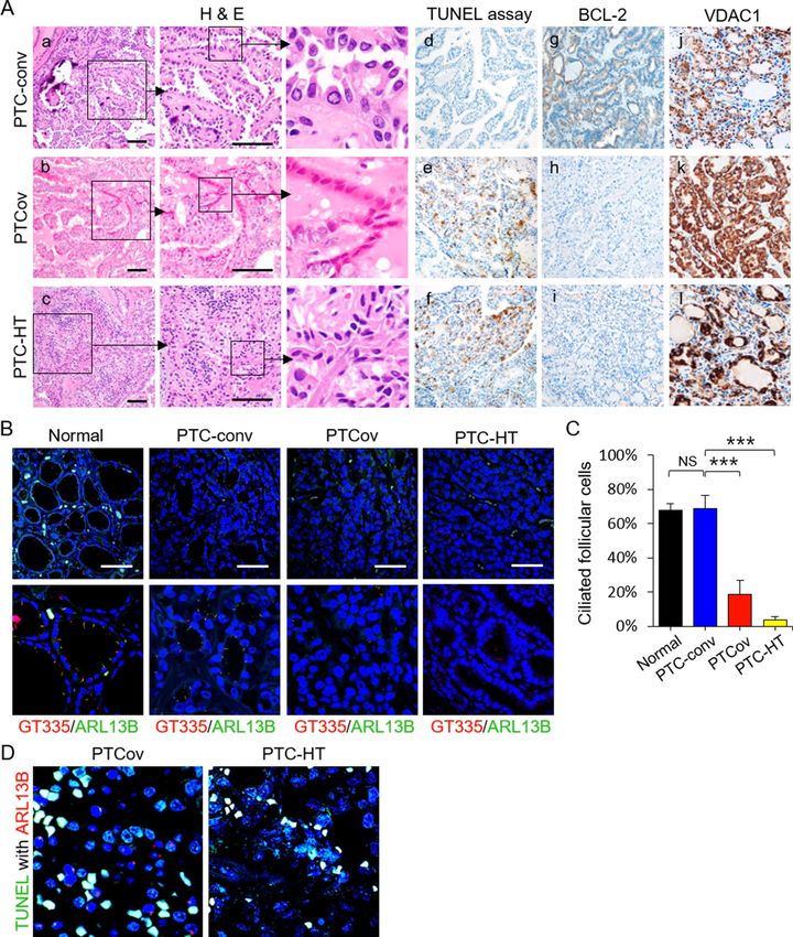
www.nature.com/scientificreports/

          Scientific Reports |   (2021) 11:4181 |   https://doi.org/10.1038/s41598-021-83418-3   2

Vol:.(1234567890)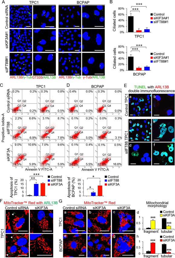
www.nature.com/scientificreports/
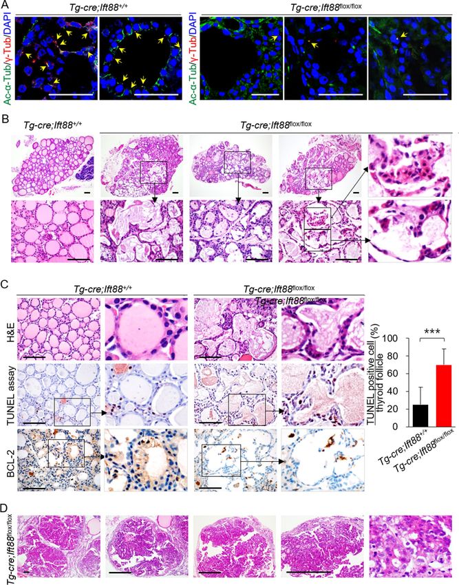
◂Figure 1. Loss of function of Ift88-mediated resulted in ciliary loss and increased apoptosis. (A)
Immunofluorescence images showing primary cilia in thyroid of Tg-Cre;Ift88+/+ and Tg-Cre;Ift88flox/flox mice.
Primary cilia were confirmed by staining with anti-acetylated α-tubulin (Ac-α-Tub, green) and anti-γ-
tubulin (γ-Tub, red) antibodies. Primary cilia are indicated by arrows. Scale bar, 10 μm. (B) The thyroid of
Tg-Cre;Ift88flox/flox mice (aged 14 weeks) shows irregular dilated follicles with a flat epithelium and luminal
colloid depletion. Scale bar, 10 μm. (C) Tg-Cre;Ift88flox/flox mice (aged 14 weeks) showed irregularly dilated
thyroid follicles comprising shrunken eosinophilic cells with compact nuclei (H&E). Scale bar, 10 μm. The
number of TUNEL-positive follicular cells per follicle in the wild-type control and Tg-Cre;Ift88flox/flox mice was
25 ± 20% and 70 ± 18%, respectively (P < 0.0001). BCL-2-positive follicular cells were rarely observed in the
irregularly dilated thyroid follicles of Tg-Cre;Ift88flox/flox mice. ***P < 0.001. (D) In 35 week-old Tg-Cre;Ift88flox/flox
mice, the dilated follicles developed into solid proliferative thyroid nodules. Scale bar, 10 μm.
glioblastoma, or induce neuron apoptosis in m ice8,9. However, the relationship between primary cilia and cell
death via activation of the mitochondrial apoptotic pathway is unclear.
Here we established a mouse model with thyrocyte-specific loss of primary cilia (Tg-Cre;Ift88flox/flox) and
human thyroid cancer cell lines with ciliary loss by silencing the KIF3A or IFT88 gene. To identify the role of
ciliogenesis with respect to the viability of normal thyrocytes and thyroid cancer cells, we examined apoptotic
cell death in murine thyroid follicular cells and human thyroid cancer cells devoid of primary cilia. We found
that mice lacking primary cilia in thyroid follicular cells showed upregulated apoptotic cell death, resulting in
altered follicular structure, and that inhibiting ciliogenesis in thyroid cancer cell lines resulted in VDAC1 oli-
gomerization following VDAC1 overexpression, leading ultimately to apoptosis. Additionally, we demonstrate
that VDAC1 is localized to the primary cilia of thyroid follicular cells. Taken together, these results establish
that LOF of primary cilia is a novel apoptogenic stimulus in thyroid cancers. Therefore, inhibiting primary cilia
might be a therapeutic target for thyroid cancers.
Results
Murine thyroid devoid of primary cilia after inactivation of the Ift88 gene shows altered folli‑
cular structure. Assembly and maintenance of primary cilia are dependent on a transport system controlled
by intraflagella transport (IFT) family p roteins10. Knockout of IFT88, an IFT retrograde complex B subunit, in
murine thyroid follicles prevents ciliogenesis3. To study the effect of thyrocyte-specific deletion of the Ift88 gene,
we used mice expressing Cre recombinase under the control of the thyroglobulin (Tg) promoter. Tg-Cre is con-
stitutively active from embryonic day 14.511,12. These Tg-Cre-expressing mice were crossed with Ift88flox/flox mice
to generate Tg-Cre;Ift88 floxed mice that exhibit thyroid follicle-specific ciliary loss.
Immunofluorescence analysis of primary cilia markers acetylated α-tubulin and γ-tubulin confirmed that
thyroid follicular cells in Tg-Cre;Ift88+/+ mice had primary cilia (Fig. 1A). By contrast, primary cilia were rarely
detected on thyroid follicles in Tg-Cre;Ift88flox/flox mice (Fig. 1A). The thyroids of 7 week-old Tg-Cre;Ift88flox/flox
mice exhibited irregularly dilated follicles with colloid depletion. These dilated follicles comprised shrunken,
hypereosinophilic cells with compact nuclei, which were morphologically compatible with apoptosis (Fig. 1B).
Terminal deoxynucleotidyl transferase dUTP nick-end labeling (TUNEL) assays revealed a higher proportion
of apoptotic follicular cells within the irregularly dilated thyroid follicles of 7 week-old Tg-Cre;Ift88flox/flox mice
than in those of wild-type control mice (Fig. 1C). In addition, thyroid follicular cells in Tg-Cre;Ift88flox/flox mice
showed lower expression of anti-apoptotic BCL-2 protein than those from control mice (Fig. 1C). Therefore, the
thyroid follicles of 7 week-old Tg-Cre;Ift88flox/flox mice show increased apoptosis.
The irregularly dilated follicles which increased with apoptosis eventually developed into papillary and solid
proliferative follicular nodules in the thyroids of 35 week-old Tg-Cre;Ift88flox/flox mice (Fig. 1D).
LOF of primary cilia in thyroid cancer cell lines results in increased apoptosis. Next, we investi-
gated whether loss of primary cilia induces apoptosis in thyroid cancer cell lines. Lactate dehydrogenase (LDH)
levels have been used as an indicator of late apoptosis in various studies. TPC1 and BCPAP cell lines had lower
LDH levels than those of other human thyroid carcinoma cell lines (PTC cell lines, TPC1 and BCPAP; anaplastic
thyroid cancer cell lines, 8505C, Hth7 and SW1736; Hürthle cell carcinoma cell line, XTC.UC1) (Supplementary
Fig. S1A) and had well-preserved primary cilia (similar to those of normal thyroid follicular cells) (Supplemen-
tary Fig. S1B)2, indicating an inverse correlation between the frequency of primary cilia and apoptosis. MTT cell
viability assays revealed that TPC1 and BCPAP cells exhibited more cell death than 8505C and Hth7 cells after
loss of primary cilia (Supplementary Fig. S1C). Based on these results, we selected TPC1 and BCPAP as the best
cell lines to demonstrate that loss of primary cilia induces apoptosis in thyroid cancer cell lines.
The primary cilium was visualized by immunofluorescence staining with an anti-ARL13B antibody (which
detect the axonemes), an anti-GT335 antibody (which detects axonemes with a basal body), and anti-γ-tubulin
(which detects the basal body)(Fig. 2A). Primary cilia were detected in 54.05 ± 9.28% of TPC1 cells and in
46.54 ± 6.58% of BCPAP cells under serum starvation conditions (Fig. 2A and B). The kinesin family member
3A (KIF3A) and intraflagellar transport 88 (IFT88) genes encode important proteins involved in cilium bio-
genesis. Knockdown (KD) of KIF3A or IFT88 by serum starvation resulted in significant decreases in the per-
centage of ciliated TPC1 (siKIF3A = 6.58 ± 5.54%; siIFT88 = 10.14 ± 5.28%) and BCPAP (siKIF3A = 8.40 ± 2.61%;
siIFT88 = 12.40 ± 2.61%) cells (Fig. 2B). The efficiency of specific siRNA-mediated KD of KIF3A or IFT88 in the
PTC cell lines is shown in Supplementary Fig. S2. This finding suggests that ciliogenesis, a process regulated by
KIF3A and IFT88, is preserved in thyroid cancer cells.
Next, we examined apoptotic cell death by performing Annexin V-FITC and PI staining and flow cytom-
etry-based quantification. We found that KD of KIF3A or IFT88 in thyroid cancer cell lines led to increased
Scientific Reports | (2021) 11:4181 | https://doi.org/10.1038/s41598-021-83418-3 3
Vol.:(0123456789)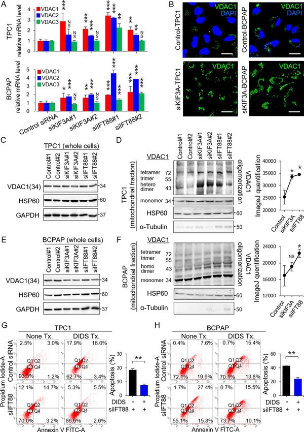
www.nature.com/scientificreports/

          Scientific Reports |   (2021) 11:4181 |   https://doi.org/10.1038/s41598-021-83418-3   4

Vol:.(1234567890)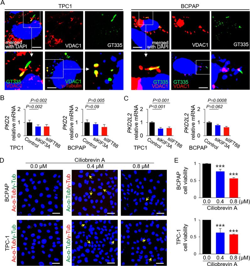
www.nature.com/scientificreports/
◂Figure 2. Loss-of-function of primary cilia increases apoptosis in thyroid cancer cell lines. (A) The number
of cells with primary cilia were determined by immunofluorescence staining with antibodies specific for
ARL13B (axoneme), anti-γ-tubulin (basal body), and GT335 (axonemes with basal bodies). Cell nuclei were
stained with DAPI. Scale bar, 10 μm. (B) Frequency of ciliated cells in the KIF3A-KD, IFT88-KD, and negative
control siRNA-transfected cell populations (TPC1 and BCPAP). *P < 0.05, **P < 0.01, ***P < 0.001, NS; not
significant. (C, D) FITC-conjugated Annexin V and PI assay to measure apoptosis of KIF3A-deficient or
IFT88-deficient TPC1 and BCPAP cells and negative control siRNA-transfected cells. Bar graphs show average
percentage of apoptotic cells (Q2 + Q4). *P < 0.05, **P < 0.01, ***P < 0.001. (E) More IFT88-KD TPC1 cells than
control TPC1 cells were TUNEL-positive (a, c). TUNEL with ARL13B double immunofluorescence staining
revealed that TUNEL-positive IFT88-KD TPC1 cells (green) had no primary cilia (red). Scale bar, 10 μm.
(F) Immunofluorescence images showing altered mitochondrial morphology and dynamics. The arrowheads
indicate the primary cilia. Scale bar, 10 μm. (G) Immunofluorescence images showing signs of apoptosis in
thyroid cancer cell lines with ciliary loss. These apoptotic cells showed highly fragmented mitochondria. Scale
bar, 10 μm. ***P < 0.001.
apoptotic cell death (Fig. 2C). Annexin V(+)/PI(+)(Q2) cells represent the late apoptotic population and
Annexin V(+)/PI(-)(Q4) cells represent the early apoptotic population. TPC1 populations with defective
IFT88 or KIF3A harbored higher numbers of cells undergoing late apoptosis (siIFT88 = 7.65 ± 1.48%, p = 0.011;
siKIF3A = 10.10 ± 0.71%, p = 0.002) and early apoptosis (siIFT88 = 7.35 ± 0.64%, p = 0.002; siKIF3A = 5.90 ± 0.00%,
p = 0.0003) than TPC transfected with negative control siRNA late apoptosis = 0.50 ± 0.42%; early apopto-
sis = 0.35 ± 0.21%)(Fig. 2C). The number of IFT88-deficient or KIF3A-deficient BCPAP cells that exhibited early
apoptosis was higher (siIFT88 = 4.05 ± 1.34%, p = 0.025; siKIF3A = 12.55 ± 5.72%, p = 0.045) than that of cells
transfected with negative control siRNA (Fig. 2D).
We then performed double detection of TUNEL and primary cilia in these cells to demonstrate that thyroid
cancer cells lacking primary cilia are apoptotic. TUNEL-positive cancer cells within the IFT88-deficient TPC1
population showed loss of primary cilia (Fig. 2E-c, d). In particular, TUNEL-positive cells showing apoptotic
nuclei lacked primary cilia (Fig. 2E-e, f).
It is widely accepted that mitochondrial fragmentation occurs during a poptosis13,14. Therefore, we used a
confocal laser scanning microscope to examine mitochondrial morphology and the dynamics of KIF3A-deficient
thyroid cancer cell lines and negative control siRNA-transfected cells stained with MitoTracker Red. TPC1 with
primary cilia showed long, tubular mitochondrial networks, while TPC1 without primary cilia showed globular
shaped mitochondria (Fig. 2F). Negative control siRNA-transfected TPC1 and BCPAP cells showed typical
tubular mitochondria (Fig. 2G-a and G-e). By contrast, KIF3A-deficient TPC1 and KIF3A-deficient BCPAP cells
showed small globular and ring-shaped mitochondria, which are indicative of increased fission and decreased
fusion (Fig. 2G-b, d and G-f, h). More cells showing signs of apoptosis (i.e., nuclear fragmentation) were noted
in thyroid cancer cell lines with ciliary loss than in thyroid cancer cells with primary cilia (Fig. 2G-c and G-g).
LOF of primary cilia in thyroid cancer cell lines increases oligomerization of VDAC1. Apopto-
sis can be initiated by one of two pathways: the intrinsic (mitochondria-dependent) pathway or the extrinsic
(death receptor-mediated) p athway15. VDAC1 plays a critical role in the mitochondria-associated apoptosis
pathway6. VDAC1 overexpression induced by various apoptogenic stimuli causes oligomerization of mitochon-
drial VDAC1, leading to cell apoptosis16,17. To explore the role of primary cilia in mitochondria-associated apop-
tosis in thyroid carcinomas, we examined expression of VDAC1, VDAC2, and VDAC3 mRNA in human PTC
cells with or without ciliary loss. Expression of VDAC1 and VDAC2 mRNA was higher in KIF3A-deficient or
IFT88-deficient TPC1 cells and BCPAP cells (Fig. 3A) than in the corresponding negative control siRNA-trans-
fected cells. These results were supported by immunofluorescence staining, which revealed that KIF3A-deficient
TPC1 and KIF3A-deficient BCPAP cells showed higher VDAC1 expression than the respective negative control
siRNA-transfected cells (Fig. 3B).
Subsequently, we examined VDAC1 protein levels by western blot analysis. Increased expression of VDAC1
mRNA expression was not mirrored by increased expression of VDAC1 protein. We found no difference in the
amount of mitochondria expressing HSP60 between thyroid cancer cells with or without primary cilia (Fig. 3C
and E). Immunofluorescence staining revealed a clear difference in VDAC1 expression. Thus, we analyzed the oli-
gomeric status of VDAC1 in KIF3A-deficient or IFT88-deficient thyroid cancer cell lines. Several distinct VDAC1
protein bands were identified by immunoblotting with anti-VDAC1 antibodies (Abcam ab15895 and ab14734),
which corresponded to VDAC1 monomers, dimers, trimers, tetramers, and multimers. VDAC1 oligomerization
increased significantly in KIF3A-deficient or IFT88-deficient TPC1 cells (siKIF3A, p = 0.030; siIFT88, p = 0.023)
(Fig. 3D). VDAC1 oligomerization increased significantly in KIF3A-deficient or IFT88-deficient BCPAP, but not
significantly in KIF3A-deficient BCPAP (siKIF3A, p = 0.188; siIFT88, p = 0.050)(Fig. 3F). Taken together, these
results indicate that loss of primary cilia from thyroid cancer cells results in VDAC1 overexpression, increased
VDAC1 oligomerization, and upregulated apoptosis. Therefore, LOF of primary cilia in thyroid cancer cells acts
as an apoptogenic stimulus for the mitochondria-dependent apoptosis pathway.
To support our conclusion that VDAC1 mediates apoptosis induced by ciliary loss after KD of KIF3A or IFT88,
we investigated whether inhibiting VDAC1 oligomerization blunts apoptosis. TPC1 or BCPAP cells were treated
with an inhibitor of VDAC1 oligomerization (DIDS). Annexin V and PI staining revealed that apoptosis was
markedly less evident in IFT88-deficient TPC1 or BCPAP treated with DIDS (late apoptosis of IFT88-deficient
TPC1 = 5.3 ± 0.28%, p = 0.001 and early apoptosis of IFT88-deficient TPC1 = 2.3 ± 0.42%, p = 0.046; late apoptosis
of IFT88-deficient BCPAP = 14.9 ± 0.85%, p = 0.001 and early apoptosis of IFT88-deficient BCPAP = 9.6 ± 0.71%,
Scientific Reports | (2021) 11:4181 | https://doi.org/10.1038/s41598-021-83418-3 5
Vol.:(0123456789)www.nature.com/scientificreports/
Scientific Reports | (2021) 11:4181 | https://doi.org/10.1038/s41598-021-83418-3 6
Vol:.(1234567890)www.nature.com/scientificreports/
◂Figure 3. Loss-of-function of primary cilia in thyroid cancer cell lines upregulates the mitochondria-dependent
apoptosis pathway. (A) Expression of VDAC1, VDAC2, and VDAC3 mRNA in human thyroid cancer cells
(TPC1 and BCPAP), with or without ciliary loss. **P < 0.01, ***P < 0.001, NS; not significant. (B) Comparison of
immunofluorescence staining of VDAC1 between negative control siRNA-transfected cells and KIF3A-deficient
TPC1 or BCPAP cells. Scale bar, 10 μm. (C) Western blot analysis of VDAC1 and HSP60 (a mitochondrial
volume marker) expression in whole-cell lysates of KIF3A-KD or IFT88-KD TPC1 compared with that in
negative control siRNA-transfected cells. (D) Analysis of the oligomeric status of VDAC1 in the mitochondrial
fractions of KIF3A-KD and IFT88-KD TPC1 cells compared with negative control siRNA-transfected cells. A
line graph was generated from the ImageJ data using arbitrary area units. *P < 0.05. (E) Western blot analysis of
VDAC1 and HSP60 expression in whole-cell lysates of KIF3A-KD and IFT88-KD BCPAP cells compared with
negative control siRNA-transfected cells. (F) Analysis of the oligomeric status of VDAC1 in the mitochondrial
fraction of KIF3A-KD and IFT88-KD BCPAP cells compared with negative control siRNA-transfected cells.
A line graph was generated from the ImageJ data using arbitrary area units. *P < 0.05, NS; not significant. (G,
H) Flow cytometry analysis of apoptosis in IFT88-deficient TPC1/BCPAP cells treated with DIDS. The levels
of apoptosis in IFT88-deficient TPC1 or BCPAP cells treated with DIDS were markedly less than those in
untreated IFT88-KD cells. Bar graphs show average percentages of apoptotic cells (Q2 + Q4). **P < 0.01.
p = 0.003) than in cells not treated with DIDS (late and early apoptosis of IFT88-deficient TPC1 = 15.2 ± 0.71%
and 3.4 ± 0.28%; late and early apoptosis of IFT88-deficient BCPAP = 27.2 ± 0.35% and 15.9 ± 0.14%) (Fig. 3G
and H). Taken together, these results suggest a strong link between increased apoptosis after ciliary loss and
VDAC1 oligomerization.
Increased apoptosis of PTCs with ciliary loss is associated with reduced tumor aggressive‑
ness. Apoptosis of thyroid follicular cells plays an important role in the pathogenesis of thyroid carcinoma
and autoimmune thyroid disorders such as Hashimoto’s thyroiditis18. In fact, apoptotic cancer cells are often
observed in PTCov and PTC-HT18,19. Likewise, we also confirmed that apoptotic cells with characteristic fea-
tures, including cell shrinkage, dark eosinophilic cytoplasm, and dense shrunken pyknotic nuclei were more fre-
quently observed in PTCov and PTC-HT (Fig. 4A-a–c). PTCov and PTC-HT tissue sections showed a marked
increase in TUNEL-positive thyroid cancer cells (Fig. 4A-d–f), whereas expression of the anti-apoptotic protein
BCL-2 was downregulated (Fig. 4A-g–i). Cancer cells in PTCov and PTC-HT tissue sections showed much
higher expression of VDAC1 protein than conventional PTC (PTC-conv) (Fig. 4A-j–l).
Subsequently, we investigated the frequency of primary cilia in cancer cells from PTCov and PTC-HT. As
expected, cancer cells from PTCov and PTC-HT rarely displayed primary cilia (Fig. 4B): normal thyroid follicles,
67.8 ± 3.6%; PTC-conv, 68.7 ± 7.8% versus PTCov 18.8 ± 7.9% (p < 0.0001); PTC-conv versus PTC-HT 3.6 ± 1.9%
(p < 0.0001) (Fig. 4C). Furthermore, we performed a TUNEL assay with ARL13B double immunofluorescence
staining to clearly establish the relationship between apoptosis and primary cilia in vivo. Compared with those
on TUNEL-negative cancer cells, primary cilia in TUNEL-positive apoptotic cancer cells were barely detectable
(Fig. 4D).
To investigate whether PTCs with apoptosis(+)/primary cilia (−) were associated with tumor behavior, we
analyzed the relationship between apoptotic cancer cells lacking cilia and clinicopathological parameters (Tables 1
and 2). PTCov and PTC-HT lacking cilia were more closely associated with increased cancer cell apoptosis
than PTC-conv. PTCs with apoptosis(+)/primary cilia(−) were inversely associated with extrathyroidal inva-
sion (Table 2). Therefore, increased apoptosis of cancer cells in PTCs with ciliary loss is associated with indolent
tumor behavior.
Extramitochondrial VDAC1 is localized in the basal body of primary cilia. During immunofluo-
rescence analysis of VDAC1 and primary cilia in thyroid cancer cells, we found that extramitochondrial VDAC1
localized in the primary cilia. Therefore, we investigated the possible interactions between VDAC1 and primary
cilia components using immunofluorescence analysis. VDAC1 co-localized with GT335-labeled primary cilia or
γ-tubulin-labeled basal bodies in TPC1 and BCPAP cells (Fig. 5A). This result indicates that VDAC1 localizes
to the basal body of primary cilia in thyroid cancer cells. This led us to hypothesize that VDAC1 expression in
the basal body is connected to overexpression of VDAC in thyroid cancer cells showing LOF of primary cilia.
a2+-signaling comparent, and ciliary membranes contain several types
Primary cilia function as a specialized C
of Ca2+ channel20. VDAC1 also possesses Ca2+-binding sites and forms the major Ca2+ ion-transport channel in
the MOM. An increase in mitochondrial C a2+ causes VDAC1 oligomerization, which then induces apoptosis by
forming a large pore to enable passage of cytochrome c21. As with mitochondrial VDAC1, VDAC1 in the basal
body may harbor Ca2+-binding sites and act as an intraciliary calcium signal messenger. Expression of mRNA
encoding polycystin-2 (PKD2), a major ciliary C a2+ channel in thyroid follicular cells, was lower in thyroid
papillary carcinoma cells with LOF of primary cilia than in those with primary cilia (Fig. 5B). Expression of
mRNA encoding polycystin 2-like 2 (PKD2L2), which functions to maintain high ciliary Ca2+ concentrations,
was significantly downregulated in thyroid papillary carcinoma cells with LOF of primary cilia (Fig. 5C). This
finding suggests that loss of ciliary function and structure results in decreased expression of calcium-regulating
genes in primary cilia. The reduced calcium sensing caused by defective ciliogenesis may lead to increased
(compensatory) expression and oligomerization of mitochondrial VDAC genes.
Next, we investigated whether pharmacological inhibition of ciliogenesis in thyroid cancer cells affects viabil-
ity. Ciliobrevin A, a Hedgehog pathway antagonist, inhibits ciliogenesis22. After treatment with ciliobrevin A
(0.4 µM or 0.8 µM), a few primary cilia were detected in thyroid cancer cells (Fig. 5D). The viability of ciliobrevin
A-treated cells was significantly lower than that of untreated cells [BCPAP: 0 µM ciliobrevin A, 98.38 ± 2.87%
Scientific Reports | (2021) 11:4181 | https://doi.org/10.1038/s41598-021-83418-3 7
Vol.:(0123456789)www.nature.com/scientificreports/
Figure 4. The proportion of apoptotic cancer cells increases in PTCs showing ciliary loss. (A) Upon histological examination
after H & E staining, apoptotic cancer cells appeared dark, with an eosinophilic cytoplasm and dense purple nuclear chromatin
fragments (insert). TUNEL analysis revealed significantly more apoptotic cancer cells in PTCov and PTC-HT tissues than in
PTC-conv tissue (d, e, and f). BCL-2 expression levels were lower in PTCov and PTC-HT tissues than in PTC-conv tissue (g,
h, and i). Immunoexpression of VDAC1 was higher in PTCov and PTC-HT than in PTC-conv (j, k, and l). Scale bar, 10 μm.
(B, C) Immunofluorescence images show labeling of primary cilia in normal human thyroid follicles and PTCs by antibodies
specific for GT335 and ARL13B. Fewer primary cilia were detected in oncocytic cancer cells in PTCov and PTC-HT tissue
than in normal thyroid follicles or conventional PTC tissue. Scale bar, 10 μm. Abbreviations: PTC-conv, conventional papillary
thyroid carcinoma; PTCov, oncocytic variant of PTC; PTC-HT, PTC with Hashimoto’s thyroiditis background. ***P < 0.001,
NS; not significant. (D) TUNEL with ARL13B double immunofluorescence staining revealed that TUNEL-positive cancer
cells (green) have no primary cilia (stained by ARL13B; red).
Scientific Reports | (2021) 11:4181 | https://doi.org/10.1038/s41598-021-83418-3 8
Vol:.(1234567890)www.nature.com/scientificreports/
PTC-conv PTCov PTC-HT
N (total = 80) 35 14 31
Age (years) 46.7 ± 12.8 50.4 ± 8.7 43.9 ± 10.3
Sex
Male 0 0 0
Female 35 14 31
Tumor size (cm) 1.83 ± 1.03 1.90 ± 0.91 1.60 ± 0.89
Apoptosis
Positive 4 (5.0%)** 12 (15.0%)* 31 (38.8%)**
Negative 31 (38.8%)** 2 (2.5%)* 0 (0.0%)**
Extrathyroidal extension
Positive 24 (30.0%) 12 (15.1%)* 9 (11.3%)**
Negative 11 (13.8%) 2 (2.5%)* 22 (27.5%)**
Multiplicity
Positive 9 (11.3%) 4 (5.0%) 6 (7.5%)
Negative 26 (32.5%) 10 (12.5%) 25 (31.3%)
Bilaterality
Positive 7 (8.8%) 3 (3.8%) 5 (6.3%)
Negative 28 (35.0%) 11 (13.8%) 26 (32.5%)
TNM stage
I 30 (37.5%) 9 (11.25%) 29 (36.25%)
II 5 (6.26%) 5 (6.25%) 2 (2.50%)
III 0 (0.0%) 0 (0.0%) 0 (0.0%)
IV 0 (0.0%) 0 (0.0%) 0 (0.0%)
Table 1. Clinicopathologic characteristics of human thyroid cancer cases. Data represent the mean ± standard
deviation. *P < 0.05, **P < 0.01. PTC-conv conventional papillary thyroid carcinoma, PTCov oncocytic variant
of PTC, PTC-HT PTC with Hashimoto’s thyroiditis.
Apoptosis with ciliary
loss
Negative Positive P value
N (total = 80) 33 47
Age at diagnosis (years) 45.1 ± 11.2 45.6 ± 9.7 0.415
Histological subtype
PTC-conv 31 (38.8%) 4 (5.0%) < 0.001
PTCov 2 (2.5%) 12 (15.0%) 0.024
PTC-HT 0 (0.0%) 31 (38.8%) < 0.001
Tumor size (cm) 1.54 ± 0.99 1.48 ± 0.95 0.420
Multiplicity 10 (12.5%) 9 (11.3%) 0.306
Bilaterality 8 (10.0%) 7 (8.8%) 0.346
Extrathyroidal invasion 24 (30.1%) 21 (26.3%) 0.044
TNM stage 0.544
I 29 (36.3%) 39 (48.7%)
II 4 (5.0%) 8 (10.0%)
III 0 (0.0%) 0 (0.0%)
IV 0 (0.0%) 0 (0.0%)
Table 2. Association between apoptosis and clinicopathological characteristics of PTCs. Data represent the
mean ± standard deviation. PTC-conv conventional papillary thyroid carcinoma, PTCov oncocytic variant of
PTC, PTC-HT PTC with Hashimoto’s thyroiditis.
versus 0.4 µM ciliobrevin A, 76.09 ± 5.96% (p < 0.0001) versus 0.8 µM ciliobrevin A, 55.84 ± 2.31% (p < 0.0001);
TPC1: 0 µM ciliobrevin A, 100.54 ± 5.05% versus 0.4 µM ciliobrevin A, 62.98 ± 12.14% (p < 0.0001) versus 0.8 µM
ciliobrevin A, 57.51 ± 1.42% (p < 0.0001)] (Fig. 5E). This means that drugs that inhibit ciliogenesis might form
the basis of a new therapeutic strategy to target differentiated thyroid cancers.
Scientific Reports | (2021) 11:4181 | https://doi.org/10.1038/s41598-021-83418-3 9
Vol.:(0123456789)www.nature.com/scientificreports/
Figure 5. Extramitochondrial VDAC1 is localized in the basal body of primary cilia in PTC cells. (A)
Immunofluorescence images show that VDAC1 localized to the basal body of primary cilia in thyroid cancer
cell lines. Primary cilia were stained by anti-GT335 antibody (axonemes with basal bodies) and anti-γ-tubulin
(basal bodies) antibodies. Scale bar, 10 μm. (B, C) Expression of PKD2 and PKD2L2 mRNA in KIF3A-deficient
or IFT88-deficient thyroid cancer cell lines (TPC1 and BCPAP) compared with that in negative control
siRNA-transfected cells. (D) BCPAP and TPC1 were incubated for 36 h with 0.4 μM or 0.8 μM ciliobrevin A.
Immunofluorescence images show primary cilia stained with anti-acetylated-α-tubulin and anti-γ-tubulin
antibodies. In the ciliobrevin A-treated groups, primary cilia are indicated by arrows. Scale bar, 10 μm. (E) The
viability of BCPAP and TPC1 was evaluated in an EZ-cytox cell viability assay. Cell viability in the ciliobrevin
A-treated groups was significantly lower than that in the untreated groups. ***P < 0.001.
Scientific Reports | (2021) 11:4181 | https://doi.org/10.1038/s41598-021-83418-3 10
Vol:.(1234567890)www.nature.com/scientificreports/
Discussion
Herein, we demonstrate the interplay between primary cilia and mitochondria-dependent apoptosis in differenti-
ated thyroid cancer cells. Genetic defects in ciliogenesis and the resulting dysfunction of primary cilia in thyroid
cancers led to marked upregulation of VDAC1 genes and proteins, VDAC1 oligomerization, and apoptotic cell
death. Thus, LOF of primary cilia in thyroid cancer cells acts as a novel apoptogenic stimulus that modulates the
mitochondria-dependent apoptosis pathway. Furthermore, pharmacological suppression of ciliogenesis reduced
the viability of thyroid cancer cells, suggesting a new therapeutic approach for differentiated thyroid cancers.
The primary cilium has a microtubule-based axoneme and a basal body. The basal body is modified from the
mother centriole during quiescence or during G1 phase of the cell cycle, and serves as a nucleation site for assem-
bly/disassembly of the axoneme microtubules23. In fact, upregulated ciliogenesis is inversely correlated with cell
cycle progression24,25. Therefore, primary cilia play a crucial role at the point where the cell cycle pathway and the
cell death pathway interact; thus primary cilia maintain the balance between cell cycle progression and apoptosis.
The primary cilia of thyroid cancer cells regulate bioenergetic metabolic r eprogramming26 and provide a
convergence point for cell cycle progression and apoptotic cell death. Primary cilia of renal tubular cells func-
tion to sense urine flow and osteocyte primary cilia are responsible for bone m echanotransduction27,28. Because
primary cilia are inserted into the apical membrane of thyroid follicular cells, they can sense the follicular
luminal environment. The sensory function of primary cilia in the thyroid follicle lumen may be lost in thyroid
cancer cells because malignant thyroid follicular cells showing loss of polarity do not form organized thyroid
follicles containing t hyroglobulin3. Moreover, when primary cilia were removed from mouse thyroid follicles,
thyroid cancer developed3. In this context, the primary cilia in thyroid cancer cells may be one of the important
components that can be reprogrammed during cancer development. We showed previously that specific forms
of thyroid cancer, such as Hürthle cell carcinoma, PTCov, and PTC-HT, show both reduced ciliogenesis and
functional alterations in mitochondria2,26. Moreover, we showed previously that ATC cell lines (SW1736, Hth7)
have a lower ciliated frequency3 and genetic loss of primary cilia in thyroid gland affects tumorigenesis and pro-
gression of thyroid c ancer26. In Tg-Cre;Ift88flox/flox mice, the thyroids of young mice are composed of irregularly
dilated follicles formed via apopotosis, which develop into proliferative solid nodules with age. Taken together,
these results suggest that loss of primary cilia may play a role in the selection of a subpopulation of thyroid cancer
cells with more malignant features.
Here, we demonstrate that marked reductions in ciliogenesis are linked to mitochondria-dependent apoptosis
via modulation of VDAC1. We also show that VDAC1 is present in the basal body of primary cilia. By exploring
functional interactions between VDAC1 in basal bodies and mitochondria under conditions of impaired cili-
ogenesis, we show that VDAC1 located in the basal body may play a role in communication with mitochondria.
The function of VDAC1 in the basal body VDAC1 remains unclear, and its role in regulating mitochondrial
VDAC in thyroid cancer cells needs to be verified. We found that defective KIF3A-mediated or defective IFT88-
mediated ciliary loss results in reduced expression of PKD2 and PKD2L2, which control calcium homeostasis.
Based on these results, we propose that reduced calcium sensing caused by defective ciliogenesis may mediate
increased (compensatory) expression and oligomerization of mitochondrial VDAC.
Ciliogenesis, particularly inhibition of ciliogenesis, is a therapeutic target for c ancer29,30. Small molecules
that inhibit ciliogenesis display anticancer activity31, although the mode of action remains unclear. Here, we
demonstrate a sequential process of cell death in thyroid cancer cells with LOF of primary cilia; this process is
characterized by VDAC1 oligomerization, cytochrome c release/increase in intracellular Ca2+ levels, and induc-
tion of apoptosis. In fact, anticancer drugs such as cisplatin, arbutin, somatostatin, and prednisolone exert anti-
tumor activity by regulating V DAC132–35. In addition, many compounds that induce apoptosis in cancer cells by
modulating VDAC are already being tested in clinical t rials36. Much research is being undertaken to find new
therapeutic targets that modulate VDAC activity. Therefore, regulation of VDAC1 by ciliogenesis inhibitors or
regulators might be a therapy for thyroid cancers. To the best of our knowledge, this is the first study to dem-
onstrate that LOF of primary cilia in PTCs acts as an apoptogenic stimulus that modulates the mitochondria-
dependent apoptosis pathway.
In conclusion, we show that LOF of primary cilia in differentiated thyroid cancer cells increases VDAC1
oligomerization and induces mitochondria-dependent apoptosis. The results provide evidence that drugs that
induce thyroid cancer cell-specific ciliary loss have potential as new therapeutics for differentiated thyroid cancer.
Materials and methods
Mice. Floxed Ift88 (Ift88flox/flox) mice and thyroglobulin-cre (Tg-Cre) mice were obtained from Dr. Kim J
(Korea Advanced Institute of Science and Technology, Daejeon, Korea) and Dr. Jukka Kero, respectively. These
mice were on a C57BL/6 genetic background. Ift88flox/flox mice were crossed with Tg-Cre transgenic mice to gen-
erate thyroid follicular cell-specific Ift88-knockout (Tg-Cre;Ift88flox/flox) mice. All animal experiments received
prior approval by the Institutional Animal Care and Use Committee of the Catholic University of Korea (approval
ID, CRCC-BE-CMC-17013391) and were performed in accordance with the guidelines and regulations of the
Catholic University of Korea.
The presence of primary cilia in the thyroid gland of adult C57BL/6 J mice was confirmed by immunofluores-
cence analysis. After removing the parathyroid gland using a stereo microscope, only thyroid tissue was excised.
The extracted thyroid was divided in half using a surgical blade (No. 11) and the cut surface was smeared onto
a glass slide. The smear slide was fixed for 20 min in 4% paraformaldehyde in PBS, followed by immunofluores-
cence staining. The primary antibodies used were specific for acetylated α-tubulin or ARL13B (axoneme), and
γ-tubulin (basal body). The axoneme of murine thyroid follicular cells was stained weakly by the anti-ARL13B
antibody, but stained intensely by the acetylated α-tubulin antibody. Interestingly, the primary cilia of murine
thyroid follicular cells had a short axoneme that was almost the same size as the basal body (Supplementary
Scientific Reports | (2021) 11:4181 | https://doi.org/10.1038/s41598-021-83418-3 11
Vol.:(0123456789)www.nature.com/scientificreports/
Fig. S3). Based on these cytology findings, it was possible to confirm the presence of primary cilia in Tg-Cre;Ift88
floxed mice (Fig. 1A).
Human papillary thyroid cancer tissues. Formalin fixation and paraffin embedded (FFPE) tissue blocks
of human thyroid (normal, conventional papillary thyroid carcinoma [PTC] oncocytic variant of PTC [PTCov],
PTC with Hashimoto’s thyroiditis [PTC-HT]) were obtained from patients that underwent a thyroidectomy
between January 2002 and December 2005 at St. Mary’s Hospital, Daejeon, South Korea. All participants gave
informed consent. The American Joint Committee on Cancer (AJCC, 8th edition) TNM classification system
was used for the thyroid cancer staging. Furthermore, we retrospectively followed the patient medical records
to evaluate tumor recurrence. A 10-year follow-up did not reveal any local recurrences or distant metastases of
thyroid cancer. The study protocol was reviewed and approved by the Institutional Review Board of Daejeon St.
Mary’s Hospital (approval ID, DC20SISI0056) and all methods were carried out in accordance with the guide-
lines of Daejeon St. Mary’s Hospital.
Culture of human thyroid cancer cell lines. TPC1 (a human RET/PTC rearrangement PTC cell line;
from Dr. Takahashi M, Nagoya University, Japan) and BCPAP (a human B RAFV600E mutant PTC cell line; from
Dr. M. Santoro, Università di Napoli Federico II, Italy) were cultured in RPMI 1640 (WELGENE Inc. Republic of
Korea) and Dulbecco’s Modified Eagle’s Medium (DMEM, WELGENE Inc. Republic of Korea), respectively, sup-
plemented with 10% fetal bovine serum (FBS, HyClone USA) and 1% penicillin/streptomycin at 37 °C/5% C O2.
Generation of thyroid cancer cell lines in which KIF3A or IFT88 were knocked down. Cells
were plated in 30-mm tissue culture dishes 24 h prior to transfection such that cells were 60% confluent at the
time of transfection. TPC1 and BCPAP cells were transfected with 20 nM siRNA specific for KIF3A (siKIF3A)
or IFT88 (siIFT88) (Invitrogen) in Opti-MEM I using Lipofectamine RNAiMAX transfection reagent (Invit-
rogen), according to the manufacturer’s optimized protocols. Negative control siRNAs containing non-specific
sequences with no homologs in the human genome were also provided by Invitrogen. All cells were used at 48 h
post-transfection. All experiments were performed in triplicate and repeated at least three times. KD efficiency
was determined by quantitative RT-PCR.
TUNEL assay to detect and quantitate apoptotic cells. To investigate apoptotic cell death, we meas-
ured apoptosis in each tissue slide using a TUNEL Assay Kit-HRP-DAB (Abcam), according to the manufac-
turer’s instructions.
The DeadEnd Fluorometric TUNEL System (Promega) was used to conduct TUNEL with double immuno-
fluorescence staining. The anti-goat ARL13B primary antibody specific for primary cilia was detected using an
anti-rabbit IgG secondary antibody conjugated to Alexa Fluor 594 (red fluorescence). This technique was used
to detect primary cilia in apoptotic thyroid follicular cells (co-labeled with green and red fluorescence).
Immunohistochemistry. FFPE tissue blocks were cut into sections (4-μm thick) and the slides were incu-
bated in an oven at 56 °C for 30 min. The immunohistochemical assay was performed using the Ventana HX
automatic BenchMark system (Ventana Medical Systems, SA, Illkirch Cedex, France). The primary antibodies
were anti-mouse Bcl-2 (BD Pharmingen) and anti-rabbit VAC1 (Abcam). Slides were cover slipped and analyzed
under an OLYMPUS BX51 microscope.
Immunofluorescence staining. Cells were plated on round coverslips in 12-well plates. After incubation
under each experimental condition, cells were washed with 1 × PBS and fixed for 20 min at room temperature
with 4% paraformaldehyde in PBS. After washing three times with 1 × PBS (10 min each), cells were permea-
bilized for 10 min with 0.5% Triton X-100 in PBS. After washing three times with 1 × PBS (10 min each time),
cells were blocked for 30 min at room temperature with 3% bovine serum albumin in PBS. Thereafter, cells were
incubated overnight at 4 °C with primary antibodies specific for acetylated α-tubulin (Cell Signaling Technol-
ogy), polyglutamylation modification (GT335, AdipoGen), ARL13B (ProteinTech Group), γ-tubulin (Sigma-
Aldrich), and VDAC1 (Abcam). After washing three times with 1 × PBS (10 min each time), cells were incubated
for 3 h at room temperature with secondary antibodies (goat anti-mouse and goat anti-rabbit secondary anti-
bodies conjugated to Alexa Fluor dyes (Invitrogen/Life Technologies)). Nuclei were stained with DAPI. After
carefully removing the coverslips from the wells, the coverslips were mounted with the cells facing towards the
microscope slide. The stained slides were observed under an Olympus FluoView FV1000 microscope equipped
with a charge-coupled device.
To observe the mitochondrial networks, MitoTracker Red (Invitrogen) was incubated with cultured live cells
for 20 min prior to paraformaldehyde fixation. To stain tissue sections, FFPE tissue blocks were sectioned (7-μm
thick), deparaffinized, and heated to 121 °C for 25 min in citrate buffer prior to antigen retrieval. After treat-
ment with 0.5% Triton X-100, the procedure was the same as that for the aforementioned cell staining method.
Preparation of mitochondrial and cytosol fractions of tumor cells. Negative control siRNA-trans-
fected TPC1 and BCPAP, KIF3A-KD TPC1 and BCPAP, and IFT88-KD TPC1 and BCPAP cells were grown
in 100-mm dishes. After transfection for 48 h, the cells were washed with 1 × PBS and harvested using 0.05%
Trypsin–EDTA solution (Gibco). Cells were centrifuged at 1000 rpm for 10 min at 4 °C. The supernatant was
discarded from the conical tube and the cell pellet was suspended in isolation buffer [210 mM Mannitol, 70 mM
Sucrose, 1 mM EGTA, 5 mM Tris–Cl (pH 7.5)] containing a protease inhibitor (cOmplete ULTRA Tablets,
Scientific Reports | (2021) 11:4181 | https://doi.org/10.1038/s41598-021-83418-3 12
Vol:.(1234567890)www.nature.com/scientificreports/
Roche). Cells were homogenized on ice using a Teflon-glass Potter–Elvehjem homogenizer and then centrifuged
at 600 × g for 10 min at 4 °C. The supernatant was recentrifuged at 17,000 × g for 10 min at 4 °C and the final
supernatant was used as the cytosolic fraction. The pellet was resuspended in RIPA lysis buffer containing pro-
tease inhibitor and used as the mitochondrial fraction.
SDS‑PAGE and western blot analyses. Cells were washed twice with cold PBS and lysed in RIPA lysis
buffer (10 mM Tris–HCl pH 8.0, 150 mM NaCl, and 1% Nonidet P-40) supplemented with a protease inhibitor.
Protein concentrations were measured using the Bradford assay and protein samples were prepared by addition
of SDS sample buffer. Samples were denatured by boiling for 5 min, loaded onto a polyacrylamide gel, and sepa-
rated by gel electrophoresis. The separated proteins were transferred to a nitrocellulose membrane using the wet
transfer method. The membrane was incubated with blocking buffer for 30 min and then incubated overnight
at 4 °C with appropriate primary antibodies specific for VDAC1 (Abcam), HSP60 (Santa Cruz Biotechnology),
and GAPDH (Abcam). After the membrane was rinsed to remove unbound primary antibody, it was exposed to
a secondary antibody for 2 h at room temperature. After washing three times in TBS/T (10 min each), the blot
was developed using a chemiluminescent detection kit (Immobilon Western Chemiluminescent HRP Substrate,
Merck Millipore).
Total RNA isolation and RT‑qPCR. The Easy-BLUE Total RNA Extraction Kit (iNtRON Bio) was used
to extract total RNA from cultured cells. M-MLV Reverse Transcriptase and oligo-dT primers (Invitrogen) were
used to synthesize complementary DNA (cDNA) from total RNA. RT-qPCR was performed using an Applied
Biosystems 7500 Real-Time PCR System (Thermo Fisher scientific, USA) and QuantiTect SYBR Green PCR
Master Mix (QIAGEN). Each reaction was carried out in triplicate. The sequences of the primers used for qPCR
were as follows: IFT88-Forward (F) 5′-TGCAAAACTCATTGCTCCTG-3′ and IFT88-Reverse (R) 5′-CACGCA
CCAATCATAACCTG-3′; KIF3A-F 5′-CTCGTCTTCTTCAGGATTCC-3′, and KIF3A-R 5′-GAGACTTTC
TTTTTTCCCCTTC-3′; VDAC1-F 5′-AAGTGAACAACTCCAGCCTGA-3′, and VDAC1-R 5′-CACCAGCAT
TGACGTTCTTG-3′; VDAC2-F 5′-CATTTCTGCAAAAGTCAACAACTC-3′ and VDAC2-R 5′-TCCCATCTA
CCAGAGCAGAGA-3′; VDAC3-F 5′-AGCCTGATTGGACTGGGTTA-3′ and VDAC3-R 5′-CTTGTGACC
TCCTGCACTGA-3′; PKD2-F 5′-CCTAGCGTATGCTCAGTTGG-3′ and PKD2-R 5′-GTAGCCCTTTCTGAT
AAGATCTGAG-3′; PKD2L2-F 5′-GCGGAGATTTGGCTGAACAAGC-3′ and PKD2L2-R 5′-CTTGAGTGA
CAGGCTGGTAGTC-3′; GAPDH-F 5′-TGCCTCCTGCACCACCAACT-3′ and GAPDH-R 5′-ACACGTTGG
CAGTGGGGACA-3’.
Analysis of apoptosis using Annexin V‑FITC and PI. Apoptosis induced by ciliary loss was assayed
using Annexin V–fluorescein isothiocyanate (FITC) and propidium iodide (PI) staining, followed by fluores-
cence-activated cell sorting (FACS) analysis. Cells were plated in 6-well plates and transfected with 5 nM KIF3A
siRNA or IFT88 siRNA for 48 h, after which the culture medium was exchanged for serum-free medium for 24 h.
Cells were harvested and centrifuged at 300 × g for 5 min at room temperature. The supernatant was discarded
and the cells were resuspended in culture medium at a concentration of 1 × 10⁶ cells/ml. The cells were then
washed with FACS buffer and stained for 20 min with 5 μl of FITC-conjugated Annexin V (BD Pharmingen)
and 5 μM PI (Sigma-Aldrich Inc.) prior to analysis using a FACS Canto-II flow cytometer (BD Biosciences).
To examine apoptosis after inhibiting VDAC1 oligomerization in thyroid cancer cell lines with ciliary loss,
cells transfected for 48 h with siRNA were treated for a further 24 h with an inhibitor of VDAC1 oligomerization
(4,4′-diisothiocyanostilbene-2,2′-disulfonic acid (DIDS); Sigma-Aldrich).
Cell viability assay. Apoptotic cell death of human thyroid cancer cell lines was assessed using the LDH
assay (Thermo Fisher Scientific Inc.). Cell viability after ciliary loss in thyroid cancer cell lines was evaluated
using the MTT cell proliferation colorimetric assay (BioVision, Inc., K301). The absorbance value at 570 nm was
read by an automatic multiwell spectrophotometer (Molecular Devices, Sunnyvale, CA, USA).
Additionally, cell viability was evaluated using the EZ-cytox Cell Viability Assay Kit (DoGenBio Co.). Briefly,
thyroid cancer cells were seeded in 96-well plates (1 × 10⁶ cells/well) and cultured for 24 h. Next, cells were treated
with ciliobrevin A (4 µM or 8 µM, Sigma-Aldrich) in serum free medium. After 36 h, cells were exposed to 10 µl
of WST solution. Absorbance was measured 0, 60, and 90 min later in a plate reader at a wavelength of 450 nm
(Molecular Devices, Sunnyvale, CA, USA).
Statistical analysis. Statistical analyses were performed using the Chi-square test (IBM SPSS Statistics
22.0). Data are presented as the mean ± standard deviation (SD). The statistical significance of the differences
between two groups was determined using Student’s t-test. A P-value < 0.05 was considered significant.
Received: 13 July 2020; Accepted: 1 February 2021
References
1. Ishikawa, H. & Marshall, W. F. Ciliogenesis: building the cell’s antenna. Nat. Rev. Mol. Cell Biol. 12, 222–234. https: //doi.org/10.1038/
nrm3085 (2011).
2. Lee, J. et al. Defective ciliogenesis in thyroid hurthle cell tumors is associated with increased autophagy. Oncotarget 7, 79117–79130.
https://doi.org/10.18632/oncotarget.12997 (2016).
Scientific Reports | (2021) 11:4181 | https://doi.org/10.1038/s41598-021-83418-3 13
Vol.:(0123456789)www.nature.com/scientificreports/
3. Lee, J. et al. Loss of primary cilia results in the development of cancer in the murine thyroid gland. Mol. Cells 42, 113–122. https
://doi.org/10.14348/molcells.2018.0430 (2019).
4. Shimizu, S., Narita, M. & Tsujimoto, Y. Bcl-2 family proteins regulate the release of apoptogenic cytochrome c by the mitochondrial
channel VDAC. Nature 399, 483–487. https://doi.org/10.1038/20959 (1999).
5. Baines, C. P., Kaiser, R. A., Sheiko, T., Craigen, W. J. & Molkentin, J. D. Voltage-dependent anion channels are dispensable for
mitochondrial-dependent cell death. Nat. Cell Biol. 9, 550–555. https://doi.org/10.1038/ncb1575 (2007).
6. Zaid, H., Abu-Hamad, S., Israelson, A., Nathan, I. & Shoshan-Barmatz, V. The voltage-dependent anion channel-1 modulates
apoptotic cell death. Cell Death Differ. 12, 751–760. https://doi.org/10.1038/sj.cdd.4401599 (2005).
7. Majumder, S., Cash, A. & Fisk, H. A. Non-overlapping distributions and functions of the VDAC family in ciliogenesis. Cells 4,
331–353. https://doi.org/10.3390/cells4030331 (2015).
8. Wang, B. et al. Loss of Tctn3 causes neuronal apoptosis and neural tube defects in mice. Cell Death Dis. 9, 520. https://doi.
org/10.1038/s41419-018-0563-4 (2018).
9. Choi, B. K. A., D’Onofrio, P. M., Shabanzadeh, A. P. & Koeberle, P. D. Stabilization of primary cilia reduces abortive cell cycle re-
entry to protect injured adult CNS neurons from apoptosis. PLoS ONE 14, e0220056. https://doi.org/10.1371/journal.pone.02200
56 (2019).
10. Taschner, M., Bhogaraju, S. & Lorentzen, E. Architecture and function of IFT complex proteins in ciliogenesis. Differentiation 83,
S12-22. https://doi.org/10.1016/j.diff.2011.11.001 (2012).
11. Kero, J. et al. Thyrocyte-specific Gq/G11 deficiency impairs thyroid function and prevents goiter development. J. Clin. Invest. 117,
2399–2407. https://doi.org/10.1172/JCI30380 (2007).
12. Undeutsch, H., Lof, C., Pakarinen, P., Poutanen, M. & Kero, J. Thyrocyte-specific Dicer1 deficiency alters thyroid follicular organi-
zation and prevents goiter development. Endocrinology 156, 1590–1601. https://doi.org/10.1210/en.2014-1767 (2015).
13. Karbowski, M. & Youle, R. J. Dynamics of mitochondrial morphology in healthy cells and during apoptosis. Cell Death Differ. 10,
870–880. https://doi.org/10.1038/sj.cdd.4401260 (2003).
14. Youle, R. J. & Karbowski, M. Mitochondrial fission in apoptosis. Nat. Rev. Mol. Cell Biol. 6, 657–663. https: //doi.org/10.1038/nrm16
97 (2005).
15. Ferri, K. F. & Kroemer, G. Organelle-specific initiation of cell death pathways. Nat. Cell Biol. 3, E255-263. https://doi.org/10.1038/
ncb1101-e255 (2001).
16. Keinan, N., Tyomkin, D. & Shoshan-Barmatz, V. Oligomerization of the mitochondrial protein voltage-dependent anion channel
is coupled to the induction of apoptosis. Mol. Cell Biol. 30, 5698–5709. https://doi.org/10.1128/MCB.00165-10 (2010).
17. Shoshan-Barmatz, V., Ben-Hail, D., Admoni, L., Krelin, Y. & Tripathi, S. S. The mitochondrial voltage-dependent anion channel
1 in tumor cells. Biochim. Biophys. Acta 2547–2575, 2015. https://doi.org/10.1016/j.bbamem.2014.10.040 (1848).
18. Lin, J. D. The role of apoptosis in autoimmune thyroid disorders and thyroid cancer. BMJ 322, 1525–1527. https: //doi.org/10.1136/
bmj.322.7301.1525 (2001).
19. Hammond, L. J. et al. Analysis of apoptosis in relation to tissue destruction associated with Hashimoto’s autoimmune thyroiditis.
J. Pathol. 182, 138–144. https://doi.org/10.1002/(SICI)1096-9896(199706)182:2%3c138::AID-PATH810%3e3.0.CO;2-F (1997).
20. Delling, M., DeCaen, P. G., Doerner, J. F., Febvay, S. & Clapham, D. E. Primary cilia are specialized calcium signalling organelles.
Nature 504, 311–314. https://doi.org/10.1038/nature12833 (2013).
21. Geula, S., Naveed, H., Liang, J. & Shoshan-Barmatz, V. Structure-based analysis of VDAC1 protein: defining oligomer contact
sites. J. Biol. Chem. 287, 2179–2190. https://doi.org/10.1074/jbc.M111.268920 (2012).
22. Firestone, A. J. et al. Small-molecule inhibitors of the AAA+ ATPase motor cytoplasmic dynein. Nature 484, 125–129. https://doi.
org/10.1038/nature10936 (2012).
23. Satir, P. & Christensen, S. T. Overview of structure and function of mammalian cilia. Annu. Rev. Physiol. 69, 377–400. https://doi.
org/10.1146/annurev.physiol.69.040705.141236 (2007).
24. Goto, H., Inaba, H. & Inagaki, M. Mechanisms of ciliogenesis suppression in dividing cells. Cell. Mol. Life Sci. CMLS 74, 881–890.
https://doi.org/10.1007/s00018-016-2369-9 (2017).
25. Rieder, C. L., Jensen, C. G. & Jensen, L. C. The resorption of primary cilia during mitosis in a vertebrate (PtK1) cell line. J. Ultra-
struct. Res. 68, 173–185. https://doi.org/10.1016/s0022-5320(79)90152-7 (1979).
26. Lee, J. et al. Loss-of-function of IFT88 determines metabolic phenotypes in thyroid cancer. Oncogene 37, 4455–4474. https://doi.
org/10.1038/s41388-018-0211-6 (2018).
27. Temiyasathit, S. & Jacobs, C. R. Osteocyte primary cilium and its role in bone mechanotransduction. Ann. N.Y. Acad. Sci. 1192,
422–428. https://doi.org/10.1111/j.1749-6632.2009.05243.x (2010).
28. Singla, V. & Reiter, J. F. The primary cilium as the cell’s antenna: signaling at a sensory organelle. Science 313, 629–633. https://doi.
org/10.1126/science.1124534 (2006).
29. Loskutov, Y. V. et al. LPA signaling is regulated through the primary cilium: a novel target in glioblastoma. Oncogene 37, 1457–1471.
https://doi.org/10.1038/s41388-017-0049-3 (2018).
30. Gradilone, S. A. et al. HDAC6 inhibition restores ciliary expression and decreases tumor growth. Cancer Res. 73, 2259–2270. https
://doi.org/10.1158/0008-5472.CAN-12-2938 (2013).
31. Jenks, A. D. et al. Primary Cilia Mediate Diverse Kinase Inhibitor Resistance Mechanisms in Cancer. Cell Rep. 23, 3042–3055. https
://doi.org/10.1016/j.celrep.2018.05.016 (2018).
32. Nawarak, J. et al. Proteomics analysis of A375 human malignant melanoma cells in response to arbutin treatment. Biochim. Biophys.
Acta 1794, 159–167. https://doi.org/10.1016/j.bbapap.2008.09.023 (2009).
33. Liu, Z. et al. Somatostatin effects on the proteome of the LNCaP cell-line. Int. J. Oncol. 30, 1173–1179 (2007).
34. Jiang, N. et al. Identification of prognostic protein biomarkers in childhood acute lymphoblastic leukemia (ALL). J. Proteomics 74,
843–857. https://doi.org/10.1016/j.jprot.2011.02.034 (2011).
35. Castagna, A. et al. A proteomic approach to cisplatin resistance in the cervix squamous cell carcinoma cell line A431. Proteomics
4, 3246–3267. https://doi.org/10.1002/pmic.200400835 (2004).
36. Magri, A., Reina, S. & De Pinto, V. VDAC1 as pharmacological target in cancer and neurodegeneration: focus on its role in apop-
tosis. Front. Chem. 6, 108. https://doi.org/10.3389/fchem.2018.00108 (2018).
Acknowledgements
JL was supported by a grant from the Basic Science Research Program through the National Research Foundation
of Korea (NRF), funded by the Ministry of Science, ICT (MISIT) (Grant No. : NRF-2019R1C1A1A02037434,
NRF-2020R1A2C2010269), and a grant from Daejeon St. Mary’s Hospital (Grant No. : CMCDJ-P-2019-011,
CMCDJ-P-2020- 003). MS was supported by a grant from the Korea Health Technology R&D Project through the
Korea Health Industry Development Institute (KHIDI), funded by the Ministry of Health and Welfare, Republic
of Korea (Grant No. : HR20C0025) and Chungnam National Univiersity (CNU) research fund.
Scientific Reports | (2021) 11:4181 | https://doi.org/10.1038/s41598-021-83418-3 14
Vol:.(1234567890)www.nature.com/scientificreports/
Author contributions
J.L. and M.S. conceived the work, designed the experiments, and wrote the manuscript. K.C.P. assisted with
qRT-PCR analysis and cell apoptosis assays. H.J.S. provided human thyroid tissue samples. K.H.K. helped to
analyze data from mice. H.J.H. assisted with immunofluorescent analysis. J.K. provided the Tg-Cre mice and
provided scientific advice.
Competing interests
The authors declare no competing interests.
Additional information
Supplementary Information The online version contains supplementary material available at (https://doi.
org/10.1038/s41598-021-83418-3).
Correspondence and requests for materials should be addressed to J.L. or M.S.
Reprints and permissions information is available at www.nature.com/reprints.
Publisher’s note Springer Nature remains neutral with regard to jurisdictional claims in published maps and
institutional affiliations.
Open Access This article is licensed under a Creative Commons Attribution 4.0 International
License, which permits use, sharing, adaptation, distribution and reproduction in any medium or
format, as long as you give appropriate credit to the original author(s) and the source, provide a link to the
Creative Commons licence, and indicate if changes were made. The images or other third party material in this
article are included in the article’s Creative Commons licence, unless indicated otherwise in a credit line to the
material. If material is not included in the article’s Creative Commons licence and your intended use is not
permitted by statutory regulation or exceeds the permitted use, you will need to obtain permission directly from
the copyright holder. To view a copy of this licence, visit http://creativecommons.org/licenses/by/4.0/.
© The Author(s) 2021
Scientific Reports | (2021) 11:4181 | https://doi.org/10.1038/s41598-021-83418-3 15
Vol.:(0123456789)You can also read