Moringin from Moringa Oleifera Seeds Inhibits Growth, Arrests Cell-Cycle, and Induces Apoptosis of SH-SY5Y Human Neuroblastoma Cells through the ...
←
→
Page content transcription
If your browser does not render page correctly, please read the page content below
International Journal of
            Molecular Sciences

Article
Moringin from Moringa Oleifera Seeds Inhibits
Growth, Arrests Cell-Cycle, and Induces Apoptosis of
SH-SY5Y Human Neuroblastoma Cells through the
Modulation of NF-κB and Apoptotic Related Factors
Santa Cirmi 1 , Nadia Ferlazzo 1 , Agnese Gugliandolo 2 , Laura Musumeci 1 , Emanuela Mazzon 2 ,
Alessia Bramanti 2,3 and Michele Navarra 1, *
 1    Department of Chemical, Biological, Pharmaceutical and Environmental Sciences, University of Messina,
      98168 Messina, Italy; scirmi@unime.it (S.C.); nferlazzo@unime.it (N.F.); lauramusumeci93@gmail.com (L.M.)
 2    IRCCS Centro Neurolesi “Bonino-Pulejo”, 98124 1Messina, Italy; agnese.gugliandolo@irccsme.it (A.G.);
      emanuela.mazzon@irccsme.it (E.M.); alessia.bramanti@gmail.com (A.B.)
 3    Eduardo Caianiello Institute of Applied Science and Intelligent Systems (ISASI), National Research Council,
      98100 Messina, Italy
 *    Correspondence: mnavarra@unime.it; Tel.: +390906466431

 Received: 8 April 2019; Accepted: 16 April 2019; Published: 19 April 2019

 Abstract: In the last decades, glucosinolates (GLs), precursors of isothiocyanates (ITCs), have been
 studied mostly for their chemopreventive and chemotherapeutic properties. The aim of our
 research was to study the antiproliferative effect of 4-(α-L-rhamnopyranosyloxy) benzyl glucosinolate
 (glucomoringin; GMG) bioactivated by myrosinase enzyme to form the corresponding isothiocyanate
 4-(α-L-rhamnopyranosyloxy) benzyl C (moringin) in SH-SY5Y human neuroblastoma cells. We found
 that moringin significantly reduced SH-SY5Y cell growth in a time and concentration-dependent
 (p < 0.05, 0.01, and 0.001 vs. ctrl, after treatment with 16.4 µM moringin for 24, 48, and 72 h, respectively)
 manner through a mechanism involving the activation of apoptotic machinery. In addition, it altered
 the normal progression of cells through the cell cycle, increasing the cell population in both G2
 and S phases, as well as decreasing that in the G1 phase. Studying the drug mechanism of action,
 we found that moringin was able to increase the expression of p53, p21, and Bax at both the protein
 and transcriptional level. Moreover, exposure of SH-SY5Y cells to moringin significantly increased the
 gene expression of both caspase 3 and 9 and enhanced their cleavage, thereby initiating an intrinsic
 apoptotic cascade. Finally, moringin inhibited nuclear translocation of NF-κB. Our study demonstrates
 the ability of moringin to reduce the growth of SH-SY5Y cells and reveals its mechanism of action,
 suggesting its promising role as an anticancer drug.

 Keywords: moringin; glucosinolates; isothiocyanate; cancer; SH-SY5Y cells; Moringa oleifera; apoptosis

1. Introduction
      Moringa oleifera Lam. is the most widely distributed plant of the Moringaceae family that grows
widely in many tropical and subtropical countries [1]. Commonly called by the name of ‘the miracle
tree’, it is a multi-use plant used as a functional food for human nutrition, animal feeding, and for
medicinal purposes [2]. The majority of its medicinal and nutritional properties have been ascribed to
some parts of the plant, such as seeds, flowers, roots, leaves and bark, which are used in traditional
medicine for the management of several diseases [3]. Indeed, extracts of different parts of Moringa
oleifera have been recognized as anti-inflammatory, anti-bacterial, anti-cancer, and hepatoprotective
remedies [4,5]. Moreover, M. oleifera is a source of several micronutrients, phenolic compounds,

Int. J. Mol. Sci. 2019, 20, 1930; doi:10.3390/ijms20081930                             www.mdpi.com/journal/ijms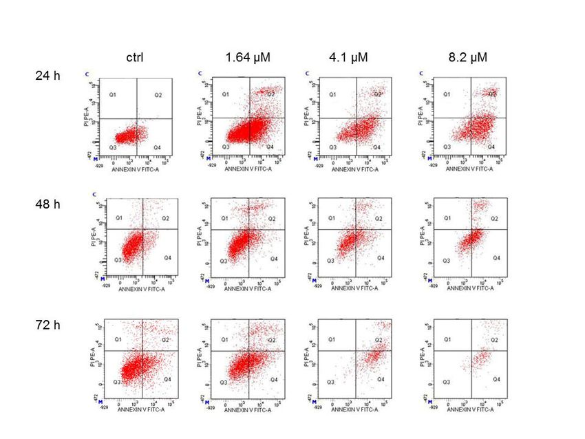
Int. J. Mol. Sci. 2019, 20, 1930 2 of 14
and glucosinolates (GLs). Generally, GLs have three moieties: a β-thioglucose moiety, a sulfonated
oxime moiety, and a variable aglycone side chain derived from an α-amino acid [6]. Furthermore,
M. oleifera possesses many unusual GLs with atypical characteristics due to a second saccharide residue
in the aglyconic side chain [6,7].
In the last decades, GLs precursor, isothiocyanates (ITCs), have been studied mostly due to
their chemopreventive and chemotherapeutic properties [8]. Observational studies have shown that
the consumption of GLs/ITCs-rich cruciferous vegetables protects against several types of human
cancer by induction of both apoptosis and cell cycle arrest. These anticancer properties have been
attributed to the high content of naturally occurring ITCs [9]. The principal GL in Moringa oleifera is
the 4-(α-L-rhamnopyranosiloxy)benzyl glucosinolate, also called glucomoringin. Due to its unusual
structure, this compound may have biological properties different from other GLs [7].
Neuroblastoma (NB) is the most common extra-cranial solid tumor of early childhood accounting
for about 28% of all cancers diagnosed in infants in the US and Europe. Annually, about 700 cases occur
in Canada and the USA as well as 1500 in Europe [10]. Even if aggressive and intensive care had some
improvements in the cure rate of NB patients, their prognosis is still poor. Moreover, conventional
cancer therapies cause serious side effects and, often, merely extend the patient’s lifespan by a few
years. Therefore, natural products to prevent cancer, and alternative approaches to its treatment are
escalating. For this purpose, due to the role of ITCs in cancer management, the aim of our study was
to evaluate the antiproliferative effect of moringin on SH-SY5Y human neuroblastoma cells, and its
molecular mechanisms of action. The natural drug resulted from myrosinase-catalyzed quantitative
hydrolysis of glucomoringin purified from the seeds of the Moringa oleifera.
2. Results
2.1. Moringin Inhibits the Growth of SH-SY5Y Human Neuroblastoma Cells
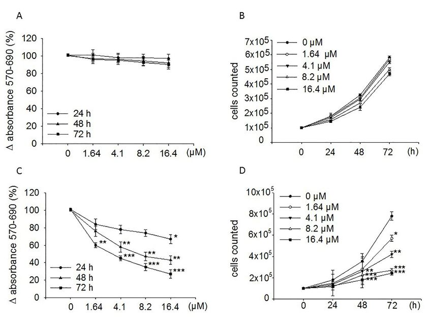
Figure 1C shows that treatment of SH-SY5Y with moringin for 24, 48, and 72 h reduced cells
proliferation in a concentration-dependent manner, achieving the greatest inhibitory effect (73%;
p < 0.001) after 72 h of exposure to 16.4 µM concentration. However, it was already active at 48
(57%; p < 0.01) and 24 h (33%; p < 0.05) of incubation. Furthermore, it is active at a concentration of
1.64 µM. MTT data were established by counting cells in a Neubauer hemocytometer chamber after
24, 48, and 72 h treatment with moringin (Figure 1D). The IC50 value at 72 h of exposure was 1.7 µM.
Contrariwise, ITC did not affect the proliferation of the WI-38 diploid fibroblast cell line (Figure 1A,B).
2.2. Cytotoxic Effect Induced by Moringin
In order to evaluate if the anti-proliferative effect induced by moringin was due to a cytotoxic
effect, the SH-SY5Y cells were exposed to different concentrations of the compound for 24 h, and then
the LDH assay and trypan blue test were performed. Figure 2 shows that, at the concentrations ranging
from 1.64 to 8.2 µM, moringin did not cause SH-SY5Y cell death (Figure 2C) or increase LDH release
(Figure 2D). On the contrary, the highest concentration of moringin tested in this study (16.4 µM)
induced significant cytotoxic effects (p < 0.05 and p < 0.01; Figure 2C,D) on SH-SY5Y cells. Figure 2A,B
show that moringin did not induce a significant increase in WI-38 cell death. Moreover, as illustrated
in Figure 3, exposure to increasing concentrations of moringin for 24 h altered the SH-SY5Y cell
morphology, which acquired a rounded shape (index of cellular suffering), detaching themselves from
the bottom of the well.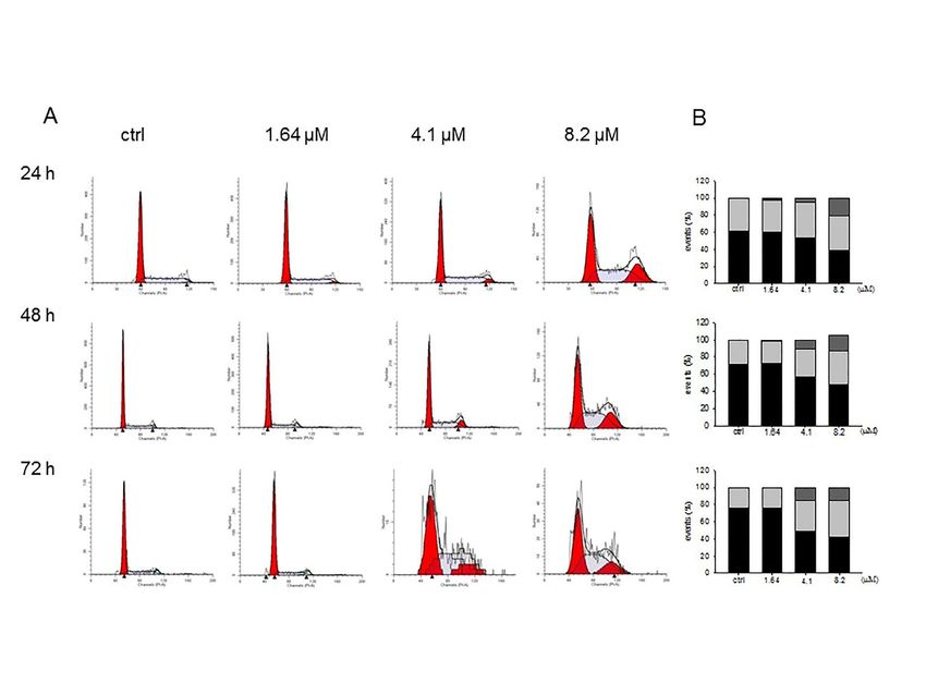
Int. J. Mol. Sci. 2019, 20, 1930                                                                                                  3 of 14
       Int. J. Mol. Sci. 2019, 20, x                                                                                         3 of 15

      Figure 1. Effects of moringin on the proliferation of WI-38 and SH-SY5Y cells. Both WI-38 (A,B) and
                Figure 1. Effects of moringin on the proliferation of WI-38 and SH-SY5Y cells. Both WI-38 (A and B)
      SH-SY5Y (C,D) cells were exposed to the drug (1.64–16.4 µM) for the indicated times. Proliferation rate
                and SH-SY5Y (C and D) cells were exposed to the drug (1.64–16.4 µM) for the indicated times.
      was performed by the MTT assay (A,C) and cell count (B,D). MTT results are expressed as percentages
                Proliferation rate was performed by the MTT assay (A and C) and cell count (B and D). MTT results
      ± SEM     areofexpressed
                       absorbance     detected in
                                  as percentages    treated
                                                 ± SEM        cells. Each
                                                          of absorbance     concentration
                                                                         detected in treatedwas
                                                                                             cells.eightfold   tested, and
                                                                                                    Each concentration     wasthree
      independent           experiments   were  carried   out. Data   from  the cell counts  were   expressed    as
                eightfold tested, and three independent experiments were carried out. Data from the cell counts were  mean   ± SEM
      ofInt. J. expressed
          three Mol.independent
                      Sci. 2019,
                              as20, x experiments
                                 mean  ± SEM of threeperformed
                                                        independentin triplicate.     < 0.05, **p
                                                                       experiments*pperformed   in <  0.01, and
                                                                                                   triplicate. *p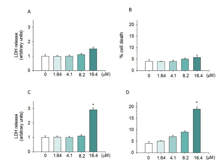
evaluated in terms of both LDH release (A and C) and cell death (B and D) after 24 h of exposure.
       LDH levels were extrapolated as the values detected in control cells which were arbitrarily expressed
       as 1. Cell death was reported as the percentage of blue stained (non-viable) vs. total cells counted.
       Data, expressed as mean ± SEM, represent the values obtained in three different sets of experiments
       made in triplicate. * p < 0.05 vs. control.
Int. J. Mol. Sci. 2019, 20, 1930                                                                                   4 of 14

    Int. J. Mol. Sci. 2019, 20, x                                                                        5 of 15
     Figure 3. Morphological analyses of the SH-SY5Y cells treated with moringin. Cell morphology
     was monitored after treatment with different concentrations (1.64–16.4 µM) of moringin for 24 h.
    2.3.
     The
         Moringin
      Figure      Induced
          morphological
                           Apoptosis
             3. Morphological
                        changes
                                     inof
                              analyses  SH-SY5Y   Neuroblastoma
                                          the SH-SY5Y
                                were observed  under ancells
                                                                   Cells
                                                             treated
                                                        inverted     with moringin.
                                                                   microscope (200×).Cell morphology was
      monitored after treatment with different concentrations (1.64–16.4 µM) of moringin for 24 h. The
         To address the way by which moringin reduced the SH-SY5Y cell growth, apoptosis was
2.3. Moringin  Induced
      morphological    Apoptosis
                    changes wereinobserved
                                    SH-SY5Y    Neuroblastoma
                                            under an inverted Cells
                                                                microscope (200×).
    detected using cytofluorimetric anlysis. After 24 h of treatment with 8.2 µM of moringin, apoptosis
     To address
    was  found inthe  way
                    78%  of by
                            thewhich  moringin reduced
                                cell population,         the SH-SY5Y
                                                  which further  reachedcell90%
                                                                             growth,
                                                                                after apoptosis wasduration
                                                                                      an elongated    detected (72
using
    h) cytofluorimetric
       of exposure to theanlysis.  After 244).
                             cells (Figure  h of treatmentthe
                                               Moreover,   with  8.2µM
                                                              16.4       of moringin, apoptosis
                                                                     µMconcentration    reduced thewasnumber
                                                                                                        found of
in 78%  of the cell population,  which  further reached 90%  after an elongated  duration  (72 h) of exposure
    cells to such a level that did not allow a correct interpretation of the results (data not shown).
to the cells (Figure 4). Moreover, the 16.4 µM concentration reduced the number of cells to such a level
that did not allow a correct interpretation of the results (data not shown).

      Figure 4. Cytofluorimetric evaluation of apoptosis on the SH-SY5Y cells exposed to moringin. Detection
      of apoptosis was performed by the Annexin V test. Representative Annexin V vs. PI dot plots of the
           Figure 4. Cytofluorimetric evaluation of apoptosis on the SH-SY5Y cells exposed to moringin.
      SH-SY5Y cells treated with 1.64–8.2 µM of moringin for 24–72 h are shown. Q3 contains the viable
      cells,Detection of apoptosis
             Q4 the cells             was performed
                           in early apoptosis,   Q2 theby  theinAnnexin
                                                        cells             V test. and
                                                                 late apoptosis,  Representative
                                                                                      Q1 containsAnnexin   V vs.
                                                                                                   the necrotic   PI dot
                                                                                                                cells.
      The plots
            FACSofanalysis
                    the SH-SY5Y     cellsistreated
                            presented              with 1.64–8.2
                                            representative        µM
                                                           of three    of moringin
                                                                    different       for 24–72 h are shown. Q3 contains
                                                                              experiments.
           the viable cells, Q4 the cells in early apoptosis, Q2 the cells in late apoptosis, and Q1 contains the
           necrotic cells. The FACS analysis presented is representative of three different experiments.

    2.4. Moringin Induced the Accumulation of G2 Phase in SH-SY5Y
           Since Brunelli et al. (2010) reported that moringin caused a strong cell cycle perturbation in the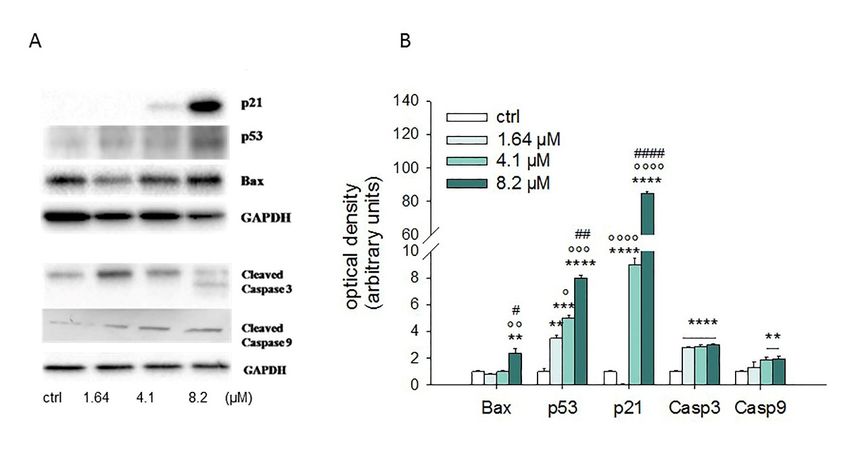
Int. J. Mol. Sci. 2019, 20, 1930                                                                                          5 of 14

2.4. Moringin Induced the Accumulation of G2 Phase in SH-SY5Y
       Since Brunelli et al. (2010) reported that moringin caused a strong cell cycle perturbation in 6the
  Int. J. Mol. Sci. 2019, 20, x                                                                         of 15
RPMI-8226 human myeloma cell line, we analyzed its effect in the SH-SY5Y cell cycle distribution.
As shown in Figure 5, moringin ranging from 1.64 to 8.2 µM increased the cell population in both G2
and S phases, while decreased the number of cells in the G1 phase. The effects of ITC are evident
already after 24 h of exposure and remained unchanged for the following 48 h.

      Figure 5. Influence of moringin on cell cycle distribution in SH-SY5Y cells. Progression of SH-SY5Y
      cells through the cell cycle was examined by flow cytometry analysis after exposure to moringin for
      24–72 h (A). The histograms on the right (B) represent the percentage of events in G1 (black bar),
      G2 (grey), and S (light grey) phases and are the mean ± SEM of three independent experiments.
      Figure 5. Influence of moringin on cell cycle distribution in SH-SY5Y cells. Progression of SH-SY5Y
2.5. Effects of Moringin on the Apoptotic Pathway
      cells through the cell cycle was examined by flow cytometry analysis after exposure to moringin for
    Apoptosis    is tightly
      24–72 h (A).           regulated
                     The histograms  onbytheseveral  factors,
                                             right (B)        including
                                                       represent         tumor suppressors
                                                                 the percentage               and
                                                                                of events in G1    inducer
                                                                                                (black bar), genes,
                                                                                                             G2
such as(grey),
         the Bcl-2
               and Sfamily   proteins,
                     (light grey)      while
                                  phases       thethe
                                          and are   caspases
                                                       mean ± (cysteine  aspartyl
                                                               SEM of three        protease)
                                                                            independent       are considered the
                                                                                         experiments.
most important executors of programmed cell death. In order to investigate the mechanism through
  2.5. Effects
which   moringinof Moringin
                      induces   onSH-SY5Y
                                   the Apoptotic    Pathway
                                                apoptotic    cell death, we evaluated the expression of the main
proteins   involved    in  the  regulation    of
        Apoptosis is tightly regulated by several apoptosis    byfactors,
                                                                    both western     blot tumor
                                                                             including     analysissuppressors
                                                                                                      and Real-Time andPCR.
                                                                                                                          inducer
      Western    blot  analyses   performed       after 24  h of  exposure    to 1.64–8.2   µM    of moringin
  genes, such as the Bcl-2 family proteins, while the caspases (cysteine aspartyl protease) are considered       showed    that it
was
  theable
       most to increase
               important  theexecutors
                                protein level    of p53, p21, Bax,
                                           of programmed           cellcleaved
                                                                        death. caspase
                                                                                 In order3,toand     9 (Figure 6).
                                                                                                  investigate    theThe  results
                                                                                                                     mechanism
ofthrough
    the Real-Time     PCR      experiments      strengthen     this  data  (Figure    7).  In  particular,
              which moringin induces SH-SY5Y apoptotic cell death, we evaluated the expression of            moringin   8.2  µM the
showed    a  1.5-fold   increase   in  Bax  at  both   gene   (p <  0.001)  and   protein   (p  <
  main proteins involved in the regulation of apoptosis by both western blot analysis and Real-Time0.01) levels,  while mRNA
ofPCR.
   Bcl-2 did not change significantly. Moreover, moringin 8.2 µM significantly raised the expression
of the Western
        gene encoding
                    blot analyses      p53 (p < 0.001),
                             proteinperformed        after 24ash well   as increasing
                                                                  of exposure             its protein
                                                                                  to 1.64–8.2    µM of amount        < 0.0001).
                                                                                                         moringin(pshowed      that
However,     p21   was  the   apoptotic   target   most  affected    by moringin,     with   a dramatic
  it was able to increase the protein level of p53, p21, Bax, cleaved caspase 3, and 9 (Figure 6). The     increase at both   the
protein
  resultsand   transcriptional
           of the   Real-Time PCR  level,experiments
                                           especially when       treatedthis
                                                          strengthen      with   4.1 (Figure
                                                                              data   and 8.2 7).µM.InIts gene expression
                                                                                                       particular,  moringin was8.2
7 µM
  and showed
       18 times ahigher      than  the  control    when   the   SH-SY5Y     cells  were   treated   with  moringin
                     1.5-fold increase in Bax at both gene (p < 0.001) and protein (p < 0.01) levels, while          at 4.1  and
8.2  µM concentrations
  mRNA       of Bcl-2 did not (p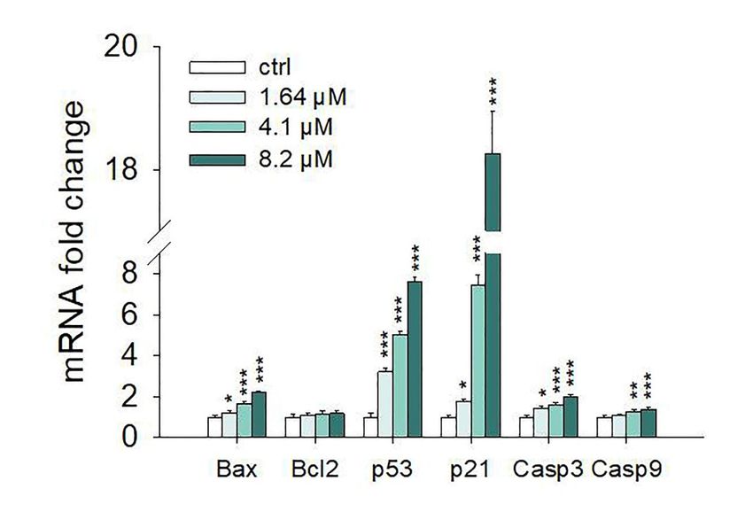
Int. J. Mol. Sci. 2019, 20, 1930                                                                                                              6 of 14
Int. J. Mol. Sci. 2019, 20, x                                                                                                                 7 of 15

      Int. J. Mol. Sci. 2019, 20, x                                                                                                 7 of 15
expression  of both
caspase 3 (4.1 µM andcaspase   3 (4.1
                          8.2 µM,   p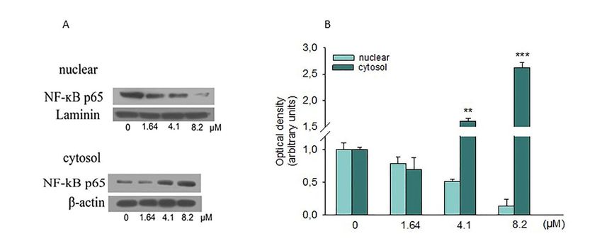
Int. J. Mol. Sci. 2019, 20, x 8 of 15
NF-κB, associated with IκB, is found in the cytoplasm as an inactive complex. When some
stimuli occur,
Int. J. Mol. these
Sci. 2019, can induce the dissociation of the NF-κB/IκB complex through the degradation
20, 1930 7 of 14
of the IκB subunit, thus permitting the translocation of NF-κB in the nucleus. There, it binds to specific
sequences of DNA, regulating the transcription of several target genes. Substantial evidence indicates
is involved
that NF-κB isininvolved
different inmolecular
differentpathways
molecularassociated
pathwayswith tumorigenesis.
associated Therefore, we
with tumorigenesis. investigated
Therefore, we
if NF-κB has been involved in the inhibition of SH-SY5Y cell growth caused by moringin.
investigated if NF-κB has been involved in the inhibition of SH-SY5Y cell growth caused by moringin. Western blot
analysis blot
Western showed a reduction
analysis showedof the nuclear
a reduction oflevels of p65levels
the nuclear revealing therevealing
of p65 ability ofthe
moringin tomoringin
ability of inhibit its
nuclear translocation (Figure 8).
to inhibit its nuclear translocation (Figure 8).
Figure 8. Effects of moringin on NF-kB activation. (A) SH-SY5Y cells were treated for 24 h with the
Figure 8. Effects of moringin on NF-ĸB activation. (A) SH-SY5Y cells were treated for 24 h with the
indicated concentration of moringin, and then, cytoplasmic and nuclear proteins were analyzed by
indicated concentration of moringin, and then, cytoplasmic and nuclear proteins were analyzed by
Western blot for the NF-kB p65 sub-unit. (B) Densitometric analysis of autoradiographic bands in
Western blot for the NF-ĸB p65 sub-unit. (B) Densitometric analysis of autoradiographic bands in
which levels of the nuclear protein were normalized for laminin and the cytosolic ones for β-actin.
which levels of the
A representative nuclear protein
immunoblot were
of three normalizedexperiments
independent for laminin and the cytosolic
is shown. ones
* p < 0.01, for***
and β-actin. A
p < 0.001
representative immunoblot of three independent experiments is shown. * p < 0.01, and *** p < 0.001
vs. control, respectively.
vs. control, respectively.
3. Discussion
3. Discussion
Numerous studies have demonstrated that natural products possess several biological activities
Numerous
widely studies havefolk,
used in traditional, demonstrated that natural products
as well as complementary possess several
and alternative medicine biological activities
[11–13]. Hence,
widely used in traditional, folk, as well as complementary and alternative
many products from the plant kingdom have been widely investigated for their therapeutic effects, medicine [11–13]. Hence,
many products
and some of these from
havetheobtained
plant kingdom have been
clinical approval orwidely investigated
are actually in clinical for their[14,15].
trials therapeutic
These effects,
include
and some
natural or of these have drugs
semisynthetic obtainedused clinical
in the approval or aresuch
field of cancer actually in clinicalvincristine,
as vinblastine, trials [14,15]. These
topotecan,
include natural (camptothecin
and irinotecan or semisynthetic drugs used
derivatives), in the field
etoposide of cancer such asderivative)
(epipodophyllotoxin vinblastine, andvincristine,
paclitaxel,
topotecan,
while others andareirinotecan (camptothecin
under preclinical derivatives),[16,17].
experimentation etoposide (epipodophyllotoxin
In this derivative) and
context, several phytochemicals,
paclitaxel,
such as GLs while
and others are under products
their breakdown preclinical(i.e.experimentation
ITCs), have been [16,17].
shown In to this context,
prevent theseveral
risk of
phytochemicals, such as ITCs
carcinogenesis [18,19]. GLs and
are their
a group breakdown
of natural products
products, (i.e.which
ITCs), are
have been
not shown to
produced as prevent
such by
the
therisk of but
plant, carcinogenesis
rather released [18,19].
afterITCs
cell are a group
damage of natural
by the enzymatic products,
action which are not produced
of myrosinase on their GLs as
such by the plant, but rather released after cell damage by the enzymatic
precursors. ITCs have long been known for their anti-tumoral and anti-inflammatory activities. action of myrosinase on
their GLs precursors.
A consistent number ofITCs have
in vitro andlong been
in vivo known
studies haveforproved
their anti-tumoral
the preventiveand andanti-inflammatory
therapeutic effects
activities.
of various A ITCsconsistent number
such as benzyl ITC,of allyl
in vitro
ITC, and in vivo
phenethyl studies
ITC, have proved
and sulforaphane the preventive
against several types andof
therapeutic effects of various ITCs such as benzyl ITC, allyl ITC, phenethyl
cancers. They act by blocking the characteristic features of cancer including cell proliferation, invasion, ITC, and sulforaphane
against severaland
angiogenesis, types of cancers.
metastasis Theyasact
as well by to
lead blocking
cell cyclethearrest
characteristic
and apoptosis features [20].of cancer including
cell proliferation,
Previously, ininvasion,
vitro andangiogenesis,
in vivo studies and havemetastasis
shown the as well as lead
anticancer to cell
effect cycle arrest
of moringin and
against
apoptosis [20]. of myeloma, carcinoma, astrocytoma, and leukemia [7,21]. Here, for the first time,
different models
Previously, the
we investigated in vitro and in vivo effect
anti-proliferative studies of have shown
moringin in anthe
in anticancer
vitro modeleffect of moringin showing
of neuroblastoma, against
different models of myeloma, carcinoma, astrocytoma, and leukemia [7,21].
that moringin reduced the growth of SH-SY5Y cells through different intracellular pathways leading Here, for the first time,to
we investigated
apoptotic cell death.the Of
anti-proliferative
note, up to 16.4effect of moringin
µM, moringin did notin an
inducein vitro model ofeffect,
any cytotoxic neuroblastoma,
since it did
showing
not causethat
cellmoringin
death or reduced
increase theLDH growth
release.of Therefore,
SH-SY5Y cells through
except for the different
highestintracellular
concentration pathways
tested in
leading to apoptotic cell death. Of note, up to 16.4 µM, moringin did not
this study, the antiproliferative effect of moringin in SH-SY5Y cells was not mediated by necrotic induce any cytotoxic effect,
cell
since
death,itasdid
wasnot cause cell
confirmed by thedeath
results orofincrease LDH release.
cytofluorimetric analyses.Therefore, except
Interestingly, for the study
a previous highestby
concentration tested in[21]
Rajan and co-workers thisshowed
study, that
the antiproliferative
moringin did not effect of moringin
affect the normal human in SH-SY5Y cells ligament
periodontal was not
tissue-derived mesenchymal stem cell (hPDLSCs) viability, as well as those of the WI-38 fibroblastsInt. J. Mol. Sci. 2019, 20, 1930 8 of 14
employed in this study. However, 24 h exposure to moringin altered SH-SY5Y cell morphology, causing
their round shape and detachment from the bottom of the well. A similar effect on cellular morphology
was reported in human pancreatic cancer cells exposed to benzyl ITC [22].
Many cancer cells are unable to undergo apoptosis, and carcinogenesis increases when apoptosis
is missing [23]. On the other hand, the goal of apoptosis is to avoid the proliferation of malignantly
transformed cells that play a crucial role in cancer. Therefore, apoptosis is considered an important
objective of several anticancer drugs. Data from the cytofluorimetric analysis performed in this study
suggest that moringin arrested the cell cycle and induced apoptosis, suggesting the mechanism through
which it exerts its antiproliferative activity.
It is well-known that p53 acts as a tumor suppressor, leading to G2 cell cycle arrest following
DNA damage [24]. Multiple downstream-targeted genes, such as p21 and Bax, are upregulated by
p53 [25]. p21 acts as a cyclin-dependent kinase inhibitory (CDKI) protein, with an affinity for both G1
and G2 cyclin-CDK complexes, highlighting the main mechanism for p53-mediated cell cycle arrest in
G1 or G2 phases. However, in contrast to what was observed by Brunelli and co-workers [7] in A2780,
NSCLC, H460 WT, and H460 S5 cells, we demonstrated that, in SH-SY5Y cells, moringin induced
cell cycle arrest in the G2 phase, probably through the involvement of p53 and p21, as suggested
by their upregulation which was induced by the moringin treatment at both the gene and protein
levels. However, our finding is in line with those of other studies showing that another ITC, the allyl
isothiocyanate, caused G2/M phase arrest in human brain malignant glioma GBM 8401 cells [26] and
prostate cancer cells [27].
The apoptotic machinery is strictly controlled by numerous factors comprising tumor inducers
and suppressor genes such as the Bcl-2 family proteins which could stimulate survival of tumor cells,
thus conferring resistance to chemotherapy (Bcl-2 and Bcl-XL) or inducing apoptosis (Bax and Bad) [28].
Results from the Western blot analysis showed that treatment of SH-SY5Y cells with 4.1 and 8.2 µM
concentrations of moringin for 24 h up-regulated some pro-apoptotic proteins such as Bax, p21, and p53.
Moreover, the Real-Time PCR data demonstrated that moringin acts at transcriptional levels modifying
the mRNA of genes related to the apoptotic process at 1.64 µM concentration. In addition, exposure of
SH-SY5Y cells to moringin significantly increased the gene expression of both caspase 3 and 9 and
enhanced their cleavage, thereby initiating the intrinsic apoptotic cascade.
The NF-κB family of transcription factors regulates growth, differentiation, and apoptosis in
several tissues [29]. In its inactive form, NF-κB is present in the cytosol as an inducible multi-subunit
complex composed of two protein subunits, p50 and p65 polypeptides that are complexed to a third
NF-κB inhibitory subunit, IκB. The activation of NF-κB is frequent in several human malignancies,
driving up-regulation of genes that codify for growth factors, anti-apoptotic genes, pro-inflammatory
cytokines, and adhesion molecules [30,31]. Many studies have shown that the downregulation of
NF-κB in the nucleus is associated with apoptosis. Hence, inhibition of NF-κB activation has been
postulated as a key target for cancer chemoprevention [32,33]. Several natural products, such as ITCs,
exert their anti-cancer effects through the suppression of one or more steps in the NF-κB signaling
pathway. In this line, our data demonstrated that moringin inhibited nuclear translocation of NF-κB in
SH-SY5Y cells, suggesting a key role of this transcription factor in the anti-adhesive and pro-apoptotic
effect of moringin in SH-SY5Y cells. Our data are in contrast with what was reported by Giacoppo
and co-workers [34] but in accordance with what was shown by Brunelli and collaborators [7] in the
RAW-NFκB cell. Therefore, our results suggest that the antiproliferative effect of moringin observed
in SH-SY5Y cells could be due, at least in part, to its suppressive effect on the NF-κB pathways and
induction of cytoprotective genes.
Overall, our study demonstrates the ability of moringin in reducing the growth of SH-SY5Y
cells, implying the knowledge on its mechanism of action, and suggests its promising role as
an anticancer drug.Int. J. Mol. Sci. 2019, 20, 1930 9 of 14
4. Materials and Methods
4.1. Sample Preparation
Glucomoringin (GMG) was isolated from M. oleifera L. (seed cake powder PKM2 provided by
Indena India Pvt. Ltd.; Bangalore, India) at the CREA-AA laboratory in Bologna in two sequential
steps, by anion exchange and size exclusion chromatography, according to a method previously
described [35]. The purity was assayed by HPLC analysis of the desulfo-derivative, yielding GMG
with a purity of about 99% (based on peak area value), and more than 95% on a weight basis, due to
its high hygroscopic properties [35]. The enzyme Myrosinase (Myr) was extracted from seeds of the
Sinapis alba L. as described by Pessina et al. [36] with some modification.
GMG powder was dissolved in culture media and then bioactivated with Myr (30 µL/mL of GMG
solution) for 30 min at 37 ◦ C to obtain the bioactive moringin (GMG-ITC). The total conversion of
GMG into moringin was confirmed by HPLC analysis of the desulfo-derivative [37].
4.2. Cell Culture and Drug Treatment
Experiments were carried out using the tumor derived SH-SY5Y human neuroblastoma cell line
as well as the fibroblast WI-38, derived from normal embryonic lung tissue. Both cell lines were
originally obtained from ATCC (Rockville, MD, USA). The cells were grown in a monolayer at 37 ◦ C in
5% CO2 humidified atmosphere. The SH-SY5Y cells were cultured in RPMI supplemented with 10%
(v/v) heat-inactivated fetal bovine serum, L-glutamine (2 mM), sodium pyruvate (1 mM), penicillin
(100 lU/mL), and streptomycin (100 µg/mL). WI-38 cell lines were grown in DMEM supplemented with
10% FBS, glutamine, and penicillin-streptomycin. All reagents were from Gibco (Life Technologies,
Monza, Italy).
Experiments performed in this study were carried out by seeding the cells in the appropriate
culture plates 24 h before each experiment. In particular, SH-SY5Y and WI-38 cells were plated in
96-well plates for both the 4,5-dimethylthiazol-2-yl)-2,5-diphenyltetrazolium bromide (MTT) test and
the lactate dehydrogenase (LDH) assay (5 × 10³ and 15 × 103 cell/well, respectively). The cell count
assay, the trypan blue dye test, and the cytofluorimetric analyses were carried out on SH-SY5Y seeded
onto a 6-well plate at a density of 10 × 103 cells/well, while for the protein expression studies we
plated 6 × 10⁵ cells/well. Finally, the mRNA profile was evaluated by seeding the SH-SY5Y in 100 mm
Petri dishes at a density of 15 × 105 cells/plate. The next day, growth media was replaced with fresh
medium with or without (untreated cultures) increased concentrations of moringin ranging from 1.64
to 16.40 µM. The length of the treatment depended on the assay performed.
4.3. Cell Proliferation Assays
In order to evaluate the anti-proliferative activity of moringin, we performed both the MTT test
and cell count assay as previously described [38]. SH-SY5Y and WI-38 cells were seeded and treated
as described above. After 24, 48, and 72 h of incubation, the plates were centrifuged at 1200 rpm for
10 min, the supernatants were removed and fresh media without phenol red containing 0.5 mg/mL of
MTT (Sigma-Aldrich, Milan, Italy), was added to each well. The plates were replaced in the incubator
for 4 h and gently shaken occasionally. Then, the plates were centrifuged at 1200 rpm for 10 min,
the supernatants were removed and crystals of formazan (MTT metabolic product) were solubilized in
100 µL of HCl/isopropanol 0.1 N lysis buffer. The absorbance was spectrophotometrically quantified
by a microplate spectrophotometer (iMark™ microplate reader, Bio-Rad Laboratories, Milan, Italy) at
a wavelength of 570 nm with reference at 690 nm. Differences in cell proliferation were measured as
a percentage of growth rates of treated cells compared to untreated cultures.
Cell growth was also detected by the cell count assay. Briefly, SH-SY5Y and WI-38 cells were seeded
and treated as described above and then harvested by trypsinization, centrifuged, and re-suspended
in a known amount of culture medium. Aliquots of cell suspensions were put in a Neubauer
hemocytometer chamber and the cells were counted by an optical microscope.Int. J. Mol. Sci. 2019, 20, 1930 10 of 14
The proliferation assays were performed in eightplicate (MTT test) or triplicate (cell count) and
repeated three different times.
4.4. Cytotoxicity Assays
Possible drug cytotoxicity was assessed by both the lactate dehydrogenase (LDH) assay and the
trypan blue test after 24 h of treatment with moringin. LDH concentrations in the medium of treated
and untreated cells were measured by a commercial kit (CytoTox 96® Non-Radioactive Cytotoxicity
Assay, Promega, Milan, Italy). Briefly, plates were centrifuged at 400× g for five minutes and then 50 µL
of supernatant from each well was transferred to the corresponding wells of clean plates together with
50 µL of fresh prepared LDH reaction solution. The plates were put on an orbital shaker for 30 min
at room temperature and then 50 µL of stop solution was added to each well. The absorbance was
quantified spectrophotometrically at 490 nm. LDH levels were extrapolated as the values detected in
control cells, which were arbitrarily expressed as 1 [39]. The trypan blue dye (0.4% w/v; TB) exclusion
assay was used to detect dead cells, that were reported as the percentage of stained (non-viable) vs.
total cells counted [39]. Both LDH and TB experiments were carried out in triplicate and repeated
three times.
4.5. Cytofluorimetric Evaluation of Apoptosis
Cell death induced by moringin was also assessed by fluorescence-activated cell sorting (FACS)
analysis exploiting the annexin-V/propidium iodide (PI) staining, a method that allows the distinction
of early apoptosis from late apoptosis and necrosis. The PI is a DNA intercalator that stains necrotic
cells because it binds to the DNA of cells with a damaged membrane. Indeed, PI cannot enters
viable cells that keep membrane integrity. Annexin-V has a high affinity to phosphatidylserines (PS),
a phospholipid component exposed on the outer leaflet of the plasma membrane of apoptotic cells.
Our experiments were performed using a commercial kit (BD Biosciences, Milan, Italy) in which the
annexin-V conjugated to fluorescein isothiocyanate (FITC) which serves as a sensitive probe of cells
that are undergoing apoptosis, compared to PI which allows the discrimination of the early from
late apoptotic cells. Therefore, based on the staining intensity, viable cells are both annexin-V and PI
negative, early apoptotic cells are annexin-V positive and PI negative, late apoptotic cells are both
annexin-V and PI positive, and necrotic cells are annexin-V negative and PI positive.
After 24, 48, or 72 h of treatment with moringin, the SH-SY5Y cells were collected by trypsinization,
washed, centrifuged, and re-suspended in the binding buffer provided by the kit at 1 × 10⁶ cell/mL
concentration. Then, both 5 µL of annexin-V-FITC and 10 µL of PI were added to 200 µL of each sample,
gently vortexed, and incubated at room temperature in the darkness for 15 min. Finally, the samples
were run on a Novocyte 2000 (ACEA Bioscences Inc., San Diego, California, USA) cytofluorimeter [40].
The experiment was repeated three times in triplicate.
4.6. Cell Cycle Analysis
Cytofluorimetric studies were also used to check the progression of cells through the cell cycle.
The SH-SY5Y cells were treated for 24, 48, and 72 h, harvested and centrifuged for 10 min at 1200 rpm.
Then, the cells were fixed in cold 70% ethanol at 4 ◦ C for 2 h, washed with cold PBS, centrifuged,
and re-suspended in 250 µL of PBS together with 5 µL of 10 mg/mL RNase A. After 1 h of incubation at
37 ◦ C, 10 µL of 1 mg/mL of PI was added to each sample and the cell suspensions were run on flow
cytometry [41]. The experiment was repeated three times in triplicate.
4.7. Western Blot Analysis
SH-SY5Y cells were seeded and treated for 24 h as described above, and then processed following
Ferlazzo and co-workers [40]. Briefly, the cells were washed with ice-cold PBS and lysed in a buffer
containing: 0.32 M sucrose, 10 mM Tris-HCl pH 7.4, 1 mM EGTA, 2 mM EDTA, 5 mM NaN3, 10 mM
2-mercaptoethanol, 50 mM NaF, and protease inhibitor (Roche Diagnostics Corporation, Indianapolis,Int. J. Mol. Sci. 2019, 20, 1930 11 of 14
USA). The homogenates were chilled on ice for 20 min, centrifuged at 9600× g at 4 ◦ C for 1 min,
and then the supernatant (cytosolic extract) was collected. Following, pellets were suspended in a lysis
buffer containing 0.1% Triton X-100, 150 mM NaCl, 10 mM Tris-HCl, pH 7.4, 1 mM EGTA, 1 mM EDTA,
and protease inhibitors (Roche Diagnostics Corporation), kept on ice for 30 min and centrifuged at
9600× g at 4 ◦ C for 10 min. The supernatant (nuclear extract) was collected and stored at −80 ◦ C until
use. Protein concentrations were determined using a Bio-Rad Protein Assay (Bio-Rad Laboratories)
using BSA as the standard. Proteins were separated on sodium dodecyl sulfate-polyacrylamide gel
electrophoresis (SDS-PAGE) and transferred onto a PVDF transfer membrane (Immobilon-P PVDF,
Merck Millipore division of Merck KGaA, Darmstadt, Germany), blocked with PBS containing 5%
non-fat dried milk for 1 h at room temperature, and subsequently probed with the following antibodies
at 4 ◦ C overnight: Cleaved-caspase 3 (1:1000), Cleaved-caspase 9 (1:1000), Bax (1:500), NFκBp65 (1:1000;
AbCam), GAPDH (1:1000), Lamin B1 (1:500) (all from Cell Signaling Technology, Inc, Danvers, MA,
USA, Cell Signaling Technology), p21 (1:1000; Merck Millipore), and p53 (1:2000; Abcam, Cambridge,
UK). Then membranes were incubated with horseradish peroxidise-conjugated goat anti-mouse or
anti-rabbit IgG secondary antibodies (1:2000; Santa Cruz Biotechnology, Inc., Dallas, Texas, USA) at
room temperature for 1 h. Protein bands were visualized using an enhanced chemiluminescence
system (Luminata™ Forte, Western HRP substrate; Millipore), acquired with the ChemiDoc™ MP
System (Bio-Rad Laboratories) and quantified with the ImageJ software.
4.8. Real-Time PCR Analysis
In order to evaluate the effect of moringin on the expression of genes encoding for apoptosis
regulatory proteins, total RNA from 24 h treated or untreated SH-SY5Y cells was extracted using
TRIzol reagent, according to the manufacturer’s protocol. Then, equal amounts of total RNA (2 µg)
were reverse transcribed using the High-Capacity cDNA Archive Kit (Applied Biosystems, Foster
City, CA). MicroRNA levels of Bax, Bcl2, p53, p21, and Caspase 3 and 9 were analyzed by SYBR green
Real-Time PCR. Quantitative PCR reactions were set up in a 96-well plate and were performed in
20 µL reactions containing 1x SYBR® Premix DimerEraser™ (TaKaRa Bio Inc., Japan), 0.1 µM specific
primers, and 25 ng RNA converted into cDNA. Real-Time PCR was carried out on a 7300 Real-Time
PCR System with the following profile: one cycle at 95 ◦ C for 10 min, followed by 40 cycles at 95 ◦ C for
15 s and 60 ◦ C for 1 min. A standard dissociation stage was added to assess primer specificity. β-Actin
was used as housekeeping control. The primer sequences used for Real-Time PCR are listed in Table 1.
Data were collected and analyzed using the 2−∆∆CT relative quantification method [42]. Values are
presented as fold change relative to untreated cells.
Table 1. Oligonucleotide primers used for Real-Time PCR.
Gene Product Primer Sequence
Forward: 50 -TTCTCCACCTAGACTGTAA-30
p21
Reverse: 50 -GCACCTGCTGTATATTCA-30
Forward: 50 -GTGTGGAGTATTTGGATGAC-30
p53
Reverse: 50 -ATGTAGTTGTAGTGGATGGT-30
Forward: 50 -GGACGAACTGGACAGTAACATGG-30
Bax
Reverse: 50 -GCAAAGTAGAAAAGGGCGACAAC-30
Forward: 50 -ATCGCCCTGTGGATGACTGAG-30
Bcl-2
Reverse: 50 -CAGCCAGGAGAAATCAAACAGAGG-30
Forward: 50 - AGCACCTGGTTATTATTCTTGG-30
Caspase 3
Reverse: 50 - GCTTGTCGGCATACTGTT-30
Forward: 50 - GCTCAGACCAGAGATTCG-30
Caspase 9
Reverse: 50 - ATCCTCCAGAACCAATGTC-30
Forward: 50 -TTGTTACAGGAAGTCCCTTGCC-30
β-Actin
Reverse: 50 -ATGCTATCACCTCCCCTGTGTG-30Int. J. Mol. Sci. 2019, 20, 1930 12 of 14
4.9. Statistical Analysis
Data were expressed as mean ± SEM and statistically evaluated for differences using one-way
analysis of variance (ANOVA), followed by the Turkey–Kramer multiple comparison test (GraphPad
Prism Software for Science, San Diego, CA, USA). P-values less than or equal to 0.05 were
considered significant.
Author Contributions: S.C. performed the experiments and drafted the manuscript; N.F. and L.M. performed the
Real-Time PCR experiments; A.G. performed the Western blot analysis; A.B. assisted in the interpretation of the
data and critically revised the manuscript; E.M. performed the statistical analyses and assisted in the design of the
experiments; M.N. conceived and designed the experiments as well as drafted the manuscript.
Funding: We did not receive any specific funding for the present research.
Conflicts of Interest: The authors declare no conflict of interest.
Abbreviations
GLs glucosinolates
ITCs isothiocyanates
MTT 3-(4,5-dimethylthiazol-2-yl)-2,5-diphenyltetrazolium bromide
LDH lactate dehydrogenase
NF-κB nuclear factor kappa-light-chain-enhancer of activated B cells
GMG glucomoringin
HPLC high-performance liquid chromatography
Myr myrosinase
PI iodide propidium
PBS phosphate buffer saline
References
1. Stohs, S.J.; Hartman, M.J. Review of the Safety and Efficacy of Moringa oleifera. Phytother. Res. 2015, 29,
796–804. [CrossRef] [PubMed]
2. Abdull Razis, A.F.; Ibrahim, M.D.; Kntayya, S.B. Health benefits of Moringa oleifera. Asian Pac. J. Cancer
Prev. 2014, 15, 8571–8576. [CrossRef] [PubMed]
3. Anwar, F.; Latif, S.; Ashraf, M.; Gilani, A.H. Moringa oleifera: A food plant with multiple medicinal uses.
Phytother. Res. 2007, 21, 17–25. [CrossRef] [PubMed]
4. Gupta, S.; Jaina, R.; Kachhwahab, S.; Kotharic, S.L. Nutritional and medicinal applications of Moringa
oleifera Lam.-Review of current status and future possibilities. J. Herb. Med. 2018, 11, 1–11. [CrossRef]
5. Lin, M.; Zhang, J.; Chen, X. Bioactive flavonoids in Moringa oleifera and their health-promoting properties.
J. Funct. Foods 2018, 47, 469–479. [CrossRef]
6. Agerbirk, N.; Olsen, C.E. Glucosinolate structures in evolution. Phytochemistry 2012, 77, 16–45. [CrossRef]
7. Brunelli, D.; Tavecchio, M.; Falcioni, C.; Frapolli, R.; Erba, E.; Iori, R.; Rollin, P.; Barillari, J.; Manzotti, C.;
Morazzoni, P.; et al. The isothiocyanate produced from glucomoringin inhibits NF-kB and reduces myeloma
growth in nude mice in vivo. Biochem. Pharmacol. 2010, 79, 1141–1148. [CrossRef]
8. Molina-Vargas, L.F. Mechanism of action of isothiocyanates. A review. Agron. Colomb. 2013, 31, 68–75.
9. Gupta, P.; Kim, B.; Kim, S.H.; Srivastava, S.K. Molecular targets of isothiocyanates in cancer: Recent advances.
Mol. Nutr. Food Res. 2014, 58, 1685–1707. [CrossRef] [PubMed]
10. Heck, J.E.; Ritz, B.; Hung, R.J.; Hashibe, M.; Boffetta, P. The epidemiology of neuroblastoma: A review.
Paediatr. Perinat. Epidemiol. 2009, 23, 125–143. [CrossRef]
11. Miroddi, M.; Navarra, M.; Quattropani, M.C.; Calapai, F.; Gangemi, S.; Calapai, G. Systematic review of
clinical trials assessing pharmacological properties of Salvia species on memory, cognitive impairment and
Alzheimer’s disease. CNS Neurosci. Ther. 2014, 20, 485–495. [CrossRef] [PubMed]
12. Cirmi, S.; Ferlazzo, N.; Lombardo, G.E.; Ventura-Spagnolo, E.; Gangemi, S.; Calapai, G.; Navarra, M.
Neurodegenerative Diseases: Might Citrus Flavonoids Play a Protective Role? Molecules 2016, 21, 1312.
[CrossRef] [PubMed]Int. J. Mol. Sci. 2019, 20, 1930 13 of 14
13. Micali, S.; Isgro, G.; Bianchi, G.; Miceli, N.; Calapai, G.; Navarra, M. Cranberry and recurrent cystitis:
More than marketing? Crit. Rev. Food Sci. Nutr. 2014, 54, 1063–1075. [CrossRef] [PubMed]
14. Mannucci, C.; Navarra, M.; Calapai, F.; Squeri, R.; Gangemi, S.; Calapai, G. Clinical Pharmacology of Citrus
bergamia: A Systematic Review. Phytother. Res. 2017, 31, 27–39. [CrossRef] [PubMed]
15. Newman, D.J.; Cragg, G.M. Natural Products as Sources of New Drugs from 1981 to 2014. J. Nat. Prod. 2016,
79, 629–661. [CrossRef] [PubMed]
16. Cirmi, S.; Ferlazzo, N.; Lombardo, G.E.; Maugeri, A.; Calapai, G.; Gangemi, S.; Navarra, M. Chemopreventive
Agents and Inhibitors of Cancer Hallmarks: May Citrus Offer New Perspectives? Nutrients 2016, 8, 698.
[CrossRef] [PubMed]
17. Cirmi, S.; Maugeri, A.; Ferlazzo, N.; Gangemi, S.; Calapai, G.; Schumacher, U.; Navarra, M. Anticancer
Potential of Citrus Juices and Their Extracts: A Systematic Review of Both Preclinical and Clinical Studies.
Front. Pharmacol. 2017, 8, 420. [CrossRef] [PubMed]
18. Fuentes, F.; Paredes-Gonzalez, X.; Kong, A.N. Dietary Glucosinolates Sulforaphane, Phenethyl
Isothiocyanate, Indole-3-Carbinol/3,30 -Diindolylmethane: Anti-Oxidative Stress/Inflammation, Nrf2,
Epigenetics/Epigenomics and In Vivo Cancer Chemopreventive Efficacy. Curr. Pharmacol. Rep. 2015,
1, 179–196. [CrossRef] [PubMed]
19. Alrawaiq, N.S.; Abdullah, A. An evaluation of sulforaphane as a potential agent for disease prevention. Res.
J. Pharm. Biol. Chem. Sci. 2014, 3, 1335–1349.
20. Grundemann, C.; Huber, R. Chemoprevention with isothiocyanates—From bench to bedside. Cancer Lett.
2018, 414, 26–33. [CrossRef]
21. Rajan, T.S.; De Nicola, G.R.; Iori, R.; Rollin, P.; Bramanti, P.; Mazzon, E. Anticancer activity of glucomoringin
isothiocyanate in human malignant astrocytoma cells. Fitoterapia 2016, 110, 1–7. [CrossRef]
22. Srivastava, S.K.; Singh, S.V. Cell cycle arrest, apoptosis induction and inhibition of nuclear factor kappa
B activation in anti-proliferative activity of benzyl isothiocyanate against human pancreatic cancer cells.
Carcinogenesis 2004, 25, 1701–1709. [CrossRef] [PubMed]
23. Hanahan, D.; Weinberg, R.A. Hallmarks of cancer: The next generation. Cell 2011, 144, 646–674. [CrossRef]
[PubMed]
24. Chen, J. The Cell-Cycle Arrest and Apoptotic Functions of p53 in Tumor Initiation and Progression. Cold
Spring Harb. Perspect. Med. 2016, 6, a026104. [CrossRef]
25. Jin, S.; Mazzacurati, L.; Zhu, X.; Tong, T.; Song, Y.; Shujuan, S.; Petrik, K.L.; Rajasekaran, B.; Wu, M.;
Zhan, Q. Gadd45a contributes to p53 stabilization in response to DNA damage. Oncogene 2003, 22, 8536–8540.
[CrossRef]
26. Chen, N.G.; Chen, K.T.; Lu, C.C.; Lan, Y.H.; Lai, C.H.; Chung, Y.T.; Yang, J.S.; Lin, Y.C. Allyl isothiocyanate
triggers G2/M phase arrest and apoptosis in human brain malignant glioma GBM 8401 cells through
a mitochondria-dependent pathway. Oncol. Rep. 2010, 24, 449–455. [PubMed]
27. Xiao, D.; Srivastava, S.K.; Lew, K.L.; Zeng, Y.; Hershberger, P.; Johnson, C.S.; Trump, D.L.; Singh, S.V.
Allyl isothiocyanate, a constituent of cruciferous vegetables, inhibits proliferation of human prostate cancer
cells by causing G2/M arrest and inducing apoptosis. Carcinogenesis 2003, 24, 891–897. [CrossRef]
28. Adams, J.M.; Cory, S. The Bcl-2 apoptotic switch in cancer development and therapy. Oncogene 2007, 26,
1324–1337. [CrossRef]
29. Xia, Y.; Shen, S.; Verma, I.M. NF-kappaB, an active player in human cancers. Cancer Immunol. Res. 2014, 2,
823–830. [CrossRef]
30. Pires, B.R.B.; Silva, R.; Ferreira, G.M.; Abdelhay, E. NF-kappaB: Two Sides of the Same Coin. Genes 2018, 9,
24. [CrossRef] [PubMed]
31. Nagel, D.; Vincendeau, M.; Eitelhuber, A.C.; Krappmann, D. Mechanisms and consequences of constitutive
NF-kappaB activation in B-cell lymphoid malignancies. Oncogene 2014, 33, 5655–5665. [CrossRef]
32. Li, F.; Zhang, J.; Arfuso, F.; Chinnathambi, A.; Zayed, M.E.; Alharbi, S.A.; Kumar, A.P.; Ahn, K.S.; Sethi, G.
NF-kappaB in cancer therapy. Arch. Toxicol. 2015, 89, 711–731. [CrossRef] [PubMed]
33. Clarke, J.D.; Dashwood, R.H.; Ho, E. Multi-targeted prevention of cancer by sulforaphane. Cancer Lett. 2008,
269, 291–304. [CrossRef] [PubMed]
34. Giacoppo, S.; Iori, R.; Rollin, P.; Bramanti, P.; Mazzon, E. Moringa isothiocyanate complexed with
alpha-cyclodextrin: A new perspective in neuroblastoma treatment. BMC Complementary Altern. Med. 2017,
17, 362. [CrossRef]Int. J. Mol. Sci. 2019, 20, 1930 14 of 14
35. Galuppo, M.; Giacoppo, S.; Iori, R.; De Nicola, G.R.; Bramanti, P.; Mazzon, E. Administration of
4-(alpha-L-rhamnosyloxy)-benzyl isothiocyanate delays disease phenotype in SOD1(G93A) rats: A transgenic
model of amyotrophic lateral sclerosis. Biomed. Res. Int. 2015, 2015, 259417. [CrossRef]
36. Pessina, A.; Thomas, R.M.; Palmieri, S.; Luisi, P.L. An improved method for the purification of myrosinase
and its physicochemical characterization. Arch. Biochem. Biophys. 1990, 280, 383–389. [CrossRef]
37. Galuppo, M.; Nicola, G.R.; Iori, R.; Dell’utri, P.; Bramanti, P.; Mazzon, E. Antibacterial activity of glucomoringin
bioactivated with myrosinase against two important pathogens affecting the health of long-term patients in
hospitals. Molecules 2013, 18, 14340–14348. [CrossRef]
38. Navarra, M.; Ferlazzo, N.; Cirmi, S.; Trapasso, E.; Bramanti, P.; Lombardo, G.E.; Minciullo, P.L.; Calapai, G.;
Gangemi, S. Effects of bergamot essential oil and its extractive fractions on SH-SY5Y human neuroblastoma
cell growth. J. Pharm. Pharmacol. 2015, 67, 1042–1053. [CrossRef] [PubMed]
39. Romeo, R.; Navarra, M.; Giofre, S.V.; Carnovale, C.; Cirmi, S.; Lanza, G.; Chiacchio, M.A. Synthesis and
biological activity of new arenediyne-linked isoxazolidines. Bioorg. Med. Chem. 2014, 22, 3379–3385.
[CrossRef] [PubMed]
40. Ferlazzo, N.; Cirmi, S.; Russo, M.; Trapasso, E.; Ursino, M.R.; Lombardo, G.E.; Gangemi, S.; Calapai, G.;
Navarra, M. NF-kappaB mediates the antiproliferative and proapoptotic effects of bergamot juice in HepG2
cells. Life Sci. 2016, 146, 81–91. [CrossRef] [PubMed]
41. Celano, M.; Maggisano, V.; De Rose, R.F.; Bulotta, S.; Maiuolo, J.; Navarra, M.; Russo, D. Flavonoid Fraction
of Citrus reticulata Juice Reduces Proliferation and Migration of Anaplastic Thyroid Carcinoma Cells. Nutr.
Cancer 2015, 67, 1183–1190. [CrossRef] [PubMed]
42. Curro, M.; Risitano, R.; Ferlazzo, N.; Cirmi, S.; Gangemi, C.; Caccamo, D.; Ientile, R.; Navarra, M. Citrus
bergamia Juice Extract Attenuates beta-Amyloid-Induced Pro-Inflammatory Activation of THP-1 Cells
Through MAPK and AP-1 Pathways. Sci. Rep. 2016, 6, 20809. [CrossRef] [PubMed]
© 2019 by the authors. Licensee MDPI, Basel, Switzerland. This article is an open access
article distributed under the terms and conditions of the Creative Commons Attribution
(CC BY) license (http://creativecommons.org/licenses/by/4.0/).You can also read


