Imprecision and DNA Break Repair Biased towards Incompatible End Joining in Leukemia
←
→
Page content transcription
If your browser does not render page correctly, please read the page content below
Published OnlineFirst December 8, 2017; DOI: 10.1158/1541-7786.MCR-17-0373

       DNA Damage and Repair                                                                                                           Molecular
                                                                                                                                       Cancer
                                                                                                                                       Research
     Imprecision and DNA Break Repair Biased towards
     Incompatible End Joining in Leukemia
     Franz Josef Gassner1,2, Maria Schubert1,2, Stefan Rebhandl1,2, Karina Spandl1,2,
     Nadja Zaborsky1,2, Kemal Catakovic1,2, Stephanie Blaimer1,2, Daniel Hebenstreit3,
     Richard Greil1,2, and Roland Geisberger1,2

     Abstract
        Cancer is a genetic disease caused by mutations and chromo-                   demonstrated that DNA repair is aberrant in CLL cells, featuring
     somal abnormalities that contribute to uncontrolled cell growth.                 perturbed DNA break structure preference with efficient joining of
     In addition, cancer cells can rapidly respond to conventional and                noncohesive ends and more deletions at repair junctions. In
     targeted therapies by accumulating novel and often specific                       addition, increased microhomology-mediated end joining
     genetic lesions leading to acquired drug resistance and relapsing                (MMEJ) of DNA substrates was observed in CLL together with
     disease. In chronic lymphocytic leukemia (CLL), however, diverse                 increased expression of MMEJ-specific repair factors. In summary,
     chromosomal aberrations often occur. In many cases, improper                     these data identify major differences in DNA repair efficiency
     repair of DNA double-strand breaks (DSB) is a major source for                   between CLL cells and normal B cells isolated from patients.
     genomic abnormalities. Therefore, this study examined the repair
     of DNA DSBs by nonhomologous end joining (NHEJ) in CLL by                        Implications: This study suggests inherently aberrant DNA DSB
     performing plasmid-based repair assays in primary CLL cells and                  repair in the acquisition of subclonal genomic structural varia-
     normal B cells, isolated from patients, as well as TALEN/Cas9–                   tions important for clonal evolution and treatment resistance in
     induced chromosomal deletions in the CLL cell line Mec1. It is                   CLL. Mol Cancer Res; 16(3); 428–38. 2017 AACR.

     Introduction                                                                     S–G2–M phase of the cell cycle, whereas NHEJ simply rejoins
                                                                                      two DNA ends and, hence, is the main repair pathway for
        Whole-genome sequencing revealed the presence of numer-
                                                                                      G1 or G0 arrested cells (7). For NHEJ, a classical and an alter-
     ous clonal and subclonal somatic mutations as well as somatic
                                                                                      native pathway are described, termed c-NHEJ and a-NHEJ,
     rearrangements in cancer genomes (1–3). Upon cancer therapy,
                                                                                      respectively. Although c-NHEJ is dependent on recognition of
     subclonal refractory cells harboring acquired novel chromo-
                                                                                      DNA ends by XRCC5, XRCC6, and DNA-PKc and ligation by
     somal aberrations or gene mutations can expand and may thus
                                                                                      DNA Ligase 4 (LIG4)/XRCC4, a-NHEJ is carried out inde-
     contribute to therapy resistance and disease relapse (4–6).
                                                                                      pendently of LIG4 and can probably be executed by a diverse
     A major source for chromosomal rearrangements are DNA
                                                                                      set of factors, including different DNA polymerases (d, q),
     double-strand breaks (DSB). DSBs occur constantly during the
                                                                                      DNA nucleases (ERCC1-XPF), and ligases (LIG1, LIG3/XRCC1;
     lifetime of any cell and are normally repaired. However, aber-
                                                                                      refs. 9–11). As a-NHEJ is frequently associated with short
     rant repair of DSBs can lead to misjoining of distant DNA ends,
                                                                                      microhomologies at the site of DNA junctions, a-NHEJ is also
     generating deletions, inversions, or complex rearrangements of
                                                                                      termed microhomology-mediated end joining (MMEJ).
     chromosomes (7, 8). DSBs are repaired by two major pathways,
                                                                                         Chronic lymphocytic leukemia (CLL) is a B-cell malignancy
     which are homologous recombination (HR) and nonhomo-
                                                                                      characterized by the progressive accumulation of clonal B cells
     logous end joining (NHEJ). HR utilizes the sister chromatide
                                                                                      in peripheral blood and lymph nodes. Clinically, the mutation
     as template for repairing DSBs and is thus primarily confined to
                                                                                      status of the B-cell receptor divides patients into subgroups
                                                                                      with poor (CLL with unmutated BCR, CLL-UM) and more
     1
       Department of Internal Medicine III with Haematology, Medical Oncology,        favorable prognosis (CLL with mutated BCR, CLL-Mut; refs. 12–
     Haemostaseology, Infectious Disease, Rheumatology, Oncologic Center,             14). Although many of the hematologic malignancies show
     Laboratory for Immunological and Molecular Cancer Research, Paracelsus           typical entity-specific chromosomal abnormalities (15), there is
     Medical University Salzburg, Austria. 2Cancer Cluster Salzburg, Salzburg,
                                                                                      no specific aberration defining CLL. Instead, in addition to
     Austria. 3School of Life Sciences, University of Warwick, Coventry, United
     Kingdom.                                                                         more common chromosomal abnormalities like del(17p), del
                                                                                      (11q), del(13q), and Tri12, conventional cytogenetics as well as
     Note: Supplementary data for this article are available at Molecular Cancer
                                                                                      novel whole-genome sequencing approaches revealed a com-
     Research Online (http://mcr.aacrjournals.org/).
                                                                                      plex set of different chromsomal aberrations in CLL, with CLL-
     F.J. Gassner, M. Schubert and S. Rebhandl contributed equally to this article.
                                                                                      UM samples showing a slight increase in the number of
     Corresponding Author: Roland Geisberger, Paracelsus Medical University           aberrations (3, 5, 6, 16–19).
                €llner Hauptstrasse 48, Salzburg 5020, Austria. Phone: 0043-
     Salzburg, Mu                                                                        In this study, we asked whether the frequent occurrence of
     57255-25847; Fax: 0043-57255-25998; E-mail: r.geisberger@salk.at
                                                                                      chromosomal abnormalities is associated with an inherently
     doi: 10.1158/1541-7786.MCR-17-0373                                               aberrant DNA DSB repair in CLL. To test this, we analyzed DNA
     2017 American Association for Cancer Research.                                  DSB repair in primary CLL cells compared with B cells from

428 Mol Cancer Res; 16(3) March 2018

         Downloaded from mcr.aacrjournals.org on February 7, 2021. © 2018 American Association for Cancer Research.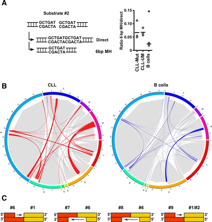
Published OnlineFirst December 8, 2017; DOI: 10.1158/1541-7786.MCR-17-0373

                                                                                                      Aberrant DNA Break Repair in CLL

healthy controls by assessing in vivo repair of artificial DNA         ccggGGTGGGTGGTGAGATCAATG-30 ; and RG1088 50 ;-aaac-
substrates carrying diverse DNA break structures using next-gen-      CATTGATCTCACCACCCACC-30 ;) into prelinearized pGuide-it-
eration sequencing (NGS) of repair junctions. These results were      ZsGreen1 Vector (Clontech). TALENs specific for chr17 position
corroborated by analyzing the induction of a large chromosomal        40,819,524-40,819,583 (KRT10 locus) were kindly provided by
deletion using TALEN (transcription activator-like effector nucle-    Oliver March (Paracelsus Private Medical University Salzburg,
ase)/Cas9 (clustered regularly interspaced short palindromic          Salzburg, Austria). The CLL cell line Mec1 (21) was incubated for 6
repeats associated protein 9) endonucleases (20). Our data show       hours in RPMI medium supplemented with or without 30 mmol/L
that DNA end joining is less precise and more promiscuous in CLL      LIG1/3 inhibitor L67 followed by Amaxa nucleofection (Lonza)
with a bias toward microhomologies as well as toward joining of       with TALEN/Cas9 constructs. Cells were kept on RPMI medium
incompatible DNA ends, likely due to increased expression levels      supplemented with or without L67 for further 2 days prior sorting
of LIG1 and XRCC1.                                                    of GFP-positive cells on a FACS AriaIII (Beckton Dickinson; purity
                                                                       94.1% GFPþ) and isolation of DNA (Qiagen). Chromosomal
Materials and Methods                                                 deletions were PCR amplified using Phusion polymerase and
                                                                      specific primers (RG1142 50 ;-TGGCATCTTCTTGGGGTTTA-30 ;
Preparation of plasmid repair substrates and transfection of
                                                                      and RG1143 50 ;-CCACATCCCCTTTTTCCATA-30 ;). For amplicon
cells
                                                                      sequencing, PCR products from 50 ng template DNA from two
   Plasmids were generated by cloning synthesized inserts (MWG
                                                                      independent experiments were pooled and sent for sequencing on
eurofins) into pAX plasmids (sequence shown in Supplementary
                                                                      the MiSeq platform (Illumina). For quantification of chromo-
Fig. S1). Plasmids were linearized using the respective restriction
                                                                      somal deletions, input DNA from cells descending from four
enzymes (Fermentas), gel purified (Qiagen), and transfected into
                                                                      independent experiments was titrated from 25 to 1.56 ng and
293 cells (genejuice, Novagen) and primary B and CLL cells using
                                                                      subjected to PCR using primers RG1142/RG1143 and HotStar
nucleofection (Amaxa, Lonza). Seventy-two hours posttransfec-
                                                                      Taq DNA polymerase (Qiagen).
tion, cells were harvested and DNA was isolated (Qiagen).
Repaired substrates were PCR amplified using high-fidelity Phu-         Cells and cell lines
sion polymerase (Biozym) and tagged 3AOX/5AOX primers                    293 cells, Mec1 cells, and primary cells were cultured in RPMI
(3AOX 50 -GCA AAT GGC ATT CTG ACA TCC-30 ; 5AOX 50 -GAC               medium supplemented with 10% FCS (Gibco). Mec1 cells were
TGG TTC CAA TTG ACA AGC-30 ). PCR products were pooled and            authenticated by DNA fingerprinting and cytometrical analysis of
sequenced on the MiSeq platform (Illumina).                           surface markers (DSMZ). After purchase, stocks of Mec1 cells were
   pmCherry was generated by cloning the PCR-amplified coding          frozen and passaged less than 6 months. 293 cells were provided
region of mCherry (RG1081 50 -CGCGGGCCCGGGATCGCCAC-                   by Stefan Hainzl (EB House Austria, Salzburg, Austria) and
CATGGTGAGCAAGGGCGAGG-30 ; and RG1082 50 ;-TCTAGA-                     periodically authenticated by morphologic inspection. Mec1 and
GTCGCGGCC TACTTGTACAGCTCGTCC-30 ;) into BamHI/NotI                    293 cells were confirmed by fingerprinting at the DMSZ on
digested pEGFP-N1 (Clontech) using In-Fusion HD cloning               October 9, 2017. Mec1 and 293 cells were tested negative for
(Clontech). Plasmids pGFP-1 and pGFP-2 were generated by              mycoplasma in September 2017 using PCR-based mycoplasma
introducing a BsmBI or an EcoRV site at position nt 137 and nt        detection from cell culture supernatants (PanReac AppliChem).
141 of the GFP coding region using PCR-based mutagenesis of the       Primary cells were isolated from peripheral blood of CLL patients
pEGFP-N1 plasmid. Plasmids were digested using BsmBI/MfeI             upon informed consent according to the national guidelines and
(pGFP-1) and EcoRV/PstI (pGFP-2) and gel extracted prior trans-       of the ethics committee and according to the Declaration of
fection together with pmCherry into 293 cells using genejuice         Helsinki (Ethics committee approval Salzburg: 415-E/1287/4–
transfection reagent or nucleofection (Amaxa) as indicated. Six       2011, 415-E/1287/8–2011). CLL cells and B cells were purified
hours before transfection, cells were incubated with highly spe-      untouched using EasySep system (StemCell Technologies) or
cific inhibitors for LIG1/3 (L67), DNA-PKc (NU7441), and PARP          MACS (Miltenyi Biotec). Patient characteristics are summarized
(Olaparib, all from Selleckchem). After transfection, cells were      in Supplementary Table S1. The determination of prognostic
incubated for further 2 days in the presence of inhibitors prior      markers and FISH analysis for trisomy 12, del11q, del13q, and
analysis for mCherry/GFP expression using flow cytometry               del17p was performed routinely at our department as described
(FC500, Beckman Coulter).                                             previously (22). In addition, FISH and karyogram analysis of
                                                                      sample 9956 (patient ID660) was performed at the laboratory for
Gene expression analysis                                              molecular biology and tumor cytogenetics at the Ordensklinikum
   RNA was isolated from purified CLL samples and B cells from         Linz (Linz, Austria).
healthy controls using High Pure RNA Isolation Kit including             For cell-cycle analysis, purified B and CLL cells were stained
DNase digestion (Roche). The same CLL samples as used for             using CD5-FITC (clone: UCHT2; eBioscience) and CD19-PE/Cy7
plasmid repair assays was used for RNA isolation. First-strand        (clone: SJ25C1; eBioscience) antibodies in PBS at room temper-
cDNA was generated using iScript (Bio-Rad). Gene expression           ature. Cells were resuspended in prewarmed RPMI without Phe-
levels in CLL, Mec1, and B cells were determined using TaqMan         nol-Red and stained with vybrant cell cycle dye (Vybrant DyeCycle
assays (Thermo Fisher Scientific) for LIG1 (Hs01553527_m1),            Violet Stain, V35003; Thermo Fisher Scientific) for 1 hour at 37 C
XRCC1 (Hs00959834_m1), PARP1 (Hs00242302_m1) with 18S                 followed by immediate analysis by flow cytometry (Gallios,
rRNA as controls (Hs99999901_s1).                                     Beckman Coulter)

Nuclease-induced chromosomal deletions                                Sequence analysis and bioinformatics
  Cas9 constructs specific for chr17 position 41,621,709 (KRT17          Paired end fastq files were acquired by eurofins genomics and
locus) were generated by cloning annealed oligos (RG1087 50 ;-        GATC-Biotech (Germany). Raw reads were trimmed and selected

www.aacrjournals.org                                                                                   Mol Cancer Res; 16(3) March 2018     429

   Downloaded from mcr.aacrjournals.org on February 7, 2021. © 2018 American Association for Cancer Research.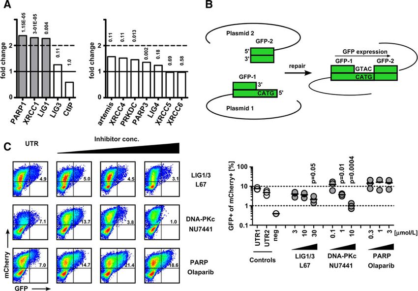
Published OnlineFirst December 8, 2017; DOI: 10.1158/1541-7786.MCR-17-0373

     Gassner et al.

     Figure 1.
     Plasmid-based DSB repair assay. A, Schematic representation of plasmid-based DSB repair. Circular plasmids are linearized upon digestion with restriction enzymes
     followed by transfection of cells where substrates are rejoined. Repair junctions are detected by PCR using conserved forward (fwd) and reverse (rev)
     primer-binding sites on the plasmids. B, DNA end structures of substrates used in our DSB repair assay (MH: microhomology of 6bp). C, B cells and CLL cells were
     transfected with our pooled repair substrates #1–#9. DNA was isolated 72 hours (left) or 0 hour and 72 hours (right) posttransfection, and repair junctions
     were PCR amplified and run on an agarose gel. Amplicons corresponding to a 300-bp band were gel excised and analyzed by NGS. Asterisks refer to bands
     corresponding to amplified undigested plasmids. Amplicons from a control PCR on TP53 are loaded to show integrity of isolated DNA. D, Number of reads obtained
     by NGS of the respective amplicons is shown.

     for paired reads using Trimmomatic v0.33 (PE mode, -phred 33,                     individual 350-bp inserts flanked by conserved primer-binding
     TRAILING:4; ref. 23), resulting in 8,378,777 paired reads surviv-                 sites (Fig. 1A; Supplementary Fig. S1). Upon digestion with
     ing (95.56% of input reads). Forward and reverse reads were                       respective restriction enzymes, the individual plasmids yield
     merged using FLASh v1.2.11 (-r 300 –f 350 –s 35; ref. 24) and                     defined DNA break structures ranging from blunt ends with and
     6,932,326 stiched sequences remained for further analysis. Only                   without a 6-bp microhomology to cohesive and noncohesive 30 ;
     merged reads flanked by forward and reverse primers were                           and 50 ; protruding ends with 4 nt overhangs (Fig. 1B). Upon
     regarded for further data analyses (only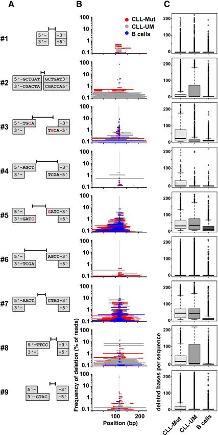
Published OnlineFirst December 8, 2017; DOI: 10.1158/1541-7786.MCR-17-0373
Aberrant DNA Break Repair in CLL
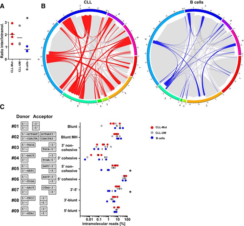
Figure 2.
DNA DSB repair in B cells and CLL cells. A, Graph shows the ratio of inter- to intramolecular repair junctions. B, Inter- and intramolecular repair events are depicted as
circos plots for CLL cells and for normal B cells. The nine segments represent the nine different plasmids used in our repair assay. The size of the segments
reflects their relative occurrence from NGS-analyzed repair junctions. Repair frequencies are indicated by size of ribbons, where orientation of repair of the respective
substrates is given by arrowheads at the end of ribbons (mean values for CLL and B cells as shown in Supplementary Table S3 were used for generating
plots; start of ribbon denotes forward primed arms; arrowhead of ribbon denotes reverse primed arm of substrates shown in Supplementary Fig. S1). Repair junctions
significantly overrepresented in CLL cells compared with B cells (and vice versa) are colored red (P < 0.05; two-tailed t test with unequal variances) and light
red (0.05 P < 0.1), and the correspondingly underrepresented repair junctions are colored blue (P < 0.05; two-tailed t test with unequal variances) and
light blue (0.05 P < 0.1), respectively. C, The frequency of intramolecular repair junctions for individual substrates is shown. Asterisks indicate significant differences
between CLL and B cells (substrate #5: P ¼ 0.004; substrate #7: P ¼ 0.005 two-tailed t test with unequal variances).
PCR-amplified repair junctions from each sample. Notably, we repair junctions in CLL samples, irrespective of the BCR mutation
observed repaired substrates immediately after nucleofection in status (CLL-Mut and CLL-UM), whereas normal B cells preferred
some CLL samples, which is in line with published data on rapid an intramolecular repair (ratio inter/intramolecular repair CLL
end joining of DNA ends (Fig. 1C; ref. 26). Upon NGS of pooled mean 1.88 0.71 STD; B cells 0.79 0.22, P ¼ 0.017 Mann–
amplicons, we obtained a total of 2,676,354 reads, ranging from Whitney test; Fig. 2A). In particular, we observed significantly
96,000 to 587,418 reads for all samples (Fig. 1D). increased intermolecular repair of incompatible DNA ends in CLL
By calculating the ratio of inter- to intramolecular repair events, cells compared with B cells (e.g. joining of plasmid #6 with
we surprisingly obtained a robust bias toward intermolecular plasmid #2 or plasmid #8 with plasmid #5), whereas compatible
www.aacrjournals.org Mol Cancer Res; 16(3) March 2018 431
Downloaded from mcr.aacrjournals.org on February 7, 2021. © 2018 American Association for Cancer Research.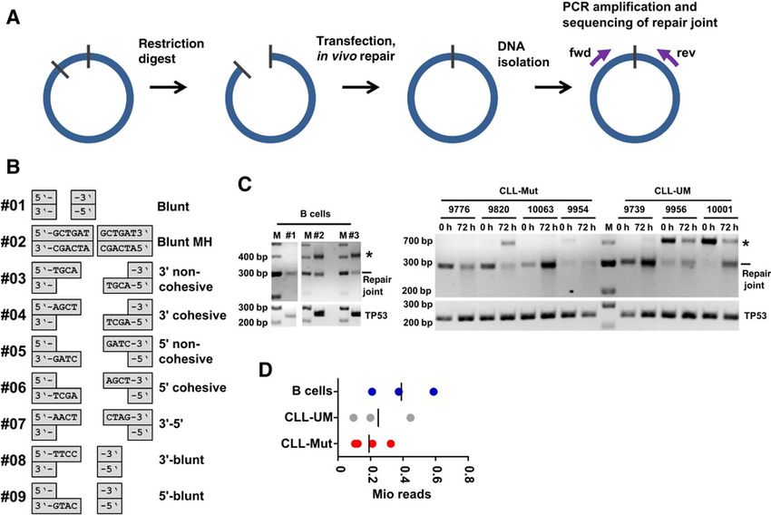
Published OnlineFirst December 8, 2017; DOI: 10.1158/1541-7786.MCR-17-0373

     Gassner et al.

     30 ; and 50 ; overhangs or blunt ends were repaired at comparable      As shown in Fig. 4A, we found that direct joining of substrate #2
     efficiency (Fig. 2B; Supplementary Table S3). None of the repair        was the preferred repair mechanism in CLL as well as in B cells;
     junctions were significantly overrepresented in B cells com-            however, repair by MMEJ was significantly increased relative to
     pared with CLL cells (Fig. 2B; Supplementary Table S3). Also,          c-NHEJ in CLL cells, irrespective of BCR mutation status (ratio
     by analyzing only intramolecular repair frequencies of the nine        microhomology-mediated/direct joining of substrate #2 for CLL:
     different DNA break structures, we observed that noncohesive           mean 0.067  0.023 SD, B cells 0.024  0.005 SD; P ¼ 0.017
     50 ; ends (substrate #5) as well as incompatible 30 ;–50 ; overhangs   Mann–Whitney test; Fig. 4A).
     (substrate #7) were repaired with significantly higher efficiency
     in CLL cells compared with B cells (substrate #5: CLL mean             Efficient joining of incompatible DNA ends in CLL is not
     percentage of reads 4.8  2.4 SD, B cells 0.7  0.5; P ¼ 0.004;        exclusively dependent on microhomologies
     substrate #7: CLL mean 12.4  5.6 SD, B cells 2.5  2.4; P ¼              Next, we tested whether CLL-specific efficient joining of incom-
     0.005 two-tailed t test assuming unequal variances; Fig. 2C;           patible DNA ends was solely dependent on microhomology-
     Supplementary Table S4).                                               based repair of end-resected substrates. To test this, we only
                                                                            considered repair junctions mapping to the most frequently
     More end resection occurs during DSB repair in CLL cells               occurring sequence of a particular repaired substrate and calcu-
        We next aimed at determining differences in the occurrence of       lated their relative frequencies, without considering repair of
     deletions at repair junctions between CLL and B cells, resulting       resected substrates. Thereby, we found that again, various incom-
     from joining of end resected DNA ends. Therefore, we first              patible DNA ends were more efficiently repaired in CLL cells
     identified the most frequent repair junction for CLL-Mut, CLL-          compared with B cells, leading to a total of 12 junctions, which
     UM, and B cells for all individual intramolecular repair events and    were significantly overrepresented in CLL samples (Fig. 4B; Sup-
     mapped all shorter sequences to the most frequent consensus. We        plementary Table S5). Again, none of the repair events were
     thereby found that the most frequent repair junctions of the nine      significantly overrepresented in B cells compared with CLL cells
     DNA substrates were the same within each sample group (CLL-            (Fig. 4B; Supplementary Table S5). Although 7 of these 12 junc-
     Mut, CLL-UM, B cells; Fig. 3A): blunt ends, irrespective of a 6 bp     tions revealed short microhomologies (1 to 5 nt, Supplementary
     microhomology, and cohesive 30 ; and 50 ; ends were preferably         Table S5), 5 of these junctions were generated by direct intermo-
     simply rejoined, whereas 1-bp microhomologies were preferen-           lecular joining of 30 ;–50 ; overhangs or 50 ; overhang-blunt ends,
     tially used when repairing noncohesive 30 ; (substrate #3; Fig. 3A)    implying that incompatible DNA ends were more efficiently
     and 50 ; overhangs (substrate #5; Fig. 3A). Incompatible 30 ;–50 ;     joined in CLL cells also independent from terminal microhomol-
     overhangs (substrate #7), 30 ; overhang/blunt and 50 ; overhang/       ogies (Fig. 4C; Supplementary Table S5).
     blunt ends (substrate #8 and #9) were preferentially joined and           To elucidate the underlying mechanism for biased DSB DNA
     the ssDNA gaps filled up (Fig. 3A).                                     repair in CLL cells, we first analysed cell-cycle stages in CLL and B
        We next graphically illustrated the occurrence of deletions at      cells (Supplementary Fig. S3). However, both CLL samples as well
     repair joints, which we discerned by NGS. In all cases, deletions      as B cells were mostly in G1 phase (range 90.2%–95.2% for CLL;
     clustered around the repair junction of the most frequent con-         87.5%–97.5% for B cells) with no apparent cell-cycle differences
     sensus sequences (Fig. 3B). By comparing the median numbers of         (Supplementary Fig. S3). We next examined expression levels of
     resected bases (deletions) at repair junctions, we found that          key factors involved in MMEJ and c-NHEJ in CLL and B cells (9).
     deletions were significantly more frequent in CLL cells compared        By extracting data from Haslinger and colleagues (28) using the
     with B cells (differences in the median number of deleted bases        oncomine database (29) (www.oncomine.org), we found that
     per sequence were significant with P < 0.0001 between CLL-Mut           several genes, including PARP1, XRCC1 (forming a complex with
     and B cells and CLL-UM and B cells for each substrate; except          LIG3), and LIG1, which all are involved in MMEJ were signifi-
     substrate #4 CLL-UM vs. B cells: P ¼ 0.13; Wilcoxon rank sum test      cantly upregulated in CLL compared with normal B cells (Fig. 5A).
     with continuity correction; Fig. 3C).                                  In contrast, within the set of factors involved in c-NHEJ, only
                                                                            DNA-PKcs (PRKDC) and PARP3 showed a slight but significant
     DNA DSB repair is skewed toward MMEJ in CLL                            overexpression in CLL, and none of the factors examined showed
        Upon analyzing the sequences harboring deletions at the repair      downregulation in CLL (Fig. 5A). To validate expression of MMEJ
     junction, we observed a high incidence of 1–9 bp microhomol-           factors in our CLL samples, we performed real-time RT-PCRs.
     ogies flanking the deleted region irrespective of the sample group      Consistently, LIG1, XRCC1, and PARP1 were all higher expressed
     (CLL and B cells; Supplementary Fig. S2). As these microhomol-         in CLL samples compared with B cells from healthy donors
     ogies could at least in part derive from repair by MMEJ and as         (Supplementary Fig. S4). As increased expression levels of
     deletions were occurring at higher incidence in CLL (Fig. 3), this     MMEJ-specific factors could likely be causative for our observed
     prompted us to test whether the MMEJ pathway is generally more         bias toward MMEJ in CLL cells, we finally wanted to test whether
     active in CLL compared with B cells. To this end, we more              PARP1, LIG1, or LIG3/XRCC1 could also contribute to micro-
     thoroughly analyzed repair junctions of substrate #2, featuring        homology-independent joining of incompatible ends, in partic-
     a 6-bp microhomology at both DNA ends. As already shown by             ular intermolecular joining of 50 ; overhangs with blunt ends,
     Verkaik and colleagues using a similar approach, repair by the         which we particularily observed to be increased in CLL samples
     MMEJ pathway leads to deletion of one of the two 6-bp micro-           (Fig. 4B and C). Therefore, we performed a 293-based assay for
     homologies, whereas repair by c-NHEJ results in direct joining of      direct joining of two GFP-based reporter constructs (pGFP-1 and
     the DNA ends, yielding a direct repeat of 6 bp within the repair       pGFP-2), which encode a functional GFP protein only upon
     junction (27). Hence, calculating the ratio of the number of           intermolecular direct joining of pGFP-1 (encoding the N-terminal
     junctions deriving from 6 bp microhomology-mediated versus             portion of GFP with a 4 nt 50 ; overhang) and pGFP-2 (encoding
     direct joining of substrate #2 reflects MMEJ versus c-NHEJ activity.    the C-terminal portion of GFP starting with a blunt end; Fig. 5B).

432 Mol Cancer Res; 16(3) March 2018                                                                                Molecular Cancer Research

        Downloaded from mcr.aacrjournals.org on February 7, 2021. © 2018 American Association for Cancer Research.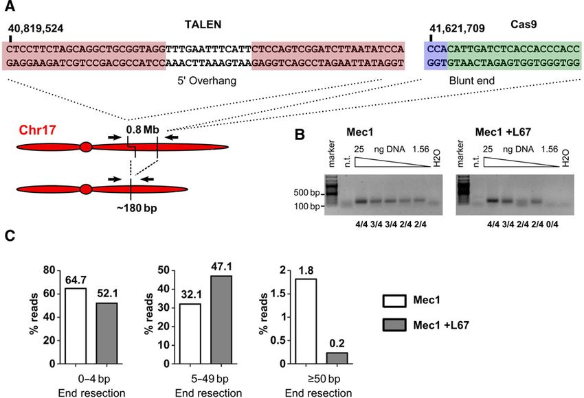
Published OnlineFirst December 8, 2017; DOI: 10.1158/1541-7786.MCR-17-0373

                                                                                           Aberrant DNA Break Repair in CLL

Figure 3.
Deletions at repair junctions. A, The most frequent
repair junction for individual substrates is indicated.
Microhomologies (1 bp) used for repair are
indicated in red. B, Graphs show the occurrence of
deletions at repaired substrates as horizontal lines.
y-axis gives the frequency of deletions and x-axis
indicates the position of the deletion within the
amplicon. C, All sequences obtained for the respective
sample cohort were analyzed on the basis
of deleted sequence lengths. Box plots show the
median length of deletions within the 25th and 75th
percentile (box) and data within a 1.5 times
interquartile distance from the median as whiskers.
Outliers (all other data points) are indicated as circles.

www.aacrjournals.org                                                                       Mol Cancer Res; 16(3) March 2018   433

    Downloaded from mcr.aacrjournals.org on February 7, 2021. © 2018 American Association for Cancer Research.
Published OnlineFirst December 8, 2017; DOI: 10.1158/1541-7786.MCR-17-0373
Gassner et al.
Figure 4.
Microhomologies at repair junctions. A,
Ratio of repair junctions deriving from
microhomology mediated joining (6 bp
microhomology, MH) versus direct
joining of substrate #2 from transfected
CLL and B-cell samples. Asterisks
indicate significant difference between
CLL and B cells (P ¼ 0.017 Mann–
Whitney test). B, The most frequently
used sequence for each of all individual
inter- and intramolecular repair events
are depicted as circos plots for CLL cells
and for normal B cells. Circos plots were
generated as indicated for Fig. 2
(sequences shown in Supplementary
Table S5). C, Repair junctions that were
significantly overrepresented in CLL (P <
0.05 from B) are schematically depicted.
The hash mark specifies the respective
forward-primed (red) and reverse-
primed (yellow) DNA substrate. Arrows
indicate gap filling by DNA polymerases.
GFP expression based on direct joining of pGFP-1/pGFP-2 was dantly expressed than in normal B cells (Supplementary Fig. S4).
analyzed in the presence of specific inhibitors targeting PARP We positively sorted transfected cells based on GFP expression
(olaparib), LIG1/3 (L67), and DNA-PKc (NU7441) as control. To from the Cas9 construct and estimated chromosomal deletion
normalize for transfection efficiencies, we cotransfected a plasmid frequencies of a large 0.8 Mb fragment by PCR on serially diluted
encoding mCherry (pmCherry). As expected, direct repair of the input DNA isolated from sorted cells (Fig. 6A and B). As estimated
GFP plasmids was strongly impeded by inhibition of DNA-PKcs, from four independent experiments, the chromosomal deletion
which are central to c-NHEJ. To a lesser extent also, inhibition of frequency of L67-treated cells was lower compared with untreated
LIG1/3 showed an effect on direct pGFP-1/pGFP-2 joining, while cells, as amplification of breakpoint junctions failed from 1.56 ng
inhibition of PARP family proteins by olaparib had no effect on input DNA of L67 treated cells in all experiments, whereas in two
DNA repair in this assay (Fig. 5C). of four experiments, PCR-amplified breakpoint junctions were
detectable in untreated cells at that DNA concentration (Fig. 6B).
Amplicon sequencing of the PCR products from two experiments
Induction of chromosomal deletions in the CLL cell line Mec1 is revealed that L67-treated Mec1 cells had an increased frequency of
impaired upon inhibition of LIG1/3 short (5–49 bp) deletions at the breakpoint junction, whereas
Finally, we tested whether LIG1 or XRCC1/LIG3 is involved in large (50 bp) deletions were reduced (Fig. 6C), again pointing to
the generation of chromosomal deletions between a 50 ; overhang involvement of LIG1/3 in the occurrence of large deletions at
DNA DSB and a blunt end DNA DSB in CLL. Therefore, we repair joints. Small (0–4 bp) deletions, likely resulting from direct
transiently transfected the CLL cell line Mec1 with constructs joining or deletions within the 50 ; overhang were also reduced in
expressing TALENs (generating a 50 ;overhang) and Cas9 (gener- L67-treated cells (Fig. 6C).
ating blunt end DSBs), which both cleave at chromosome 17, with Thus, we infer that the described overexpression of LIG1 or
or without treatment with 30 mmol/L LIG1/3 inhibitor L67 (Fig. XRCC1 in CLL could explain the observed bias toward both,
6A). Of note, real-time RT-PCR revealed that in Mec1 cells, MMEJ- MMEJ as well as increased repair of incompatible DNA ends with
specific factors LIG1, XRCC1, and PARP1 were also more abun- increased deletions at repair junctions.
434 Mol Cancer Res; 16(3) March 2018 Molecular Cancer Research
Downloaded from mcr.aacrjournals.org on February 7, 2021. © 2018 American Association for Cancer Research.Published OnlineFirst December 8, 2017; DOI: 10.1158/1541-7786.MCR-17-0373
Aberrant DNA Break Repair in CLL
Figure 5.
DNA DSB repair of nonresected DNA substrates. A, Expression values for factors involved in MMEJ (left) and c-NHEJ (right) are shown as fold change between
CLL and B lymphocytes. Gray shading, >2-fold differences. Significances are indicated above each bar (CLL n ¼ 100; B cells n ¼ 11). B, Schematic representation of
plasmids (GFP-1 and GFP-2) used to detect intermolecular repair of blunt/50 overhang junctions. Direct intermolecular repair of nonresected plasmids
leads to GFP expression. C, 293 cells were transfected using plasmids GFP-1 and GFP-2 shown in B together with a plasmid expressing mCherry. Representative FACS
plots show GFP/mCherry expression in the presence of inhibitors. Data within plots give percentage of GFPþ cells within all mCherryþ cells. Graph shows
the results from three independent FACS experiments. UTR1, untreated cells transfected using GeneJuice (controls for NU7441, olaparib-treated cells). UTR2,
untreated cells transfected using nucleofection (controls for L67-treated cells). Neg, cells transfected with mCherry plasmids only. Significant differences in
comparison with untreated controls is indicated above the respective dataset (two-tailed t test with unequal variances).
Discussion revealed a complex set of substantial differences in efficiency as
well as precision of DNA repair in CLL versus B cells. In addition to
Aberrant repair of DSB can lead to genetic changes, driving
the frequent occurrence of deletions at the repair junction, which
carcinogenesis and clonal evolution of cancer. Here, we describe
was already described for leukemic cells (30), our assay revealed
several differences of DSB repair in B cells versus CLL cells by
that noncohesive protruding ends were joined significantly more
applying a plasmid-based repair assay analyzed by NGS as well as
efficiently in CLL cells compared with B cells.
by assessing repair of TALEN/Cas9–induced chromosomal dele-
In our study, efficient joining of incompatible ends was attrib-
tions in the Mec1 cell line. Although plasmid-based assays have
uted to increased MMEJ activity, reflected by the presence of short
been used in the past to investigate DSB repair in cell lines and
microhomologies within repair junctions of end resected incom-
cancer cells (27, 30, 31), our NGS-based approach allowed
patible DNA substrates. Concurrently, a 6-bp microhomology
simultaneous analysis of efficiency and accuracy of DSB repair
was preferentially used in CLL cells for repair of DNA substrates.
in regard to diverse DNA break structures and the cellular context
These results indicate that DNA DSB repair is skewed toward
at single-nucleotide level. In general, our assay revealed that
MMEJ in CLL, which is corroborated by overexpression of MMEJ-
complementary 50 ; protruding ends as well as blunt ends exhib-
specific factors in CLL. Notably, this skewing toward MMEJ does
ited highest repair efficiency and 30 ; cohesive and 30 ; noncohesive
not result from cell-cycle differences, as nucleofected CLL cells in
staggered ends showing weakest joining efficiency. Weak repair of
our analysis were also G1 arrested. We further observed CLL-
30 ; overhangs was noted in earlier studies (31), probably because
specific increased joining of incompatible DNA ends independent
30 ; overhangs rather promote HR as they are required for strand
from end resection and usage of microhomologies. Strikingly, this
exchange reaction in HR (32, 33). However, our repair assay
increased joining efficiency was especially pronounced in
www.aacrjournals.org Mol Cancer Res; 16(3) March 2018 435
Downloaded from mcr.aacrjournals.org on February 7, 2021. © 2018 American Association for Cancer Research.Published OnlineFirst December 8, 2017; DOI: 10.1158/1541-7786.MCR-17-0373
Gassner et al.
Figure 6.
Induction of chromosomal deletions by TALEN/Cas9 generated DNA DSBs are dependent on LIG1/3. A, Schematic representation of chromosomal deletions induced
by TALENs/Cas9 constructs. Binding sites of TALENs and Cas9 constructs are shaded red and green, respectively. Genomic position and sequence of binding
sites are shown in 50 -30 orientation. The protospacer adjacent motif is colored blue. Specific primers for amplification of breakpoint junctions are given
as black arrows on chromosome 17, yielding an approximate 180-bp PCR product in case a 0.8 Mb fragment of chr17 is deleted. B, Limiting dilution of genomic DNA to
estimate the frequency of chromosomal deletions after expression of TALEN/Cas9 constructs in Mec1 cells treated with 30 mmol/L LIG1/3 inhibior L67 or
untreated controls. PCR amplification was performed with serial dilutions of genomic DNA from four independent experiments as template (25, 12.5, 6.25, 3.125, and
1.56 ng). An agarose gel from one experiment is shown. The number of times a PCR fragment was detected from four independent experiments is indicated
below the gel. n.t., Mec1 cells not transfected with TALEN/Cas9 constructs. C, PCR products from two experiments were amplicon sequenced and fragment lengths
were analyzed (Mec1: 1,191,602 reads; Mec1 L67 treated: 177,319 reads). The percentage of reads with the respective amount of end resection is depicted
in separate graphs, demonstrating the occurrence of about 8 times more fragments with large deletions (50 bp) at breakpoint junctions. Percentages are indicated
above the bars (reads with insertions at breakpoints are not depicted).
intermolecular repair events, indicating a somehow increased increased MMEJ but also to direct joining of incompatible DNA
promiscuity in DNA end joining. Although MMEJ classically ends. LIG4-deficient cell lines are still able to repair DNA DSBs,
involves end resection of DNA overhangs after stabilization of albeit the number of direct joints are decreased, and microho-
the paired homologous region by several nucleases (34, 35), mology usage is increased due to activity of LIG1/3 (41, 42).
direct joining of incompatible protruding ends can be mediated Although sole activity of LIG1/3 in LIG4-deficient cells led to a
by two possibilities: either DNA ends are blunted prior to DNA higher frequency of endonuclease-induced translocations in
joining (refilled or resected) or otherwise DNA is resynthesised rodent cell lines, the frequency in human cell lines was decreased,
across a stabilized DNA junction of two staggered DNA ends by showing that efficiency and kinetics of DNA DSB repair is likely
polymerase m or l, followed by ligation of DNA nicks (36–38). depending on cell type and species specific expression levels of
Although ligation of blunted dsDNA ends is normally mediated repair factors (41, 43). Our own data suggest that LIG1/3 may
by LIG4/XRCC4, nick sealing is efficiently performed by LIG1 and significantly contribute to the formation of TALEN/Cas9–induced
LIG3/XRCC1 complexes (39, 40). Our pGFP-1/pGFP-2 repair large chromosomal deletions in the CLL cell line Mec1 as we
assay revealed that although most repair events are catalyzed by observed a reduction in their occurrence upon LIG1/3 inhibition,
c-NHEJ involving DNA-PKcs, repair could partly be mediated by together with a reduction in large deletions at repair joints. Hence,
polymerase-dependent DNA synthesis across direct pGFP-1/ our results would propose that CLL-specific increased expression
pGFP-2 junctions followed by nick-sealing by LIG1/3. In that levels of the MMEJ factors LIG1 and XRCC1 could contribute to
way, overexpression of LIG1/3 could not only contribute to increased propensity to acquire chromosomal aberrations during
436 Mol Cancer Res; 16(3) March 2018 Molecular Cancer Research
Downloaded from mcr.aacrjournals.org on February 7, 2021. © 2018 American Association for Cancer Research.Published OnlineFirst December 8, 2017; DOI: 10.1158/1541-7786.MCR-17-0373
Aberrant DNA Break Repair in CLL
DNA DSB repair. This fits with the observation that chromosomal LIG1/3 inhibitors during CLL treatment would not only potentiate
aberrations are frequently found to be subclonal in CLL, pointing cytotoxicity of DNA-damaging agents (50) but might also impede
to their successive generation during disease progression (6). In clonal evolution by acquisition of novel genomic rearrangements.
addition, an increased MMEJ activity could also explain the
formation of small deletions and microdeletions frequently Disclosure of Potential Conflicts of Interest
observed in CLL (44) and also in other cancer entities (45). These No potential conflicts of interest were disclosed.
deletions often exhibit sequence homologies at breakpoint junc-
tions, suggesting that they likely arise due to MMEJ of DNA DSBs. Authors' Contributions
Alternatively, they could occur by polymerase slippage during Conception and design: S. Rebhandl, R. Geisberger
replication (46, 47). As we observed several differences in end Development of methodology: M. Schubert, S. Rebhandl, S. Blaimer,
joining between CLL and B cells, it would be certainly interesting R. Geisberger
to screen DNA repair quality/quantity in therapy-sensitive versus Acquisition of data (provided animals, acquired and managed patients,
provided facilities, etc.): F.J. Gassner, M. Schubert, S. Rebhandl, N. Zaborsky,
refractory samples as well as in other clinically relevant CLL
S. Blaimer, R. Greil, R. Geisberger
subsets (such as samples with complex karyotype). Deriano and Analysis and interpretation of data (e.g., statistical analysis, biostatistics,
colleagues already reported increased error-prone end joining of computational analysis): F.J. Gassner, M. Schubert, S. Rebhandl, N. Zaborsky,
DNA ends in CLL cells obtained from a therapy-resistant patient K. Catakovic, D. Hebenstreit, R. Greil, R. Geisberger
(48). Hence, it is conceivable that CLL subset or risk group– Writing, review, and/or revision of the manuscript: F.J. Gassner, M. Schubert,
specific differences in DNA DSB repair could contribute to diverse S. Rebhandl, N. Zaborsky, D. Hebenstreit, R. Greil, R. Geisberger
Administrative, technical, or material support (i.e., reporting or organizing
clinical outcomes.
data, constructing databases): F.J. Gassner, M. Schubert, S. Rebhandl,
Although a portion of CLL patients exhibits mutations at DNA K. Spandl, N. Zaborsky, K. Catakovic, R. Geisberger
repair genes (6), our data rather suggest an inherently aberrant Study supervision: R. Geisberger
DNA DSB repair activity in CLL due to different expression levels
of key factors for MMEJ, in particular as we excluded del(11q) and Acknowledgments
del(17p) patients (harboring deleted ATM and TP53 genes) and This work was supported by the SCRI-LIMCR, the Province of Salzburg,
as all samples included in our analyses showed a very uniform the City of Salzburg, and grants from the Austrian Science Fund FWF to
pattern of repair junction formation, irrespective of the BCR R. Geisberger (FWF; P24619 and P28201).
mutation status. The authors thank Oliver March (Salzburg) for providing TALENs and
Markus Steiner (Salzburg) for cell sorting.
Summarizing, we could identify profound differences in effi-
ciency and precision of DSB repair between CLL cells and normal B
The costs of publication of this article were defrayed in part by the payment
cells. Considering a reported increased formation of DNA DSBs in of page charges. This article must therefore be hereby marked advertisement
precancerous lesions and cancers (49), their imprecise repair due to in accordance with 18 U.S.C. Section 1734 solely to indicate this fact.
increased LIG1 and XRCC1 levels could thus continuously enhance
the formation of genomic aberrations, contributing to clonal Received July 13, 2017; revised September 28, 2017; accepted November 9,
evolution and chemoresistance. Hence, concomitant usage of 2017; published OnlineFirst December 8, 2017.
References
1. Alexandrov LB, Nik-Zainal S, Wedge DC, Aparicio SA, Behjati S, Biankin AV, 10. Kent T, Chandramouly G, McDevitt SM, Ozdemir AY, Pomerantz RT.
et al. Signatures of mutational processes in human cancer. Nature 2013; Mechanism of microhomology-mediated end-joining promoted by
500:415–21. human DNA polymerase theta. Nat Struct Mol Biol 2015;22:230–7.
2. Stephens PJ, McBride DJ, Lin ML, Varela I, Pleasance ED, Simpson JT, et al. 11. Meyer D, Fu BX, Heyer WD. DNA polymerases delta and lambda cooperate
Complex landscapes of somatic rearrangement in human breast cancer in repairing double-strand breaks by microhomology-mediated end-
genomes. Nature 2009;462:1005–10. joining in Saccharomycescerevisiae. Proc Natl Acad Sci U S A 2015;112:
3. Stephens PJ, Greenman CD, Fu B, Yang F, Bignell GR, Mudie LJ, et al. E6907–E6916.
Massive genomic rearrangement acquired in a single catastrophic event 12. Hamblin TJ, Davis Z, Gardiner A, Oscier DG, Stevenson FK. Unmutated Ig V
during cancer development. Cell 2011;144:27–40. (H) genes are associated with a more aggressive form of chronic lympho-
4. Burger JA, Landau DA, Taylor-Weiner A, Bozic I, Zhang H, Sarosiek K, cytic leukemia. Blood 1999;94:1848–54.
et al. Clonal evolution in patients with chronic lymphocytic leukae- 13. Hartmann TN, Pleyer L, Desch P, Egle A, Greil R. Novel therapeutics
mia developing resistance to BTK inhibition. Nat Commun 2016;7: approaches to chronic lymphocytic leukemia based on recent biological
11589. insights. Discov Med 2009;8:157–64.
5. Landau DA, Tausch E, Taylor-Weiner AN, Stewart C, Reiter JG, Bahlo J, et al. 14. Pleyer L, Egle A, Hartmann TN, Greil R. Molecular and cellular mechan-
Mutations driving CLL and their evolution in progression and relapse. isms of CLL: novel therapeutic approaches. Nat Rev Clin Oncol 2009;
Nature 2015;526:525–30. 6:405–18.
6. Landau DA, Carter SL, Stojanov P, McKenna A, Stevenson K, Lawrence MS, 15. Rowley JD.Chromosomal translocations: revisited yet again. Blood 2008;
et al. Evolution and impact of subclonal mutations in chronic lymphocytic 112:2183–9.
leukemia. Cell 2013;152:714–26. 16. Dicker F, Schnittger S, Haferlach T, Kern W, Schoch C. Immunostimulatory
7. Lieber MR. The mechanism of double-strand DNA break repair by the oligonucleotide-induced metaphase cytogenetics detect chromosomal
nonhomologous DNA end-joining pathway. Annu Rev Biochem 2010; aberrations in 80% of CLL patients: a study of 132 CLL cases with
79:181–211. correlation to FISH, IgVH status, and CD38 expression. Blood 2006;
8. Lieber MR, Gu J, Lu H, Shimazaki N, Tsai AG. Nonhomologous DNA end 108:3152–60.
joining (NHEJ) and chromosomal translocations in humans. Subcell 17. Haferlach C, Dicker F, Schnittger S, Kern W, Haferlach T. Comprehensive
Biochem 2010;50:279–96. genetic characterization of CLL: a study on 506 cases analysed with
9. Chiruvella KK, Liang Z, Wilson TE. Repair of double-strand breaks by end chromosome banding analysis, interphase FISH, IgV(H) status and immu-
joining. Cold Spring Harb Perspect Biol 2013;5:a012757. nophenotyping. Leukemia 2007;21:2442–51.
www.aacrjournals.org Mol Cancer Res; 16(3) March 2018 437
Downloaded from mcr.aacrjournals.org on February 7, 2021. © 2018 American Association for Cancer Research.Published OnlineFirst December 8, 2017; DOI: 10.1158/1541-7786.MCR-17-0373
Gassner et al.
18. Puente XS, Pinyol M, Quesada V, Conde L, Ordonez GR, Villamor N, et al. share the initial end resection step to repair DNA double-strand
Whole-genome sequencing identifies recurrent mutations in chronic lym- breaks in mammalian cells. Proc Natl Acad Sci U S A 2013;110:
phocytic leukaemia. Nature 2011;475:101–5. 7720–5.
19. Puente XS, Bea S, Valdes-Mas R, Villamor N, Gutierrez-Abril J, Martin- 36. Smith J, Baldeyron C, De OI, Sala-Trepat M, Papadopoulo D. The influence
Subero JI, et al. Non-coding recurrent mutations in chronic lymphocytic of DNA double-strand break structure on end-joining in human cells.
leukaemia. Nature 2015;526:519–24. Nucleic Acids Res 2001;29:4783–92.
20. Kim H, Kim JS. A guide to genome engineering with programmable 37. Mahajan KN, Nick McElhinny SA, Mitchell BS, Ramsden DA. Associa-
nucleases. Nat Rev Genet 2014;15:321–34. tion of DNA polymerase mu (pol mu) with Ku and ligase IV: role for pol
21. Stacchini A, Aragno M, Vallario A, Alfarano A, Circosta P, Gottardi D, mu in end-joining double-strand break repair. Mol Cell Biol 2002;
et al. MEC1 and MEC2: two new cell lines derived from B-chronic 22:5194–202.
lymphocytic leukaemia in prolymphocytoid transformation. Leuk Res 38. Pryor JM, Waters CA, Aza A, Asagoshi K, Strom C, Mieczkowski PA, et al.
1999;23:127–36. Essential role for polymerase specialization in cellular nonhomologous
22. Tinhofer I, Rubenzer G, Holler C, Hofstaetter E, Stoecher M, Egle A, et al. end joining. Proc Natl Acad Sci U S A 2015;112:E4537–45.
Expression levels of CD38 in T cells predict course of disease in male 39. Cannan WJ, Rashid I, Tomkinson AE, Wallace SS, Pederson DS. The
patients with B-chronic lymphocytic leukemia. Blood 2006;108:2950–6. Human ligase IIIalpha-XRCC1 protein complex performs DNA nick
23. Bolger AM, Lohse M, Usadel B. Trimmomatic: a flexible trimmer for repair after transient unwrapping of nucleosomal DNA. J Biol Chem
Illumina sequence data. Bioinformatics 2014;30:2114–20. 2017;292:5227–38.
24. Magoc T, Salzberg SL. FLASH: fast length adjustment of short reads to 40. Chafin DR, Vitolo JM, Henricksen LA, Bambara RA, Hayes JJ. Human
improve genome assemblies. Bioinformatics 2011;27:2957–63. DNA ligase I efficiently seals nicks in nucleosomes. EMBO J 2000;19:
25. Katoh K, Standley DM. MAFFT multiple sequence alignment software 5492–501.
version 7: improvements in performance and usability. Mol Biol Evol 41. Ghezraoui H, Piganeau M, Renouf B, Renaud JB, Sallmyr A, Ruis B, et al.
2013;30:772–80. Chromosomal translocations in human cells are generated by canonical
26. Mao Z, Bozzella M, Seluanov A, Gorbunova V. Comparison of nonho- nonhomologous end-joining. Mol Cell 2014;55:829–42.
mologous end joining and homologous recombination in human cells. 42. Masani S, Han L, Meek K, Yu K. Redundant function of DNA ligase 1 and 3
DNA Repair 2008;7:1765–71. in alternative end-joining during immunoglobulin class switch recombi-
27. Verkaik NS, Esveldt-van Lange RE, van HD, Bruggenwirth HT, Hoeijmakers nation. Proc Natl Acad Sci U S A 2016;113:1261–6.
JH, Zdzienicka MZ, et al. Different types of V(D)J recombination and end- 43. Lu G, Duan J, Shu S, Wang X, Gao L, Guo J, et al. Ligase I and ligase III
joining defects in DNA double-strand break repair mutant mammalian mediate the DNA double-strand break ligation in alternative end-joining.
cells. Eur J Immunol 2002;32:701–9. Proc Natl Acad Sci U S A 2016;113:1256–60.
28. Haslinger C, Schweifer N, Stilgenbauer S, Dohner H, Lichter P, Kraut N, 44. Marinelli M, Peragine N, Di Maio V, Chiaretti S, De Propris MS, Raponi S,
et al. Microarray gene expression profiling of B-cell chronic lymphocytic et al. Identification of molecular and functional patterns of p53 alterations
leukemia subgroups defined by genomic aberrations and VH mutation in chronic lymphocytic leukemia patients in different phases of the disease.
status. J Clin Oncol 2004;22:3937–49. Haematologica 2013;98:371–5.
29. Rhodes DR, Yu J, Shanker K, Deshpande N, Varambally R, Ghosh D, et al. 45. Rothenberg SM, Mohapatra G, Rivera MN, Winokur D, Greninger P, Nitta
ONCOMINE: a cancer microarray database and integrated data-mining M, et al. A genome-wide screen for microdeletions reveals disruption of
platform. Neoplasia 2004;6:1–6. polarity complex genes in diverse human cancers. Cancer Res 2010;70:
30. Gaymes TJ, Mufti GJ, Rassool FV. Myeloid leukemias have increased activity 2158–64.
of the nonhomologous end-joining pathway and concomitant DNA mis- 46. Verdin H, D'haene B, Beysen D, Novikova Y, Menten B, Sante T, et al.
repair that is dependent on the Ku70/86 heterodimer. Cancer Res 2002; Microhomology-mediated mechanisms underlie non-recurrent disease-
62:2791–7. causing microdeletions of the FOXL2 gene or its regulatory domain. PLoS
31. Poplawski T, Pastwa E, Blasiak J. Non-homologous DNA end joining in Genet 2013;9:e1003358.
normal and cancer cells and its dependence on break structures. Genet Mol 47. Watson CT, Marques-Bonet T, Sharp AJ, Mefford HC. The genetics of
Biol 2010;33:368–73. microdeletion and microduplication syndromes: an update. Annu Rev
32. Broderick R, Nieminuszczy J, Baddock HT, Deshpande RA, Gileadi O, Paull Genomics Hum Genet 2014;15:215–44.
TT, et al. EXD2 promotes homologous recombination by facilitating DNA 48. Deriano L, Merle-Beral H, Guipaud O, Sabatier L, Delic J. Mutagenicity
end resection. Nat Cell Biol 2016;18:271–80. of non-homologous end joining DNA repair in a resistant subset of
33. Liu T, Huang J. DNA end resection: facts and mechanisms. Genomics human chronic lymphocytic leukaemia B cells. Br J Haematol 2006;
Proteomics Bioinformatics 2016;14:126–30. 133:520–5.
34. Davis AJ, Chen DJ. DNA double strand break repair via non-homologous 49. Halazonetis TD, Gorgoulis VG, Bartek J. An oncogene-induced DNA
end-joining. Transl Cancer Res 2013;2:130–43. damage model for cancer development. Science 2008;319:1352–5.
35. Truong LN, Li Y, Shi LZ, Hwang PY, He J, Wang H, et al. Micro- 50. Tomkinson AE, Howes TR, Wiest NE. DNA ligases as therapeutic targets.
homology-mediated end joining and homologous recombination Transl Cancer Res 2013;2:1219.
438 Mol Cancer Res; 16(3) March 2018 Molecular Cancer Research
Downloaded from mcr.aacrjournals.org on February 7, 2021. © 2018 American Association for Cancer Research.Published OnlineFirst December 8, 2017; DOI: 10.1158/1541-7786.MCR-17-0373
Imprecision and DNA Break Repair Biased towards Incompatible
End Joining in Leukemia
Franz Josef Gassner, Maria Schubert, Stefan Rebhandl, et al.
Mol Cancer Res 2018;16:428-438. Published OnlineFirst December 8, 2017.
Updated version Access the most recent version of this article at:
doi:10.1158/1541-7786.MCR-17-0373
Supplementary Access the most recent supplemental material at:
Material http://mcr.aacrjournals.org/content/suppl/2017/12/08/1541-7786.MCR-17-0373.DC1
Cited articles This article cites 50 articles, 18 of which you can access for free at:
http://mcr.aacrjournals.org/content/16/3/428.full#ref-list-1
E-mail alerts Sign up to receive free email-alerts related to this article or journal.
Reprints and To order reprints of this article or to subscribe to the journal, contact the AACR Publications Department at
Subscriptions pubs@aacr.org.
Permissions To request permission to re-use all or part of this article, use this link
http://mcr.aacrjournals.org/content/16/3/428.
Click on "Request Permissions" which will take you to the Copyright Clearance Center's (CCC)
Rightslink site.
Downloaded from mcr.aacrjournals.org on February 7, 2021. © 2018 American Association for Cancer Research.You can also read