Successful gene therapy of Diamond-Blackfan anemia in a mouse model and human CD34+ cord blood hematopoietic stem cells using a clinically ...
←
→
Page content transcription
If your browser does not render page correctly, please read the page content below
Successful gene therapy of Diamond-Blackfan anemia
in a mouse model and human CD34+ cord blood
hematopoietic stem cells using a clinically applicable
lentiviral vector

by Yang Liu, Maira Dahl, Shubhranshu Debnath, Michael Rothe, Emma M. Smith, Tan Hooi
Min Grahn, Sarah Warsi, Jun Chen, Johan Flygare, Axel Schambach, and Stefan Karlsson

Haematologica 2021 [Epub ahead of print]

Citation: Yang Liu, Maira Dahl, Shubhranshu Debnath, Michael Rothe, Emma M. Smith, Tan Hooi
Min Grahn, Sarah Warsi, Jun Chen, Johan Flygare, Axel Schambach, and Stefan Karlsson.
Successful gene therapy of Diamond-Blackfan anemia in a mouse model and human CD34+ cord blood
hematopoietic stem cells using a clinically applicable lentiviral vector.
Haematologica. 2021; 106:xxx
doi:10.3324/haematol.2020.269142

Publisher's Disclaimer.
E-publishing ahead of print is increasingly important for the rapid dissemination of science.
Haematologica is, therefore, E-publishing PDF files of an early version of manuscripts that
have completed a regular peer review and have been accepted for publication. E-publishing
of this PDF file has been approved by the authors. After having E-published Ahead of Print,
manuscripts will then undergo technical and English editing, typesetting, proof correction and
be presented for the authors' final approval; the final version of the manuscript will then
appear in print on a regular issue of the journal. All legal disclaimers that apply to the
journal also pertain to this production process.
Successful gene therapy of Diamond-Blackfan anemia in a mouse model and human CD34+
cord blood hematopoietic stem cells using a clinically applicable lentiviral vector

Yang Liu1,*, Maria Dahl1, Shubhranshu Debnath1, Michael Rothe2, Emma M. Smith1, Tan Hooi Min
Grahn1, Sarah Warsi1, Jun Chen1, Johan Flygare1, Axel Schambach2,3, Stefan Karlsson1,*

1. Molecular Medicine and Gene Therapy, Lund Stem Cell Center, Lund University, Lund 22184,
Sweden;
2. Institute of Experimental Hematology, Hannover Medical School, Hannover 30625, Germany;
3. Division of Hematology/Oncology, Boston Children’s Hospital, Harvard Medical School, Boston,
USA
*Corresponding author. Email: stefan.karlsson@med.lu.se; yang.liu@med.lu.se

                                                                                             1
ABSTRACT
Diamond-Blackfan anemia (DBA) is an inherited bone marrow failure disorder with pure red blood
cell aplasia associated with physical malformations and a predisposition to cancer. Twenty-five
percent of patients with DBA have mutations in a gene encoding ribosomal protein S19 (RPS19).
Our previous proof-of-concept studies demonstrated that DBA phenotype could be successfully
treated using lentiviral vectors in Rps19-deficient DBA mice. In our present study, we developed a
clinically applicable single gene self-inactivating lentiviral vector, containing the human RPS19
cDNA driven by the human elongation factor 1α short promoter, that can be used for clinical gene
therapy development for RPS19-deficient DBA. We examined the efficacy and safety of the vector
in a Rps19-deficient DBA mouse model and in human primary RPS19-deficient CD34+ cord blood
cells. We observed that transduced Rps19-deficient bone marrow cells could reconstitute mice long-
term and rescue the bone marrow failure and severe anemia observed in Rps19-deficient mice, with a
low risk of mutagenesis and a highly polyclonal insertion site pattern. More importantly, the vector
can also rescue impaired erythroid differentiation in human primary RPS19-deficient CD34+ cord
blood hematopoietic stem cells. Collectively, our results demonstrate the efficacy and safety of using
a clinically applicable lentiviral vector for the successful treatment of Rps19-deficient DBA in a
mouse model and in human primary CD34+ cord blood cells. These findings show that this vector
can be used to develop clinical gene therapy for RPS19-deficient DBA patients.

INTRODUCTION
DBA is a congenital bone marrow (BM) failure disorder with erythroid hypoplasia that presents early
in infancy1-3. The classic hematologic profile of DBA consists of macrocytic anemia with
reticulocytopenia, normal or decreased levels of neutrophils, and a variable platelet count3, 4.
Additionally, patients with DBA can also manifest with non-hematological symptoms such as
physical abnormalities and cancer predisposition2, 5.
The majority of DBA cases (60-70%) are caused by heterozygous loss-of-function mutations in
genes coding for ribosomal proteins (RP), resulting in functional RP haploinsufficiency2. Recent
studies have also identified mutations in erythroid transcriptional factors GATA1 and TSR2 (a direct
binding partner of RPS26) as a cause of the DBA phenotype6-8. In addition, mutations in RPS19 are
the most common among patients with putative causal mutations, contributing to over 25% of cases2.
Corticosteroids are the main therapeutic option for DBA patients, with more than 80% of the subjects
responding well during early stages of treatment. However, half of these patients become non-
responsive to corticosteroids therapy over prolonged treatment and have to be treated with blood
transfusions9. Importantly, none of the currently available treatments are curative, and there are often

                                                                                                      2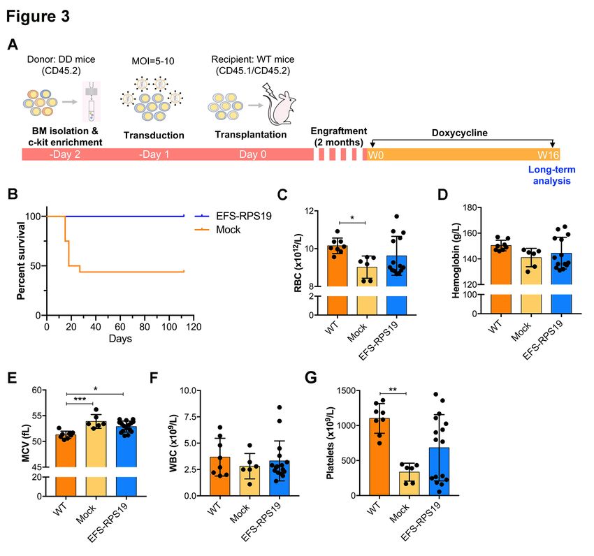
serious complications accompanying the treatments1,        2, 10
                                                                   . Hematopoietic stem cell (HSC)
transplantation is currently the sole curative option for the treatment of DBA. This treatment is ,
however, limited by the availability of suitable donors and the potential for serious immunological
complications11. Gene therapy, using gene-corrected HSCs, would be a potential alternative
therapeutic strategy as highlighted in our previous studies12-16. For clinical applications using this
approach, the efficiency of transgene expression and safety aspects, including potential insertional
mutagenesis, should be addressed17-19. Our group recently demonstrated that correction of Rps19-
deficient BM cells using lentiviral vectors containing a clinically relevant promoter could rescue BM
failure and defects in erythroid development, whilst exhibiting limited risk of insertional
mutagenesis12.
In our previous study, we utilized a lentiviral vector containing the RPS19 gene as well as a GFP
marker. We have subsequently designed a clinically applicable single gene lentiviral self-inactivating
(SIN) vector for the clinical development of gene therapy for Rps19-deficient DBA patients. This
vector harbors a codon-optimized human RPS19 cDNA driven by the short human elongation factor
1alpha promoter and lacks a fluorescent marker. In this study, we demonstrate that this vector can
rescue the anemia and lethal BM failure observed in mouse models of Rps19-deficient DBA, with a
low risk insertion profile and no evidence of clonal expansion associated with vector integration near
cancer-associated genes. We also observed the rescue of impaired erythroid differentiation in human
RPS19-deficient CD34+ cord blood cells treated with this vector. Our results demonstrate the
feasibility and preclinical efficacy for treatment of RPS19-deficient DBA using a clinically
applicable SIN lentiviral vector, which opens the possibility for the development of clinical gene
therapy for RPS19-deficient DBA patients.

METHODS
Lentiviral vector constructs
                                                                                        20
The SIN lentiviral vector is derived from the pRRL.PPT.PGK. vector backbone                  . A codon-
                                                                        21
optimized human RPS19 cDNA was designed as described previously and inserted downstream of
the EFS promoter. Lentiviral vectors were produced by the Vector Unit at Lund University as
previously described12.

Mice and transplantations
Mice were maintained at the Lund University animal facility and all animal experiments were
approved by the Lund University animal ethics committee. The homozygous doxycycline-inducible

                                                                                                      3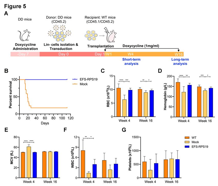
Rps19-deficient mouse model used in the study was established as previously described15. A detailed
description of transplantations is provided in the Online Supplementary Methods.
Transduction
c-kit+ or lineage negative (Lin-) cells isolated from BM of transgenic mice were enriched by using
CD117 or Lin- microbeads and magnetic-activated cell sorting separation columns (all from Miltenyi
Biotec) according to manufacturer’s protocol. After enrichment, cells were pre-stimulated for 24
hours in StemSpan serum-free expansion medium (Stem Cell Technologies), supplemented with
penicillin/streptomycin (Gibco), murine stem cell factor (SCF, 100 ng/mL; PeproTech), and human
thrombopoietin (50 ng/mL; PeproTech) in six-well plates at the concentration of 0.5x10⁶ cells/mL.
For transduction, retronectin-coated (20 ng/mL; Takara) 12-well plates were preloaded with the viral
vectors (MOI=5-10) and followed by seeding of 0.5 million cells into each well that filled with 1 mL
pre-stimulation medium.

Flow cytometry
A complete description of all antibodies used is listed in the Online Supplementary Methods.

Human primary cord blood cells and erythroid differentiation
Human cord blood samples were obtained from the maternity wards of Helsingborg General Hospital
and Skåne University Hospital in Lund and Malmö, Sweden, after informed, written consent
according to guidelines approved by the regional ethical committee. Mononuclear cells were
separated through density-gradient centrifugation. CD34+ cells were magnetically isolated according
to manufacturing description (Milteny Biotec, Cat. 130-046-702). Cells were cultured in serum-free
expansion medium (SFEM, Stem Cell Technologies), supplemented with human SCF (hSCF),
thrombopoietin (TPO), and FLT3-ligand (FLT3L) at 100 ng/ml from Peprotech. Full descriptions of
transduction and erythroid differentiation are provided in the Online Supplementary Methods.

Other experimental details
Full descriptions of qRT-PCR, determination of transduction efficiency and vector copy number
(VCN) measurements are provided in the Online Supplementary Methods.

Insertion site analysis
Whole BM cells were isolated at 16 weeks after transplantation. Genomic DNA was isolated from
the BM of flushed femurs using the DNA Blood & Tissue kit (Qiagen). Vector-genome junction was

                                                                                                  4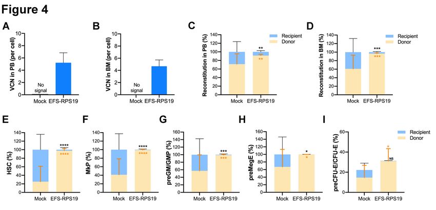
amplified using the INtegration Site PIpeline for paIRed-End reaDs (INSPIIRED) workflow as
described by Sherman and colleagues24.

Statistical analysis
T-test and One-way ANOVA with the Tukey multiple-comparison test were used to determine
statistical significance using GraphPad Prism (version 6; GraphPad Software).

RESULTS
High transduction efficiency of the EFS-RPS19 vector
We first studied the transduction efficiency of the vector in BM progenitor cells isolated from our
established Rps19-deficient DBA mouse model15. This model contains the Rps19-targeting shRNA
expressed under a doxycycline-responsive promoter located downstream of the collagen A1 gene
(Figure 1A). Experimental animals were bred to be either heterozygous (D/+) or homozygous (D/D)
for the shRNA to generate two models with intermediate or severe Rps19 deficiency (Figure 1B).
Rps19 mRNA expression was reduced by approximately 50%, and a trend toward more efficient
knockdown in D/D mice compared to D/+ mice was seen, as shown in our previous studies15. Upon
induction with doxycycline, transplanted recipients receiving D/D BM cells developed an acute and
lethal BM failure, while recipients receiving D/+ BM cells developed a mild chronic anemia15. As
D/D mice develop lethal BM failure shortly after doxycycline induction, the more severely affected
D/D mice were used in the present study to investigate whether the lethal phenotype could be fully
rescued with the vector.
The clinically applicable single gene lentiviral vector was developed using a SIN lentiviral vector
design harboring the codon-optimized human RPS19 cDNA driven by an internal EFS promoter
(named as EFS-RPS19, Figure 1C). Compared to the human codon-optimized RPS19 cDNA, there
are 6 mismatches in the shRNA construct for generating the mouse model. Because of this, gene
expression derived from the human codon-optimized RPS19 cDNA is not affected by the shRNA.
We first examined the transduction efficiency of the vector. In our previous study, we transduced
cells at an MOI of 10-20 and demonstrated the rescue of the anemia and BM failure with a reduced
risk of insertional mutagenesis12. To investigate the therapeutic effects of a lower average VCN per
cell, we decided to use an MOI of 5-10. As shown in the Online Supplementary Figure S1, the
transduction efficiency was 75% on average in c-kit+ cells isolate from D/D mice. We next examined
both endogenous Rps19 and vector-derived RPS19 mRNA expression in c-kit+ cells isolated from
D/D and +/+ mice at an MOI of 5. As shown in the Online Supplementary Figure S2, the
endogenous Rps19 mRNA expression levels were significantly decreased in the cells isolated from

                                                                                                  5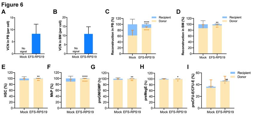
D/D mice compared to the cells from +/+ mice after doxycycline administration. Cells transduced
with EFS-RPS19 exhibited a 2.5-fold higher expression level of human RPS19 mRNA compared to
the endogenous Rps19 expression in the D/D group. Interestingly, transduced cells isolated from +/+
mice showed a significantly lower level of human RPS19 mRNA expression than the transduced
cells isolated from D/D mice at the same MOI, indicating the internal physiological regulation of
excess RPS19 production as reported by others29, 30
. The overall results indicated that cells
transduced with the EFS-RPS19 vector could successfully express the human RPS19 transgene.
Gene-corrected BM cells can rescue the DBA phenotype in Vivo
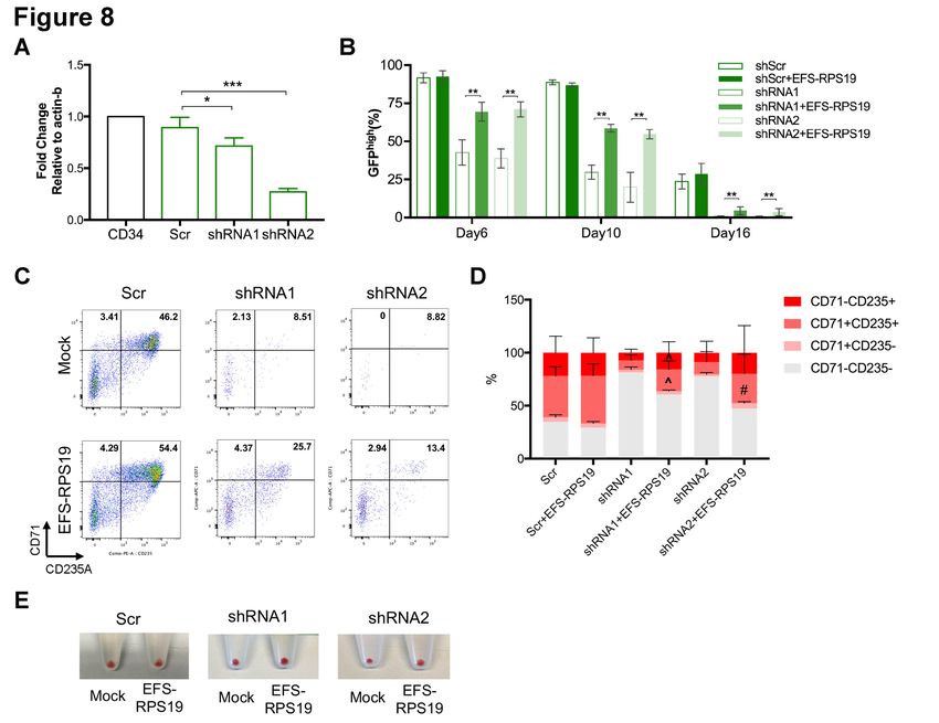
We next assessed the function of gene-corrected BM cells using EFS-RSP19 vector in vivo. As
shown in Figure 2A, uninduced (no doxycycline) c-kit+ BM cells from D/D mice (CD45.2) were
transduced with the EFS-RPS19 vector (MOI=5-10), and then transplanted into lethally irradiated
wild-type B6SJL recipient mice (CD45.1/CD45.2, named as EFS-RPS19 group). Recipients
receiving uninduced c-kit+ BM cells without vector transduction were regarded as the mock group
(negative control). Following engraftment and stable donor-derived reconstitution of the
hematopoietic system, doxycycline was administrated to all recipients to induce the DBA phenotype.
To see whether the vector-treated cells could achieve a full correction, age-matched B6SJL wild-type
mice receiving no irradiation and no transplantation but obtained the same doxycycline
administration were used as the WT control group.
Before doxycycline administration, both the mock and EFS-RPS19 groups showed high overall
donor reconstitution, indicating minimal to absent recipient-derived hematopoiesis (Online
Supplementary Figure S3). After induction with doxycycline for 2 weeks, recipients in the mock
group showed a dramatic decrease in red blood cell counts, mean corpuscular volume (MCV), white
blood cell and platelets counts, indicating that mice developed BM failure shortly after doxycycline
administration (Figure 2B-F). In contrast, recipients in the EFS-RPS19 vector-treated group showed
normal blood cellularity compared to the WT group. To assess the long-term therapeutic effects,
recipients were administrated with doxycycline for 16 weeks (Figure 3A). As shown in Figure 3B,
most of the recipients in the mock group died (9 out of 16) due to severe anemia or BM failure (data
not shown) at 2-3 weeks after doxycycline administration. The few remaining recipients exhibited a
macrocytic anemia phenotype with significantly reduced red blood cell counts and increased MCV at
16 weeks. The hemoglobin levels and platelet counts were also decreased compared to the WT group
(Figure 3C-G). As expected, there was a significantly decreased expression of endogenous Rps19
expression in donor-derived BM cells from both the mock and EFS-RPS19 groups after 16 weeks of
doxycycline administration (Online Supplementary Figure S4). Strikingly, all the mice in the EFS-
6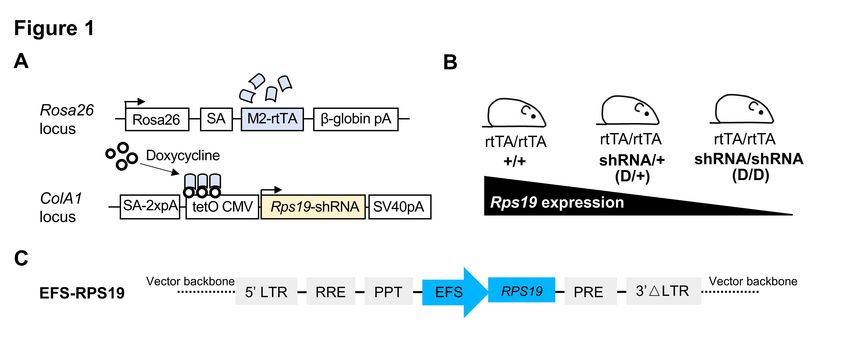
RPS19 group survived without any signs of anemia and with normal BM cellularity compared to the
WT group. These results indicate that the lethal bone marrow failure can be prevented by the vector.
The VCN at 16 weeks after transplantation was 5.2 ± 1.6 and 4.7 ± 1.0 on average in gene-corrected
cells isolated from peripheral blood (PB) and BM, respectively (Figure 4A-B). In addition, we also
analyzed the fraction of myeloid-erythroid compartments by flow cytometry (Online Supplementary
Figure S5). The donor-derived hematopoiesis was observed in the EFS-RPS19 group, and the mean
percentage of donor cells (CD45.2) in every progenitor population was significantly higher in the
EFS-RPS19 group than the mock group (Figure 4C-I). In contrast to the EFS-RPS19 group, limited
reconstitution ability of transplanted cells was observed in the mock group. In addition, we observed
a significantly higher reconstitution of resident recipient cells (CD45.1/CD45.2) in the few surviving
mice in the mock group than the EFS-RPS19 group, perhaps explaining why these animals did not
develop severe BM failure.

EFS-RPS19 vector-treated Rps19-deficient BM cells provide long-term reconstitution
We next asked whether the EFS-RPS19 vector-treated Rps19-deficient BM cells could generate
long-term engraftment and reconstitution in doxycycline-induced lethally irradiated wild-type
recipients (Figure 5A). To this end, D/D mice were induced with doxycycline for one week and red
blood cell counts and hemoglobin levels were measured to confirm the DBA phenotype (Online
Supplementary Figure S6). The isolated Lin- BM cells from doxycycline-induced mice were
transduced with the vector and transplanted into the doxycycline-induced lethally irradiated wild-
type recipients (EFS-RPS19 group). Recipients receiving untransduced Rps19-deficient Lin- BM
cells from doxycycline-induced D/D mice were regarded as the mock group. The setting of the WT
group was the same as described above. Doxycycline was administrated to all the recipients directly
after transplantation. After induction with doxycycline for 2-3 weeks, the majority of mice in the
mock group died (13 out of 16 animals) due to severe anemia or BM failure (Figure 5B & Online
Supplementary Figure S7). The few surviving mice exhibited significantly decreased levels of
endogenous Rps19 expression in donor-derived BM cells at 16 weeks after doxycycline
administration (Online Supplementary Figure S8) with the concomitant development of a severe
anemia phenotype in the mock group (Figure 5C-G). In contrast, all recipients in the EFS-RPS19
group survived with normal blood cellularity compared to the WT group. The VCN was on average
8.3 ± 4.0 or 10.9 ± 3.9 in gene-corrected Rps19-deficient cells isolated from PB or BM, respectively
(Figure 6A-B). By analyzing the fraction of myeloid-erythroid compartments at 16 weeks, we
observed almost complete donor-derived hematopoiesis in the EFS-RPS19 group, which was
significantly higher than those in the mock group (Figure 6C-I). Taken together, our results

                                                                                                    7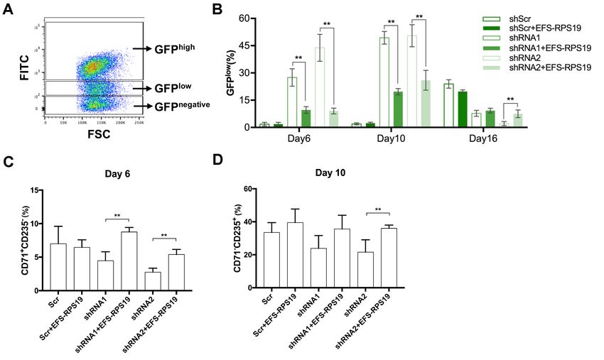
demonstrate that the EFS-RPS19 vector-treated group obtained full correction of the anemia and BM
failure compared to wild-type mice.
Gene-corrected BM cells showed polyclonal hematopoiesis and have a typical lentiviral
insertion profile
The risk of insertional mutagenesis is a major concern for future applications of gene therapy in the
clinic. To assess the safety of the EFS-RPS19 vector integration profile, as well as the clonal
dynamics of the transduced cells, insertion site analysis was performed using the INtegration Site
PIpeline for paIRed-End reaDs (INSPIIRED) workflow24. BM cells from uninduced donors (Figure
3A) of cohort 1 (animals 5-8) and cohort 2 (animal 13-16), or those from doxycycline-induced
donors (Figure 5A) of cohorts 3 (animals 7-11) and cohort 4 (animals 17-20) were isolated 16 weeks
after disease induction in the recipients. Detailed information of pool size estimation and sequence
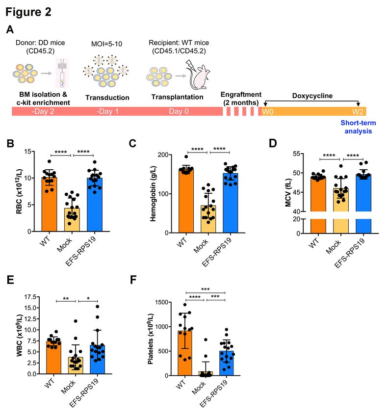
diversity in each sample were shown in the Online Supplementary Table S2-S3. Particularly, the top
ten integrations with the highest sequence contribution in each sample are depicted in Figure 7A and
Online Supplementary Figure S9A. Overall, EFS-RPS19 transduced cells showed a highly polyclonal
insertion site pattern reflecting the overall integration preferences of lentiviral vectors. No integration
site contributed with more than 5.37% in uninduced gene-corrected cells (Figure 7A) or 4.4% in
gene-corrected Rps19-deficient cells (Online Supplementary Figure S9A) to the overall sequence
pool, and there were no integrations in or close to known high-risk proto-oncogenes (Lmo2, Ccnd2
or Hmga2). However, one integration at 16.5 kb upstream of the high-risk locus Ikzf1 was detected
(account for 1.65%), and another integration at 20.7 kb upstream of the high-risk gene Mecom was
found (account for 0.04%) in recipients receiving gene-corrected Rps19-deficient BM cells. For the
analysis of overlaps between integrations in or near the same genes among BM samples, we
observed common lentiviral integration sites identified in previous integration site analysis (Online
Supplementary Table S4-S5)31, 32. We identified 11 integrations in or near the same refSeq genes
between cohorts 1+2 and cohorts 3+4 (Online Supplementary Figure S10). Four of these shared
common insertion sites in or near Hgf, Kdm6a, Lnpep and Mef2c were also as proto-oncogenes listed
in the All Onco database25. The sole occurrence of the integration sites in or near high-risk loci was
not an indication of a higher risk for insertional mutagenesis in case no dominant clones were
detected. The detected integrations might simply reflect that lentiviral vectors were capable of
integrating at these genomic sites. We performed analysis of insertion site profile including
parameters of the integration site preferences close to CpG islands, GC rich regions, in or near
transcription units, the transcriptional start site of genes, gene boundaries or proto-oncogenes (Figure
7B-D & Online Supplementary Figure S9B-D). More integrations were detected in a distance of 100
8
kb relative to CpG islands, marking actively transcribed regions but not in the direct vicinity of CpG
islands (1-10 kb), hence not close to the promoter region. Our data showed that GC rich regions
(marking promoter regions of genes) and long intergenic regions were generally disfavored by the
vector. The integrations inside of transcriptional units and in or close to proto-oncogenes (within a
100 kb window = onco.100k) are displayed relative to the matched random controls (Figure 7B &
Online Supplementary Figure S9B).

EFS-RPS19 vector rescued impaired erythroid differentiation of human RPS19-deficient
CD34+ cord blood cells
We next examined the therapeutic effects of the EFS-RPS19 vector using human primary CD34+
cord blood cells. Since primary CD34+ cells from DBA patients are difficult to obtain, we utilized
previously validated lentiviral shRNA vectors that silence RPS19 expression in human CD34+ cells
to induce a DBA phenotype23.
Two lentiviral vectors expressing shRNAs (shRNA1 and shRNA2) targeting different regions of the
human RPS19 mRNA sequence were used to induce the DBA phenotype, and a vector expressing a
scrambled shRNA sequence (Scr) was used as a healthy control. To ensure that the human construct
in the vector would not be degraded by the targeting shRNA, alignments were performed and there
are 2 mismatches (out of 19 nucleotides) for shRNA1 and 4 mismatches (out of 19 nucleotides) for
shRNA2. Thus, it is very unlikely that the gene expression derived from the human codon-optimized
RPS19 cDNA would be affected by either shRNA. Since the shRNA-vectors also contain a GFP
marker gene, transduced GFP+ cells were sorted for further examination. As shown in Figure 8A,
both shRNA1 and shRNA2 significantly decreased RPS19 mRNA expression, with slightly more
efficient knockdown being obtained with shRNA1 than shRNA2, which is similar to our previous
observations in D/+ and D/D mouse models15. In order to examine the function of EFS-RPS19, we
transduced sorted GFP+ cells and cultured in erythroid differentiation medium 48 hours after
transduction. The PCR results indicated successful integration of the vector into human cells as
shown in the Online Supplementary Figure S11. It is a well-known phenomenon that lentiviral
vector-mediated transgene expression is silenced in a fraction of CD34+ cells, and that this fraction
increases during differentiation of cells, likely due to the changes in chromatin accessibility. As
shown in Figure 8B, transgene silencing was evident in a fraction of the Scr-transduced CD34+ cells
where the fraction of GFPhigh shRNA expressing cells decreased from 100% in the sorted cells to
around 90% on day 6 and day 10, with a further reduction to around 25% at day 16. However, in the
RPS19-deficient groups, the loss of GFPhigh cells was evident from day 6, with an increased fraction
of GFPlow and GFPnegative cells (Online Supplementary Figure S12A-B). By further analysis of cell

                                                                                                    9
distribution, significantly decreased outputs of CD71+CD235A- cells on day 6 and CD71-CD235A+ cells on day 10 in GFPhigh populations of RPS19-deficient groups (Online Supplementary Figure S12C-D) were observed. The impaired differentiation was rescued by EFS-RPS19, with significantly increased GFPhigh populations (1.6-fold for shRNA1 and 1.8-fold for shRNA2, Figure 8B) and progenitor populations (CD71+CD235A-, Online Supplementary Figure S12C) compared to the RPS19-deficient groups on day 6. Particularly, during terminal erythropoiesis on day 16, cells in the RPS19-deficient groups showed reduced red blood cell production (especially in the shRNA2 group) and few GFPhigh cells (
copy insertion of the therapeutic gene in the target cells is suggested to avoid the risk of genotoxicity
in clinical gene therapy manipulation19, we plan to examine the therapeutic effects using transduced
cells with a lower MOI (e.g. MOI=1) in our future studies. Moreover, our results also demonstrate
the vector could rescue the impaired erythroid differentiation of RPS19-deficient cord blood cells by
increasing red blood cell production. Overall, we demonstrate that the EFS-RPS19 vector could
rescue the anemia and BM failure of RPS19-deficient DBA.
Apart from efficacy, vector safety is the other essential factor to assess when applying gene therapy.
The risk of insertional mutagenesis is a concern for future applications of gene therapy in the clinic.
To prevent this, we utilized a third-generation SIN lentiviral vector that lacks potent enhancers in the
LTR regions, since such vectors have been shown to exhibit a safer integration profile in previous
clinical trials19,   34, 36
                              and also in our previous animal studies12,   15
                                                                            . By using the state-of-the-art
INSPIIRED workflow, which can provide better quantification on clonal abundance compared to a
linear amplification-mediated PCR approach, we found that gene-corrected BM cells in both models
exhibited a low risk of mutagenesis with no evidence of clonal expansion associated with vector
integration near cancer-associated genes. As shown in our study, no hematologic abnormalities were
observed due to enforced expression of RPS19. The results collectively demonstrate the safety of
EFS-RPS19 vector for clinical gene therapy development. The bioinformatic pipeline of the
INSPIIRED workflow is a more automated approach, making it well suited for monitoring patients
in gene therapy trials in the future.
The increased MCV is a classic clinical observation in patients with DBA due to macrocytic anemia.
This is, at least in part, caused by the stabilization of p53 and activation of p53 targets (e.g. p21, Bax),
which are responsible for cell cycle arrest in G0/G1 phases leading to larger cell size9, 37. We also
observed a significantly increased MCV level in the mock group after induction with doxycycline for
more than 4 weeks (Figure 3E & 5E). In particular, during the initial phase after doxycycline
induction, the majority of red blood cells in the circulation were produced before the DBA phenotype
was induced. Since old red blood cells are smaller than the newly produced cells, the MCV is
decreased compared to normal for a short period during the first 2 weeks after doxycycline induction
(Figure 2E). In addition to the severe anemia phenotype after induction of Rps19 deficiency, we
observed decreased white blood cell and platelet counts in the mock group. The features of mild
thrombocytopenia and neutropenia (low levels of neutrophilic granulocytes) have also been observed
in about 25% of DBA patients during the course of the disease4, 37, 38. In addition, there are several
cases in patients with RPS19-deficient DBA develop into myelodysplastic syndrome with
multilineage cytopenia, which suggests the multilineage defect37. This observation correlates with
the absolute reduction in numbers of hematopoietic stem and progenitor cells in BM due to the

                                                                                                         11
Rps19 deficiency15, which led to the lethal BM failure we’ve observed in untreated animals shortly
after doxycycline administration. This is also supported by the limited reconstitution ability of
progenitor compartments in the mock group (Figure 4C-I) and impaired erythroid differentiation of
human primary RPS19-deficient cord blood cells. RPS19-deficient patients who develop
thrombocytopenia and neutropenia also experience similar progressive phenotypes of
38, 39
hypocellularity in the BM . Moreover, DBA is a very heterogeneous disease. There are unknown
reasons that family members with the same genetic mutation in RPS19 have very different
phenotypes ranging from no anemia to severe anemia with progression to multilineage bone marrow
failure40. The current understandings of phenotype-genotype correlations are far from comprehensive
and need to be studied further.
Although the majority of mice died in the mock group, there were still a few mice surviving until the
planned endpoint at 16 weeks. One of the possible reasons for this may be due to the emergence of
resident hematopoietic stem cells derived from the recipients, which contribute to the reconstitution
of hematopoiesis as a protective mechanism against stress-induced exhaustion in BM41-43. In support
of this, recipient-derived hematopoietic stem cells were observed in BM of the mock group at 16
weeks post-transplantation, even though full lethal irradiation was performed prior to transplantation.
In addition, other unknown reasons may also contribute to this observation and the underlying
mechanism is unknown.
As mentioned before, the majority of DBA patients have mutations in genes coding for ribosomal
proteins, and 25% of them are RPS19-deficient (mostly point mutations or small deletions). Our
findings indicate the possibility of developing SIN lentiviral vectors tailored also for other DBA
mutations (e.g. RPL5) in the future.
In conclusion, our data show the safety and efficacy of a clinically applicable SIN lentiviral vector
for the successful treatment of Rps19-deficient DBA in our mouse model and in human primary
CD34+ cord blood cells. We do not observe any hematologic abnormalities in vivo due to enforced
expression of RPS19. Our present study suggests that the clinically applicable SIN lentiviral vector,
EFS-RPS19, has the potential to be employed in a clinical gene therapy strategy for RPS19-deficient
DBA patients.
AUTHOR CONTRIBUTIONS
S.K. and Y. L. conceptualized the project and directed the research; Y.L., M.D., M.R., E.S.,
T.H.M.G., S.W., and J.C. performed the experiments; Y.L., M.R., A.S. and S.K. analyzed the data;
and Y.L. and S.K. wrote the manuscript and other co-authors provided feedback on the manuscript.
12CONFLICTS OF INTERESTS
The authors declare no competing financial interests.
ACKNOWLEDGEMENTS
The authors thank Beata Lindqvist and Xiaojie Xian for lentivirus production and Zhi Ma for
technical assistance. We thank Alexander Doyle for English language editing. This work was
supported by a Hemato-Linne grant from the Swedish Research Council Linnaeus, project grants
from Swedish Research Council, the Swedish Cancer Society and the Swedish Children’s Cancer
Society (to S.K.), the Tobias Prize awarded by the Royal Swedish Academy of Sciences financed by
the Tobias Foundation, a clinical research grant from Lund University Hospital (to S.K.), European
Union project grants STEMEXPAND and PERSIST (to S.K.), grant from The Royal Physiographic
Society of Lund, Sweden (to Y.L), and grant from Stiftelsen Lars Hiertas Minne (to Y.L.).
REFERENCES
1. Lipton JM, Ellis SR. Diamond-Blackfan anemia: diagnosis, treatment, and molecular
pathogenesis. Hematol Oncol Clin North Am. 2009;23(2):261-282.
2. Ulirsch JC, Verboon JM, Kazerounian S, et al. The Genetic Landscape of Diamond-Blackfan
Anemia. Am J Hum Genet. 2018;103(6):930-947.
3. Vlachos A, Muir E. How I treat Diamond-Blackfan anemia. Blood. 2010;116(19):3715-3723.
4. Willig TN, Niemeyer CM, Leblanc T, et al. Identification of new prognosis factors from the
clinical and epidemiologic analysis of a registry of 229 Diamond-Blackfan anemia patients. DBA
group of Societe d'Hematologie et d'Immunologie Pediatrique (SHIP), Gesellshaft fur Padiatrische
Onkologie und Hamatologie (GPOH), and the European Society for Pediatric Hematology and
Immunology (ESPHI). Pediatr Res. 1999;46(5):553-561.
5. Narla A, Vlachos A, Nathan DG. Diamond Blackfan anemia treatment: past, present, and
future. Semin Hematol. 2011;48(2):117-123.
6. Gripp KW, Curry C, Olney AH, et al. Diamond-Blackfan anemia with mandibulofacial
dystostosis is heterogeneous, including the novel DBA genes TSR2 and RPS28. Am J Med Genet A.
2014;164A(9):2240-2249.
7. Ludwig LS, Gazda HT, Eng JC, et al. Altered translation of GATA1 in Diamond-Blackfan
anemia. Nat Med. 2014;20(7):748-753.
8. Sankaran VG, Ghazvinian R, Do R, et al. Exome sequencing identifies GATA1 mutations
resulting in Diamond-Blackfan anemia. J Clin Invest. 2012;122(7):2439-2443.
9. Vlachos A, Ball S, Dahl N, et al. Diagnosing and treating Diamond Blackfan anaemia: results
of an international clinical consensus conference. Br J Haematol. 2008;142(6):859-876.
10. Boria I, Garelli E, Gazda HT, et al. The ribosomal basis of Diamond-Blackfan Anemia:
mutation and database update. Hum Mutat. 2010;31(12):1269-1279.
11. Myers KC, Davies SM. Hematopoietic stem cell transplantation for bone marrow failure
syndromes in children. Biol Blood Marrow Transplant. 2009;15(3):279-292.
12. Debnath S, Jaako P, Siva K, et al. Lentiviral Vectors with Cellular Promoters Correct Anemia
and Lethal Bone Marrow Failure in a Mouse Model for Diamond-Blackfan Anemia. Mol Ther.
2017;25(8):1805-1814.
1313. Flygare J, Olsson K, Richter J, Karlsson S. Gene therapy of Diamond Blackfan anemia
CD34(+) cells leads to improved erythroid development and engraftment following transplantation.
Exp Hematol. 2008;36(11):1428-1435.
14. Hamaguchi I, Ooka A, Brun A, Richter J, Dahl N, Karlsson S. Gene transfer improves
erythroid development in ribosomal protein S19-deficient Diamond-Blackfan anemia. Blood.
2002;100(8):2724-2731.
15. Jaako P, Flygare J, Olsson K, et al. Mice with ribosomal protein S19 deficiency develop bone
marrow failure and symptoms like patients with Diamond-Blackfan anemia. Blood.
2011;118(23):6087-6096.
16. Naldini L. Ex vivo gene transfer and correction for cell-based therapies. Nat Rev Genet.
2011;12(5):301-315.
17. Anguela XM, High KA. Entering the Modern Era of Gene Therapy. Annu Rev Med.
2019;70:273-288.
18. Biffi A, Bartolomae CC, Cesana D, et al. Lentiviral vector common integration sites in
preclinical models and a clinical trial reflect a benign integration bias and not oncogenic selection.
Blood. 2011;117(20):5332-5339.
19. Cavazzana M, Bushman FD, Miccio A, Andre-Schmutz I, Six E. Gene therapy targeting
haematopoietic stem cells for inherited diseases: progress and challenges. Nat Rev Drug Discov.
2019;18(6):447-462.
20. Dull T, Zufferey R, Kelly M, et al. A third-generation lentivirus vector with a conditional
packaging system. J Virol. 1998;72(11):8463-8471.
21. Moreno-Carranza B, Gentsch M, Stein S, et al. Transgene optimization significantly
improves SIN vector titers, gp91phox expression and reconstitution of superoxide production in X-
CGD cells. Gene Ther. 2009;16(1):111-118.
22. Jaako P, Debnath S, Olsson K, et al. Gene therapy cures the anemia and lethal bone marrow
failure in a mouse model of RPS19-deficient Diamond-Blackfan anemia. Haematologica.
2014;99(12):1792-1798.
23. Flygare J, Kiefer T, Miyake K, et al. Deficiency of ribosomal protein S19 in CD34+ cells
generated by siRNA blocks erythroid development and mimics defects seen in Diamond-Blackfan
anemia. Blood. 2005;105(12):4627-4634.
24. Sherman E, Nobles C, Berry CC, et al. INSPIIRED: A Pipeline for Quantitative Analysis of
Sites of New DNA Integration in Cellular Genomes. Mol Ther Methods Clin Dev. 2017;4:39-49.
25. Berry CC, Nobles C, Six E, et al. INSPIIRED: Quantification and Visualization Tools for
Analyzing Integration Site Distributions. Mol Ther Methods Clin Dev. 2017;4:17-26.
26. Chao A, Chazdon RL, Colwell RK, Shen TJ. Abundance-based similarity indices and their
estimation when there are unseen species in samples. Biometrics. 2006;62(2):361-371.
27. Haemmerle R, Phaltane R, Rothe M, et al. Clonal Dominance With Retroviral Vector
Insertions Near the ANGPT1 and ANGPT2 Genes in a Human Xenotransplant Mouse Model. Mol
Ther Nucleic Acids. 2014;3:e200.
28. De LD. On the estimation of biological populations. Biometrics. 1947;3(4):145-167.
29. Lam YW, Lamond AI, Mann M, Andersen JS. Analysis of nucleolar protein dynamics
reveals the nuclear degradation of ribosomal proteins. Curr Biol. 2007;17(9):749-760.
30. Devlin EE, Dacosta L, Mohandas N, Elliott G, Bodine DM. A transgenic mouse model
demonstrates a dominant negative effect of a point mutation in the RPS19 gene associated with
Diamond-Blackfan anemia. Blood. 2010;116(15):2826-2835.
31. Cesana D, Ranzani M, Volpin M, et al. Uncovering and dissecting the genotoxicity of self-
inactivating lentiviral vectors in vivo. Mol Ther. 2014;22(4):774-785.
32. Deichmann A, Brugman MH, Bartholomae CC, et al. Insertion sites in engrafted cells cluster
within a limited repertoire of genomic areas after gammaretroviral vector gene therapy. Mol Ther.
2011;19(11):2031-2039.
1433. Da Costa L, O'Donohue MF, van Dooijeweert B, et al. Molecular approaches to diagnose Diamond-Blackfan anemia: The EuroDBA experience. Eur J Med Genet. 2018;61(11):664-673. 34. High KA, Roncarolo MG. Gene Therapy. N Engl J Med. 2019;381(5):455-464. 35. Dunbar CE, High KA, Joung JK, Kohn DB, Ozawa K, Sadelain M. Gene therapy comes of age. Science. 2018;359(6372):eaan4672. 36. Marktel S, Scaramuzza S, Cicalese MP, et al. Intrabone hematopoietic stem cell gene therapy for adult and pediatric patients affected by transfusion-dependent ss-thalassemia. Nat Med. 2019;25(2):234-241. 37. Da Costa L, Leblanc T, Mohandas N. Diamond-Blackfan anemia. Blood. 2020;136(11):1262- 1273. 38. Giri N, Kang E, Tisdale JF, et al. Clinical and laboratory evidence for a trilineage haematopoietic defect in patients with refractory Diamond-Blackfan anaemia. Br J Haematol. 2000;108(1):167-175. 39. Casadevall N, Croisille L, Auffray I, Tchernia G, Coulombel L. Age-related alterations in erythroid and granulopoietic progenitors in Diamond-Blackfan anaemia. Br J Haematol. 1994;87(2):369-375. 40. Engidaye G, Melku M, Enawgaw B. Diamond Blackfan Anemia: Genetics, Pathogenesis, Diagnosis and Treatment. EJIFCC. 2019;30(1):67-81. 41. Shi W, Vu T, Boucher D, et al. Ssb1 and Ssb2 cooperate to regulate mouse hematopoietic stem and progenitor cells by resolving replicative stress. Blood. 2017;129(18):2479-2492. 42. Baumgartner C, Toifl S, Farlik M, et al. An ERK-Dependent Feedback Mechanism Prevents Hematopoietic Stem Cell Exhaustion. Cell Stem Cell. 2018;22(6):879-892.e6. 43. Singh SK, Singh S, Gadomski S, et al. Id1 Ablation Protects Hematopoietic Stem Cells from Stress-Induced Exhaustion and Aging. Cell Stem Cell. 2018;23(2):252-265.e8. FIGURE LIGENDS Figure 1. The inducible Rps19-deficient mouse model and structure of the EFS-RPS19 self- inactivating lentiviral vector (A) Overview of modified loci. Black arrowheads indicate the transcriptional start sites. (B) Breeding strategy to adjust the level of Rps19 downregulation. Homozygous mice (D/D mice) are used in the project. (C) The self-inactivating lentiviral vectors harboring a codon optimized human RPS19 cDNA driven by human elongation factor 1α short (EFS) promoter. LTR, long terminal repeat; pA, polyadenylation signal; PPT, polypurine tract; RRE, Rev response element; SA, splice acceptor. Figure 2. Effective correction of the anemia by the EFS-RPS19 vector at two weeks after induction of DBA phenotype (A) The scheme of the uninduced gene-corrected cell transplantation model and plan for examining short-term therapeutic effects. (B-F) Blood cellularity at 2 weeks after doxycycline induction (n=13- 16, error bars represent the SD, *p
Figure 3. Effective long-term correction of the anemia and BM failure in mice treated with the
EFS-RPS19 vector
(A) The scheme of induced gene-corrected cell transplantation model and the plan for examining
long-term therapeutic effects. (B) Surviral rate analysis. (C-G) Blood cellularity at 16 weeks after
doxycycline induction (n=13-16, error bars represent the SD, *p
in black indicates comparison of the recipients-derived cells between the mock and EFS-RPS19
group, asterisk in orange indicates comparison of the donors-derived cells between the mock and
EFS-RPS19 group, *p
Supplementary Appendix

Supplementary Methods
Mice
The homozygous doxycycline-inducible Rps19-deficient mouse model used in the study was
established as previously described15. Rps19 deficiency was induced by doxycycline in drinking water
(1 mg/mL or 2 mg/mL doxycycline; Sigma-Aldrich) supplemented with 10 mg/mL sucrose (Sigma-
Aldrich). Mice were maintained at the Lund University animal facility and all animal experiments
were performed with consent from the Lund University animal ethics committee.

Blood and BM analysis
Peripheral blood (PB) was collected from the tail vein into the microvette tube (Sarstedt) and was
analyzed using Sysmex XE-5000. Red blood cells were lysed using ammonium chloride for 10 min at
room temperature. To evaluate the contribution toward various blood lineages by flow cytrometry
following BM transplantation, samples were stained with the following antibodies for 30 min on ice
in the dark: CD45.1 (110730; Biolegend), CD45.2 (47-0454-82; eBioscience). Experiments were
performed using a FACS LSR Fortessa cytometer (BD Biosciences) and were analyzed by FlowJo
software (version 10.5.3; Tree Star). FACS analysis of the myeloerythroid compartments in BM was
performed as previously described22. Briefly, BM cells were harvested by crushing the femur and tibia
in phosphate-buffered saline (PBS) containing 2% fetal bovine serum (FBS) (Gibco). Fresh cells were
stained with the following antibodies: CD45.1 (740889; BD Biosciences), CD45.2 (109806;
Biolegend), CD41 (12-0411-83; eBioscience), GR1 (108410; Biolegend), CD11b (101210;
Biolegend), B220 (103210; Biolegend), CD3 (100310; Biolegend), c-kit (47-1171-82; eBioscience),
CD105 (120404; Biolegend), Ter119 (25-5921-82; eBioscience), and Sca-1 (122520; Biolegend).
Streptavidin was purchased from Life Technologies (Q10101MP). Propidium iodide (Life
Technologies) was used to exclude dead cells. Experiments were performed using a FACS LSR
Fortessa cytometer (Becton Dickinson) and were analyzed by FlowJo software (version 10.5.3; Tree
Star).

Transplantation of hematopoietic progenitor cells
For the transplantation of uninduced gene-corrected BM cells, after incubation for 1 day, 0.5x10⁶ bulk
transduced c-kit+ cells were resuspended in 300µL PBS and transplanted into the tail vein of lethally
irradiated (900 cGy) wild-type recipients (CD45.1/45.2). For the transplantation of gene-corrected
Rps19-deficient BM cells, after incubation for 1 day with doxycycline (1 μg/ml), 0.5x10⁶ bulk
transduced Lin- cells and 1x10⁶ untransduced Lin+ cells were resuspended in 300µL PBS and
transplanted into the tail vein of lethally irradiated (900 cGy) wild-type recipients (CD45.1/45.2).

Transduction of human primary cord blood cells and erythroid differentiation
The shRNAs along with GFP marker were designed as previously described23. For transduction, cells
were transduced with shRNAs at an MOI of 5. The sequences for shRNAs are shown in the
Supplementary Table S1. After 48 hours of transduction, GFP+ cells were sorted and transduced with
or without EFS-RPS19 at an MOI of 5. To check the successful integration of the vector into human
cells, we used genomic DNA isolated from cells with or without EFS-RPS19 transduction as templates,
and primers specifically targeting at the lentiviral vector WPRE region were used for the PCR array
(the ALB gene was used as an internal reference gene). The primer sequences are shown in the
Supplementary Table S1. For erythroid differentiation, an equal number of CD34+ cells (20,000) were
cultured in erythroid differentiation medium supplemented with different cytokines for three stages of
differentiation. The base medium for all three differentiation phases comprises: IMDM, 15% FBS, 1%
BSA, 500mg/ml human holo-transferrin, 1% Insulin-Transferrin-Selenium and 1% penicillin–
streptomycin and ß-mercaptoethanol. At stage I (day 1 to day 6), cells were cultured in base medium
plus 50 ng/ml hSCF, 10 ng/ml human IL-3 (hIL-3) and 6U/ml Erythropoietin. At stage II (day 7 to day
10), hIL-3 was removed from the medium. At stage III (day 10 to day 16), both hIL-3 and hSCF were
removed from the medium.

Real-time quantitative reverse transcription polymerase chain reaction (qRT-PCR)
RNA was isolated using RNeasy Micro Kit (Qiagen, USA), followed with reverse transcription using
SuperScript™ III (Thermo Fiesher, USA). qRT-PCR was performed with SYBR Green PCR Master
Mix (Applied Biosystems, USA) without specific indications. All reactions were performed in
triplicates. The primer sequences are shown in the Supplementary Table S1.

Determination of transduction efficiency
The c-kit+ cells were enriched and transduced (MOI=5-10) as mentioned above. At 48 hours after
transduction, 40x103 c-kit+ transduced cells were seeded in 1.5 mL of M3434 methylcellulose (Stem
Cell Technologies) and single colonies were picked after 14 days of culture. DNA was isolated from
each colony, followed by an PCR assay to detect the inserted vector. We determined the vector-
transduced colonies based on the positive detection of WPRE region of the vector. Transduction
efficiency was determined by the total number of colonies carrying the targeted WPRE region of the
vector backbone relative to the total colony number. The experiments were performed in triplicates
(100 colonies were picked each time).

Determination of vector copy number (VCN)
Whole BM or PB cells were isolated at 16 weeks after transplantation. Genomic DNA was isolated
with the DNA Blood & Tissue kit (Qiagen). The mean VCN was determined by qRT-PCR using
TaqMan Gene Expression Assay (Thermo Fisher Scientific). The number of viral sequences was
normalized to the genomic reference sequence. We used WPRE element to detect the viral sequences
(as shown in the Supplementary Table S1), and Tfrc gene (4458367; Thermo Fisher Scientific) was
used for genomic DNA normalization.

Insertion site analysis
Vector-genome junction was amplified using the INtegration Site PIpeline for paIRed-End reaDs
(INSPIIRED) workflow as described by Sherman and colleagues24. In brief, genomic DNA was
purified using AMPure beads and fragmented with a Covaris 220 sonicator. After an additional round
of AMPure purification, DNA was end-repaired and subjected to linker ligation. For each sample, a
unique linker was used to prevent cross-contamination between samples during batch processing.
Primers specific for the SIN long-terminal repeat (LTR) region of the lentivirus in combination with
linker-specific primers were used to amplify vector genome junctions. The amplification from the LTR
into the vector (instead of the genome) was controlled and largely prohibited using blocking oligos. In
a second nested PCR, LTR-specific index primers together with linker-specific primers were used to
attach next-generation sequencing adapters for Illumina paired-end sequencing. After Bioanalyzer
quality assessment, the libraries were loaded on MiSeq Nano flow cells. The sequencing reads were
deconvoluted according to the primer index used during the second nested PCR step. The individual
sequences were quality filtered, aligned to the target genome and quantified by sonic abundance as
described in the INSPIIRED bioinformatics pipeline25. The iPSC-clones C14 or HD2 were used as
monoclonal control samples as indicated. The pool size estimation and sequence diversity were
analyzed as previously described26-28. Bioinformatic steps were generally performed as described25.
Particularly, we allowed a total of 5 bp below the quality threshold per amplicon. A quality sliding
window of 10 bp was chosen. A maximum fragment length of 2500 bp, a minimal overlap with the
reference genome of 20 bp with 95% homology and a start of the alignment no later than 5 bp after
the LTR were set in the processing parameters of the INSPIIRED pipeline. The individual files were
aligned and annotated to the mouse (mm9, for all mouse samples) and human genome (hg38, for C14
or HD2).
Supplementary Figures

Supplementary Figure S1. Transduction efficiency of the EFS-RPS19 vector in uninduced c-kit+ BM
cells isolated from D/D mice

Supplementary Figure S2. Comparison of endogenous Rps19 and transgene RPS19 expressions in c-
kit+ BM cells isolated from D/D and +/+ mice on day 4 after transduction (average of 3 independent
experiments with 3 technical replicates in each group, error bars represent the SD, *p
Supplementary Figure S4. Inhibition of mouse Rps19 expression in the mock and gene-corrected
cells induced with doxycycline for 16 weeks (n=3-6 in each group, error bars represent the SD,
**p
Supplementary Figure S6. Anemia phenotype in Rps19-deficient mice receiving doxycycline
administration for 1 week (n=9 for +/+ group, n=22 for D/D group, **p
Supplementary Figure S9. Gene-corrected Rps19-deficient BM cells show a vector integration
pattern that indicates low risk of mutagenesis and a highly polyclonal insertion site pattern
(A) Top 10 integration sites in each sample (*indicates that the integration was within a transcription
unit, ~ indicates that the insertion was within 50 kb of a cancer-related gene). (B-C) Percent of all
integrations inside of transcriptional units (B) and percent of integrations within 100 kb of proto-
oncogenes compared to matched random control sites (C). (D) Genomic heatmap analysis of insertion
site profile. (mrc: matched random control, ***p< 0.001 by unpaired t-test).
Supplementary Figure S10. Venn diagram displaying the shared common insertion sites in cohorts
1+2 and cohorts 3+4. In case the integration overlapped with a proto-oncogene listed in the AllOnco
database, the gene symbol is marked in red, in case it was described as a previous common insertion
site it is marked in orange, if both parameters apply, the gene symbol is marked in purple.

Supplementary Figure S11. Successful integration of the vector into human RPS19-deficient CD34+
cord blood cells by PCR array (untreated CD34+ cord blood cells were regarded as control; Human
ALB was used as an internal reference gene)
Supplementary Figure S12. Impaired erythroid differentiation of RPS19-deficient CD34+ cord blood
cells
(A) The applied FACS strategy according to GFP intensity. (B) Percentage of GFPlow populations in
RPS19-deficient CD34+ cells treated with or without EFS-RPS19 during erythroid differentiation from
stage I to stage III. (C) Percentage of indicated cell outputs of GFPhigh populations on day 6. (D)
Percentage of indicated cell outputs of GFPhigh populations on day 10 (data are shown as mean±SD,
**p
Supplementary Tables

 For qRT-PCR                       5' to 3'
 human codon-optimized RPS19   F   AAGAAAAGCGGCAAACTCAAG
                               R   CCGTAGATCTTGGTCATGCT

 human RPS19                   F   GCCTGGAGTTACTGTAAAAGACG
                               R   CCCATAGATCTTGGTCATGGAGC

 human ACTIN                   F   AGAAAATCTGGCACCACACC
                               R   GGGGTGTTGAAGGTCTCAAA

 mouse Rps19                   F   GCAGAGGCTCTAAGAGTGTGG
                               R   CCAGGTCTCTCTGTCCCTGA

 mouse Actin                   F   ATGGTGGGAATGGGTCAGAA
                               R   CCATGTCGTCCCAGTTGGTAA

 WPRE                          F   TTCTGGGACTTTCGCTTTCC
 WPRE                          R   CCGACAACACCACGGAATTA
 Taqman PROBE                      ATCGCCACGGCAGAACTCATCG

 For RT-PCR                        5' to 3'
 WPRE                          F   GAGGAGTTGTGGCCCGTTGT
 WPRE                          R   TGACAGGTGGTGGCAATGCC

 mouse Actin                   F   GCTAATGAGGCTGGTGATAAGTGG
                               R   CACGCTCGGTCAGGATCTTCAT

 human ALB                     F   TGAAACATACGTTCCCAAAGAGTTT
                               R   CTCTCCTTCTCAGAAAGTGTGCAT

 shRNA sequences                   5' to 3'
 human RPS19 shRNA-1               GCACAAAGAGCTTGCTCCC
 human RPS19 shRNA-2               GAGATCTGGACAGAATCGC
 Scramble                          GACACGCGACTTGTACCAC
Supplementary Table S1. Sequences for qRT-PCR, RT-PCR and shRNAs (F: Forward; R: Reverse;
ALB: ALBUMIN)
SID# Sample Name Unique Sites Chapman Coverage S.chao1 Gini Shannon UC50
1 HD2 Clone 7 8 58% 5 0.705 0.897 1
2 Animal 5 882 887 58% 1411 0.840 5.025 32
3 Animal 6 878 856 61% 1963 0.874 4.732 17
4 Animal 7 987 976 64% 1506 0.884 4.523 14
5 Animal 8 1304 1411 48% 3108 0.889 4.990 31
6 Animal 13 933 922 79% 2164 0.873 4.727 18
7 Animal 14 957 934 68% 1359 0.872 4.883 23
8 Animal 15 1115 1083 67% 1670 0.839 5.287 44
9 Animal 16 985 979 62% 1559 0.884 4.757 22
Supplementary Table S2. Analysis of the pool size (Chapman, S.chao1), clonality (Gini) and
diversity (Shannon) of gene-corrected BM cells from cohorts 1+2
SID# Sample Name Unique Sites Chapman Coverage S.chao1 Gini Shannon UC50
1 C14 2 2 67% 3 0.497 0.023 1
2 Animal 7 248 234 89% 610 0.604 4.880 35
3 Animal 8 317 294 83% 999 0.758 4.603 22
4 Animal 9 275 253 83% 1187 0.758 4.412 15
5 Animal 10 301 282 86% 680 0.715 4.745 30
6 Animal 11 532 501 84% 868 0.734 5.219 41
7 Animal 17 338 313 83% 934 0.699 4.903 34
8 Animal 18 346 326 88% 734 0.726 4.786 23
9 Animal 19 296 274 79% 634 0.771 4.473 21
10 Animal 20 309 290 83% 662 0.727 4.708 24
Supplementary Table S3. Analysis of the pool size (Chapman, S.chao1), clonality (Gini) and
diversity (Shannon) of gene-corrected Rps19-deficient BM cells from from cohorts 3+4
Animal 13
Animal 14
Animal 15
Animal 16
Animal 5
Animal 6
Animal 7
Animal 8
Overlaps
Gene Symbol
Lrrc4c ● ● ● ● ● ● ● ● 8
Mir101c ● ● ● ● ● ● ● ● 8Ncam2 ● ● ● ● ● ● ● ● 8
Vegfc ● ● ● ● ● ● ● ● 8
Cylc2 ● ● ● ● ● ● ● 7
Dach1 ● ● ● ● ● ● ● 7
Dlg2 ● ● ● ● ● ● ● 7
Pcmtd1 ● ● ● ● ● ● ● 7
Phtf2 ● ● ● ● ● ● ● 7
Rgs18 ● ● ● ● ● ● ● 7
Tfec ● ● ● ● ● ● ● 7
Trps1 ● ● ● ● ● ● ● 7
1700019E08Rik ● ● ● ● ● ● 6
1700019M22Rik 2.1% ● ● ● ● ● 6
4930529K09Rik ● ● ● ● ● ● 6
AA545190 ● ● ● ● ● ● 6
Alcam ● ● ● ● ● ● 6
Angpt1 ● ● ● ● ● ● 6
Arhgap18 ● ● ● ● ● ● 6
Ascc3 ● ● ● ● ● ● 6
Caap1 ● 1.1% ● ● ● ● 6
Cntnap2 ● ● ● ● ● ● 6
Csmd3 ● ● ● ● ● ● 6
Ddi1 ● ● ● ● ● ● 6
Diaph2 ● ● ● ● ● ● 6
Epha7 2.0% ● ● ● ● ● 6
Gm11917 ● ● ● ● ● ● 6
Gm20125 ● ● ● ● ● ● 6
Gm20756 ● ● ● ● ● ● 6
Gm35496 ● ● ● ● ● ● 6
Gm6578 ● ● ● ● ● ● 6
Hgf ● ● ● ● ● ● 6
Ikzf2 1.0% ● ● ● ● ● 6
Inpp4b ● ● ● ● ● ● 6
Animal 13
Animal 14
Animal 15
Animal 16
Animal 5
Animal 6
Animal 7
Animal 8
Overlaps
Gene Symbol
Kdm6a ● ● ● ● ● ● 6
Lnpep ● ● ● ● ● ● 6
Lrrn3 ● ● ● ● ● ● 6
Mir6368 ● ● ● ● ● ● 6
Nrxn1 ● ● ● ● ● ● 6
Pla2g4a ● ● ● ● ● ● 6
Rab38 ● ● ● ● ● ● 6
Stau2 ● ● ● ● ● ● 6
Tbc1d5 ● ● ● ● ● ● 6Wac ● ● ● ● ● ● 6
Wapl ● ● ● ● ● ● 6
1700128A07Rik ● ● ● ● ● 5
4930559C10Rik ● ● ● ● ● 5
4933422A05Rik ● ● ● ● ● 5
A930001A20Rik ● ● ● ● ● 5
Adamts6 ● ● ● ● ● 5
Adgrl3 ● ● ● ● ● 5
Akap13 ● ● ● ● ● 5
Arap2 ● ● ● ● ● 5
Arhgap6 ● ● ● ● ● 5
Asxl2 ● ● ● ● ● 5
B3galt2 ● ● ● ● ● 5
Ccser2 ● ● ● ● ● 5
Cdk14 ● ● ● ● ● 5
Cdk8 ● ● ● ● ● 5
Celf2 ● ● ● ● ● 5
Cfap47 ● ● ● ● ● 5
Dcun1d5 ● ● ● ● ● 5
Ddx10 ● ● 2.8% ● ● 5
Fam107b ● ● ● ● ● 5
Fam174a ● ● ● ● ● 5
Gm19782 ● ● ● ● ● 5
Gm6634 ● ● ● ● ● 5
Gria3 ● ● ● ● ● 5
Hs3st1 ● ● ● ● ● 5
Mctp2 ● 1.1% ● ● ● 5
Mef2c ● ● ● ● ● 5
Micu3 ● ● ● ● ● 5
Mir1931 ● ● ● ● ● 5
Mir3961 ● ● ● ● ● 5
Mir6350 ● ● ● ● ● 5
Animal 13
Animal 14
Animal 15
Animal 16
Animal 5
Animal 6
Animal 7
Animal 8
Overlaps
Gene Symbol
Mir6411 2.4% ● ● ● ● 5
Mllt10 ● ● ● ● ● 5
Mmp16 ● ● ● ● ● 5
Myo10 ● ● ● ● ● 5
Nipbl ● ● ● ● ● 5
Nudt12 ● ● ● ● ● 5
Pcdh7 ● ● ● ● ● 5
Pisd-ps3 ● ● ● ● ● 5
Plxdc2 ● ● ● ● ● 5Prex2 ● ● ● ● ● 5
Rab10os ● ● ● ● ● 5
Rasa1 ● ● ● ● ● 5
Rb1cc1 ● ● ● ● ● 5
Rnpc3 ● ● ● ● ● 5
Sema3a ● ● ● ● ● 5
Sfi1 ● ● ● ● ● 5
Shank3 ● ● 1.9% ● ● 5
Shprh 2.9% ● ● ● ● 5
Slc4a7 ● ● ● ● ● 5
Smurf2 ● ● ● ● ● 5
Spata6 ● ● ● ● ● 5
Tab2 ● ● ● ● ● 5
Tbl1xr1 ● ● ● ● ● 5
Tlr4 ● ● 1.1% ● ● 5
Ttc37 ● ● ● ● ● 5
Ube3a ● ● ● ● ● 5
Utrn ● ● ● ● ● 5
Vav3 ● ● ● ● ● 5
Vwc2 ● ● ● ● ● 5
Zmym4 ● ● ● ● ● 5
Supplementary Table S4. Common insertion sites in or near the same genes in cohort 1 (animal 5-8)
and cohort 2 (animal 13-16) of mice receiving gene-corrected BM cells from uninduced donors. In
case the integration overlapped with a proto-oncogene listed in the AllOnco database, the gene symbol
is marked in red, in case it was described as a previous common insertion site it is marked in orange,
if both parameters apply, the gene symbol is marked in purple.
Animal 10
Animal 11
Animal 17
Animal 18
Animal 19
Animal 20
Animal 7
Animal 8
Animal 9
Overlaps
Gene_Symbol
Pisd-ps3 ● ● ● ● ● ● ● ● 8
Diaph3 ● ● ● ● ● ● ● 7
Lrrc4c ● ● ● ● ● ● ● 7
Nrxn1 ● ● ● ● ● 1.9% ● 7
1700019M22Rik ● 1.4% ● ● ● ● 6
Trps1 ● ● ● ● ● ● 6
Agbl1 ● ● ● ● ● 5
Epha5 3.2% 1.5% ● ● ● 5
Hgf ● ● ● 1.5% ● 5
Kdm6a ● ● 2.9% ● ● 5You can also read