Identification of rare HIV-1-infected patients with extreme CD4+ T cell decline despite ART-mediated viral suppression - JCI Insight
←
→
Page content transcription
If your browser does not render page correctly, please read the page content below
Identification of rare HIV-1–infected patients
with extreme CD4+ T cell decline despite ART-
mediated viral suppression
Andrea Lisco, … , Claire Deleage, Irini Sereti
JCI Insight. 2019;4(8):e127113. https://doi.org/10.1172/jci.insight.127113.

 Clinical Medicine   AIDS/HIV

BACKGROUND. The goal of antiretroviral therapy (ART) is to suppress HIV-1 replication
and reconstitute CD4+ T cells. Here, we report on HIV-infected individuals who had a
paradoxical decline in CD4+ T cells despite ART-mediated suppression of plasma HIV-1
load (pVL). We defined such an immunological outcome as extreme immune decline
(EXID).

METHODS. EXID’s clinical and immunological characteristics were compared to
immunological responders (IRs), immunological nonresponders (INRs), healthy controls
(HCs), and idiopathic CD4+ lymphopenia (ICL) patients. T cell immunophenotyping and
assembly/activation of inflammasomes were evaluated by flow cytometry. PBMC
transcriptome analysis and genetic screening for pathogenic variants were performed.
Levels of cytokines/chemokines were measured by electrochemiluminescence. Luciferase
immunoprecipitation system and NK-mediated antibody-dependent cellular cytotoxicity
(ADCC) assays were used to identify anti-lymphocyte autoantibodies.

RESULTS. EXIDs were infected with non-B HIV-1 subtypes and after 192 weeks of
consistent ART-mediated pVL suppression had a median CD4+ decrease of 157 cells/µl,
compared with CD4+ increases of 193 cells/µl and 427 cells/µl in INR and IR, respectively.
EXID had reduced naive CD4+ T cells, but similar proportions of cycling CD4+ T cells and
HLA-DR +CD38+CD8+ T cells compared with IR and INR. Levels of inflammatory cytokines
were […]

Find the latest version:
http://jci.me/127113/pdf
CLINICAL MEDICINE

                                       Identification of rare HIV-1–infected
                                       patients with extreme CD4+ T cell decline
                                       despite ART-mediated viral suppression
                                       Andrea Lisco,1 Chun-Shu Wong,1 Silvia Lucena Lage,1 Itzchak Levy,2 Jason Brophy,3 Jeffrey Lennox,4
                                       Maura Manion,1 Megan V. Anderson,1 Yolanda Mejia,1 Christopher Grivas,1 Harry Mystakelis,1
                                       Peter D. Burbelo,5 Ainhoa Perez-Diez,1 Adam Rupert,6 Craig A. Martens,7 Sarah L. Anzick,7
                                       Caryn Morse,1 Shanna Chan,8 Claire Deleage,9 and Irini Sereti1
                                       Laboratory of Immunoregulation, HIV Pathogenesis Section, National Institute of Allergy and Infectious Diseases (NIAID),
                                       1

                                       NIH, Bethesda, Maryland, USA. 2Sheba Medical Center, Tel Hashomer and the Sackler Medical School, Tel Aviv, Israel.
                                       3
                                           Children’s Hospital of Eastern Ontario, Ottawa, Canada. 4Grady Memorial Hospital, Emory University, Atlanta, Georgia,
                                       USA. 5Dental Clinical Research Core, National Institute of Dental and Craniofacial Research, NIH, Bethesda, Maryland,
                                       USA. 6AIDS Monitoring Laboratory, Leidos Biomedical Research, Frederick, Maryland, USA. 7Rocky Mountain Laboratory,
                                       Genomics Unit, NIAID, NIH, Hamilton, Montana, USA. 8Winnipeg Regional Health Authority, Manitoba, Canada. 9Tissue
                                       Analysis Core, AIDS and Cancer Virus Program, Leidos Biomedical Research Inc., Frederick National Laboratory for Cancer
                                       Research, Frederick, Maryland, USA.

                                                   BACKGROUND. The goal of antiretroviral therapy (ART) is to suppress HIV-1 replication and
                                                   reconstitute CD4+ T cells. Here, we report on HIV-infected individuals who had a paradoxical decline
                                                   in CD4+ T cells despite ART-mediated suppression of plasma HIV-1 load (pVL). We defined such an
                                                   immunological outcome as extreme immune decline (EXID).

                                                   METHODS. EXID’s clinical and immunological characteristics were compared to immunological
                                                   responders (IRs), immunological nonresponders (INRs), healthy controls (HCs), and idiopathic
                                                   CD4+ lymphopenia (ICL) patients. T cell immunophenotyping and assembly/activation of
                                                   inflammasomes were evaluated by flow cytometry. PBMC transcriptome analysis and genetic
                                                   screening for pathogenic variants were performed. Levels of cytokines/chemokines were measured
                                                   by electrochemiluminescence. Luciferase immunoprecipitation system and NK-mediated
                                                   antibody-dependent cellular cytotoxicity (ADCC) assays were used to identify anti-lymphocyte
                                                   autoantibodies.

                                                   RESULTS. EXIDs were infected with non-B HIV-1 subtypes and after 192 weeks of consistent
                                                   ART-mediated pVL suppression had a median CD4+ decrease of 157 cells/μl, compared with CD4+
                                                   increases of 193 cells/μl and 427 cells/μl in INR and IR, respectively. EXID had reduced naive CD4+
                                                   T cells, but similar proportions of cycling CD4+ T cells and HLA-DR+CD38+CD8+ T cells compared
                                                   with IR and INR. Levels of inflammatory cytokines were also similar in EXID and INR, but the IL-7
                                                   axis was profoundly perturbed compared with HC, IR, INR, and ICL. Genes involved in T cell and
                                                   monocyte/macrophage function, autophagy, and cell migration were differentially expressed
                                                   in EXID. Two of the 5 EXIDs had autoantibodies causing ADCC, while 2 different EXIDs had an
                                                   increased inflammasome/caspase-1 activation despite consistently ART-suppressed pVL.
Conflict of interest: Jeffrey Lennox
reports grants from ViiV Healthcare                CONCLUSIONS. EXID is a distinct immunological outcome compared with previously described
and personal fees from Gilead.                     INR. Anti–CD4+ T cell autoantibodies and aberrant inflammasome/caspase-1 activation despite
                                                   suppressed HIV-1 viremia are among the mechanisms responsible for EXID.
Copyright: © 2019 American Society
for Clinical Investigation

Submitted: January 3, 2019
Accepted: March 12, 2019
Published: April 18, 2019.             Introduction
Reference information: JCI Insight.    The goal of antiretroviral therapy (ART) is the suppression of HIV-1 replication and the ensuing reconsti-
2019;4(8):e127113. https://doi.        tution of CD4+ T cells. The improvement in morbidity and mortality associated with adequate CD4+ T cell
org/10.1172/jci.insight.127113.        reconstitution with ART has dramatically changed the natural history of HIV-1 infection. Nonetheless, a

insight.jci.org   https://doi.org/10.1172/jci.insight.127113                                                                                                       1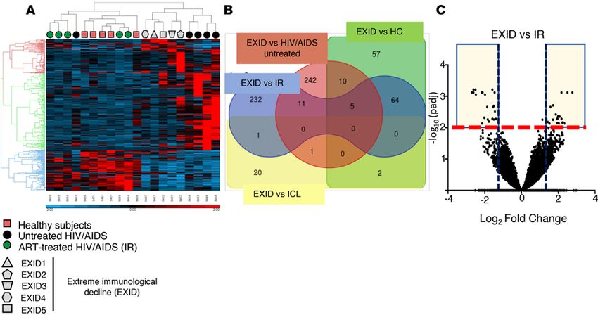
CLINICAL MEDICINE
considerable proportion of ART-treated individuals can only achieve a modest increase of CD4+ T cells
despite suppression of HIV-1 replication. These subjects are defined immunological nonresponders (INRs)
and have a significantly higher mortality compared with immunological responders (IRs) (1–3). Although
demographic (older age, male gender) and immunological variables (low nadir CD4, immune activation,
lymphoid tissue fibrosis, and impaired homeostatic signaling) have been associated with a blunted immu-
nological response to ART, the mechanisms underlying such phenomena remain elusive and therefore no
adjunctive therapy improving CD4+ T cell reconstitution is currently available (4, 5).
Here, we report on HIV-1–infected individuals who, despite consistent ART-mediated suppression
of HIV-1 replication, developed a paradoxical decline in CD4+ T cells. We present the epidemiological
and immunological findings of such immunological outcome and propose what we believe are novel
mechanisms associated with this extreme immune decline (EXID) in ART-treated subjects.
Results
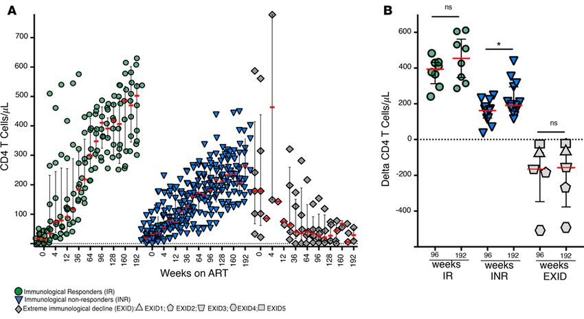
Immunological response to ART. Five subjects were referred to the NIH Clinical Center for declining CD4+ T
cells despite consistent suppression of plasma HIV-1 load (pVL) on different ART regimens and extensive
work-up ruling out any concomitant malignancy. The demographic, clinical, and immunological charac-
teristics of these subjects are summarized in Table 1 and supplemental material. The median age at time of
enrollment was 25 years (range 13–49) and all subjects were infected with non-B HIV-1 subtypes. The medi-
an CD4+ T cell count before ART was 179 cells/μl (IQR 67–414), but after 192 weeks of ART, despite con-
sistent suppression of pVL on different regimens, the median CD4+ T cells decreased to 36 cells/μl (IQR
17–60) (Figure 1). This paradoxical decline of CD4+ T cells despite consistent ART-mediated pVL suppres-
sion was profoundly different from the immunological responses in ART-naive subjects with nadir CD4+
less than 100 cells/μl on ART for 192 weeks. The median difference in CD4+ T cells between baseline and
week 192 of ART in EXID was –157 cells/μl (IQR –376 to –40) compared with an increase of 193 cells/
μl (IQR 161–300) and 427 cells/μl (IQR 300–568) in INR (n = 15) and IR (n = 8), respectively (Figure 1B).
We defined this unexpected immunological outcome as extreme immune decline (EXID), because not
only was it in sharp contrast with IR, it was even inferior to INR.
Distinct T cell immunophenotype and cytokine/chemokine profile in EXID. Because the proportions of CD4+
T cell maturation subsets and of activated T cells have been proposed as correlates of poor CD4+ T cell
recovery (4), we evaluated the distribution of different T cell subsets in healthy controls (HC, n = 13) as well
as in IR, INR, and EXID after 96 weeks of ART.
The median proportion of naive CD4+ T cells was not significantly different between IR and HC (43%
and 43%, respectively), while it was significantly lower in EXID compared with IR and HC (4% compared
with 43%, Supplemental Figure 1 and Supplemental Figure 2; supplemental material available online with
this article; https://doi.org/10.1172/jci.insight.127113DS1). Similarly, the median proportion of central
memory CD4+ T cells, which was not different between IR, INR, and HC (43%, 45%, and 50%, respective-
ly), was significantly reduced in EXID compared with HC and INR (15%). The lower proportion of naive
and central memory CD4+ T cells observed in EXID was associated with a relative increase in the effector
memory CD4+ T cells (66%) compared with HC and IR (5% and 8% respectively, Supplemental Table 1
and Supplemental Figure 2). EXID was also associated with a lower proportion of naive and central mem-
ory and relative increase in effector and effector memory CD8+ T cells compared with HC (Supplemental
Table 1 and Supplemental Figure 3), but the differences in the proportions of these CD8+ T cell subsets
between EXID and IR or INR were not statistically significant.
An increased proportion of cycling CD4+ T cells and activated T cells has been associated with INR (4)
and, in fact, we found that the proportion of cycling memory CD4+ T cells (CD45RO+Ki67+) and activated
(HLA-DR+CD38+) CD4+ and CD8+ T cells was significantly increased in INR compared with HC (Figure 2 and
Supplemental Table 1). In contrast, EXID was not associated with a higher proportion of cycling memory CD4+
T cells or activated CD8+ T cells compared with HC, IR, or INR (Figure 2 and Supplemental Table 1), but rather
with a marked increase in the frequency of PD1+ memory CD4+ T cells (59% vs. 8%) and of activated (HLA-
DR+CD38+) CD4+ T cells (7% vs. 0.6%) compared with HC (Figure 2 and Supplemental Table 1).
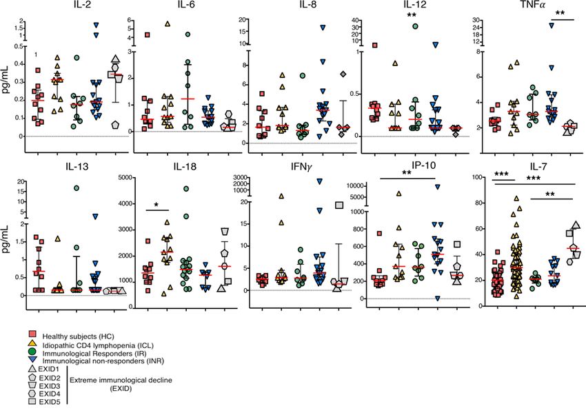
To further characterize the immunological milieu in EXID, we compared the plasma/serum levels of
21 cytokines/chemokines in EXID, HC, IR, INR, and patients with idiopathic CD4 lymphopenia (ICL,
n = 11), a heterogeneous clinical syndrome with low CD4+ T cells (median 27 cells/μl [IQR 2–217]) in
the absence of HIV-1 infection or any other known primary or acquired immunodeficiency (6). The cyto-
insight.jci.org https://doi.org/10.1172/jci.insight.127113 2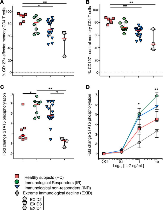
CLINICAL MEDICINE

Table 1. General characteristics of the subjects with extreme immune decline (EXID)

                               EXID 1                  EXID 2                 EXID 3A                EXID 4                  EXID 5            EXID (all) median
Age at enrollment               49                     18                      13                      25                     40                    25
Gender                           F                     M                       M                       M                       F                60% M/40% F
HIV-1 subtype                CRF BC                     C                      C                    CRF AG                    D                     na
Country                      Zambia                  Congo                  Congo                    Israel                Zambia                   na
ART (wks)                      536                    593                     357                     159                    254                    357
                          TFV/FTC/EFV            AZT/3TC/NLF             AZT/3TC/rLPV            TFV/FTC/RPV             TFV/FTC/EFV
                          ABC/3TC/rATZ           D4T/3TC/rLPV            TFV/FTC/cEVG            TFV/FTC/rLPV
ART                                                                                                                                               3 regimens
                          TFV/FTC/rATZ           ABC/3TC/rLPV            TFV/FTC/rDRV            AZT/3TC/rLPV
                            EFV/RAL            ABC/EFV/rATZ/RAL
CD4 before ART                 102                    315                      179                    587                     22                      179
CD4 week 192 ART                36                     52                      26                      68                     9                       36
                           A*30, 4301                A*68                   A*29, 68                A*03, 24               A*02, 68
                             B*18, 57               B*13, 18               B*07:05:01G              B*07, 18               B*42, 01
HLA
                            CW*07, 07             CW*04, 06                                        CW*07, 15                CW*17
                          DRB1*0804, 11           DRB1*11, 13                                     DRB1*12, 14             DRB1*08, 13
Peak HIV-1 pVL              >100,000                82,000                    13,163                  660                  >100,000                 82,000
(copies/ml)
A
 Perinatally infected subject. ABC, abacavir; AZT, zidovudine; cEVG, cobicistat/elvitegravir; D4T, stavudine; EFV, efavirenz; FTC, emtricitabine; FTV, ; NLF,
nelfinavir; RAL, raltegravir; rATZ, ritonavir/atazanavir; rDRV, ritonavir/darunavir; rLPV, ritonavir/loprinavir; RPV, rilpivirine; 3TC, lamivudine; TFV, tenofovir.

                                     kine profile of EXID was distinct, with lower concentrations of TNF-α compared with INR, and IL-12
                                     compared with HC. The most apparent difference in EXID, however, was that the levels of IL-7 were sig-
                                     nificantly higher compared with HC and IR (Figure 3 and Supplemental Table 2). We documented a neg-
                                     ative correlation between the serum IL-7 levels and the CD4+ T cell counts among HIV-1–infected subjects
                                     (Spearman’s ρ –0.53 [CI 95% –0.76 to –0.17], P < 0.004, Supplemental Figure 4) and IR, INR, and EXID
                                     segregated as 3 distinct clusters in relation to these 2 variables.
                                          Impaired IL-7/IL-7 receptor axis in EXID. To investigate the integrity of the IL-7 signaling axis, we
                                     compared the expression of the IL-7 receptor α-chain (CD127) and STAT5 phosphorylation status in
                                     response to IL-7 between the 3 EXID patients for which sufficient material was available (EXID2, -3,
                                     and -4) and other patients’ groups.
                                          The proportion of CD127+ effector memory CD4+ T cells was 55% in EXID, 94% in HC, 82% in IR, and
                                     71% in INR (Figure 4A and Supplemental Table 1). Similar differences in CD127 expression were also observed
                                     in the central memory CD4+ T cell subset (Figure 4B and Supplemental Table 1), but not in naive CD4+ T cells
                                     of the 4 groups. A significantly lower expression of CD127 was observed in CD8+ T cells from EXID compared
                                     with HC, IR, and INR (Supplemental Table 2). The transcriptional effects of IL-7 are mediated by STAT5
                                     phosphorylation (p-STAT5) and its subsequent nuclear translocation; stimulation with 10 ng of IL-7 resulted in
                                     a median 3.8-fold (IQR 3.5–4.5) increase in p-STAT5 mean fluorescence intensity (MFI) in CD4+ T cells from
                                     HC. Although INR and IR were both associated with a larger increase in p-STAT5 MFI in response to IL-7
                                     compared with HC (INR 5.8 [IQR 5.5–6.8]; IR 6.9 [IQR 5.7–7.2]), EXID had a blunted p-STAT5 induction of
                                     3.3-fold (IQR 2.4–3.4), significantly lower compared with HC, INR, and IR (Figure 4, C and D). Similar differ-
                                     ences were also noted with stimulation at lower concentrations of IL-7 (1 ng/ml, Figure 4D).
                                          EXID is associated with a distinct transcriptional profile. We next compared the transcriptional profile of periph-
                                     eral blood mononuclear cells (PBMCs) from EXID patients with those of HC, ICL (median CD4+ T cells 115
                                     cells/μl), and paired HIV/AIDS untreated subjects before ART (median HIV-1 pVL 196,418 copies/ml; CD4+
                                     T cells 45 cells/μl) and after 96 weeks of ART (median HIV-1 pVL < 40 copies/ml; CD4+ T cells 317 cells/μl).
                                          No differences in the number of differentially expressed genes (DEGs) were found between ART-treat-
                                     ed subjects and HC; in contrast, a considerable number of genes were differentially regulated in EXID
                                     compared with HC, HIV/AIDS untreated, and ART-treated IR (n = 138, n = 269, n = 313, respectively),
                                     while a more limited group of genes distinguished ICL and EXID transcriptomes (n = 24, Figure 5, A and
                                     B, and Supplemental Table 3). The EXID transcriptome was distinct from HC, HIV/AIDS untreated, and
                                     ART-treated IR; hierarchical clustering of a group of 320 genes with functional roles in innate or adaptive

insight.jci.org   https://doi.org/10.1172/jci.insight.127113                                                                                                       3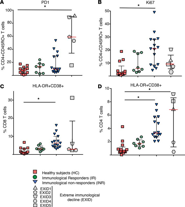
CLINICAL MEDICINE

Figure 1. CD4+ T cell trends after ART initiation. (A) CD4+ T cell count in immunological responders (IRs), immunological nonresponders (INRs), and
extreme immunological decline (EXID) after initiation of ART. The median (red bar), IQR (error bar), and each available CD4+ T cell count measurement
(symbols) is presented at each time point for IR (n = 8), INR (n = 15), and EXID (n = 5). (B) The median (red bar), IQR (error bar), and the difference in CD4+ T
cell count between week 0 (ART initiation) and week 96 or week 192 (symbols) is presented for each IR (n = 8), INR (n = 15), and EXID (n = 5) subject. Each
EXID subject is identified by a different gray-filled shape. *P ≤ 0.05 in the comparison indicated by the black horizontal line as determined by Mann-Whit-
ney U test; ns, nonsignificant difference.

                                    immune responses revealed 3 clusters of genes, differentially regulated in EXID, HC, HIV/AIDS untreated,
                                    and ART-treated IR subjects (red, green, and blue clusters in Figure 5A and Supplemental Tables 4 and 5).
                                         In addition, the list of the top best-scoring DEGs (log2 fold change [FC] ≥ 1.5 [18 genes] or ≤ –1.5 [20
                                    genes], adjusted P value ≤ 0.005; Figure 5C) in EXID compared with ART-treated IR subjects was enriched with
                                    genes involved in T cell function, TCR signaling, monocyte/macrophage function, autophagy, and cell migra-
                                    tion (Supplemental Tables 4 and 5, and Supplemental Figure 5). The DEGs between EXID and ART-treated
                                    IR were different from the DEGs found between EXID and HIV/AIDS untreated; in fact, 118 and 61 genes
                                    were respectively downregulated or upregulated in EXID compared with HIV/AIDS untreated subjects but
                                    only 1 of these genes was among the DEGs between EXID with ART-treated IR (TNFAIP2; Figure 5, B and C,
                                    and Supplemental Figure 5). This analysis demonstrated that the transcriptome in EXID is different from both
                                    HIV/AIDS untreated subjects with severe CD4+ T cell depletion (median 45 cells/μl) as well as from paired
                                    ART-treated subjects with CD4+ T cell reconstitution (317 cells/μl). Such a distinct transcriptome characterized
                                    by the differential regulation of genes and pathways involved in T cell and macrophage/monocyte functions
                                    as well as autophagy and inflammation prompted additional genetic work-up to evaluate the presence of rare
                                    hypomorphic genetic variants in genes involved in the control of similar immunological functions.
                                         Genetic screening for variants associated with primary immunodeficiencies in EXID. We performed targeted next-gen-
                                    eration sequencing (NGS) on 3 of the 5 EXID subjects for which sufficient material was available. EXID2
                                    had a heterozygous variant in the gene RTEL1 (regulator-of-telomeres elongation helicase-1, NM_016434-co-
                                    don_1322G>A, protein_W441*, combined annotation-dependent depletion [CADD] score 36), introducing a
                                    premature stop codon possibly resulting in nonsense-mediated decay. EXID2 did not have laboratory or clinical
                                    findings consistent with autosomal dominant telomeropathies classically associated with pathogenic RTEL1
                                    variants (dyskeratosis congenita, Online Mendelian Inheritance in Man [OMIM#615190] and pulmonary fibro-
                                    sis and/or bone marrow failure [OMIM#616373]) (7), but had a significant reduction in telomere length (TL)
                                    in total naive T cells (median TL: 6.5 vs. 8.1 kilobases [kb] in control of same age [CSA], 1–10 percentile),
                                    memory T cells (median TL: 3.8 vs. 6.5 kb in CSA,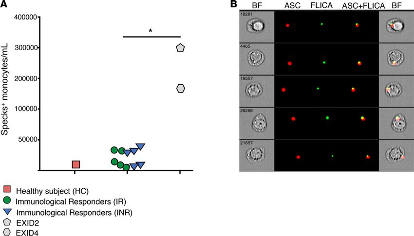
CLINICAL MEDICINE

                                  Figure 2. T cell immunophenotyping in healthy controls, IR, INR, and EXID. (A) Proportion of PD1-expressing memory
                                  CD4+ T cells (CD45RO+). (B) Proportion of Ki67-expressing memory CD4+ T cells (CD45RO+). (C) Proportion of HLA-
                                  DR+CD38+ CD8+ T cells. (D) Proportion of HLA-DR+CD38+ CD4+ T cells. The median (red bar), IQR (error bar), and each
                                  subject (symbols) is presented for healthy controls (HC) (n = 13), IR (n = 8), INR (n = 15), EXID (n = 5). Each EXID subject
                                  is identified by a different gray-filled shape. *P ≤ 0.05 in the comparison indicated by the black horizontal line as deter-
                                  mined by Kruskal-Wallis test followed by Dunn’s post-hoc test.

                                  (Supplemental Figure 6). Genetic screening in EXID3 did not reveal obvious causative variants but a heterozy-
                                  gous missense variant of unknown significance was identified in the gene ATG9A (autophagy-related protein
                                  9A, NM_024085-codon_2473G>A, protein_E825K, CADD score 33) (8). In EXID4 we identified a hetero-
                                  zygous pathogenic variant in the gene MEFV (pyrin, NM_000243-codon2080A>G, protein_M694V, CADD
                                  score 0.01) associated with Mediterranean fever (9, 10) and a rare variant in the gene NLRP7 (NLR family pyrin
                                  domain–containing 7, NM_001127255-codon_2156C>T, protein_A719V, CADD score 15.89) (11).
                                       Anti-lymphocyte autoantibodies in EXID. Because cytopenias are seen in autoimmune conditions and in
                                  primary immunodeficiencies (12, 13), we evaluated the presence of anti-lymphocyte autoantibodies pos-
                                  sibly causing autoimmune destruction, impaired homeostatic expansion, or altered trafficking of CD4+ T
                                  cells in EXID, IR, or INR. Luciferase immunoprecipitation system (LIPS) immunoassay did not identify
                                  autoantibodies against several T cell surface proteins (CD3δ, CD3ε, CD3γ, CD8α, CD8β, CD4, CTL4,
                                  CD127, IFN-γ receptor-1, and IL-2 receptor γ-chain) nor against Ro52/TRIM21, a common target of
                                  autoantibodies in several autoimmune diseases, in any of the subjects. As an alternative strategy, we devel-
                                  oped an NK cell–mediated antibody-dependent cellular cytotoxicity (ADCC) assay to reveal autoantibod-
                                  ies against any other epitopes expressed on CD4+ T cells. By this assay we found that CD4+ T cells were

insight.jci.org   https://doi.org/10.1172/jci.insight.127113                                                                                                 5
CLINICAL MEDICINE
Figure 3. Cytokine/chemokine levels in HC, IR, INR, and EXID and idiopathic CD4 lymphopenia. The median (red bar), IQR (error bar), and each subject
(symbols) is presented for HC (n = 10), idiopathic CD4 lymphopenia (ICL) (n = 11), IR (n = 8), INR (n = 15), and EXID (n = 5). IL-7 levels were measured in serum
for HC (n = 39), IR (n = 8), INR (n = 13), EXID (n = 5), and ICL (n = 60). Each EXID subject is identified by a different gray-filled shape. *P ≤ 0.05, **P ≤ 0.01,
***P ≤ 0.001 in the comparison indicated by the black horizontal line as determined by Kruskal-Wallis test followed by Dunn’s post-hoc test.
specifically depleted when incubated with plasma from EXID1 and EXID5 but not with PBS, HC, IR, or
INR plasma, nor plasma of other EXID patients (Figure 6). NK-mediated ADCC of CD4+ T cells was
more evident at higher effector/target (E/T) ratios, reaching an average of 50% of target CD4+ T cells at a
40:1 E/T ratio. In addition, CD4+ T cell killing was not observed when EXID1 or EXID5 plasma was IgG
depleted, while the IgG eluate was sufficient to provide CD4+ T cell NK-mediated ADCC (Figure 6). The
same subjects (EXID1 and EXID5) also had evidence of NK-mediated ADCC of CD19+ B cells in plasma;
such ADCC activity was abolished by IgG depletion of plasma but was not associated with a decrease in
the number of CD19-expressing B cells in peripheral blood.
Long-term follow-up of EXID subjects. Long-term clinical and immunological follow-up was available for
all EXID subjects. EXID2 and EXID3 did not have evidence of opportunistic infections, immune reconsti-
tution, nor specific clinical events.
The CD4+ T cells of EXID1 remained below the pre-ART level, 36 cells/μl (5%), with 1.25% naive after
approximately 5 years of consistent suppression of HIV-1 pVL, but in the following 7 years progressively
increased, reaching 607 cells/μl (19%), with 17% naive (Supplemental Figure 7). We evaluated the presence
of NK-mediated ADCC in plasma samples collected before and after the CD4+ T cell recovery (week 188 and
week 551 after ART initiation, respectively). No NK-mediated ADCC CD4+ T cell killing was found in the latter
samples (Figure 6), supporting the causative role of such autoantibodies in hampering immune reconstitution.
EXID5’s CD4+ T cells, after approximately 226 weeks of ART, started to progressively increase (high-
est CD4+ T cell counts: 119 cells/μl, 12% on week 249 after ART) (Supplemental Figure 7). Alongside such
CD4+ T cell reconstitution, EXID5 developed mental status decline with radiological evidence of basilar
arteritis and cerebral ischemic lesions. A clinical diagnosis of CNS tuberculosis was formulated but the
patient was lost to follow-up after initiation of antituberculous therapy.
insight.jci.org https://doi.org/10.1172/jci.insight.127113 6CLINICAL MEDICINE
Figure 4. IL-7 signaling axis in HC, IR, INR, and EXID. (A) Proportion of CD127-expressing effector memory CD4+ T cells
(CD27–CD45RO+). (B) Proportion of CD127-expressing central memory CD4+ T cells (CD27+CD45RO+). (C) Fold change in
STAT5 phosphorylation upon IL-7 stimulation (10 nM). (D) Dose-response of STAT5 phosphorylation at different concen-
trations of IL-7. The median (red bar), IQR (error bar), and each subject (symbols) is presented for HC (n = 6), IR (n = 8),
INR (n = 15), and EXID (n = 3). Each EXID subject is identified by a different gray-filled shape; sufficient material for the
analysis was available from EXID2, -3, and -4. *P ≤ 0.05, **P ≤ 0.01 in the comparison indicated by the black horizontal
line as determined by Kruskal-Wallis test followed by Dunn’s post-hoc test.
EXID4 maintained stable CD4+ T cells below the pre-ART level, 68 cells/μl (11%) with 4.3% naive
(Supplemental Material Case Reports), despite 3 years of consistent suppression of HIV-1 pVL, but
eventually developed right eye pain due to an infiltrating, hypermetabolic right orbital mass causing
displacement of the right superior extraocular muscle and modeling of the superior right orbital bone
as seen on MRI and PET-CT. A biopsy of the mass documented dense fibrosis and granulomas of epi-
thelioid myeloid cells consistent with idiopathic orbital inflammation (IOI) (Supplemental Figure 8).
This rare clinical entity has unknown etiology, but is associated with other inflammatory diseases and
can cause a local infiltrating, destructive sclerosing process (14, 15). His inguinal lymph node histology
and quantitative imaging documented a prominent IFN-α signature, increased collagen-1 deposition
with accumulation of myeloid cells, and overall preserved CD4+ T cell density in the follicular area and
T cell zone compared with HC and paired HIV/AIDS untreated and ART-treated IR subjects (Supple-
mental Figures 8 and 9). Infliximab treatment, indicated in IOI (16), resulted in regression of the orbital
insight.jci.org https://doi.org/10.1172/jci.insight.127113 7CLINICAL MEDICINE
Figure 5. Transcriptional profile of peripheral blood mononuclear cells from EXID, HC, and paired HIV/AIDS subjects before and after ART. (A) Hier-
archical clustering of 320 genes with functional role in innate or adaptive immune responses in HC (n = 5), EXID (n = 5), HIV/AIDS untreated (n = 5), and
ART-treated IR subjects (n = 5). Each EXID subject is identified by a different gray-filled shape. (B) Edwards-Venn 4-set diagram depicting the number and
the intersection of differentially regulated genes (DEGs) in 4 comparisons (EXID vs. HC [green], EXID vs. ICL [yellow], EXID vs. IR [blue], and EXID vs. HIV/
AIDS untreated [red]). (C) Volcano plot of DEGs in EXID compared with ART-treated HIV/AIDS subjects. The shaded area identifies the top differentially
downregulated (log2 fold change ≤ 1.5, 20 genes) or upregulated (log2 fold change ≤ 1.5, 18 genes) genes (listed in Supplemental Table 5).
mass, and was associated with a progressive recovery of CD4+ T cells reaching 454 cells/μl (24%),
with proportions of naive and effector memory CD4+ T cells of 0.21% and 80.9%, respectively, after 12
months of infliximab (Supplemental Figures 8 and 9).
In the context of myeloid granulomas and an underlying inflammatory disorder possibly causing CD4+
T cell pyroptosis (17, 18), we performed an analysis of inflammasome and caspase-1 activation in mono-
cytes and T cells of EXID2 and EXID4 and found that monocytes had an increased number of apopto-
sis-associated speck-like protein containing a C-terminal caspase recruitment domain (ASC) colocalizing
with caspase-1 compared with HC, IR, and INR (EXID2 124,852 cells/ml, EXID4 69,858 cells/ml, com-
pared with 3096 cells/ml, 7710 cells/ml, and 6159 cells/ml, in HC, IR, and INR, respectively; Figure 7, A
and B). CD4+ T cells had a higher level of caspase-1 activation in EXID and INR compared with IR and
HC (EXID2 7.3%, EXID4 2.8%, and INR 4.7%, compared with 2.5% and 1.9% in HC and IR, respective-
ly); conversely, caspase-1 activation was negligible and not significantly different in CD8+ T cells from HC,
IR, INR, or EXID (Supplemental Figure 10).
Discussion
Herein, we report, for the first time to our knowledge, on a paradoxical decline in CD4+ T cells occurring
after initiation of ART, which persisted despite consistent suppression of HIV pVL and changes in ART
regimens. A comprehensive prospective clinical, immunological, and genetic characterization of these
patients led to the identification of what we believe are novel mechanisms causing CD4+ T cell depletion in
ART-treated individuals, specifically autoimmunity and inflammasome/caspase-1 activation.
The hallmark of HIV/AIDS, in the context of uncontrolled HIV-1 viral replication, is the progres-
sive depletion of CD4+ T cells; direct viral cytopathic effects, apoptosis, pyroptosis, and immune-mediated
lysis are among the mechanisms evoked to explain such depletion. Irrespective of the specific role of each
of these mechanisms, ART-mediated suppression of HIV-1 replication results in reconstitution of CD4+
T cells in all HIV-1–infected subjects. In fact, although the extent of immune reconstitution can vary, a
decline of CD4+ T cells on ART has never been described to the best of our knowledge, except as drug-re-
lated cytotoxicity of combined tenofovir-didanosine treatment (19).
insight.jci.org https://doi.org/10.1172/jci.insight.127113 8CLINICAL MEDICINE
Figure 6. NK cell–mediated antibody-dependent cellular cytotoxicity of EXID1 plasma. Plotted is the percentage of killing (percentage difference in
cell counts between untreated control and any experimental condition) of (A) CD4+ T cells, (B) CD8+ T cells, and (C) CD19+ B cells incubated with PBS,
rituximab, and plasma from a heathy subject, IR (n = 3), INR (n = 4), or EXID (n = 5) at an effector/target ratio of 10:1 (PBS and rituximab) or 40:1 for
all other experimental conditions. Each EXID subject is identified by a different gray-filled shape. Antibody-dependent cellular cytotoxicity (ADCC)
activity was noted only in plasma of EXID1 and EXID5 for which additional experimental conditions were included.
Therefore, we identified an immunological outcome of ART, extreme immunological decline or
EXID, in subjects infected with non-B HIV-1 subtypes, an observation suggesting a role for a specific
combination of both host and viral factors in the pathogenesis of such rare and perplexing immunologi-
cal outcome. EXID was not associated with previously described predictors of poor immune response, as
it occurred in young individuals, with higher nadir CD4+ and without any consistent increase in cycling
CD4+ T cells or activated CD4+ or CD8+ T cells. In fact, rather than an extreme form of INR, EXID
appears distinct from INR, as it is characterized by the following: decline of CD4+ T cell counts after
initiation of ART, which remained consistently below the pre-ART level for at least 192 weeks despite
constant suppression of pVL below the limit of detection; profound reduction of naive and relative
expansion of effector memory CD4+ T cells; distinct immunophenotypic (low Ki67, high PD-1) and
plasma biomarker profile (low TNF and IL-12); and high serum IL-7 levels with downregulated IL-7
receptor expression and impaired STAT5 phosphorylation.
The perturbation in the IL-7 axis and the lack of adequate CD4+ T cell expansion in response to
increased IL-7 levels in EXID was not associated with an increase in inflammatory cytokines IL-6 and
IL-1β, as proposed in INR (5), and appears to be more in line with those observed in ICL (20). It is con-
ceivable that the blunted IL-7 axis can contribute to EXID, but it is also possible that it develops in response
to a persistent compensatory mechanism that fails to achieve the homeostatic expansion of CD4+ T cells
because of additional mechanisms preventing adequate T cell reconstitution (i.e., antilymphocyte autoanti-
bodies, aberrant inflammatory responses in genetically predisposed individuals).
The transcriptome analysis further documented a distinct transcriptional signature of EXID compared
with paired viremic and ART-treated immunological responders characterized by different regulation of
genes involved in T cell function, TCR signaling, monocyte/macrophage function, autophagy, and cell
migration. In contrast, EXID’s transcriptional signature was similar to that of subjects with ICL, suggest-
ing some shared mechanisms responsible for the depletion of CD4+ T cells possibly independent from
HIV-1 direct cytopathic effects. Because EXID, ICL, and viremic HIV-1 individuals had a similar level of
lymphopenia it is unlikely the changes in transcriptional signature between these groups were driven by the
differences in relative numbers of CD4+ T cell counts.
insight.jci.org https://doi.org/10.1172/jci.insight.127113 9CLINICAL MEDICINE
Figure 7. Quantification of canonical inflammasome activation in CD14+ monocytes. PBMCs from IR (n = 5), INR (n = 5), EXID2, EXID4, and a healthy
subject were incubated with a fluorochrome-labeled inhibitor of caspase-1 (FAM-FLICA), stained for monocyte identification with antibodies against
CD14 and the intracellular apoptosis-associated speck-like protein containing a C-terminal caspase recruitment domain (ASC), and evaluated by imaging
flow cytometry. (A) The number of monocytes showing spontaneous ASC speck formation was quantified after application of the feature Area Threshold
versus Modulation Morphology in the CD14-expressing monocyte gate, by ImageStream Data Exploration and Analysis Software (IDEAS 6.2.64.0, Milli-
poreSigma). Each EXID subject is identified by a different gray-filled shape. *P ≤ 0.05 in the comparison indicated by the black horizontal line as deter-
mined by Mann-Whitney U test. (B) Representative images showing colocalization of active caspase-1 with ASC specks were selected after Bright Detail
Similarity was applied in the ASC speck gate. Images show, respectively: brightfield (BF), ASC and FLICA fluorescence followed by a composite image
containing BF, and the fluorescence of ASC and FLICA merged.
The clinical follow-up provided important clues to the pathogenesis of EXID; a sustained recovery of
CD4+ T cell counts was associated with the loss of anti-lymphocyte autoantibodies causing NK-mediated
ADCC of CD4+ T cells in EXID1. An identical mechanism was also demonstrated in EXID5. The role
of ADCC causing CD4+ T cell depletion has been previously documented in HIV-1–infected individuals
(21–23) but its role in the pathogenesis of AIDS or immunological failure remains elusive. The evidence
of recovery of CD4+ T cell counts after loss of such anti-lymphocyte autoantibodies has implication for
the spontaneous reversibility of such a condition or for potential therapeutic interventions (i.e., rituximab
or plasmapheresis). Importantly, the loss of anti-lymphocyte autoantibodies and consequent spontaneous
recovery of CD4+ T cells occurred after more than 5 years of consistent suppression of pVL and in one case
(EXID5) was also associated with symptoms and radiological findings consistent with immune reconstitu-
tion inflammatory syndrome that led to significant morbidity.
A spontaneous recovery of CD4+ T cell counts was not observed in any of the other 3 EXID subjects.
However, the long-term follow-up of EXID4 revealed a different mechanism responsible for the CD4+ T cell
decline; an orbital inflammatory disorder (IOI) prompted initiation of infliximab that resulted not only in the
expected improvement of the orbital inflammatory mass but also in an unexpected reconstitution of CD4+
T cells. The CD4+ T cell recovery on infliximab, its pattern (lack of naive and expansion of effector mem-
ory CD4+ T cells), the increased inflammasome/caspase-1 activation along with lymph node fibrosis, and
rare variants in genes controlling inflammasome/caspase-1 activation (NLRP7 and MEFV) (9–11), suggest
a perturbed inflammatory response resulting in altered CD4+ T cell trafficking and homeostasis rather than
autoimmune phenomena, which were in fact not documented in this subject. Interestingly, in a nonhuman
primate model of acute SIV infection, adalimumab (TNF-α–blocking monoclonal antibody similar to inflix-
imab) reduced lymph node fibrosis and preserved CD4+ T cells (24), while studies on TNF-α blockade in
HIV-1–infected patients documented the safety of such intervention and an increase in CD4+ T cell counts
in some case reports (25–28). EXID2 was found to have a remarkable inflammasome/caspase-1 activation
along with severe decrease in TL, with an RTEL1 frameshift variant introducing an early stop codon. Such a
insight.jci.org https://doi.org/10.1172/jci.insight.127113 10CLINICAL MEDICINE
clinical scenario may resemble the recently reported novel quantitative and qualitative primary T cell immu-
nodeficiency in which telomere shortening caused by specific genetic defects reaches a critical cellular thresh-
old causing depletion of CD4+ T cells from intrinsic and extrinsic apoptosis (29). It is conceivable that, as seen
in this model, in EXID2, the telomere attrition caused by T cell replication in a perinatal HIV-1 infection with
persistent vigorous inflammasome/caspase-1 activation could not be fully compensated for in the context
of the frameshift variant in RTEL1 and reached a critical threshold in naive and memory T cells (7, 29, 30).
Despite the long-term follow-up and the prospective nature of our study, the small sample size and
the variability in some immunological correlates imposed by the differences in the mechanism respon-
sible for EXID limited our ability to generalize our findings. Nevertheless, we identified 2 mechanisms
contributing to CD4+ T cell depletion in subjects with suppressed HIV-1 replication, namely (a) autoim-
mune depletion of CD4+ T cells, and (b) pyroptosis, impaired trafficking, and expansion of CD4+ T cells
in altered inflammasome/caspase-1 activation.
EXID is a rare immunological outcome of ART that may occur in patients with specific host and viral
factors; EXID results in unique clinical challenges but can also reveal and help elucidate the specific mech-
anisms determining CD4+ T cell depletion during uncontrolled HIV-1 replication from the ones limiting the
degree of reconstitution of CD4+ T cells on ART.
Methods
Study participants. Patients referred to the NIH Clinical Center were enrolled in NIAID IRB–approved proto-
cols after informed consent was obtained (NCT02147405, NCT00867269, NCT00001467, NCT00001281,
NCT00286767, and NCT00789009). Written informed consent was provided for the patient’s photo appearing
in the supplemental material. IRs and INRs were defined as subjects with less than 100 CD4+ T cells/μl on
enrollment who achieved greater than or less than 270 CD4+ T cells/μl after 96 weeks of ART, respectively.
EXID was defined as a progressive and persistent decrease in CD4+ T cell counts despite 192 weeks of ART in
the absence of malignancy and regardless of pre-ART CD4+ T cell counts. ICL is defined by less than 300 CD4+
T cells/μl in the absence of HIV-1 infection or any other known primary or acquired immunodeficiency.
Immunophenotyping and cytokine/chemokine quantification. PBMCs were stained with fluorescently
labeled antibodies against cell surface markers subsequently detected by flow cytometry. Sufficient material
for the analysis of CD127 expression and STAT5 phosphorylation in response to IL-7 stimulation was
available from EXID2, -3, and -4. Biomarkers were measured by electrochemiluminescence with a custom
multiplex kit (Meso Scale Discovery).
Transcriptome analysis and targeted NGS. RNA was extracted from PBMCs of 5 EXID, 5 HC, 5 ICL,
and 5 HIV/AIDS subjects prior to start and after 96 weeks of ART. The libraries were run on an Illumina
HiSeq 2500 sequencer and reads were mapped to the human genome assembly NCBI-hg38 using Hisat2
(31). RNA sequence information reported in this study is deposited in NCBI’s Gene Expression Omnibus
(GEO GSE125223). The networks were generated through the use of IPA (QIAGEN Inc., https://www.
qiagenbioinformatics.com/products/ingenuity-pathway-analysis).
Targeted NGS of exons and flanking regions of 318 genes causing primary immunodeficiencies was
performed as previously described (32). Variants with a population frequency of less than 0.5% in different
data sets (1000-Genomes, EXAC, NHLBI-6500, GnomAD) were scored using the CADD score.
LIPS for measuring anti-lymphocyte antibodies and ADCC assays. Autoantibodies against CD8α, CD8β,
CD3δ, CD3ε, CD3γ, CD4, CTL4, IL-7 receptor, IFN-γ receptor-1, IL-2 receptor γ-chain, and Ro52/TRIM21
were measured by LIPS employing luciferase-tagged antigens as previously described (33). For the ADCC
assay, NK cells were isolated from healthy subjects by immunomagnetic negative selection (EasySep Human
NK Cell Isolation Kit, Stemcell Technologies) and incubated overnight with 1,000 U/ml IL-2. PBMCs from
the same donor were used as targets after resting and CFSE labeling. Targets were incubated with or without
NK in the presence of PBS, anti-CD20, or patient’s plasma, then stained for CD3, CD4, CD8, and CD19 and
analyzed by flow cytometry. Cells were enumerated using counting beads and percentage killing was calcu-
lated as the difference in the absolute number of CFSE-labeled targets incubated with and without NK. Total
IgG was purified from plasma with Protein A/G agarose affinity resin (Thermo Fisher Scientific).
Immunohistochemistry, quantitative imaging, and inflammasome analysis. Immunohistochemical staining
and quantification were performed as previously described (34, 35). Briefly, immunohistochemistry was
performed on 5-μm tissue sections after heat-induced epitope retrieval followed by incubation with anti-
body against myxovirus resistance gene A (MxA), or collagen 1, or CD4 in combination with CD163 and
insight.jci.org https://doi.org/10.1172/jci.insight.127113 11CLINICAL MEDICINE
CD68. Slides were washed, blocked, and incubated with rabbit or mouse Polink-1 or -2 horseradish per-
oxidase and developed with Immpact DAB (3,3′-diaminobenzidine; Vector Laboratories). All slides were
washed, counterstained, mounted, and scanned using the ScanScope CS System (Aperio Technologies),
yielding high-resolution data from the entire tissue section. The percentage area positive for MxA, collagen
1, and CD4+ T cells was quantified using CellProfiler version 3.1.5.
For the inflammasome activation analysis, cells were acquired using an Amnis ImageStreamX Mark II
imaging flow cytometer and the integrated software INSPIRE (MilliporeSigma) was used for data collection.
Additional information on immunohistochemistry and inflammasome analysis is in the supplemental material.
Statistics. Comparisons of continuous variables between different groups was performed by using non-
parametric tests (Kruskal-Wallis test, Mann-Whitney U test). All hypothesis were 2-tailed and the criterion for
statistical significance for these comparisons was set to P ≤ 0.05; adjustment for multiplicity with Dunn’s mul-
tiple-comparisons test was applied for the immunophenotypic analysis of T cell subsets and the analysis of
the cytokines’ plasma/serum concentration whenever more than 2 groups were compared. Spearman’s rank
correlation was used to measure the linear association between IL-7 serum levels and CD4+ T cell counts.
Differential expression analysis was performed using the Bioconductor package DESeq2 (36). Genes
were determined to be significantly differentially expressed if they passed multiple test correction using the
Benjamini-Hochberg (37) adjusted P value of ≤0.05.
Author contributions
AL and IS designed the study, provided clinical care to the enrolled patients, analyzed data, and wrote the
manuscript. CSW, SLL, CG, HM, PDB, APD, AR, CAM, SLA, and CD conducted experiments, analyzed
data, and/or acquired data. IL, JB, JL, MM, MVA, YM, CM, and SC provided clinical care to the enrolled
patients. All authors reviewed and approved the manuscript prior to submission.
Acknowledgments
This work has been supported in part by the intramural research program of the NIAID, NIH and in part
with federal funds from the National Cancer Institute, NIH, under Contract No. HHSN261200800001E.
The content of this publication does not necessarily reflect the views or policies of the Department of
Health and Human Services, nor does mention of trade names, commercial products, or organizations
imply endorsement by the US Government.
Address correspondence to: Andrea Lisco or Irini Sereti, 10 Center Drive, Building 10, Room 6D44G (A.
Lisco) or Room 11B17 (I. Sereti), Bethesda, Maryland 20892. USA. Phone: 301.761.7122; Email: Andrea.
lisco@nih.gov (A. Lisco). Phone: 301.496.5533; Email: Irini.Sereti@nih.gov (I. Sereti).
1. Engsig FN, et al. Long-term mortality in HIV patients virally suppressed for more than three years with incomplete CD4 recov-
ery: a cohort study. BMC Infect Dis. 2010;10:318.
2. Kelley CF, et al. Incomplete peripheral CD4+ cell count restoration in HIV-infected patients receiving long-term antiretroviral
treatment. Clin Infect Dis. 2009;48(6):787–794.
3. Robbins GK, et al. Incomplete reconstitution of T cell subsets on combination antiretroviral therapy in the AIDS Clinical Trials
Group protocol 384. Clin Infect Dis. 2009;48(3):350–361.
4. Lederman MM, et al. Immunologic failure despite suppressive antiretroviral therapy is related to activation and turnover of
memory CD4 cells. J Infect Dis. 2011;204(8):1217–1226.
5. Shive CL, et al. Inflammatory cytokines drive CD4+ T-cell cycling and impaired responsiveness to interleukin 7: implications for
immune failure in HIV disease. J Infect Dis. 2014;210(4):619–629.
6. Zonios DI, et al. Idiopathic CD4+ lymphocytopenia: natural history and prognostic factors. Blood. 2008;112(2):287–294.
7. Deng Z, et al. Inherited mutations in the helicase RTEL1 cause telomere dysfunction and Hoyeraal-Hreidarsson syndrome. Proc
Natl Acad Sci USA. 2013;110(36):E3408–E3416.
8. Xie Z, Klionsky DJ. Autophagosome formation: core machinery and adaptations. Nat Cell Biol. 2007;9(10):1102–1109.
9. Marek-Yagel D, et al. Clinical disease among patients heterozygous for familial Mediterranean fever. Arthritis Rheum.
2009;60(6):1862–1866.
10. Jéru I, et al. The risk of familial Mediterranean fever in MEFV heterozygotes: a statistical approach. PLoS One.
2013;8(7):e68431.
11. Khare S, et al. An NLRP7-containing inflammasome mediates recognition of microbial lipopeptides in human macrophages.
Immunity. 2012;36(3):464–476.
12. Seidel MG. Autoimmune and other cytopenias in primary immunodeficiencies: pathomechanisms, novel differential diagnoses,
and treatment. Blood. 2014;124(15):2337–2344.
13. Henriksson G, Manthorpe R, Bredberg A. Antibodies to CD4 in primary Sjögren’s syndrome. Rheumatology (Oxford).
insight.jci.org https://doi.org/10.1172/jci.insight.127113 12CLINICAL MEDICINE
2000;39(2):142–147.
14. Yuen SJ, Rubin PA. Idiopathic orbital inflammation: distribution, clinical features, and treatment outcome. Arch Ophthalmol.
2003;121(4):491–499.
15. Monaghan TM, Albanese G, Kaye P, Thomas JD, Abercrombie LC, Moran GW. Orbital inflammatory complications of
Crohn’s disease: a rare case series. Clin Med Insights Gastroenterol. 2018;11:1179552218757512.
16. Garrity JA, Coleman AW, Matteson EL, Eggenberger ER, Waitzman DM. Treatment of recalcitrant idiopathic orbital inflam-
mation (chronic orbital myositis) with infliximab. Am J Ophthalmol. 2004;138(6):925–930.
17. Doitsh G, et al. Cell death by pyroptosis drives CD4 T-cell depletion in HIV-1 infection. Nature. 2014;505(7484):509–514.
18. Muñoz-Arias I, Doitsh G, Yang Z, Sowinski S, Ruelas D, Greene WC. Blood-derived CD4 T cells naturally resist pyroptosis
during abortive HIV-1 infection. Cell Host Microbe. 2015;18(4):463–470.
19. Negredo E, et al. Unexpected CD4 cell count decline in patients receiving didanosine and tenofovir-based regimens despite
undetectable viral load. AIDS. 2004;18(3):459–463.
20. Puronen CE, et al. Decreased interleukin 7 responsiveness of T lymphocytes in patients with idiopathic CD4 lymphopenia. J Infect
Dis. 2012;205(9):1382–1390.
21. Weinhold KJ, Lyerly HK, Stanley SD, Austin AA, Matthews TJ, Bolognesi DP. HIV-1 GP120-mediated immune suppression
and lymphocyte destruction in the absence of viral infection. J Immunol. 1989;142(9):3091–3097.
22. Luo Z, et al. Pathological role of anti-CD4 antibodies in HIV-infected immunologic nonresponders receiving virus-suppressive
antiretroviral therapy. J Infect Dis. 2017;216(1):82–91.
23. Lyerly HK, et al. Anti-GP 120 antibodies from HIV seropositive individuals mediate broadly reactive anti-HIV ADCC. AIDS
Res Hum Retroviruses. 1987;3(4):409–422.
24. Tabb B, et al. Reduced inflammation and lymphoid tissue immunopathology in rhesus macaques receiving anti-tumor necrosis
factor treatment during primary simian immunodeficiency virus infection. J Infect Dis. 2013;207(6):880–892.
25. Hsu DC, et al. A paradoxical treatment for a paradoxical condition: infliximab use in three cases of mycobacterial IRIS. Clin
Infect Dis. 2016;62(2):258–261.
26. Calabrese LH, Zein N, Vassilopoulos D. Safety of antitumour necrosis factor (anti-TNF) therapy in patients with chronic viral
infections: hepatitis C, hepatitis B, and HIV infection. Ann Rheum Dis. 2004;63 Suppl 2:ii18–ii24.
27. Habib SF, Hasan MZ, Salam I. Infliximab therapy for HIV positive Crohn’s disease: A case report. J Crohns Colitis.
2009;3(4):302–304.
28. Beltrán B, Nos P, Bastida G, Iborra M, Hoyos M, Ponce J. Safe and effective application of anti-TNF-alpha in a patient infected
with HIV and concomitant Crohn’s disease. Gut. 2006;55(11):1670–1671.
29. Wagner CL, et al. Short telomere syndromes cause a primary T cell immunodeficiency. J Clin Invest. 2018;128(12):5222–5234.
30. Côté HC, et al. Leukocyte telomere length in HIV-infected and HIV-exposed uninfected children: shorter telomeres with uncon-
trolled HIV viremia. PLoS One. 2012;7(7):e39266.
31. Kim D, Langmead B, Salzberg SL. HISAT: a fast spliced aligner with low memory requirements. Nat Methods. 2015;12(4):357–360.
32. Stoddard JL, Niemela JE, Fleisher TA, Rosenzweig SD. Targeted NGS: a cost-effective approach to molecular diagnosis of
PIDs. Front Immunol. 2014;5:531.
33. Burbelo PD, Lebovitz EE, Notkins AL. Luciferase immunoprecipitation systems for measuring antibodies in autoimmune and
infectious diseases. Transl Res. 2015;165(2):325–335.
34. Schuetz A, et al. Initiation of ART during early acute HIV infection preserves mucosal Th17 function and reverses HIV-related
immune activation. PLoS Pathog. 2014;10(12):e1004543.
35. Carpenter AE, et al. CellProfiler: image analysis software for identifying and quantifying cell phenotypes. Genome Biol. 2006;7(10):R100.
36. Love MI, Huber W, Anders S. Moderated estimation of fold change and dispersion for RNA-seq data with DESeq2. Genome
Biol. 2014;15(12):550.
37. Benjamini Y, Drai D, Elmer G, Kafkafi N, Golani I. Controlling the false discovery rate in behavior genetics research. Behav
Brain Res. 2001;125(1–2):279–284.
insight.jci.org https://doi.org/10.1172/jci.insight.127113 13You can also read


