JBC Papers in Press. Published on April 6, 2018 as Manuscript RA118.003200 The latest version is at ...
←
→
Page content transcription
If your browser does not render page correctly, please read the page content below
JBC Papers in Press. Published on April 6, 2018 as Manuscript RA118.003200
  The latest version is at http://www.jbc.org/cgi/doi/10.1074/jbc.RA118.003200
                                       Oxidants, Mitochondrial Bioenergetics and MPTP

       Oxidative stress alters mitochondrial bioenergetics and modifies pancreatic cell death
            independently of cyclophilin D, resulting in an apoptosis-to-necrosis shift
   2
   Jane A. Armstrong*, 1Nicole J. Cash*, 1Yulin Ouyang, 1Jack C. Morton, 1Michael
 Chvanov, 2Diane Latawiec, 2Muhammad Awais, Alexei V. Tepikin, 2Robert Sutton and
                                  1
                                    David N. Criddle

    Departments of 1Cellular & Molecular Physiology and 2Molecular and Clinical Cancer
        Medicine, Institute of Translational Medicine, University of Liverpool, UK,

                 Running title: Oxidants, Mitochondrial Bioenergetics and MPTP

Correspondence to Dr. David N. Criddle, Department of Cellular & Molecular Physiology,
Institute of Translational Medicine, University of Liverpool, UK. E-mail: criddle@liv.ac.uk.
Telephone: ++44 151 794 5304. Fax: ++44 151 794 5327.

* contributed equally to this study

Keywords: Oxidative stress, mitochondrial dysfunction, cyclophilin D, MPTP, Seahorse,

                                                                                                         Downloaded from http://www.jbc.org/ by guest on October 30, 2018
pancreatic acinar cell

ABSTRACT                                             of Δᴪm, and cell death were not ameliorated
Mitochondrial dysfunction lies at the core of        by genetic deletion of CypD or by its acute
acute pancreatitis (AP). Diverse AP stimuli          inhibition with cyclosporine A. These results
induce Ca2+-dependent formation of the               indicate that oxidative stress alters
mitochondrial permeability transition pore           mitochondrial bioenergetics and modifies
(MPTP), a solute channel modulated by                pancreatic acinar cell death. A shift from
cyclophilin D (CypD), whose formation                apoptosis to necrosis appears to be associated
causes ATP depletion and necrosis. Oxidative         with     decreased     mitochondrial     spare
stress reportedly triggers MPTP formation            respiratory capacity and ATP production,
and is elevated in clinical AP, but how              effects that are independent of CypD-
reactive oxygen species influence cell death         sensitive MPTP formation.
is unclear. Here, we assessed potential MPTP
involvement in oxidant-induced effects on            INTRODUCTION
pancreatic acinar cell bioenergetics and fate.                Acute pancreatitis (AP) is a severe
H2O2 application promoted acinar cell                inflammatory disorder, triggered primarily by
apoptosis at low concentrations (1–10 μM),           excessive gallstones or excessive alcohol
whereas higher levels (0.5–1 mM) elicited            consumption, that can lead to a systemic
rapid necrosis. H2O2 also decreased the              inflammatory response syndrome, multiple
mitochondrial NADH/FAD+ redox ratio and              organ failure and death of the patient (1). It is
Δᴪm in a concentration-dependent manner (10          one of the most common causes of
μM–1 mM H2O2), with maximal effects at               emergency hospital          admission from
500 μM H2O2. H2O2 decreased the basal O2             gastrointestinal causes in the US, with an
consumption rate of acinar cells, with no            annual cost of U$ 2.6 billion (2). However,
alteration of ATP turnover at ˂ 50 μM H2O2.          the underlying pathophysiology of AP is
However, higher H2O2 levels (≥ 50μM)                 incompletely understood and currently there
diminished spare respiratory capacity and            is no specific therapy (3). The initial focus of
ATP turnover, and bioenergetic collapse,             damage is considered to be the pancreatic
ATP depletion, and cell death ensued.                acinar cell, which manifests pathological
Menadione exerted detrimental bioenergetic           changes including premature protease
effects similar to those of H2O2, which were         activation, vacuolisation and necrotic cell
inhibited by the antioxidant N-acetylcysteine.       death pathway activation (4). Diverse AP
Oxidant-induced bioenergetic changes, loss           precipitants have been shown to induce Ca2+-

                                                 1
Oxidants, Mitochondrial Bioenergetics and MPTP
dependent mitochondrial depolarisation, loss H2O2 (1 µM - 1 mM) to pancreatic acinar cells
of ATP production and acinar cell necrosis. caused a time-dependent increase of
Recent studies have demonstrated the central intracellular ROS that attained a maximal
role of mitochondrial permeability transition response at 500 µM (Figure 1A). The
pore (MPTP) formation in AP (5,6), a Ca2+- characteristics of the response varied with the
sensitive channel modulated by cyclophilin D H2O2 concentration applied; rapid elevations
that allows movement of solutes ˂1.5kD in were detected in pancreatic acinar cells at
and out of the mitochondria (7). Although higher (500 µM - 1 mM) H2O2 levels,
elevation of mitochondrial matrix Ca2+ is the whereas lower levels (1 - 10 µM) induced
principal trigger for MPTP formation, more slowly developing rises. Accordingly,
oxidative stress has been implicated in pore stimulation of acinar cells with H2O2 elicited
opening (7,8). For example, fibroblasts and cell death pathway activation that varied
hepatocytes from mice lacking CypD (Ppif-/-) according to the severity of insult applied.
were partially protected from H2O2 induced Lower concentrations (1 - 10 µM) of H2O2
cell death (9-11), although whether the MPTP preferentially promoted time- and
is modulated by oxidative stress in the concentration-dependent apoptotic cell death
pancreas is unclear. pathway activation, with relatively little
Oxidative stress is a prominent induction of necrosis until later time-points
feature of AP in preclinical and clinical (Figure 1B). Conversely, higher H2O2
Downloaded from http://www.jbc.org/ by guest on October 30, 2018
studies (12). Increases of reactive oxygen concentrations (500 µM - 1 mM) induced
species (ROS) and their by-products were rapid necrosis with minimal transient
detected in patients, concurrent with a elevation of apoptotic cell death; necrotic cell
suppression of antioxidant defences, which death exceeded twice the control value at 2
correlated with disease severity (13,14). hours and was sustained over the
However, the precise role of ROS in experimental period (Figure 1B).
pancreatic pathophysiology remains unclear
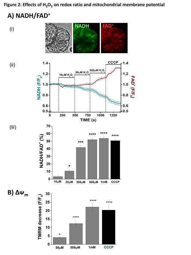
and clinical trials of antioxidant therapy have Concentration-dependent inhibitory
demonstrated no clear benefit in the treatment effects of H2O2 on the redox ratio
of AP (12). We have shown that generation of (NADH/FAD+) and mitochondrial membrane
ROS may constitute a protective mechanism potential (∆ᴪm) - In confocal microscopy
that disposes of stressed pancreatic acinar experiments cellular NADH and FAD+
cells, since bile acid-induced ROS production autofluorescence was distributed in a typical
increased apoptosis with a concomitant mitochondrial arrangement, as described
reduction of necrosis (15). The extent of previously in isolated pancreatic acinar cells
pancreatic necrosis is linked to more severe (17) (Figure 2Ai). Application of H2O2 (10
clinical disease (16), and the µM - 1 mM) induced concentration-
apoptosis/necrosis balance may therefore be dependent decreases of NADH, effects
an important determinant of AP progression. mirrored by increases of FAD+ (Figures
The aim of the present study was to 2Aii). These bioenergetic changes were
determine the effects of oxidants on maximal at 500 µM; subsequent application
pancreatic acinar cell bioenergetics, of the protonophore CCCP (10 µM) produced
mitochondrial dysfunction and cell death no further changes in the NADH/FAD+ ratio
using multiple approaches; the role of CypD (Figure 2Aiii). In separate experiments, H2O2
was evaluated using a knockout mouse model at concentrations 50 µM induced a
(Ppif-/-) and pharmacological inhibition. Our concentration-dependent diminution of the
data demonstrate that the level of oxidative mitochondrial membrane potential that was
stress applied modified mitochondrial maximal at the highest concentration of H2O2
bioenergetics and determined cell death (1 mM). Addition of CCCP induced no
patterns independently of CypD-sensitive further depolarisation (Figure 2B).
MPTP formation.
Concentration-dependent inhibitory
RESULTS effects of H2O2 and menadione on acinar cell
Concentration-dependent inhibitory bioenergetics - Seahorse flux analysis
effects of H2O2 on apoptotic and necrotic cell showed that freshly isolated pancreatic acinar
death pathway activation - Application of cells exhibited a basal O2 consumption rate
2Oxidants, Mitochondrial Bioenergetics and MPTP
(OCR) of 462.24 ± 28.78 pmol/min, (n = 10 compared to the control at 30 mins (Figures
independent experiments); this was 4A-C). In common with H2O2, menadione
approximately 56 % of the maximal oxygen induced a significant elevation of ECAR at all
consumption achievable, measured by use of concentrations within 5 mins (Figure 4D), an
the uncoupling agent FCCP. This finding effect maintained at lower concentrations
indicates the presence of a significant reserve throughout the application.
(spare respiratory capacity: SRC) in this cell
type that is available when bioenergetic Inhibitory effects of H2O2 and
demand is increased. The pancreatic acinar menadione on bioenergetics reduce ATP
cells exhibited a basal extracellular production and cell viability - In order to
acidification rate (ECAR) of 10.81 ± 0.91 determine whether the detrimental changes of
mpH/min, an indirect index of glycolysis bioenergetics induced by oxidants in acinar
reflecting cellular lactate production (n = 10 cells resulted in a reduction of ATP levels,
independent experiments). separate luciferase-based plate-reader assays
Application of H2O2 (30 - 100 µM) were conducted. Application of H2O2 (10 -
caused a concentration-dependent decrease in 100 µM) and menadione (5 - 50 µM) to
basal respiration, an effect commencing pancreatic acinar cells caused concentration-
within 5 mins of application that was dependent decreases of cellular ATP (Figures
sustained over a 30 minute period (Figures 5Ai and ii). Addition of the ATP synthase
Downloaded from http://www.jbc.org/ by guest on October 30, 2018
3A-C). This depression of OCR was inhibitor oligomycin was used to show
accompanied by a concentration-dependent maximal blockade of mitochondrial
increase in ECAR within 5 min, however, this respiration (Figure 5Aii). Application of
elevation was not sustained over 30 minutes H2O2 and menadione in separate plate-reader
(Figure 3D). Use of the respiratory function assays induced concentration-dependent
("stress") test, requiring sequential addition decreases of pancreatic acinar cell viability at
of inhibitors of the electron transport chain, concentrations 30 µM and 10 µM,
revealed the inhibitory effects of H2O2 on respectively (Figures 5bi and ii).
mitochondrial bioenergetics. The
mitochondrial ATP turnover capacity, Oxidant-induced bioenergetic
visualised after addition of oligomycin, was changes and cell death are inhibited by N-
concentration-dependently decreased by acetylcysteine - In order to confirm that the
H2O2 (Figure 3E). At concentrations 50 µM observed actions of exogenously applied
H2O2 significantly reduced the spare oxidants on cellular bioenergetics and fate
respiratory capacity (Figure 3F), with no were mediated through ROS generation, the
effect on proton leak (Figure 3G). effects of N-acetylcysteine (NAC; 250 µM)
The oxidant menadione, which has on menadione-induced changes on OCR were
been shown to generate ROS in pancreatic assessed. NAC inhibited the stimulation of
acinar cells via a redox cycle that consumes basal respiration elicited by 10 µM
NADH (18), exerted predominantly menadione (Figures 6A-B) and increase in
inhibitory actions on mitochondrial ECAR (data not shown). Similarly, NAC
bioenergetics. In common with H2O2, application significantly inhibited oxidant-
menadione (5 - 30 µM) greatly decreased the induced decrease of ATP turnover and spare
acinar cell spare respiratory capacity in a respiratory capacity. Furthermore, the
concentration-dependent manner with a reductions of cellular ATP measured by
maximal effect observed at 30 µM (Figures luciferase-based plate-reader assay induced
4A, B and F). At concentrations 10 µM by menadione were significantly inhibited by
menadione also decreased ATP turnover 250 µM NAC (Figure 6E). In separate
capacity (Figure 4E). However, in contrast to experiments NAC (250 µM) reversed the
the suppression of basal respiration observed increase of apoptosis induced by low (10 µM)
with H2O2, menadione caused a slight H2O2 back to basal levels, but did not prevent
increase of OCR per se, an effect that the necrosis caused by high (500 µM) H2O2
gradually declined over the 30 min (Figure 7).
application. At the highest concentration
tested (30 µM) menadione caused a H2O2- and menadione-induced
significant decrease in basal respiration effects on bioenergetics, ∆ᴪm and cell death
3Oxidants, Mitochondrial Bioenergetics and MPTP
are MPTP-independent - ROS may be the MPTP is currently unknown and
an important trigger for MPTP formation in controversial, with recent evidence
excitable tissues including cardiac muscle suggesting that it may comprise a dimer of the
(19) and neurones (20), therefore possible ATP synthase (21-23). However, other
involvement of the pore in pancreatic acinar studies have cast doubt this, including
cells was assessed by comparing the effects evidence that deletion of ATP synthase
of H2O2 on bioenergetics and cell death in subunits did not prevent permeability
cyclophilin D knockout (Ppif-/-) and wild type transition (24-26) and there is no consensus.
(C57BL6) mice. In confocal microscopy Although the primary trigger for MPTP
experiments application of H2O2 (50 and 500 formation is high mitochondrial matrix Ca2+,
µM) induced concentration-dependent oxidative stress has also been implicated
decreases of NADH and ∆Ψm in pancreatic (7,8); it has been suggested that CypD may
acinar cells (Figure 8A-D). These responses act as a redox sensor (27) and/or that the
were not significantly altered by genetic redox environment may directly gate the
deletion of CypD. Furthermore, acute voltage sensor of the pore (28). Since the
inhibition of CypD with cyclosporine A MPTP can be formed independently of
(CsA) did not prevent the effects of H2O2 CypD, this protein appears modulatory rather
(Figure 8). than a core structural component (29,30),
Bioenergetics evaluations using the with CypD-deficient mitochondria able to
Downloaded from http://www.jbc.org/ by guest on October 30, 2018
Seahorse XF analyser demonstrated that the undergo permeability transition, albeit with a
detrimental changes on respiration induced decreased sensitivity to Ca2+ (7,9,31).
by H2O2 (10 & 30 µM) were not significantly Few investigations, however, have
different between Ppif-/- and wild type mice used genetic deletion of Ppif to specifically
(Figure 9). Thus, the concentration- evaluate whether CypD mediates ROS-
dependent decreases of basal respiration, induced MPTP formation. Studies have
ATP turnover and spare respiratory capacity shown that cultured fibroblasts and
were unaffected by the absence of CypD. hepatocytes from Ppif-/- mice were partially
Similarly, the profile of cell death induced by protected from Ca2+-overload and oxidative
H2O2 (10 & 100 µM) was not significantly stress-induced cell death (9-11). In contrast,
different between Ppif-/- and wild type mice; CypD deletion affected the sensitivity of the
apoptotic cell death, preferentially promoted MPTP in primary hepatocytes to Ca2+ but not
by lower (10 µM) H2O2, and necrotic cell to oxidative stress, with mitochondria from
death by higher (100 µM) H2O2, was similar Ppif-/- mice as sensitive to the MPTP-
in the presence and absence of CypD (Figures inducing effects of thiol oxidants as wildtype
10Ai and Bi). Furthermore, apoptotic and animals (31). Our current results clearly
necrotic pancreatic acinar cell death in wild demonstrate that oxidant effects in exocrine
type mice were not inhibited by pancreas did not involve CypD-dependent
pharmacological inhibition of CypD with MPTP opening, since bioenergetic changes,
CsA (Figures 10Aii and Bii, respectively). ∆ᴪm and cell death were not different between
In separate experiments, addition of Ppif-/- and wildtype mice. Furthermore, the
menadione (30 µM) caused mitochondrial CypD inhibitor CsA, previously shown to
depolarisation and a reduction of NADH in protect against acinar cell necrosis induced by
pancreatic acinar cells that was not inhibited AP precipitants (5), did not prevent oxidant-
by genetic deletion of CypD or acute induced bioenergetics changes and associated
inhibition with CsA (Figures 11A and B). cell death. Our results in pancreatic acinar
Similarly, patterns of cell death evoked by the cells are therefore in agreement with the view
oxidant were not significantly different that CypD modulates the sensitivity of the
between WT and Ppif-/- mice (Figures 11C MPTP to Ca2+ but not to oxidative stress (31).
and D). A study using pancreatic acinar cells from
Sprague-Dawley rats, however, has
DISCUSSION previously shown sensitivity of H2O2-
Our results demonstrate that oxidants induced mitochondrial depolarisation to CsA,
affected pancreatic acinar cell bioenergetics although a genetic deletion of CypD was not
and fate independently of CypD-sensitive assessed (32); an apparent discrepancy with
MPTP formation. The precise composition of our current results may reflect species
4Oxidants, Mitochondrial Bioenergetics and MPTP
variation. A CypD-independent opening of UCP-2 reported in AP models that correlated
the MPTP has been shown in isolated rat liver with disease severity (40).
mitochondria and in CEM and HL60 cells Importantly, the level of oxidative
(33); increased production of ROS generated stress applied determined the bioenergetic
by cytochrome bc1 activated a CsA- profile and pattern of acinar cell death; at low
insensitive MPTP, whereas Ca2+-induced oxidant concentrations there was a
MPTP opening was inhibited by the CypD preferential induction of apoptotic cell death,
inhibitor. The existence of both regulated whereas higher levels rapidly caused
(Ca2+-activated and CsA-sensitive) and necrosis. Our findings in primary cells are
unregulated (CsA-insensitive) MPTP has therefore consistent with studies showing that
previously been proposed (34,35). such oxidants exerted differential effects on
A pivotal role of MPTP formation apoptosis and necrosis in cultured T-
has been demonstrated in mediating lymphoma Jurkat cells (41) and AR42J cells
pancreatic necrosis during AP (5,6). (42), and strongly indicate an important role
Inhibition of CypD, by both genetic knockout of redox status in determining pancreatic
and pharmacological inhibition, was acinar cell death patterns. We have
protective in multiple in vivo AP models and previously shown that mitochondrial ROS
of human and murine pancreatic acinar generation by menadione and bile acid
necrosis in vitro. In contrast, apoptotic cell promoted apoptotic death in human and
Downloaded from http://www.jbc.org/ by guest on October 30, 2018
death was unaffected by CypD inhibition, murine pancreatic acinar cells by Ca2+-
consistent with previous findings in liver (11) independent and Ca2+-dependent
and eosinophils (36) showing that necrotic mechanisms, respectively (15,18).
but not apoptotic cell death was MPTP- Manipulation of ROS levels affected the
dependent (11). Previously, MPTP- apoptosis-necrosis balance; scavenging of
dependent pancreatic acinar cell death ROS caused a relative increase in necrosis
induced by menadione was reported (37), and decrease in apoptosis, whereas
although no genetic knockout model was potentiation of ROS elicited the converse
used and conclusions were based solely on (15). Since the extent of necrosis determines
the effects of bongkrekic acid, an inhibitor of the severity of experimental AP, induction of
the adenine nucleotide transporter (ANT). It apoptosis by ROS may be beneficial
was subsequently demonstrated that the ANT (3,43,44). Under low stress conditions, in
is not a core MPTP component since which slight elevations of ROS are associated
mitochondria lacking ANT were capable of with relatively mild bioenergetic inhibition,
permeability transition, while hepatocytes preferential promotion of apoptosis may be
deficient in Ant1 and Ant2 exhibited an efficient means of disposing of
enhanced Ca2+-induced mitochondrial compromised acinar cells without instigation
swelling and cell death (38). However, the of necrosis that drives inflammation.
previous study in acinar cells (37) and our The likely trigger for change in cell
current data may point to a CypD- death modality was inhibition of
independent mechanism of MPTP formation mitochondrial ATP production (45) since the
in the exocrine pancreas. Although the basis shift from apoptotic to necrotic cell death at
is currently unclear, direct lipid peroxidation higher oxidant concentrations was coincident
and/or formation of products such as 4- with a marked diminution of ATP turnover.
hydroxynonenal known to damage cellular Interestingly, in contrast to H2O2, which
membranes may contribute to the progressive acutely inhibited basal respiration,
inhibition of bioenergetics caused by menadione caused a transient elevation of
oxidants in the present study. However, no OCR before inhibitory effects ensued. Unlike
significant increase of proton leak was H2O2, quinones undergo fast redox cycles that
detected in response to either H2O2 or generate ROS and consume NAD(P)H (18);
menadione, and more specific actions of ROS raised OCR may therefore indicate a
may be involved. Interestingly, elevation of compensatory boost of metabolism as NADH
matrix ROS has also been shown to activate is used. However, the predominant action of
uncoupling protein 2 (UCP-2) in kidney ROS elevation by both oxidants was to
mitochondria (39), with upregulation of depress mitochondrial bioenergetics.
Mitochondrial dysfunction is a central feature
5Oxidants, Mitochondrial Bioenergetics and MPTP
of AP pathophysiology (3,46), with diverse
AP precipitants inducing sustained elevations EXPERIMENTAL PROCEDURES
of cytosolic and mitochondrial Ca2+ (15,47- Pancreatic acinar cell preparation
49) that compromise ATP production and solutions - Cyclophilin D-deficient mice
(47,50). Our study has demonstrated that the were generated by targeted disruption of the
spare respiratory capacity in acinar cells was Ppif gene and generously provided by Dr D.
progressively diminished as the level of Yellon (University College London, UK) and
oxidative stress increased, an action linked to Dr M.A. Forte (Oregon Health and Sciences
reduced cell viability. This important University, USA). Genotyping of mice was
bioenergetic parameter is decreased under performed using standard PCR with a specific
pathophysiological conditions, including primer set (Exon3-F: CTC TTC TGG
cardiac and neurodegenerative damage (51- GCA AGA ATT GC, Neo-F: GGC TGC
53), and indicates an insufficiency of the TAA AGC GCA TGC TCC, Exon4-R: ATT
acinar cell to meet its metabolic demands GTG GTT GGT GAA GTC GCC) to confirm
under conditions of elevated stress. In deletion of CypD (Figure S1A). Furthermore,
addition, the action of oxidants on respiration mitochondria isolated from these animals
was associated with a boost of glycolysis, showed a typical resistance to MPTP
potentially as a compensatory mechanism to formation in a classical Ca2+ retention assay
maintain cellular ATP. Nevertheless, the fall (Figure S1B). Fresh pancreatic acinar cells
Downloaded from http://www.jbc.org/ by guest on October 30, 2018
in cellular ATP observed at higher oxidant were isolated using standard collagenase
levels would compromise caspase activation (Worthington Biochemical Corporation,
integral to execution of apoptosis; caspase Lakewood, NJ) from pancreata of young (8-
inhibition exacerbated pancreatic acinar cell 12 week old) adult C57BL/6 (wild type) and
necrosis and worsened experimental AP (54). Ppif-/- mice as previously described (5,15).
Other mechanisms may also contribute to cell The animals were humanely sacrificed by
death patterns since caspases are directly cervical dislocation (Schedule 1 procedure)
regulated by the cellular redox state. Thus, in in accordance with the Animals (Scientific
Jurkat cells caspase-3 activity was Procedures) Act (1986) under Establishment
differentially regulated according to the Licence 40/2408 and with approval by the
extent of H2O2-induced ROS generation, with University of Liverpool Animal Welfare
lower levels increasing activity whereas Committee and ethical review body
higher concentrations were inhibitory (55). (AWERB). The extracellular solution
Consequently, an apoptotic phenotype would contained (mM): 140 NaCl, 4.7 KCl, 1.13
be compatible with low oxidative stress, MgCl2, 1 CaCl2, 10 D-glucose, and 10
whereas high ROS levels would shift cell HEPES (adjusted to pH 7.25 using NaOH).
death toward necrosis (56). Consistent with H2O2 and menadione (Sigma) were applied to
our present findings, H2O2 inhibited pancreatic acinar cells to induce oxidative
plasmalemmal Ca2+-ATPase in pancreatic stress and N-acetylcysteine (Sigma) utilised
acinar cells at concentrations above 50µM as an antioxidant.
(32), implying a role for excessive oxidative
stress in the Ca2+ overload that drives necrosis Confocal Microscopy - Confocal
(57). imaging was performed using a Zeiss
In conclusion, our results LSM510 system (Carl Zeiss Jena GmbH,
demonstrate that oxidants altered Germany). Freshly dispersed acinar cells
mitochondrial bioenergetics in pancreatic were loaded with tetramethyl rhodamine
acinar cells and modified cell fate, resulting methyl ester (TMRM 37 nM; excitation 543
in a shift from apoptosis to necrosis, nm and emission 560-650 nm) for 30 min at
independently of CypD-sensitive MPTP room temperature for mitochondrial
formation. Since oxidative stress is a feature membrane potential (∆ᴪm) measurements.
of clinical acute pancreatitis, it is likely to Mitochondrial metabolism was assessed in
exert an important influence on local cell unloaded cells by NADH (excitation 363 nm
death patterns underlying disease and emission 390-450 nm) and FAD+
pathophysiology, and be an important factor (excitation 458 nm and emission 505-560nm)
with respect to potential therapeutic autofluorescence simultaneously. The redox
intervention via MPTP inhibition (5,58). ratio was determined by calculating the ratio
6Oxidants, Mitochondrial Bioenergetics and MPTP
of the measured fluorescence intensities of glucose, 2 mM L-glutamine and 2 mM
NADH and FAD+ (59,60). Fluorescence sodium pyruvate (Sigma-Aldrich). The
measurements are expressed as changes from optimum number of cells/well for detection
basal fluorescence (F/F0 ratio), where F0 of changes in OCR and ECAR was
represents the initial fluorescence recorded at determined to be 75,000/0.32 cm2. A
the start of the experiment and F the mitochondrial respiratory function "stress"
fluorescence recorded at specific time points test protocol was implemented to measure
(“n” represents the number of cells studied indices of mitochondrial function with and
for each experimental protocol). without oxidative stress applied. Oligomycin,
FCCP, antimycin A and rotenone were
Detection of Reactive Oxygen Species - For injected sequentially through ports of the
ROS measurement isolated pancreatic acinar Seahorse Flux Pak cartridges to achieve final
cells were loaded with chloromethyl 2', 7'- concentrations of 1 µg/ml, 0.3 µM and 2
dichlorodihydrofluorescein diacetate (CM- µg/ml respectively. Using these agents, the
H2DCFDA, 5 µM) for 30 min at 37 oC. Cells basal OCR, oxygen consumption linked to
were plated at a density of 300,000 per well ATP production, level of non-ATP-linked
and ROS detected using a POLARstar Omega oxygen consumption (proton leak), maximal
Plate Reader (excitation 488 nm and emission and spare respiration capacity and non-
520 nm; BMG Labtech, Germany). mitochondrial oxygen consumption were
Downloaded from http://www.jbc.org/ by guest on October 30, 2018
Triplicates were run for each condition and determined. Since H2O2 is an oxidant and
fluorescence intensity normalised to negative may potentially interfere with the Seahorse
controls for each mouse. O2 sensor, control experiments were
performed in which H2O2 (10 µM – 1mM)
Detection of Apoptotic and Necrotic Cell was added to empty wells and OCR
Death Pathways - For detection of necrosis measured; no changes were observed
and apoptosis, a POLARstar Omega indicating suitability of this experimental
fluorescence microplate reader (BMG protocol (Figure S2).
Labtech, Germany) was employed for time-
course experiments at 370C. Flat bottomed Intracellular ATP determination - Pancreatic
96-well microplates (Greiner Bio-One Ltd) acinar cells (1 x 10⁶/condition) were
were used to seed cells at a density of 300,000 pretreated with oxidant for 30 minutes and
per well. Propidium Iodide (PI) was used to washed with buffer A (25 mM Tris-HCl,
detect necrosis and loaded at a final 10 mM KH2PO4, 150 mM KCl, 5 mM MgCl2,
concentration of 10µg/ml. Excitation was set 0.1% BSA, pH 7.8). The cells were then
at 520nm and emission collection at > 590nm. covered and permeabilized with 200 µl 1×
For apoptosis measurements CellEvent® ATP-releasing reagent in buffer A (Sigma-
Caspase-3/7 Green Ready Probes® Reagent Aldrich) per well for 2 min. Then 20 µl of
was added to the acinar cell suspension at 40 each supernatant was transferred to a white
µl/ml. Excitation was 485 nm and emission at plate and the measurement protocol was
530nm. The fluorescence intensity was started immediately using a POLARstar
normalised to negative controls for each Omega Plate Reader (BMG Labtech,
mouse. Germany). 80 µl mastermix, consisting of
0.3 mM luciferin potassium salt and
Oxygen Consumption and Lactate luciferase (Sigma-Aldrich), was injected per
Production Analysis - The XF24 Analyzer well and the luminescence emission was
(Seahorse Biosciences, North Billerica, MA, recorded for 15 min. Addition of the ATP
USA) was used to measure bioenergetic synthase inhibitor oligomycin was used to
function in pancreatic acinar cells. The XF24 show maximal blockade of mitochondrial
measures OCR (oxygen consumption rate) respiration. The chemiluminescence intensity
and ECAR (extracellular acidification rate) in was normalised to negative controls for each
unloaded cells, monitored in real time. Prior mouse/run.
to bioenergetic measurements, the isolation
medium was changed to unbuffered LDH Assay - Pancreatic acinar cells were
Dulbecco's modified Eagle's medium pretreated with oxidant for 30 minutes,
(DMEM, pH 7.4) supplemented with 10mM centrifuged and washed before using a
7Oxidants, Mitochondrial Bioenergetics and MPTP
Lactate Dehydrogenase Activity Assay Kit Statistical analysis - Prism 5.0 software
(Sigma-Aldrich). Samples were added per (GraphPad Software Inc., La Jolla, CA) was
well and the absorbance measurement was used to perform statistical analyses. All data
started immediately using a POLARstar were analyzed using analysis of variance
Omega Plate Reader (BMG Labtech, (ANOVA) and Tukey’s post-test and are
Germany). presented as mean ± SEM. All experiments
were repeated at least three times.
Acknowledgments: The work was supported by the Wellcome Trust (JM: 102381/Z/13/Z),
Medical Research Council (UK), China Scholarship Council (YO) and by the National Institute
for Health Research (UK) grant to the NIHR Liverpool Pancreas Biomedical Research Unit.
Conflict of interest: The authors declare that they have no conflicts of interest with the contents
of this article.
Author contributions: JAA, NJC, JM, MC, MA, DL and YO conducted the experiments and
analysed the results. DNC conceived the idea for the project, designed experiments and
interpreted data. JAA and DNC wrote the paper. AT, MC and RS interpreted data and critically
Downloaded from http://www.jbc.org/ by guest on October 30, 2018
reviewed the paper.
8Oxidants, Mitochondrial Bioenergetics and MPTP
References
1. Pandol, S. J., Saluja, A. K., Imrie, C. W., and Banks, P. A. (2007) Acute
pancreatitis: bench to the bedside. Gastroenterology 133, 1056-1056
2. Peery, A. F., Crockett, S. D., Barritt, A. S., Dellon, E. S., Eluri, S., Gangarosa,
L. M., Jensen, E. T., Lund, J. L., Pasricha, S., Runge, T., Schmidt, M., Shaheen,
N. J., and Sandler, R. S. (2015) Burden of Gastrointestinal, Liver, and Pancreatic
Diseases in the United States. Gastroenterology 149, 1731-1741
3. Criddle, D. N. (2016) Reactive oxygen species, Ca(2+) stores and acute
pancreatitis; a step closer to therapy? Cell Calcium 60, 180-189
4. Lerch, M. M., and Gorelick, F. S. (2013) Models of acute and chronic
pancreatitis. Gastroenterology 144, 1180-1193
5. Mukherjee, R., Mareninova, O. A., Odinokova, I. V., Huang, W., Murphy, J.,
Chvanov, M., Javed, M. A., Wen, L., Booth, D. M., Cane, M. C., Awais, M.,
Gavillet, B., Pruss, R. M., Schaller, S., Molkentin, J. D., Tepikin, A. V.,
Petersen, O. H., Pandol, S. J., Gukovsky, I., Criddle, D. N., Gukovskaya, A. S.,
Sutton, R., and Unit, N. P. B. R. (2016) Mechanism of mitochondrial
permeability transition pore induction and damage in the pancreas: inhibition
Downloaded from http://www.jbc.org/ by guest on October 30, 2018
prevents acute pancreatitis by protecting production of ATP. Gut 65, 1333-1346
6. Shalbueva, N., Mareninova, O. A., Gerloff, A., Yuan, J., Waldron, R. T.,
Pandol, S. J., and Gukovskaya, A. S. (2013) Effects of oxidative alcohol
metabolism on the mitochondrial permeability transition pore and necrosis in a
mouse model of alcoholic pancreatitis. Gastroenterology 144, 437-446 e436
7. Elrod, J. W., and Molkentin, J. D. (2013) Physiologic Functions of Cyclophilin
D and the Mitochondrial Permeability Transition Pore. Circ J 77, 1111-1122
8. Bernardi, P. (2013) The mitochondrial permeability transition pore: a mystery
solved? Front Physiol 4, 95
9. Baines, C. P., Kaiser, R. A., Purcell, N. H., Blair, N. S., Osinska, H., Hambleton,
M. A., Brunskill, E. W., Sayen, M. R., Gottlieb, R. A., Dorn, G. W., Robbins,
J., and Molkentin, J. D. (2005) Loss of cyclophilin D reveals a critical role for
mitochondrial permeability transition in cell death. Nature 434, 658-662
10. Schinzel, A. C., Takeuchi, O., Huang, Z. H., Fisher, J. K., Zhou, Z. P., Rubens,
J., Hetz, C., Danial, N. N., Moskowitz, M. A., and Korsmeyer, S. J. (2005)
Cyclophilin D is a component of mitochondrial permeability transition and
mediates neuronal cell death after focal cerebral ischemia. P Natl Acad Sci USA
102, 12005-12010
11. Nakagawa, T., Shimizu, S., Watanabe, T., Yamaguchi, O., Otsu, K., Yamagata,
H., Inohara, H., Kubo, T., and Tsujimoto, Y. (2005) Cyclophilin D-dependent
mitochondrial permeability transition regulates some necrotic but not apoptotic
cell death. Nature 434, 652-658
12. Armstrong, J. A., Cash, N., Soares, P. M., Souza, M. H., Sutton, R., and Criddle,
D. N. (2013) Oxidative stress in acute pancreatitis: lost in translation? Free
Radic Res 47, 917-933
13. Tsai, K., Wang, S. S., Chen, T. S., Kong, C. W., Chang, F. Y., Lee, S. D., and
Lu, F. J. (1998) Oxidative stress: an important phenomenon with pathogenetic
significance in the progression of acute pancreatitis. Gut 42, 850-855
14. Curran, F. J., Sattar, N., Talwar, D., Baxter, J. N., and Imrie, C. W. (2000)
Relationship of carotenoid and vitamins A and E with the acute inflammatory
response in acute pancreatitis. Br.J.Surg. 87, 301-305
2Oxidants, Mitochondrial Bioenergetics and MPTP
15. Booth, D. M., Murphy, J. A., Mukherjee, R., Awais, M., Neoptolemos, J. P.,
Gerasimenko, O. V., Tepikin, A. V., Petersen, O. H., Sutton, R., and Criddle,
D. N. (2011) Reactive oxygen species induced by bile acid induce apoptosis and
protect against necrosis in pancreatic acinar cells. Gastroenterology 140, 2116-
2125
16. Beger, H. G., and Rau, B. M. (2007) Severe acute pancreatitis: Clinical course
and management. World J Gastroenterol 13, 5043-5051
17. Johnson, P. R., Dolman, N. J., Pope, M., Vaillant, C., Petersen, O. H., Tepikin,
A. V., and Erdemli, G. (2003) Non-uniform distribution of mitochondria in
pancreatic acinar cells. Cell Tissue Res 313, 37-45
18. Criddle, D. N., Gillies, S., Baumgartner-Wilson, H. K., Jaffar, M., Chinje, E.
C., Passmore, S., Chvanov, M., Barrow, S., Gerasimenko, O. V., Tepikin, A.
V., Sutton, R., and Petersen, O. H. (2006) Menadione-induced Reactive Oxygen
Species Generation via Redox Cycling Promotes Apoptosis of Murine
Pancreatic Acinar Cells. J.Biol.Chem. 281, 40485-40492
19. Zorov, D. B., Filburn, C. R., Klotz, L. O., Zweier, J. L., and Sollott, S. J. (2000)
Reactive oxygen species (ROS)-induced ROS release: a new phenomenon
accompanying induction of the mitochondrial permeability transition in cardiac
Downloaded from http://www.jbc.org/ by guest on October 30, 2018
myocytes. J Exp Med 192, 1001-1014
20. Juhaszova, M., Wang, S., Zorov, D. B., Nuss, H. B., Gleichmann, M., Mattson,
M. P., and Sollott, S. J. (2008) The identity and regulation of the mitochondrial
permeability transition pore: where the known meets the unknown. Ann N Y
Acad Sci 1123, 197-212
21. Bonora, M., Wieckowski, M. R., Chinopoulos, C., Kepp, O., Kroemer, G.,
Galluzzi, L., and Pinton, P. (2015) Molecular mechanisms of cell death: central
implication of ATP synthase in mitochondrial permeability transition.
Oncogene 34, 1475-1486
22. Giorgio, V., von Stockum, S., Antoniel, M., Fabbro, A., Fogolari, F., Forte, M.,
Glick, G. D., Petronilli, V., Zoratti, M., Szabo, I., Lippe, G., and Bernardi, P.
(2013) Dimers of mitochondrial ATP synthase form the permeability transition
pore. Proc Natl Acad Sci U S A 110, 5887-5892
23. Alavian, K. N., Beutner, G., Lazrove, E., Sacchetti, S., Park, H. A., Licznerski,
P., Li, H., Nabili, P., Hockensmith, K., Graham, M., Porter, G. A., Jr., and Jonas,
E. A. (2014) An uncoupling channel within the c-subunit ring of the F1FO ATP
synthase is the mitochondrial permeability transition pore. Proc Natl Acad Sci
U S A 111, 10580-10585
24. Zhou, W., Marinelli, F., Nief, C., and Faraldo-Gomez, J. D. (2017) Atomistic
simulations indicate the c-subunit ring of the F1Fo ATP synthase is not the
mitochondrial permeability transition pore. Elife 6
25. He, J., Ford, H. C., Carroll, J., Ding, S., Fearnley, I. M., and Walker, J. E. (2017)
Persistence of the mitochondrial permeability transition in the absence of
subunit c of human ATP synthase. Proc Natl Acad Sci U S A 114, 3409-3414
26. He, J., Carroll, J., Ding, S., Fearnley, I. M., and Walker, J. E. (2017)
Permeability transition in human mitochondria persists in the absence of
peripheral stalk subunits of ATP synthase. Proc Natl Acad Sci U S A
27. Linard, D., Kandlbinder, A., Degand, H., Morsomme, P., Dietz, K. J., and
Knoops, B. (2009) Redox characterization of human cyclophilin D:
Identification of a new mammalian mitochondrial redox sensor? Archives of
Biochemistry and Biophysics 491, 39-45
3Oxidants, Mitochondrial Bioenergetics and MPTP
28. Petronilli, V., Costantini, P., Scorrano, L., Colonna, R., Passamonti, S., and
Bernardi, P. (1994) The voltage sensor of the mitochondrial permeability
transition pore is tuned by the oxidation-reduction state of vicinal thiols.
Increase of the gating potential by oxidants and its reversal by reducing agents.
J.Biol.Chem. 269, 16638-16642
29. Bernardi, P., Rasola, A., Forte, M., and Lippe, G. (2015) The Mitochondrial
Permeability Transition Pore: Channel Formation by F-Atp Synthase,
Integration in Signal Transduction, and Role in Pathophysiology. Physiological
Reviews 95, 1111-1155
30. De Marchi, U., Basso, E., Szabo, I., and Zoratti, M. (2006) Electrophysiological
characterization of the Cyclophilin D-deleted mitochondrial permeability
transition pore. Mol Membr Biol 23, 521-530
31. Basso, E., Fante, L., Fowlkes, J., Petronilli, V., Forte, M. A., and Bernardi, P.
(2005) Properties of the permeability transition pore in mitochondria devoid of
Cyclophilin D. J.Biol.Chem. 280, 18558-18561
32. Baggaley, E. M., Elliott, A. C., and Bruce, J. I. (2008) Oxidant-induced
inhibition of the plasma membrane Ca2+-ATPase in pancreatic acinar cells: role
of the mitochondria. Am J Physiol Cell Physiol 295, C1247-1260
Downloaded from http://www.jbc.org/ by guest on October 30, 2018
33. Armstrong, J. S., Yang, H., Duan, W., and Whiteman, M. (2004) Cytochrome
bc(1) regulates the mitochondrial permeability transition by two distinct
pathways. The Journal of biological chemistry 279, 50420-50428
34. He, L., and Lemasters, J. J. (2003) Heat shock suppresses the permeability
transition in rat liver mitochondria. The Journal of biological chemistry 278,
16755-16760
35. He, L., and Lemasters, J. J. (2002) Regulated and unregulated mitochondrial
permeability transition pores: a new paradigm of pore structure and function?
FEBS letters 512, 1-7
36. Zhu, X., Hogan, S. P., Molkentin, J. D., and Zimmermann, N. (2016)
Cyclophilin D regulates necrosis, but not apoptosis, of murine eosinophils.
American journal of physiology Gastrointestinal and liver physiology 310,
G609-617
37. Gerasimenko, J. V., Gerasimenko, O. V., Palejwala, A., Tepikin, A. V.,
Petersen, O. H., and Watson, A. J. (2002) Menadione-induced apoptosis: roles
of cytosolic Ca(2+) elevations and the mitochondrial permeability transition
pore. J.Cell Sci. 115, 485-497
38. Kokoszka, J. E., Waymire, K. G., Levy, S. E., Sligh, J. E., Cai, J., Jones, D. P.,
MacGregor, G. R., and Wallace, D. C. (2004) The ADP/ATP translocator is not
essential for the mitochondrial permeability transition pore. Nature 427, 461-
465
39. Echtay, K. S., Murphy, M. P., Smith, R. A., Talbot, D. A., and Brand, M. D.
(2002) Superoxide activates mitochondrial uncoupling protein 2 from the matrix
side. Studies using targeted antioxidants. J Biol Chem 277, 47129-47135
40. Segersvard, R., Rippe, C., Duplantier, M., Herrington, M. K., Isaksson, B.,
Adrian, T. E., Erlanson-Albertsson, C., and Permert, J. (2005) mRNA for
pancreatic uncoupling protein 2 increases in two models of acute experimental
pancreatitis in rats and mice. Cell Tissue Res. 320, 251-258
41. Saito, Y., Nishio, K., Ogawa, Y., Kimata, J., Kinumi, T., Yoshida, Y., Noguchi,
N., and Niki, E. (2006) Turning point in apoptosis/necrosis induced by hydrogen
peroxide. Free Radic Res 40, 619-630
4Oxidants, Mitochondrial Bioenergetics and MPTP
42. Sata, N., Klonowski-Stumpe, H., Han, B., Haussinger, D., and Niederau, C.
(1997) Menadione induces both necrosis and apoptosis in rat pancreatic acinar
AR4-2J cells. Free Radic Biol Med 23, 844-850
43. Kaiser, A. M., Saluja, A. K., Sengupta, A., Saluja, M., and Steer, M. L. (1995)
Relationship between severity, necrosis, and apoptosis in five models of
experimental acute pancreatitis. Am.J.Physiol 269, C1295-C1304
44. Bhatia, M., Wallig, M. A., Hofbauer, B., Lee, H. S., Frossard, J. L., Steer, M.
L., and Saluja, A. K. (1998) Induction of apoptosis in pancreatic acinar cells
reduces the severity of acute pancreatitis. Biochem.Biophys.Res.Commun. 246,
476-483
45. Nicotera, P., Leist, M., and Ferrando-May, E. (1998) Intracellular ATP, a switch
in the decision between apoptosis and necrosis. Toxicol.Lett. 102-103, 139-142
46. Gukovsky, I., Pandol, S. J., and Gukovskaya, A. S. (2011) Organellar
dysfunction in the pathogenesis of pancreatitis. Antioxid Redox Signal 15, 2699-
2710
47. Criddle, D. N., Raraty, M. G., Neoptolemos, J. P., Tepikin, A. V., Petersen, O.
H., and Sutton, R. (2004) Ethanol toxicity in pancreatic acinar cells: mediation
by nonoxidative fatty acid metabolites. Proc.Natl.Acad.Sci.U.S.A 101, 10738-
Downloaded from http://www.jbc.org/ by guest on October 30, 2018
10743
48. Kim, M. S., Hong, J. H., Li, Q., Shin, D. M., Abramowitz, J., Birnbaumer, L.,
and Muallem, S. (2009) Deletion of TRPC3 in mice reduces store-operated
Ca2+ influx and the severity of acute pancreatitis. Gastroenterology 137, 1509-
1517
49. Huang, W., Booth, D. M., Cane, M. C., Chvanov, M., Javed, M. A., Elliott, V.
L., Armstrong, J. A., Dingsdale, H., Cash, N., Li, Y., Greenhalf, W., Mukherjee,
R., Kaphalia, B. S., Jaffar, M., Petersen, O. H., Tepikin, A. V., Sutton, R., and
Criddle, D. N. (2014) Fatty acid ethyl ester synthase inhibition ameliorates
ethanol-induced Ca2+-dependent mitochondrial dysfunction and acute
pancreatitis. Gut 63, 1313-1324
50. Voronina, S. G., Barrow, S. L., Simpson, A. W., Gerasimenko, O. V., da Silva
Xavier, G., Rutter, G. A., Petersen, O. H., and Tepikin, A. V. (2010) Dynamic
changes in cytosolic and mitochondrial ATP levels in pancreatic acinar cells.
Gastroenterology 138, 1976-1987
51. Sansbury, B. E., Jones, S. P., Riggs, D. W., Darley-Usmar, V. M., and Hill, B.
G. (2011) Bioenergetic function in cardiovascular cells: the importance of the
reserve capacity and its biological regulation. Chem Biol Interact 191, 288-295
52. Yadava, N., and Nicholls, D. G. (2007) Spare respiratory capacity rather than
oxidative stress regulates glutamate excitotoxicity after partial respiratory
inhibition of mitochondrial complex I with rotenone. J Neurosci 27, 7310-7317
53. Kramer, P. A., and Darley-Usmar, V. M. (2015) The emerging theme of redox
bioenergetics in health and disease. Biomed J 38, 294-300
54. Mareninova, O. A., Sung, K. F., Hong, P., Lugea, A., Pandol, S. J., Gukovsky,
I., and Gukovskaya, A. S. (2006) Cell death in pancreatitis: caspases protect
from necrotizing pancreatitis. J Biol Chem 281, 3370-3381
55. Hampton, M. B., and Orrenius, S. (1997) Dual regulation of caspase activity by
hydrogen peroxide: implications for apoptosis. FEBS Lett. 414, 552-556
56. Mari, M., Colell, A., Morales, A., von Montfort, C., Garcia-Ruiz, C., and
Fernandez-Checa, J. C. (2010) Redox Control of Liver Function in Health and
Disease. Antioxid Redox Sign 12, 1295-1331
5Oxidants, Mitochondrial Bioenergetics and MPTP
57. Bruce, J. I., and Elliott, A. C. (2007) Oxidant-impaired intracellular Ca2+
signaling in pancreatic acinar cells: role of the plasma membrane Ca2+-ATPase.
Am J Physiol Cell Physiol 293, C938-950
58. Shore, E. R., Awais, M., Kershaw, N. M., Gibson, R. R., Pandalaneni, S.,
Latawiec, D., Wen, L., Javed, M. A., Criddle, D. N., Berry, N., O'Neill, P. M.,
Lian, L. Y., and Sutton, R. (2016) Small Molecule Inhibitors of Cyclophilin D
To Protect Mitochondrial Function as a Potential Treatment for Acute
Pancreatitis. J Med Chem 59, 2596-2611
59. Chance, B., Schoener, B., Oshino, R., Itshak, F., and Nakase, Y. (1979)
Oxidation-reduction ratio studies of mitochondria in freeze-trapped samples.
NADH and flavoprotein fluorescence signals. Journal of Biological Chemistry
254, 4764-4771
60. Ostrander, J. H., McMahon, C. M., Lem, S., Millon, S. R., Brown, J. Q.,
Seewaldt, V. L., and Ramanujam, N. (2010) Optical Redox Ratio Differentiates
Breast Cancer Cell Lines Based on Estrogen Receptor Status. Cancer Research
70, 4759-4766
Downloaded from http://www.jbc.org/ by guest on October 30, 2018
6Oxidants, Mitochondrial Bioenergetics and MPTP FIGURE LEGENDS FIGURE 1. Effects of H2O2 on intracellular ROS levels and cell death. Concentration- dependent effects of H2O2 (1 μM - 1 mM) on (A) intracellular ROS levels (CM-H2DCFDA), (B(i)) apoptosis (Caspase-3/7 Green) and (B(ii)) necrosis (propidium iodide) in isolated murine pancreatic acinar cells measured over a 13 hr period (H2O2 was applied at time = 0). Changes are normalised increases in fluorescence from the baseline (F/F0) and expressed as the mean ± SEM (n = 6). (Significant differences from the control are shown as * p
Oxidants, Mitochondrial Bioenergetics and MPTP mins and (B) after 30 mins, (C) ATP turnover capacity and (D) Spare respiratory capacity (n = 3). (E) The effects of NAC (250 M) on menadione (10 and 30 M)-induced reductions of ATP levels measured via luciferase assay. Data are shown as the mean ± SEM (n = 4). (Significant differences from the control are shown as * p
Oxidative stress alters mitochondrial bioenergetics and modifies pancreatic cell
 death independently of cyclophilin D, resulting in an apoptosis-to-necrosis shift
 Jane A Armstrong, Nicole J Cash, Yulin Ouyang, Jack C Morton, Michael Chvanov,
  Diane Latawiec, Muhammad Awais, Alexei V Tepikin, Robert Sutton and David N
                                     Criddle
J. Biol. Chem. published online April 6, 2018

Access the most updated version of this article at doi: 10.1074/jbc.RA118.003200

Alerts:
     • When this article is cited
     • When a correction for this article is posted

Click here to choose from all of JBC's e-mail alerts

                                                                                     Downloaded from http://www.jbc.org/ by guest on October 30, 2018
You can also read