PEBBLE nanosensors for in vitro bioanalysis
←
→
Page content transcription
If your browser does not render page correctly, please read the page content below
PEBBLE nanosensors for in vitro bioanalysis

Eric Monson, Murphy Brasuel, Martin A. Philbert* and Raoul Kopelman

 University of Michigan, Department of Chemistry
*Department of Environmental Health Sciences

Preface

I
    n medical and biochemical research, when the domain of the sample is reduced to micrometer
    regimes, e.g. living cells or their subcompartments, the real-time measurement of chemical
    and physical parameters with high spatial resolution and negligible perturbation of the sample
becomes extremely challenging. A traditional strength of chemical sensors (optical, electrochemical,
etc.) is the minimization of chemical interference between sensor and sample, achieved with
the use of inert, “biofriendly” matrices or interfaces. However, when it comes to penetrating
individual live cells, even the introduction of a sub-micron sensor tip can cause biological
damage and resultant biochemical consequences. In contrast, individual molecular probes (free
sensing dyes) are physically small enough but usually suffer from chemical interference between
probe and cellular components. Our recently developed PEBBLE sensors (Probes Encapsulated
By Biologically Localized Embedding) are nano-scale spherical devices consisting of sensor
molecules entrapped in a chemically inert matrix. This protective coating eliminates interferences
such as protein binding and/or membrane/organelle sequestration, which alter dye response.
Conversely, the nanosensor matrix also provides protection to the cellular contents, enabling dyes
that would usually be toxic to cells to be used for intracellular sensing. In addition, the inclusion
of reference dyes allows quantitative, ratiometric fluorescence techniques to be used. Furthermore,
the matrix phase allows the implementation of synergistic sensing schemes. PEBBLEs have been
used to measure analytes such as calcium, potassium, nitric oxide, oxygen, chloride, sodium and
glucose.

                                                     Acknowledgements
                                                     The authors would like to acknowledge the
                                                     contributions of Dr. Heather Clark, Dr. Jon
                                                     Aylott, James Sumner, Hao Xu, Dr. Steve
                                                     Parus, Dr. Ron Tjalkens, Terry Miller and
                                                     Dr. Marion Hoyer, as well as the support of
                                                     NIH Grant R01-GM-50-300 and DARPA
                                                     Grant MDA972-97-1-006.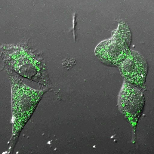
include reference dyes to allow ratiometric imaging, or
                              Bound                                                   ionophore/chromoionophore combinations that allow the
      Ion+
                              Targets
                                                              Acrylamide              use of highly selective, non-fluorescent ionophores. Both
                                                            + Molecular probe
   H+
                      Ionophore
                                                             + Reference Dye          the protection and the powerful sensing flexibility come in
                                                                + Dextran
                                                                                      a nano-package, which, in terms of minimal mechanical
               Fluorescent
                                Enzyme
                                                            Liquid Polymer            and physical perturbation, is closer to “free molecular
                Indicator                               (PVC or Decyl Methacrylate)
                                                          + Ionophore & Indicator
                                                                                      dyes” than most other sensing platforms. However, the
 hn
                             (-)         Targeting Ab
                                                                  + PEG
                                                                                      nanosensor preserves the excellent chemical sensing
                              Ionic
                                         or Peptide
                                                                 Sol Gel
                                                                                      and biocompatibility of macro-sensors and surpasses
             Sensitizing     Additive
      hn        Dye                                             + Enzyme              their performance in terms of response time and absolute
                                                             + Reference Dye
                                    hn                            + PEG               detection limit.
                                                                                           PEBBLEs are a direct outgrowth of the pulled optical
Figure 1: Schematic diagram of a PEBBLE nanosensor                                    fiber nano-technology developed for biosensing by Tan
showing many options available within this flexible, integrated                       et al. [1, 2] and continuing in the work of Rosenzweig
device platform. On the right, current matrix materials are
                                                                                      [3, 4], Shortreed [5, 6], and Barker [7, 8]. In their
presented with typical constituents.
                                                                                      paper, Dourado and Kopelman formalized the specific
                                                                                      advantages of having nano-scale dimension sensors
1. Introduction                                                                       [9]. In most instances, there is an explicit functional
                                                                                      dependence of optode characteristics on the sensor radius
P   EBBLE nanosensors (Probes Encapsulated By
    Biologically Localized Embedding) are sub-micron
sized optical sensors designed specifically for minimally
                                                                                      (r). For instance, the absolute detection limit decreases
                                                                                      with r3 (good!) and the response time is reduced as r2
                                                                                      (good!). The signal to noise ratio, though, decreases
invasive analyte monitoring in viable, single cells
                                                                                      with r (bad!) but not r3 (luckily!) under standard working
with applications for real-time analysis of drug, toxin,
                                                                                      conditions. Other features that improve, as sensors get
and environmental effects on cell function. PEBBLE
                                                                                      smaller include sample volume, sensitivity, invasiveness,
is a general term that describes a family of matrices
                                                                                      spatial resolution, dissipation of heat in sensor and/or
and nano-fabrication techniques used to miniaturize
                                                                                      sample, toxicity and materials cost. Features that may
many existing optode technologies. The main classes
                                                                                      worsen include fluorophore leaching and photo-damage
of PEBBLE nano-sensors are based on matrices of
                                                                                      to sensor and/or sample.
polyacrylamide hydrogel, sol gel silica, and cross-linked
decyl methacrylate. These matrices have been used to
fabricate sensors for H+, Ca2+, K+, Na+, Mg2+, Zn2+, Cl–, 2. Practical Concept Examples

                                                                                      I
NO2–, O2, NO, and Glucose that range from 30 to 600 nm           t is useful to point out concrete examples of the features
in size. A host of delivery techniques have been used to         discussed above before delving into the details of
successfully deliver PEBBLE nanosensors into mouse PEBBBLE production and application. All PEBBLEs
oocytes, rat alveolar macrophages, rat C6 glioma, and must be well characterized before use, including such
human neuroblastoma cells.                                     measures as nanoparticle size and response calibration.
     PEBBLEs were developed specifically for biological Other essential metrics include tests for constituent
applications, and fill a niche that lies between pulled leaching, ratiometric stability, response time and
micro-optodes and free molecular probes (naked sensitivity to interference from similar analytes and non-
indicator dye molecules). The strength of the PEBBLE specific protein binding.
concept lies in two related but distinct
roles. First and foremost the PEBBLE                                                                                                        0.12

protects the cell from the toxicity inherent
                                                                                                             Differential Number Fraction

in some free molecular dyes, and at the                                                                                                     0.08

same time protects indicator dyes from
cellular interferents such as protein
binding. The second role, which is possible
                                                                                                                                            0.04

because the PEBBLE matrix creates a
separate sensing phase, distinct from the                                                                                                   0.00
                                                                                                                                                   0   50   100   150   200

cellular environment, is that multiple                                                                  Diameter

                                               Figure 2: Left: Typical Scanning Electron Microscope (SEM) image of sol gel
dyes, ionophores, and other components
                                               PEBBLEs. Note the 500 nm scale bar in the image legend used to determine
can be combined to create complex ~160 nm average particle size. Right: Light scattering results for (left to right)
sensing schemes. These schemes can one polyacrylamide and two different sol gel PEBBLE formulations.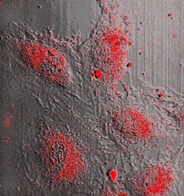
450000
Green has good selectivity over intracellular ions, the
dye itself is prone to artifacts resulting from non-specific
3.0
Fluorescence Intensity (arb. units)
400000
2.5
350000
300000 2.0
binding of proteins, such as bovine serum albumin
(BSA), as shown in Figure 4 (left). Monitoring the
R 0/R -1
250000
1.5
peak of Newport Green at 530 nm, there is a substantial
200000
1.0
150000
100000
50000
0.5 increase in the peak intensity with each successive
0
500 550 600 650 700
0.0
0 10 20 30 40 50
addition of BSA. The PEBBLEs containing the Newport
Wavelength (nm)
O xy g e n C o n c e n tra tio n (p p m )
Green dye, however, are unaffected by the additions of
Figure 3: Left: Aqueous phase emission spectra of sol gel BSA. As little as 0.02% BSA causes an intensity increase
oxygen PEBBLEs excited at 488 nm: top line: PEBBLE of over 200% in the naked Newport Green dye, but the
solution purged with N2; middle line: PEBBLE solution purged intensity of the Newport Green embedded in the sensor
with air; bottom line: PEBBLE solution purged with O2. Right: remains unchanged, even at BSA concentrations above
Stern-Volmer plot of relative fluorescence intensity ratios for 0.10% [11].
ratiometric sol gel oxygen PEBBLEs in aqueous phase. Dashed
Figure 4 (right) demonstrates the advantage of
line denotes biologically relevant range.
using an integrated ratiometric device over a single
intensity-based dye. Four different excitation light
2.1 Ratiometric sol gel oxygen sensor: Size, signal
levels were used for zinc sensing. Although the absolute
and calibration.
intensity of fluorescent emission for each dye decreased
D epending on the size and matrix material, TEM,
SEM and light scattering measurements are used
for PEBBLE size characterization. The sol gel, hybrid
with decreasing illumination power, the ratio of peak
intensities, of Newport Green and Texas Red, remained
constant. It is evident from this that fluctuations in the
organic/inorganic silica matrix is typically produced in intensity of either a laser or arc lamp would complicate
the 50 – 200 nm size range, as shown by Figure 2. quantitative analysis for intensity-based measurements,
These sol gel PEBBLEs contain a ruthenium-based while the ratiometric PEBBLEs eliminate the artifacts
dye, [Ru(dpp)3]2+, which has an intensity decrease due resulting from power fluctuations. The equivalent would
to excited state quenching in the presence of molecular be true, as well, for insensitivity to fluctuations in the
oxygen. As a spectrally separated intensity reference, the local PEBBLE concentration.
PEBBLEs also include Oregon Green-488® (Molecular
Probes), which is insensitive to changes in local
oxygen concentrations. Figure 3 shows spectra of these
3. PEBBLE production techniques.
PEBBLEs in aqueous solution, in the presence of varying
concentrations of oxygen. It is very clear that the Oregon
Green reference peak, on the left, remains constant while
P EBBLEs represent an advance in nano-optode
technology. The science of nano-optode production
relies on advances in nano-scale production, using
the Ru peak, on the right, changes in intensity. Also emulsion and dispersion fabrication techniques. The
shown (right) is the Stern-Volmer (calibration) plot of nano-emulsion/dispersion process for preparing
fluorescence intensity ratio vs. oxygen concentration. PEBBLEs is subtle and there is no universal method
Although the performance of the sol gel PEBBLEs is for making hydrophilic, hydrophobic, and amphiphilic
slightly reduced in the aqueous phase, as opposed to the
gas phase, the sensors still demonstrate good reversibility
3.5
3.0 0uM Zinc 23uM Saturation
Intensity Ratio (546 / 604 nm)
and reproducibility [10]. The dashed line in Figure 3 2.5 0.90
Naked D y e
(right) shows the extent of the biologically relevant 2.0
0.80
I/I0
oxygen concentrations. The sensors showed at least 95%
1.5
0.70
1.0
recovery each time that the sensing environments were 0.5 Acry lam ide PE BBL E s
0.60
0.50
changed among air, O2, or N2 saturated sensor solutions. 0.0
0 0.02 0.04 0.06 0.08 0.10 0.12
0 1 2 3 4
Power (mW)
B S A (w /v % )
2.2 Ratiometric zinc PEBBLE insensitive to Figure 4: Left: Normalized Newport Green emission (530 nm)
protein interference after addition of successive aliquots of a 10 % (w/v) bovine
H
serum albumin solution. As little as 0.02 % BSA causes a
ere, the PAA zinc sensor, based on Newport Green®
greater than 200% increase in Newport Green free (naked)
(Molecular Probes), a zinc sensitive dye, and Texas dye intensity, but the intensity of the dye embedded in a PAA
Red, a spectrally distinct intensity reference, shows the PEBBLE remains unchanged. Right: Fluorescence emission
advantages of PEBBLEs. Quantitative measurements intensity ratio (545 nm / 604 nm) from a 10 mg/ml PEBBLE
show these sensors to be insensitive to changes in suspension in 10 mM Tris buffer monitored using neutral
excitation intensity as well as providing protection from density filters (1.0, 0.5, and 0.3) to attain varied excitation
non-specific protein interference. Although Newport powers at three different zinc concentrations.
nanospheres that contain the right matrix and right stable than polymer matrices. The preparation of sol gel
chemical components in their proper proportions. Thus, “glasses” is technically simple, and tailoring the physico-
switching from single dye containing hydrophilic chemical properties (i.e. pore size or inner-surface
polyacrylamide nanospheres to multi-component, hydrophobicity) of sensor materials can be achieved
hydrophobic, liquid polymer sensors, or to inert glass, sol easily by varying the processing conditions and the
gel sensors is not yet a routine procedure. However, the concentration or type of reactants used. This enables the
production methods, once optimized for a given matrix pore sizes to be optimized such that the analyte is able
and its constituents, are based on relatively simple wet to diffuse easily and interact with the sensing molecules,
chemistry techniques, as opposed to many complicated while the latter are prevented from leaking out of the
physical and chemical nanotechnology schemes. Specific matrix (also true for polyacrylamide-based sensors).
methods for producing sensors from all these matrices Furthermore, this “glass” is produced under so-called soft
are described below as well as the related response chemical conditions, i.e. low temperatures and relatively
mechanisms for each type of sensor.                         mild pH conditions, allowing the inclusion of organic
                                                            dyes and even biomolecules. It may also be “hybridized”
3.1 Polyacrylamide (PAA hydrogel)                           with organic polymers, as shown in the example below.

I  n polyacrylamide (PAA) polymer PEBBLEs, a dye                 The reaction solution for the production of oxygen
   that has a chromometric response to an analyte is        sensitive     sol gel PEBBLEs consists of the organic,
entrapped in the matrix pores. Extraction of analyte ions   hydrophilic        polymer, polyethylene glycol (PEG) MW
into the hydrogel is not a consideration, though, because   5000   monomethyl           ether (3 g), ethanol (200 proof, 6
water and small ions diffuse freely through the hydrogel.   ml),  Oregon         Green-dextran        MW 10,000 (0.1 mM),
What does occur is the formation of a chromoionophore-      [Ru(dpp)    3
                                                                          ] 2+
                                                                                (0.4   mM),   and   30%    wt. ammonia water
analyte complex, similar to the response of the “naked”     (3.9  ml)   with     ammonia      serving    as catalyst and water
dye in solution. The dynamic range and selectivity of the   being   one    of   the   reactants.  Upon    mixing,    the solution
PEBBLE is dependent on the KD of the dye with respect       becomes       transparent       and   the   inorganic    “monomer”
to the analyte and any interfering ions.                    tetraethyl    orthosilicate     (TEOS)     (0.5  ml)  is  added drop-
     The production of acrylamide PEBBLEs is based on       wise  to  initiate     the  hydrolysis   of  TEOS.   The   solution is
the nano-emulsion techniques studied by Daubresse [12].     then  stirred    at  room    temperature    for 1 hour   to allow the
Some control over particle size and shape can be gained     sol gel  reaction       (analogous    to polymerization)     to reach
by adjusting surfactant to water ratios in the emulsion.    completion.      A   liberal  amount   of  ethanol  is then  added  to
The typical polymerization solution consists of 0.4 mM      the reaction      solution    and the  mixture  is transferred   to an
fluorescent ionophore (any hydrophilic dye selective for Amicon ultrafiltration cell (Millipore Corp., Bedford, MA).
the analyte of interest), 27% acrylamide (monomer), 3% A 100 kD membrane is used to separate the reacted sol
N,N-methylenebis(acrylamide) (cross-linker), all in 0.1 gel particles (PEBBLEs) from the unreacted monomers,
M phosphate buffer, pH 6.5. One milliliter of this solution PEG, ammonia and dye molecules, under a pressure of
is then added to a solution containing 20 ml hexane, 1.8 10 psi. The PEBBLEs are further rinsed with 500 ml
mmole dioctyl sulfosuccinate sodium salt (surfactant), ethanol to ensure that all unreacted chemicals have been
and 4.24 mmole Brij 30 (surfactant). The solution is removed. The PEBBLE solution is then passed through
stirred under nitrogen for 20 min, while cooling in an a suction filtration system (Fisher, Pittsburgh, PA) with a
ice bath. The polymerization is initiated with 24 µl of a 2 µm filter membrane to separate the larger size particles
10% ammonium persulfate solution and 12 µl TEMED from the smaller ones. The filtrate (containing the smaller
(initiators), then the solution is allowed to stir at room particles) is filtered again, this time with a 0.02 µm filter
temperature for 2 hours. Hexane is removed by rotary membrane, to collect the particles which are then dried to
evaporation, then the probes are rinsed of surfactant with yield a final product consisting of sol gel PEBBLEs in the
ethanol, to give a majority 40 nm probes [13, 14].          size range of 100 – 400 nm in diameter [10].

3.2 Sol Gel (silica/organic hybrid)                               3.3 Decyl Methacrylate (hydrophobic liquid
                                                                  polymer)
S    ol gel glass has also been used as the matrix for
     the fabrication of PEBBLE nanosensors, because
of the superior properties it has for some applications          T
                                                           he use of fluorescent indicator molecules in
                                                           encapsulated form (acrylamide PEBBLEs), has
over organic polymers. Sol gel glass is a porous, high proven valuable in the study of a number of intracellular
purity, optically transparent and homogeneous material analytes [11, 13, 14, 16] (H , Ca , Mg , Zn , O 2),
                                                                                        +     2+    2+    2+

[15], thus making it an ideal choice as a sensor matrix however, there are many ions for which no fluorescent
for quantitative spectrophotometric measurements. Also, indicator dye is sufficiently selective or even available.
it is chemically inert, and more photo- and thermally An alternate class of tandem optical nano-sensors is thus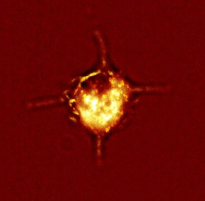
required, driving the development of decyl methacrylate              PEBBLEs
(hydrophobic) liquid polymer PEBBLEs.                                                                       Helium

     A batch of decyl methacrylate PEBBLE sensors                                                                      Rupture Disk
is typically made from 210 mg of decyl methacrylate, Liposomes                                                         Carrier Disk
180 mg hexanedioldimethacrylate, 300 mg of dioctyl                                                                     PEBBLEs
                                                                       Cell
sebacate (DOS), with 10 – 30 mmole/kg each of                                                                           Petri Dish
ionophore, chromoionophore, and ionic additives added                                                                   with Cells

after spherical particle synthesis. The spherical particles
are prepared by dissolving decyl methacrylate, hexanedi                    PEBBLEs                 Mouse Oocyte
oldimethacrylate, and dioctyl sebacate in 2 ml of hexane.
To a 100 ml round bottom flask, in a water bath on a                                                            PEBBLEs
                                                                             Phagosomes
hot plate stirrer, 75ml of pH 2 HCl is added along with            Cell
                                                                                                                                 Injection
1,793 mg of PEG 5000 monomethyl ether and stirred and                                              Holder                        Pipette
degassed. The hexane-dissolved monomer cocktail is then                                            Pipette

added to the reaction flask (under nitrogen), stirred at full Figure 5: Range of delivery methods currently available for
speed, and water bath temperature is raised to 80º C over PEBBLE nanosensors into single cells for bioanalysis. Moving
30 – 40 minutes. 6.0 mg of potassium peroxodisulfate clockwise from the upper left: Liposomal delivery, gene gun,
is then added to the reaction and stirring is reduced to picoinjection, and phagocytosis.
medium speed. The temperature is kept at 80º C for two
more hours, and then the reaction is allowed to return to plastic disk into a cell culture. The gene gun can be
room temperature and stir for 8 – 12 hours. The resulting used to deliver one to thousands of PEBBLEs per cell
polymer is suction filtered through a glass microanalysis into a large number of cells very quickly (dependent
vacuum filter holder with a Whatman Anodisc filter (0.2 on the concentration of PEBBLEs on the delivery disk)
µm pore diameter). The polymer is rinsed three times [10, 13, 16, 17]. Cell viability is excellent, 98% viability
with water and three times with ethanol to remove excess compared to control cells [16], for small numbers
PEG and unreacted monomer. THF is then used to leach of PEBBLEs, and hinges directly on the number of
out the DOS and then the PEBBLEs are again filtered PEBBLEs delivered, the delivery pressure, and the
and rinsed. They are allowed to dry in a 70º C oven chamber vacuum. The PEBBLE momentum determines
overnight. Dry polymer is then weighed out, and DOS, whether the PEBBLEs are mainly internalized in the
ionophore, chromoionophore and ionic additive are cytoplasm or in the nucleus.
added to this dry polymer, so that the resulting polymer            Picoinjection is used to inject picoliter (pl) volumes
will have 40% DOS, 20 mmole/kg ionophore, 10 mmole/           of  PEBBLE        containing solution into single cells (Figure
kg chromoionophore, and 10 mmole/kg ionic additive.           5). This   method    of delivery is dependent on the fabrication
Enough THF is added to this mixture so as to just wet the     of   pulled     capillary    “needles”, through the use of a
PEBBLEs. The PEBBLEs are allowed to swell for eight           pipette-puller      and   a  micro-forge.     The smallest volume
hours and then the THF is removed by rotary evaporation.      deliverable      is 10 pl  and   the most    concentrated     PEBBLE
The resulting PEBBLE sensors are rinsed with doubly           solution    to   work  in  the  pulled   capillary   syringe    is 5 mg/
distilled water and allowed to air dry.                       ml   PEBBLEs.        The  maximum        number    of  PEBBLEs         one
                                                              can put in is dependent on the volume of solution that
                                                              can be injected without damaging the cell. Picoinjection
4. Delivery Methods                                           can give a wide range of PEBBLE concentrations in the

O     ne of the most important considerations when cell, and cell viability is good (if done by an expert),
      applying PEBBLE nanosensors to single cell studies but because each cell must be individually injected, the
is the (non-invasive) delivery of the PEBBLEs to the cell. method is time consuming and tedious [16].
The many methods that have been explored include gene               Commercially available liposomes can also be
gun, picoinjection, liposomal delivery, and sequestration     used    to deliver PEBBLEs to cells. The liposomes are
(phagocytosis and pinocytosis) into macrophages. All of       prepared      in a solution of PEBBLEs and then placed in
these methods are summarized in Figure 5.                     the   cell  culture   where the liposomes fuse with the cell
     The method of PEBBLE delivery by gene gun can            membranes          and  empty their contents (the PEBBLE
best be thought of as a shotgun method. PEBBLEs are           containing      solution)   into the cell. Three factors play a key
dried on a plastic (delivery) disk, and this disk is set      role   in  determining      the number of PEBBLEs delivered
in front of a rupture disk. Helium pressure is built up       to  each   cell  with  this  method: The original concentration
behind the rupture disk, which ruptures at a specific         of  the   PEBBLEs,      the   concentration of liposomes placed
helium pressure and propels the PEBBLEs from the              in  the   cell  culture,  and   the length of time the liposomes
2000
NoPEBBLEs
macrophage overnight. Macrophage images were then
1750 WithPEBBLEs
ConA10min taken on a confocal microscope and spectra of the
Fluorescence Intensity
1500 ConA20min
1250
same cells were obtained on the fluorescent microscope
1000 (shown in Figure 6). Acrylamide PEBBLEs selective
750
for calcium (containing Calcium Crimson in the
500
250
acrylamide matrix) [16] were used in order to monitor
0
calcium in phagosomes within rat alveolar macrophage,
because of the ease in which macrophage phagocytose
500 550 600 650 700 750
Wavelength (nm)
Figure 6: Confocal microscope image (left) of alveolar particles. This method for delivering the PEBBLEs
macrophage containing phagocytosed polyacrylamide into cells provided a simple, yet important, test of the
PEBBLEs containing Calcium-Crimson dye. Fluorescence PEBBLE sensors in a challenging (acidic) intracellular
spectra (right) show an increase in intracellular calcium after
environment. Macrophage that had phagocytosed 20
cells have been challenged by Concanavalin A (Con A).
nm calcium-selective PEBBLE sensors were challenged
are left with the cells [14, 16, 18]. The parameters must with a mitogen, Concanavalin A (Con A), inducing a slow
be tailored for each cell line used in order to obtain the increase in intracellular calcium, which was monitored
desired concentration of PEBBLEs in the cells. While it over a period of 20 minutes. PEBBLE clusters confined
would be difficult to deliver a single PEBBLE to each cell to the phagosome enabled correlation of ionic fluxes with
with this method, it does seem that a low end of between stimulation of this organelle.
10 – 50 PEBBLEs per cell would be possible, with the The calcium PEBBLE in the macrophage experiment
high end being the maximum number of PEBBLEs clearly demonstrates a time resolved observation of a
the cell could take without losing viability. Liposomal biological phenomenon in a single, viable cell. One
Delivery is useful for delivering PEBBLEs to a lot of can clearly obtain relevant time domain data with a
cells simultaneously. The challenge is in tailoring the fluorescence microscope, spectrograph and CCD. With
delivery, for the concentrations desirable and for the cell a confocal microscope system and the appropriate
line being used. Cell viability is excellent. Obviously, the dye/filter sets one can attain both temporal and spatial
PEBBLE size needs to be small enough for this method, resolution, as demonstrated below.
and delivery is essentially limited to the cell cytoplasm. Calcium PEBBLEs have also been developed
Macrophages, a specialized immune system cell, take utilizing “Calcium Green-1” (Molecular Probes)
up PEBBLEs automatically. The number of PEBBLEs dye, in combination with sulforhodamine dye, as
that each macrophage takes up is dependent on the sensing components. We note that Calcium Green
concentration of the PEBBLE solution and the amount of fluorescence increases in intensity with increasing
time the macrophages are allowed to stay in the PEBBLE calcium concentrations, while the sulforhodamine
solution. The advantage of this delivery method is fluorescence intensity remains unchanged, regardless
that one can easily deliver varying concentrations of of biologically relevant concentration of ions, pH, or
PEBBLEs to macrophages. The disadvantages are that other cellular component; thus, the ratio of the Calcium
it is mainly useful for macrophages (which are hard to Green/sulforhodamine intensity gives a good indication
culture) and that PEBBLEs are only internalized into of cellular calcium levels regardless of dye or PEBBLE
certain cell regions. This method also provides excellent concentration or fluctuations of light source intensity.
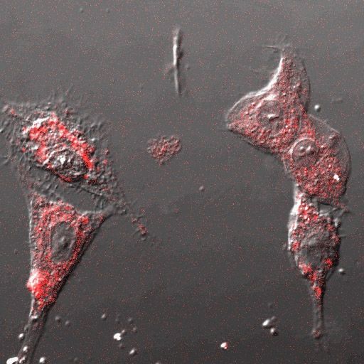
cell viability [16]. Figure 7 shows a confocal microscope image of human
C6 glioma cells containing calcium green/sulforhodamine
PEBBLEs. The top image of the pair is the light intensity
5. In vitro bioanalysis
5.1 Calcium (PAA) PEBBLEs
T he first PEBBLEs produced were acrylamide-based,
and one of the first examples of their successful
application to cells was with macrophages. Alveolar
macrophages were recovered from rat lung lavage using
Krebs-Henseleit buffer. Macrophage were maintained
in a 5% CO2, 37° C incubator in Dulbecco’s Modified Figure 7: Confocal microscope image, split into green
Eagle Medium (DMEM) containing 10% fetal bovine (top) and red (bottom) channels, of human C6 glioma cells
serum and 0.3% penicillin, streptomycin and neomycin. containing Calcium Green / sulforhodamine (reference dye)
PEBBLE suspensions ranging from 0.3 – 1.0 mg/ml PEBBLEs (toxin diffusing left to right as seen by lack of green
were prepared in DMEM and incubated with alveolar on right side of image).from the green (calcium sensitive) fluorescence, and the
bottom shows the red intensity (reference), both dyes
confined in the same PEBBLEs. The PEBBLEs were
delivered by liposomes to the cytoplasm of the cells. The
toxin, m-dinitrobenzene (DNB), was introduced to the
left side of the image and allowed to diffuse to the right.
The effect of DNB is the disruption of mitochondrial
function, followed by the uncontrolled release of calcium
associated with onset of the mitochondrial permeability
transition (MPT) [18]. Calcium PEBBLEs were used to Figure 8: Confocal images of rat C6 glioma cells loaded with
sol gel PEBBLEs by gene-gun injection. Nomarski illumination
determine that the half-maximal rate of calcium release
image overlaid with Oregon Green fluorescence (reference, left)
(EC50) occurred at a 10-fold lower concentration of m- and [Ru(dpp) ]2+ fluorescence (right) of the same ratiometric
3
DNB in human SY5Y neuroblastoma cells than in human PEBBLEs inside cells.
C6 glioma cells [18].
also some in the nucleus.
5.2 Aqueous oxygen (sol gel) PEBBLEs After gene gun injection, the cells were immersed
S ol gel, the newest PEBBLE matrix, gives the flexibility in DPBS (Dulbecco’s Phosphate Buffered Saline)
of being able to tailor the properties of the matrix to and a spectrum was taken of these cells, using 480 ±
accept either hydrophilic or hydrophobic dyes. Also, for 10 nm excitation light. The air-saturated DPBS was
oxygen, their dynamic range is much wider than that of then replaced by nitrogen-saturated DPBS, to cause a
similar acrylamide PEBBLEs [10]. It is also proven as a decrease in the intracellular oxygen concentration, and
matrix compatible with the use of protein based sensors the response of the oxygen PEBBLE sensors inside
[15]. Using the gene gun, sol gel PEBBLEs were inserted the cells was monitored during a time period of 2+ 2
into rat C6 glioma cells, so as to monitor oxygen. A minutes. The fluorescence intensity of [Ru(dpp) 3
]
ratiometric sol gel PEBBLE sensor ([Ru(dpp)3]2+ oxygen went up successively, indicating that the oxygen level
sensitive dye and Oregon Green 488-dextran reference inside the cells decreased. Average intracellular oxygen
dye) was used [10]. Figure 8 shows the confocal images concentrations were determined on the basis of a Stern-
of C6 glioma cells containing sol gel PEBBLEs under Volmer calibration curve, obtained using the fluorescence
Nomarski illumination overlaid with: (Left) The green microscope-Acton spectrometer system [10], and are
fluorescence of Oregon Green 488-dextran and (Right) summarized in Table 1. The comparatively large errors
the red fluorescence of [Ru(dpp)3]2+. It can be seen that are due to the low resolution of the spectrometer. We note
the cells still maintained their morphology after the gene that the measured intracellular oxygen value (when cells
gun injection of PEBBLEs and showed no sign for cell were in air saturated DPBS) is comparable with the value
death. The dyes were excited, respectively, by reflecting of ~7.1 ppm measured electrochemically inside the much
the 488 nm (Ar-Kr) and the 543 nm (He-Ne) laser lines larger islets of Langerhans [19]. These results show that
onto the specimen, using a double dichroic mirror. The the PEBBLE sensors are responsive when loaded into
Oregon Green fluorescence from the PEBBLEs inside cells and that they retain their spectral characteristics,
the cells (Figure 8 left) was detected by passage through enabling a ratiometric measurement to be made [10].
a 510 nm long-pass and a 530 nm short-pass filter, and 5.3 Potassium (Decyl Methacrylate) PEBBLEs
the fluorescence of [Ru(dpp)3]2+ (Figure 8 right) through
a 605 nm (45 nm band-pass) barrier filter. A 40X, 1.4 NA
oil immersion objective was used to image the Oregon
Green and [Ru(dpp)3]2+ fluorescence. The distribution
T he acrylamide PEBBLE matrix has proven to work
with any hydrophilic sensing components. However,
it is not able to take advantage of the rich history of
of PEBBLEs in overlaid images demonstrated that the electrochemical sensors where there exist a host of highly
green and red fluorescence in Figure 8 were truly from selective, hydrophobic ionophores. In many cases the
PEBBLEs inside cells. It should be noted that most of the selectivity of these ionophores has yet to be matched by
PEBBLEs were loaded into the cytoplasm, but there were hydrophilic dyes (chromoionophores). Highly selective
intracellular (and extracellular) hydrophilic indicator
Table 1: Experimental ratiometric in vitro oxygen results dyes are limited to a small set of analytes, such as pH and
Avg. intracellular O2 concentrations (ppm) calcium. While the use of PEBBLEs instead of traditional
(Air saturated buffer solution = 8.8 ± 0.8) free “naked” indicators results in protection beneficial to
Cells in air saturated buffer 7.9 ± 2.1 both the cell and the dye, it does not solve the selectivity
Cells in N2 saturated buffer (after 25 sec) 6.5 ± 1.7 problems. For instance, hydrophilic potassium indicators
Cells in N2 saturated buffer (after 120 sec) ≤ 1.5 will not work in the presence of significantly higher1.0
of ion-exchange sensors developed by
0.8 Simon, Bakker and colleagues [5, 21-
0.6 23]. For the incorporation of a selective
P
0.4 neutral ionophore (BME-44) into a matrix,
0 .0 1 M K C l
1000
F l1 F l2 0 .0 5 M
0.2
along with a selective chromoionophore
Fluorescent Intensity
(ETH 5350) for indirect ion monitoring
0 .2 0 M
0 .5 0 M
800 0
4 8
(ion exchange sensors), the metal ion
2 .0 M 2 6 10
Log(a X+ /aH+ )
600 1.0
activity (aK+) in solution is a function of
400
0.8
the hydrogen ion activity in solution (aH+),
0.6 the interfering cation activity (a Na+) and
P
200
0.4 the constants [Ltot], [Ctot], [Rtot-], which are
0 0.2 total ionophore (ligand) concentration, total
550 600 650 700 750 800
0 chromoionophore concentration, and total
Wavelength 2 4 6 8 10
Log(a K+ /aH+ )
lipophilic charge site concentration, in the
Figure 9: Left: Normalized emission spectra from suspended K+ PEBBLE membrane. Note that [CH] is the protonated
sensors using the pH chromoionophore ETH5350 for ion-correlation chromoionophore concentration and [C] is
spectroscopy in tandem with BME-44. Spectra show response from 10 mM
the free base concentration. The parameter
to 2.0 M KCl (well beyond sensor saturation), all in 10 mM Tris buffer, pH
7.2. Right top: Response of same PEBBLEs to K+(ο), and Na+(∆), along with
Π has been defined [5-9] as the relative
theoretical curves. The lines delimit values for log (aK /aH ) typically found
+ + portion of the protonated chromoionophore,
in intracellular (solid) and extracellular (dashed) media [28]. Right bottom: Π = [CH] / [Ctot].
Response to additions of KCl in Tris buffer (ο) compared to a similar experiment Calibration of a K + sensor based on
run in a constant background of 0.5 M Na+ ( ). Solid lines are calculated (not these principles is shown in figure 9 (right
fit) theoretical curves for the K+ response in the presence of 0.5 M interfering top) along with normalized spectra (left). For
Na+ using the experimentally determined log selectivity value of –3.3. potassium sensing, the chromoionophore is
sodium concentrations, and conversely, sodium ETH 5350, the ionophore is BME-44, and
indicators will not work in the presence of high the lipophilic additive is KTFPB [6, 17]. The data points
potassium concentrations [20]. Obviously, this has for potassium and sodium responses are plotted along
serious implications for both intracellular (e.g. high with corresponding theoretical curves. Dashed lines
potassium/sodium ion ratios) and extracellular (e.g. high delimit typical extracellular activity ratios and the solid
sodium/potassium) applications. Moreover, for many lines delimit the intracellular levels (log (aK+/aH+)) [28].
important analyte ions, such as nitrite, no satisfactory It was found that the response matches well with the
color indicators are available. The above problem has theory, which is gratifying, considering the small size
been solved in optodes by using in tandem an optically of the systems. The dynamic range at pH 7.2 extends
silent ionophore (which is highly selective) and a next- from 0.63 mM to 0.63 M aK+. The log of the selectivity
door optically visible agent that plays the role of a for potassium vs. sodium, determined by measuring the
spectator, or reporter dye. While the principles of such horizontal separation of the response curves at Π = 0.5,
tandem sensing schemes were worked out by Bakker and is –3.3. This selectivity value can be used, along with
Simon [21-23], Suzuki [24, 25], and Wolfbeis [26, 27], the mathematical theory for this sensing mechanism, to
the first demonstration of such a sensing scheme on the calculate what the K +
response of the PEBBLEs should be
nanoscale occurred with the pulled optodes developed by in the presence of 0.5 M interfering Na +
. Figure 9 (lower
Shortreed et al. [5, 6]. The extension of these principles right) shows these calculated theoretical curves (not fits)
to PEBBLEs required the optimization of a new liquid along with the corresponding experimental data. This
polymer matrix, decyl methacrylate [17]. shows a selectivity similar to or better than that obtained
The work described here takes advantage of an for other and larger matrices incorporating BME-44, e.g. -
indicator with two fluorescence emission maxima (λ1,λ2), 3.1 in PVC based fiber optic work, and –3.0 in PVC based
giving a relative intensity that changes with the degree microelectrodes [6, 17]. It also exactly matches the value
of protonation (Π). This degree of protonation, Π, can given in the review by Buhlmann, Pretsch, and Bakker
be evaluated in terms of the ratio of the protonated [23] for a thin PVC film sensor. This selectivity should
chromoionophore intensity F λ2 to the deprotonated be more than sufficient for measurements in intracellular
chromoionophore intensity Fλ1 (See figure 9 for spectra) media where potassium concentration [28] is about 100
based on an analytically derived relationship [5]. mM and sodium is about 10 mM.
The degree of protonation (Π) of the indicator spectra The first application of this liquid polymer class of
obtained from the PEBBLE calibration is related to the PEBBLEs was the observation of potassium uptake in rat
analyte concentration by using the theoretical treatment C6 glioma cells [17]. Decyl methacrylate PEBBLEs were7.8
References
7.6
1. W. Tan, Z.-Y. Shi, S. Smith, D. Birnbaum, and R. Kopelman, Science
Log (aK+/aH+)
7.4 258, 778 (1992).
7.2 2. W. Tan, R. Kopelman, S. L. R. Barker, and M. T. Miller, Analytical
Chemistry 71, 606A (1999).
7.0
3. Z. Rosenzweig and R. Kopelman, Analytical Chemistry 67, 2650
6.8 (1995).
0 20 40 60 80 100 120 4. Z. Rosenzweig and R. Kopelman, Analytical Chemistry 68, 1408
Time in seconds (1996).
Figure 10: Left: Confocal image of decyl methacrylate K + 5. M. Shortreed, E. Bakker, and R. Kopelman, Analytical Chemistry
PEBBLE fluorescence, overlaid with Nomarski image of rat C6 68, 2656 (1996).
glioma cells (488 nm excitation, 580 nm long pass emission). 6. M. R. Shortreed, S. Dourado, and R. Kopelman, Sensors and
Right: Ratio data of decyl methacrylate K+ PEBBLEs in C6- Actuators B 38-39, 8 (1997).
glioma cells during the addition of kainic acid (50 µl of 0.4 mg/ 7. S. L. R. Barker, M. R. Shortreed, and R. Kopelman, Analytical
Chemistry 69, 990 (1997).
ml) at 20 s and at 60 s. Ratios were converted to log (aK+/aH+)
8. S. L. R. Barker, B. A. Thorsrud, and R. Kopelman, Analytical
using solution calibration of the PEBBLEs. Log (aK+/aH+) is
Chemistry 70, 100 (1998).
seen to increase after kainic acid addition (and subsequent K+
9. S. Dourado and R. Kopelman, Spie (Int. Soc. Opt. Eng.) Proc 2836,
channel openings). 2 (1996).
10. H. Xu, J. W. Aylott, R. Kopelman, T. J. Miller, and M. A. Philbert,
delivered by gene gun using a BioRad (Hercules, CA) Analytical Chemistry 73, 4124 (2001).
Biolistic PDS-1000/He system, with a firing pressure of 11. J. P. Sumner, J. W. Aylott, E. Monson, and R. Kopelman, Analyst
650 psi, and a vacuum of 15 torr applied to the system. (2001).
Immediately following PEBBLE delivery, cells were 12. C. Daubresse, C. Granfils, R. Jerome, and P. Teyssie, Journal of
placed on an inverted fluorescent microscope. The Colloid and Interface Science 168, 222 (1994).
gating software for the CCD was set to take continuous 13. H. A. Clark, S. L. R. Barker, R. Kopelman, M. Hoyer, and M. A.
Philbert, Sensors and Actuators B 51, 12 (1998).
spectra at 1.3 second intervals. After 20 seconds, and 14. H. A. Clark, M. Hoyer, M. A. Philbert, and R. Kopelman, Analytical
after 60 seconds, 50 µl of 0.4 mg/ml kainic acid was Chemistry 71, 4831 (1999).
injected into the microscope cell. Kainic acid is known 15. D. R. Uhlmann, G. Teowee, and J. Boulton, Journal of Sol-Gel
to stimulate cells by causing the opening of ion channels. Science and Technology 8, 1083 (1997).
Figure 10 (left) shows the confocal fluorescent image 16. H. A. Clark, M. Hoyer, S. Parus, M. Philbert, and R. Kopelman,
Mikrochimica Acta 131, 121 (1999).
of the PEBBLEs, overlaid with a Nomarski differential
17. M. Brasuel, R. Kopelman, T. J. Miller, R. Tjalkens, and M. A.
interference contrast image of the cells [17]. Image Philbert, Analytical Chemistry 73, 2221 (2001).
analysis indicated that the PEBBLE sensors were 18. H. A. Clark, R. Kopelman, R. Tjalkens, and M. A. Philbert,
localized in the cytoplasm of the glioma cells. Figure Analytical Chemistry 71, 4837 (1999).
10 (right) shows the PEBBLE sensors inside the cells 19. S.-K. G. Jung, Waldemar; Aspinwall, Craig A.; Kauri, Lisa M.,
responding to the kainic acid addition. One can see that Anal. Chem. 71, 3642 (1999).
log (aK+/aH+) increases, indicating either an increase 20. R. P. Haugland, “Molecular Probes Handbook of Fluorescent
Probes and Research Chemicals.” Molecular Probes, Inc, Eugene,
in K+ concentration or a decrease in H+ concentration OR, 1993.
(increase in pH). The amount of kainic acid added is not 21. E. Bakker and W. Simon, Analytical Chemistry 64, 1805 (1992).
known to affect the pH of cells in culture and kainic acid 22. W. E. Morf, K. Seiler, B. Lehmann, C. Behringer, K. Hartman, and
by itself has no effect on the sensors. Thus the change is W. Simon, Pure & Applied Chemistry 61, 1613 (1989).
likely due to increasing intracellular concentration of K+, 23. P. Buhlmann, E. Pretsch, and E. Bakker, Chemical Reviews 98,
which is the expected trend. The membrane of C6 glioma 1593 (1998).
24. K. Kurihara, M. Ohtsu, T. Yoshida, T. Abe, H. Hisamoto, and K.
cells can initiate an inward rectifying K+ current, induced Suzuki, Analytical Chemistry 71, 3558 (1999).
by specific K+ channels, a documented role in the control 25. K. Suzuki, H. Ohzora, K. Tohda, K. Miyazaki, K. Watanabe, H.
of extracellular potassium [29]. Thus, when stimulated Inoue, and T. Shirai, Analytica Chimica Acta 237, 155 (1990).
with a channel opening agonist, the K+ concentration 26. G. J. Mohr, F. Lehmann, R. Ostereich, I. Murkovic, and O. S.
within the glioma cells is indeed expected to increase. Wolfbeis, Fresenius Journal of Analytical Chemistry 357, 284
(1997).
27. G. J. Mohr, I. Murkovic, F. Lehmann, C. Haider, and O. S. Wolfbeis,
Sensors and Actuators B-Chemical 39, 239 (1997).
28. D. Ammann, “Ion-Selective Microelectrodes.” Springer, Berlin,
1986.
29. A. Emmi, H. J. Wenzel, and P. A. Schwartzkroin, Journal of
Neuroscience 20, 3915 (2000).You can also read


