Quercetin can reduce viral RNA level of O'nyong nyong virus and resulting innate immune cytokine responses in cultured human synovial fibroblasts ...
←
→
Page content transcription
If your browser does not render page correctly, please read the page content below
www.nature.com/scientificreports

                OPEN             Quercetin can reduce viral RNA
                                 level of O’nyong‑nyong virus
                                 and resulting innate immune
                                 cytokine responses in cultured
                                 human synovial fibroblasts
                                 Axelle Septembre‑Malaterre1*, Yosra Bedoui1, Claude Giry1, Philippe Gasque1,2,
                                 Pascale Guiraud1 & Jimmy Sélambarom1*

                                 O’nyong-nyong virus is an alphavirus closely related to chikungunya virus, causing arthralgia, rash and
                                 fever. Alphaviruses mainly target synovial fibroblasts and persists in the joints of patients, possibly
                                 leading to chronic arthritis. To date, no specific antiviral treatment is available for ONNV infection
                                 and induced-inflammation. Primary human synovial fibroblasts cells were used to assess infection by
                                 ONNV and the resulting cytokine responses. Phenolics (gallic acid, caffeic acid and chlorogenic acid,
                                 curcumin and quercetin) and a curcuminoids-rich extract from turmeric were tested for their antiviral
                                 and anti-inflammatory capacities. We showed that infection occurred in HSF cells and increased
                                 gene expression and protein secretion of two major proinflammatory CCL-2 and IL-1β markers. In
                                 ONNV-infected HSF cells (MOI 1), we found that non-cytotoxic concentrations of phenolics (10 µM)
                                 reduced the level of viral RNA (E1, E2, nsP1, nsP2) and downregulated CCL-2 and IL-1β expression and
                                 secretion. These results highlighted the high value of the flavonol quercetin to reduce viral RNA levels
                                 and inflammatory status induced by ONNV in HSF cells.

                                 Abbreviations
                                 CaA	Caffeic acid
                                 ChA	Chlorogenic acid
                                 CRE	Curcuminoids-rich extract
                                 CU	Curcumin
                                 CHIKV	Chinkungunya virus
                                 GaA	Gallic acid
                                 DAPI	4′,6-Diamidino-2-phenylindole
                                 HSF	Human synovial fibroblasts
                                 LDH	Lactate dehydrogenase
                                 MOI	Multiplicity of infection
                                 ONNV	O’nyong-nyong virus
                                 PFU	Plaque Forming Unit
                                 QU	Quercetin
                                 RT-PCR	Real-time polymerase chain reaction
                                 qRT-PCR	Quantitative RT-PCR
                                 ELISA	Enzyme-linked immunosorbent assay

                                 O’nyong-nyong virus (ONNV) is a mosquito-borne virus belonging to the genus Alphavirus in the Togaviridae
                                 family. This RNA virus, closely related antigenically, genetically and clinically to chikungunya virus (CHIKV),

                                 1
                                  Université de La Réunion, Unité de recherche Etudes Pharmaco‑Immunologie (EPI), CHU La Réunion site Félix
                                 Guyon, Allée des Topazes, CS11021, 97400 Saint Denis de La, Réunion, France. 2Laboratoire d’immunologie
                                 clinique et expérimentale de la zone de l’océan indien (LICE‑OI, CHU La Réunion site Félix Guyon, Allée
                                 des Topazes, CS11021, 97400 Saint Denis de La, Réunion, France. *email: axelle.malaterre-septembre@
                                 univ-reunion.fr; jimmy.selambarom@univ-reunion.fr

Scientific Reports |   (2021) 11:6369               | https://doi.org/10.1038/s41598-021-85840-z                                              1

                                                                                                                                         Vol.:(0123456789)
www.nature.com/scientificreports/
was first isolated in 1959 in Uganda during the first epidemic in East Africa which affected more than 2 mil-
lion people1. ONNV is endemic in sub-Saharan Africa and its reemergence is reflected by several outbreaks
(1984–1985, 1996–1997, 2003) in eastern and western A frica2. Vertebrate reservoir hosts of ONNV remain
largely unknown though the virus is known to be transmitted by Anopheles mosquitoes, which also are vectors of
malaria parasites3. The importation of ONNV to Europe by an infected traveler from Kenya in 2013 highlights the
potential for worldwide introduction of ONNV by travelers4. As recently experienced with CHIKV, reemergence
of ONNV with large outbreaks should be considered5.
ONNV causes symptoms similar to those caused by CHIKV including fever, rash and a rthralgia6–8. Due to
their antigenic and clinical similarities, the development of vaccines or drugs may serve in both cases9, as recently
reported with a serogrouping-protection vaccine using mouse models10. In vivo studies in mouse model support
the importance of innate responses in the initial control of ONNV infection, while the adaptive immune system
mainly operates in the acute infection5. Chronic manifestations of ONNV infection are poorly u nderstood11,
there is evidence that alphavirus infection may cause persistent arthralgia and arthritis from residual virus in
target cells and subsequent accumulation of inflammatory m ediators12,13.
ONNV is classified as an arthritogenic alphavirus due to joint symptoms resulting from cartilage and bone
damage14. In-depth investigations of alphavirus pathogenesis and immunity have established the critical role of
synovial fibroblasts which are part of connective tissue (synovium) around human joints and are the primary tar-
get of alphavirus infection and its chronic manifestations12. Synovial fibroblasts are established as innate immune
cells by recognizing invading pathogens via pattern recognition receptors (PRR)15 and are involved in alphavirus
chronic manifestations and joint damage16. Furthermore, synovial fibroblasts are enrolled in cell signaling via
interactions with other major immune cells including monocytes and macrophages17. These synovial-derived
cells have therefore gained growing interest during the past decade to study in vitro alphavirus infection and
management, with a recent attempt to clarify their r ole18.
To date, there are no available antiviral drugs or vaccines for the management of arthritogenic alphavirus
infection11. Identification of antiviral effectors remains a continuous c hallenge19,20. The available treatment of
symptomatic alphavirus disorders involves treatment with analgesics, antipyretics and non-steroidal anti-inflam-
matory drugs. More attention is being paid to the possible adverse effects of in-depth treatments. Methotrexate,
the most common conventional disease modifying anti-rheumatic drug, is applied to relieve chronic CHIKV-
induced arthritis, and without evidence for adverse immunological e ffects21,22. In contrast, the antimalarial
chloroquine was used to control CHIKV replication but was found to increase chronic clinical m anifestations23.
Natural immunomodulators have gained relevance for the treatment of non-immune and immune chronic
inflammatory diseases24 and their development is supported by preclinical evaluation and clinical trials25. This
new option includes naturally occurring p henolics26 also recognized as the most abundant antioxidants in human
diet27 and established for various biological activities including anti-inflammatory, antiviral and anti-atherogenic
effects28–30. The anti-inflammatory potential of phenolic compounds is exploited in folk medicine31 and mainly
relies on their interference with the redox system and cytokine pathways32. From cumulative in vitro, in vivo
and clinical data, the predominant innate immune responses against arthritogenic alphavirus infection involves
a broad range of inflammatory mediators, as mainly evidenced from CHIKV but with a notable exception
for ONNV12,20. Thus, identification of pro-inflammatory markers during ONNV infection on HSF cells may
guide therapeutic strategies and pathogenesis investigations. Focusing on a first-line immune response during
alphavirus infection, we selected the pro-inflammatory chemokine CCL-2 (or monocyte chemoattractant pro-
tein 1, MCP-1) for its active recruitment of monocytes to the site of inflammation, as well as the cytokine IL-1β,
which is produced in response to cell infection and tissue injury but also as a marker of arthralgia. Chronic
inflammation has been assigned to a prolonged released of cytokines and others proinflammatory mediators,
notably CCL-2 and IL-1β, during persistent viral replication in synovial tissue33. In the case of ONNV, secretion
of pro-inflammatory CCL-2 and IL-1β mediators are involved in the initiation and progression of arthritis34.
Here, we assess the antiviral and anti-inflammatory effects of phenolics in vitro in human synovial fibroblasts
cells (HSF) infected with ONNV including three phenolic acids (gallic acid, GaA; caffeic acid, CaA; chlorogenic
acid, ChA), a curcuminoid (curcumin, CU), a flavonoid (quercetin, QU) and a curcuminoid-rich extract (CRE)
from turmeric (Curcuma longa, Reunion island).
Materials and methods
Chemicals. GaA, CaA, ChA, CU and QU (Fig. 1) were purchased from Sigma-Aldrich and CRE was obtained
from turmeric provided by an essential oils company (Extraits de Bourbon, Reunion Island, France). CRE from
turmeric was prepared as follows: 2 g of turmeric (Curcuma longa) was added to 10 mL of aqueous ethanol (70%,
v/v). After incubation at 4 °C for 90 min, sample was centrifuged at 3500 rpm for 20 min at 4 °C. Supernatant
was collected and stored at -80 °C until analysis. As previously reported using an ultra-performance liquid chro-
matography tandem mass spectrometry (UPLC-MS) our analyses led to the identification of curcumin and both
derivatives demethoxycurcumin and bisdemethoxycurcumin in CRE s ample35. The identity and quantity agreed
with literature data reported in Curcuma longa extracts from India36.
Cell culture and virus. HSF cells were obtained from ScienCell research laboratory (ScienCell, 4700 Cli-
nisciences) and were grown in Modified Eagle’s Medium (MEM, PAN Biotech P0408500) supplemented with
10% heat-inactivated fetal bovine serum (FBS; PAN Biotech, 3302 P290907), 2 mM of L-glutamine (Biochrom
AG, K0282), 0.1 mg/mL penicillin–streptomycin (PAN Biotech, P0607100), 1 mM of sodium pyruvate (PAN
Biotech, P0443100) and 0,5 µg/mL of amphotericin B (PAN Biotech, P0601001). Cells were maintained at 37 °C
with 5% CO2. A clinical and passage-limited isolate of ONNV was obtained from the National Reference Center
(CNR arbovirus, Marseille, France) and titrated on Vero cells at 107 PFU/mL.
Scientific Reports | (2021) 11:6369 | https://doi.org/10.1038/s41598-021-85840-z 2
Vol:.(1234567890)www.nature.com/scientificreports/
Phenolic acids
O OH OH
HO
O O
HO HO OH
O OH
OH
HO OH
O
OH HO HO
Gallic acid (GaA) Caffeic acid (CaA) Chlorogenic acid (ChA)
Cucurminoids Flavonoids
OH
O OH OH
RO OR'
HO O
HO OH
OH
OH O
R = R' = -CH3 : Curcumin (CU)
R = H ; R' = -CH3 : Demethoxycurcumin Quercetin (QU)
R = R' = H : Bisdemethoxycurcumin
Figure 1. Phenolics with related chemical structural classes.
To determine the PFU, Vero cells (3 × 10⁶) were plated in plate 6-well. After 24 h incubation, the medium was
discarded and 1 mL of diluted viral suspension (dilution factor: 10) was carefully spread over the cell monolayer.
2 h later, 1 mL of carboxyl methyl cellulose (0.4%) in medium was added. Plate was incubated for 2 days at 37 °C
with 5% C O2. Supernatants were discarded and cells were fixed with Paraformaldehyde (3.7%) 10–20 min. Then,
cells were stained with crystal violet (0.5%, Ethanol 20%) to visualize plaque during 5 min. The plates formed
were counted and expressed in PFU (Plate Forming Unit)/mL.
Cell infection and/or treatment. HSF cells were placed in a 96-, 24- or 6-well plates and maintained at
37 °C in a humid atmosphere with 5% CO2. The medium was replaced twice a week. Cells were allowed to grow
to 80–90% confluency. Cells were then infected by ONNV to multiplicity of infection 1 (MOI 1) in the presence
or not of phenolics (10 µM) for 24 h at 37 °C in a humid atmosphere with 5% CO2. In order to infect at MOI
1, cells were counted before each infection. HSF cells were counted with a hemocytometer using trypan blue
exclusion. method37.
Cytotoxicity assay. Cytotoxicity assay were performed by quantitative release of lactate dehydrogenase
(LDH) from damaged cells using a colorimetric-based kit (ref. G1781, CytoTox 96 Non-Radioactive Cytotoxic-
ity Assay, Promega). Cells were grown as previously described and infected with escalating MOIs of ONNV
(MOI 10–3 to 1) or treated with phenolics (1 to 100 µM) during 24 h. Culture medium was collected and cells
were lysed following the manufacturer’s instructions. Released LDH in culture medium after treatments was
compared to the maximum LDH release (intracellular LDH induced by addition of Triton 1%). Cytotoxicity was
expressed relative to maximum LDH release with the formula: % cytotoxicity = 100 × experimental LDH release/
maximum LDH release Control (CTRL; refers to LDH release from untreated cells).
Immunofluorescence staining and microscopy. Cells were grown on glass coverslips in a 24-well plate
as previously described, then treated with phenolics (10 µM) and exposed or not to ONNV (MOI 1) for 24 h.
Coverslips were then fixed and permeabilized with cold 99% ethanol at room temperature for 10 min. Cells
were incubated overnight with primary antibody Alpha SC 293,153 anti-alphavirus capsid (3582, Santa Cruz
Biotechnology). Alexa Fluor488-conjugated donkey anti-mouse was used as secondary antibody (A-21202, Inv-
itrogen, Thermo Fischer Scientific). Nuclei were revealed by staining with the nuclear fluorochrome 4′,6-diami-
Scientific Reports | (2021) 11:6369 | https://doi.org/10.1038/s41598-021-85840-z 3
Vol.:(0123456789)www.nature.com/scientificreports/
Target gene Forward Reverse Detection
CCL-2 CTGCTCATAGCAGCCACCTT CTTGAAGATCACAGCTTCTTTGGG Sybergreen
IL-1β TTGCTCAAGTGTCTGAAGCAG GGTGGTCGGAGATTCGTAGC Sybergreen
E1 CACCGTCCCCGTACGTAAAA GGCTCTGTAGGCTGATGCAA Sybergreen
E2 CCCCTGACTACACGCTGATG CCTTCATTGGAGCCGTCACA Sybergreen
nsP1 GAGAAAACTTGCGTCAGCCG GACGGCGTAGACGTCTTGAT Sybergreen
nsP2 GCGGAGCAGGTAAAAACGTG TAGAACACGCCCGTCGTATG Sybergreen
GAPDH TGCGTCGCCAGCCGAG AGTTAAAAGCAGCCCTGGTGA Sybergreen
Table 1. Primers used for qRT-QPCR analysis.
dino-2-phenylindole (DAPI, Sigma-Aldrich, Germany). The coverslips were mounted with Vectashield (Vector
Labs; Clinisciences) and fluorescence was observed using a Nikon Eclipse E2000-U microscope (Nikon, Tokyo,
Japan). Images were captured and processed using a Hamamatsu ORCA-ER camera and the imaging software
NIS-Element AR (Nikon, Tokyo, Japan). Magnification X20.
ELISA assay. Culture media collected from cells infected (MOI 1) co-treated or not with phenolics (10 µM)
as previously described, was analyzed by ELISA. IL-1β and CCL-2 concentrations in HSF cells supernatants
were measured using commercially available ELISA kits for CCL-2 (Peprotech, cat. no. 900-T31) and IL-1β
(Peprotech, cat. no. 900-K95), according to the manufacturer’s instructions. Samples were analyzed from three
independent biological experiments.
qRT‑PCR analysis. Total RNA from HSF cells exposed to phenolics (10 µM) in the presence or absence
of ONNV (MOI 1) during 24 h, was extracted directly from harvested cell culture (in 6-well plates) using a
QIAamp RNA Blood Mini Kit (QIAGEN, Cat No 52304). 350 μL of lysis buffer from the kit was added to each
well (culture media were collected before), collected after 5 min and kept at – 80 °C until use. qRT-PCR experi-
ments were done using the One Step Prime Script Syber Green RT-PCR kit from TAKARA (Cat No RR066A).
qRT-PCR was performed in a final volume of 5 μL containing 1 μL of extracted total RNA per reaction, 2.7 μL of
enzyme mix and 1.3 μL of primers mix with final primer concentration of 250 nM. qRT-PCR was carried out in
Quantstudio 3 PCR thermocycler (Thermo Fisher Scientific) with the following steps: a reverse transcription at
42 °C for 5 min and 40 cycles comprising a denaturation step at 95 °C for 5 s, annealing step at 58 °C for 15 s and
extension step at 72 °C for 15 s. Fluorescence data were collected at 520 nm during the extension step. Relative
gene expression was calculated using GAPDH as a reference g ene38. Samples were analyzed from three inde-
pendent biological experiments. Primer sequences related to the genes coding for CCL-2, IL-1β, E1, E2, nsP1,
nsP2 and GAPDH are listed in Table 1 below.
Statistical analysis. Data were expressed as means ± SEM. All assays were performed in triplicate inde-
pendent biological experiments. Statistical analysis was achieved using Graph Prism 3 software. Significant dif-
ferences (p < 0.05) between the means were determined by analysis of variance (two-way ANOVA) procedures
followed by a multiple comparison test (Bonnferroni).
Results
Cytotoxicity assessment. LDH release assays was performed to determine the capacity of ONNV
and phenolic to affect HSF cells viability after 24 h of exposure. As shown in Fig. 2, a basal release of LDH
(5.10 ± 1.28) was found in culture supernatants of untreated cells (CTRL). Escalating MOI ( 10–3 to 1) of ONNV
did not significantly affect LDH release from HSF cells when compared to the basal release (CTRL). In contrast,
a dose-dependent effect was observed by increasing phenolics concentrations (1 to 100 µM). Significant LDH
release compared to untreated cells (CTRL) was observed for the highest doses of gallic acid (100 µM; p < 0.01),
caffeic acid (> 25 µM; p < 0.001), chlorogenic acid (> 50 µM; p < 0.01 and 0.001), quercetin (> 50 µM; p < 0.01 and
0.001) and curcumin (100 µM; p < 0.01), while CRE led to an insignificant effect on LDH release for all the tested
concentrations. Based on the overall results, the further experiments on HSF cells were performed using ONNV
at a MOI of 1 (to allow the virus to infect all cells) for infection by ONNV and the concentration of 10 µM (the
highest concentration that can be found in the bloodstream that does not show a cytotoxic effect in our results)
for treatment with phenolics39.
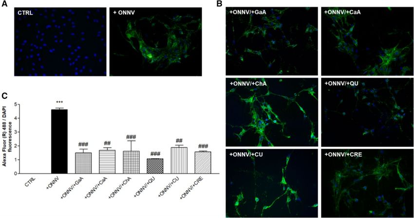
Reduction of ONNV infection. Immunofluorescence and microscopy were used to study the effect of
the phenolics on HSF cells infection by ONNV. Overlay micrographs reported in Fig. 3 were obtained from
DAPI staining of nuclei (blue) and fluorescent specific alphavirus capsid antibody binding (green) (Fig. 3A). As
shown in Fig. 3B, ONNV infected cells and treated by phenolics visually led to a reduced viral antigen detection,
as particularly evidenced for quercetin (+ ONNV/ + QU). Ratio Alexa Fluor 488 / DAPI fluorescence intensity
(Fig. 3C) confirmed the micrograph data given that infected cells had an overall staining level of (+ ONNV:
4.61 ± 0.09, *** p < 0.001) when compared infected-QU-treated cells (+ ONNV/ + QU: 1.06 ± 0.02, ### p < 0.001).
Moreover, we found that all other phenolics significantly decreased viral antigen detection in HSF cells. (###
p < 0.001 for GaA, ChA, CRE; ## p < 0.01 for CaA and CU).
Scientific Reports | (2021) 11:6369 | https://doi.org/10.1038/s41598-021-85840-z 4
Vol:.(1234567890)www.nature.com/scientificreports/
Figure 2. Concentration-dependent cytotoxic effects of phenolics or ONNV on cultured HSF cells. Cells were
infected by ONNV (MOI 1 0–3 to 1) or phenolics (1 to 100 µM) for 24 h. Released LDH in culture medium was
measured using a colorimetric-based method (CytoTox 96 Non-Radioactive Cytotoxicity Assay) and expressed
relative to the maximum release by application of a lysis buffer (Triton 1%). Reported values are means ± SEM
of three independent experiments and p value was calculated using the Bonferroni multiple comparison test
(**p < 0.01; ***p < 0.001) with untreated cells (CTRL).
Figure 3. Quercetin impairs HSF cells infection by ONNV. HSF cells were exposed to ONNV (MOI 1)
co-treated or not with phenolics (10 µM) for 24 h. Cells were probed with primary antibody Alpha SC 58,088
anti-alphavirus before nuclei staining by DAPI (blue) and antibody binding by Alexa Fluor 488-conjugated
donkey anti-mouse IgG antibody (green) (A). Untreated cells (CTRL) and infected cells (+ ONNV) (B) Alexa
Fluor 488/DAPI overlay upon phenolic treatment. (C) Ratio Alexa Fluor 488 / DAPI fluorescence intensity with
at least 10,000 DAPI stained cells. Reported values are means ± SEM of three independent experiments. p value
was calculated using the Bonferroni multiple comparison test (***p < 0.001) between infected cells (+ ONNV)
and untreated cells (CTRL) or (##p < 0.01, ###p < 0.001) between infected cells (+ ONNV) and infected cells upon
treatment with phenolics.
Reduction of viral RNA levels. We assessed by qRT-PCR the effect of phenolics (10 µM) on viral RNA
levels (E1, E2, nsP1 and nsP2) in ONNV-infected HSF cells (MOI 1) as well as in the supernatants. First in cells
and as shown in Fig. 4, viral RNA levels increased significantly in infected cells (+ ONNV) for all tested genes
(***p < 0.001). Treatment with phenolics of uninfected cells was carried out to exclude possible cross-contami-
nation during experiments. No viral RNA was identified in these samples (data not shown). In infected cells, the
phenolics were able to reduce viral RNA significantly as compared to the positive control (+ ONNV alone). E1
and nsP1 gene expression levels were significantly decreased by all tested phenolics. No significant reduction of
viral RNA levels was observed when applied GaA, CaA or CRE for E2, and ChA or CRE for nsP2. A cross-anal-
Scientific Reports | (2021) 11:6369 | https://doi.org/10.1038/s41598-021-85840-z 5
Vol.:(0123456789)www.nature.com/scientificreports/
Figure 4. Quercetin impairs ONNV replication in HSF cells. HSF cells were infected by ONNV (MOI 1)
co-treated or not with phenolic compounds (10 µM) for 24 h. Then, RNA was collected and E1, E2, nsP1
and nsP2 gene expression levels were determined by qRT-PCR. Reported values are means ± SEM of three
independent experiments. p value was calculated using the Bonferroni multiple comparison test: *p < 0.05,
**p < 0.01, ***p < 0.001 when compared to non-infected and non-treated cells (CTRL) and #p < 0.05, ##p < 0.01,
###
p < 0.001 when compared to infected cells (+ ONNV).
Figure 5. Quercetin impairs ONNV replication in supernatants in HSF cells. HSF cells were infected by
ONNV (MOI 1) co-treated or not with phenolic compounds (10 µM) for 24 h. Then, RNA was collected in
the supernatants and E1, E2, nsP1 and nsP2 gene expression levels were determined by qRT-PCR. Reported
values are means ± SEM of three independent experiments. p value was calculated using the Bonferroni multiple
comparison test: *p < 0.05, **p < 0.01, ***p < 0.001 when compared to non-infected and non-treated cells (CTRL)
and #p < 0.05, ##p < 0.01 when compared to infected cells (+ ONNV).
ysis of these results indicates a broad capacity of QU and CU to reduce all viral RNA levels, and more intensively
nsP1 (QU: 0.001 ± 9.20*10–5 to 0.0003 ± 0.0001, ### p < 0.001; CU: 0.001 ± 9.20*10–5 to 0,0001, ### p < 0.001).
For the experiments testing viral RNA levels in supernatants ONNV infection and replication was evidenced
for all tested genes Fig. 5 (*p < 0.05 and **p < 0.01). Only QU was able to counteract ONNV infection by decreas-
ing Viral RNA for both E1 (0.0045 ± 0.0005 to 0.0005 ± 0.0002, ## p < 0.01) and nsP1 (0.57 ± 0.03 to 0.32 ± 0.09,
# p < 0.05). No statiscal significant effect were observed for E2 and nsP2.
Scientific Reports | (2021) 11:6369 | https://doi.org/10.1038/s41598-021-85840-z 6
Vol:.(1234567890)www.nature.com/scientificreports/
Figure 6. Quercetin reduces CCL-2 and IL-1β pro-inflammatory mediators production in ONNV infected HSF
cells. HSF cells were infected by ONNV (MOI 1) and/or treated with phenolics (10 µM) for 24 h. Then, RNA
was collected and CCL-2 and IL-1β gene expression levels were determined by qRT-PCR, (A) Pro-inflammatory
mediators RNA levels. Then, culture media were collected CCL-2 and IL-1β levels evaluated by ELISA, (B) Pro-
inflammatory mediators levels. Reported values are means ± SEM of three independent experiments. p value was
calculated using the Bonferroni multiple comparison test: *p < 0.05, **p < 0.01, ***p < 0.001 when compared to
non-infected and non-treated cells (CTRL) and #p < 0.05, ##p < 0.01, ###p < 0.001 when compared to infected cells
(+ ONNV).
Downregulation of CCL‑2 and IL‑1β pro‑inflammatory mediators. ONNV causes an arthralgia-
rash and inflammatory syndrome that can persist for months. The inflammation is produced by a release of
pro-inflammatory chemokines and cytokines such as IL-1β and CCL-2 which are involved in the initiation and
progression of arthritis34. qRT-PCR and ELISA assays were performed to determine levels of pro-inflamma-
tory chemokine CCL-2 and cytokine IL-1β when phenolics (10 µM) were applied on non-infected or infected
cells (MOI 1). As shown in Fig. 6A,B, significantly increased levels of both gene expression and secretion of
CCL-2 and IL-1β were observed in infected cells (+ ONNV) when compared to the basal level from non-treated
and non-infected cells (CTRL). Phenolics are not affecting the inflammatory status on their own. Indeed, they
caused no significant change in CCL-2 and IL-1β expression and secretion levels in non-infected cells (+ GaA, +
CaA, + ChA, + QU, + CU or + CRE) in comparison to control (CTRL). Application of phenolics in infected cells
resulted in significant downregulation of both gene expression and secretion levels in all cases when compared
to infected cells (+ ONNV). Analyses of these data demonstrated a broad ability of QU to reduce viral RNA and
to exert anti-inflammatory activity by reducing the expression and secretion of two cytokines tested (p < 0.001).
Discussion
The lack of specific treatment for ONNV infection makes the identification of readily available, safe and efficient
antiviral agents a great imperative. Phenolics are of growing interest for their broad-spectrum antiviral a ctivity40
and for their anti-inflammatory potentiality in chronic diseases41. The aim of this study was to evaluate the effects
of phenolics against ONNV infection in HSF cells. Our major result ascertained the peculiar capacity of the
flavonol (QU) to control for viral RNA level and the inflammation following exposure to ONNV.
Synovial fibroblasts are the main target for alphaviruses infections including during the chronic stage with
persistent inflammation and arthritic disease. This makes synovial fibroblasts an appropriate cell model to assess
both acute and chronic alphavirus infection and treatment, as demonstrated previously in our t eam21. Our
present in vitro experiments showed that viable HSF cells exposed to ONNV infection up to MOI 1 (Fig. 2)
are appropriate for study of viral replication (Figs. 3, 4 and 5) and major pro-inflammatory reactions by CCL-2
and IL-1β markers (Fig. 6). Thus, HSF cells can be used to achieve efficient assessment of phenolics (10 µM) on
critical endpoints for an antiviral and/or anti-inflammatory strategy against ONNV.
enefits42 and QU is the most abun-
Phenolics have been established as ‘functional nutrients’ for their health b
43
dant phenolic provided by human diet . Our overall results on HSF cells support the reduction of viral RNA
and anti-inflammatory capacities of phenolics at 10 µM against ONNV, without significant toxicity. However,
the benefits of phenolics in medicinal use rely on their bioavailability that refers to their plasmatic concentra-
tions after food ingestion, mostly ranging from 0.072 to 5 μM44. The bioavailability of quercetin is considered
as significant but strongly affected by many factors45, thereby requiring high dosage forms in a pharmaceuti-
cal development to obtain the relevant plasmatic concentrations for expected biological effects. An improved
formulation of quercetin for oral absorption has been recently described from a clinical s tudy46. Additionally,
quercetin at concentration up to 10 µM inhibits the ATPase of proteins associated with multi-drug resistance47.
This may support the applicability of quercetin for pharmaceutical purposes.
Scientific Reports | (2021) 11:6369 | https://doi.org/10.1038/s41598-021-85840-z 7
Vol.:(0123456789)www.nature.com/scientificreports/
Figure 7. Antiviral mechanism of Phenolics.
Broad-spectrum antiviral activity of quercetin has been reported for hepatitis C virus48, porcine epidemic
diarrhea virus49,50, herpes simplex virus type 1 and type 2 51, rhinovirus52 and dengue virus type 2 53. Quercetin
remains attractive against (re)-emerging pathogens including Mayaro virus54, influenza A virus55, CHIKV56, Mid-
dle East respiratory syndrome c oronavirus57, the severe acute respiratory syndrome c oronavirus58 and it’s more
recent worldwide epidemic form SARS-CoV-259. To the best of our knowledge, quercetin antiviral capacity has
not been reported yet against ONNV infection. Our results show that quercetin at 10 µM may impair viral infec-
tion in HSF cells. The broad-range activities of quercetin support its capacity to interfere with major key-points
of the replication cycle but few data are available in the case of alphaviruses60. Inhibition of the viral replication
was previously demonstrated for Mayaro virus in Vero c ells54. In contrast, increasing doses (6.25–200 µg/mL)
of quercetin were proved to be crudely less efficient than the phenolic silymarin to inhibit CHIKV-induced
cytopathic effects (CPE) in Vero and BHK-21 (Baby hamster kidney) cells56. As first evidence from our results,
down-regulation of viral replication occurs for ONNV in HSF cells using quercetin which, therefore, may oper-
ate as inhibitor of the viral replication. Alphaviruses are small enveloped RNA viruses whose genome consists
of a single-stranded, positive-sense mRNA. The non-structural polyprotein of this genome is cleaved into four
different proteins (nsP1, nsP2, nsP3 and nsP4) that are necessary for the transcription and translation of viral
mRNA in the cytoplasm of host cells. The structure of the nsP1 protein, which is an mRNA-capping enzyme, is
essential for the translation of viral m RNA61. nsP1 is involved in the recruitment of other n sps62 and its mem-
brane association with the host cell is crucial for the replication of the v irus63. Therefore, nsP1 is an interesting
target for drug development. Additionally, the marked effect of quercetin on nsP1 is of interest for its druggable
status claimed for CHIKV64,65. Our data suggest that phenolics and especially QU act on nsP1. They could act
either by modifying the structure of nsP1 which would disrupt the translation of viral RNA, or by disrupting
the affinity of nsP1 for the cell membrane which would stop the recruitment of other nsPs, thus preventing viral
replication (Fig. 7).
The identification of pro-inflammatory probes during ONNV infection on HSF cells may guide therapeutic
strategies and pathogenesis investigations. Focusing on a first-line immune response during alphavirus infection,
we selected the pro-inflammatory chemokine CCL-2 (or monocyte chemoattractant protein 1, MCP-1) for its
active recruitment of monocytes to the site of inflammation, as well as the cytokine IL-1β produced in response
to cell infection and tissue injury but also a marker of arthralgia. Chronic inflammation has been assigned to a
prolonged released of cytokines and others proinflammatory mediators, noticeably CCL-2 and IL-1β, during
persistent viral replication in synovial t issue33. In the case of ONNV, secretion of pro-inflammatory CCL-2 and
IL-1β mediators are involved in the initiation and progression of arthritis34. We showed that ONNV infection of
HSF cells increases the levels of both CCL-2 and IL-1β, but treatment with phenolics results in their downregu-
lation (Fig. 6). The marked effect of quercetin was consistent with its reported prominent capacity to alleviate
chronic inflammation by different w ays66.
Conclusion
Our unprecedent findings are related to the capacity of common phenolic compounds to reduce viral replication
of ONNV and inflammation occurring on the key effectors HSF cells. We gave evidence for the high value of the
flavonol quercetin to achieve at low and non-toxic dose both inhibition of viral replication and downregulation of
two key pro-inflammatory markers (CCL-2 and IL-1 β). This study provides preliminary assessment of the role
of phenolic treatment in alleviating ONNV infection in vitro. Further studies are required to confirm the use of
Scientific Reports | (2021) 11:6369 | https://doi.org/10.1038/s41598-021-85840-z 8
Vol:.(1234567890)www.nature.com/scientificreports/
phenolics in treating ONNV infection both in vitro and in vivo, as well as investigations to assess the potential
action of quercetin (QU) against other viruses.
Received: 4 November 2020; Accepted: 8 March 2021
References
1. Williams, M. C., Woodall, J. P., Corbet, P. S. & Gillett, J. D. O’nyong-Nyong fever: an epidemic virus disease in East Africa. 8. Virus
isolations from anopheles mosquitoes. Trans. R. Soc. Trop. Med. Hyg. 59, 300–306 (1965).
2. Olivia, L. W. et al. Global emergence of Alphaviruses that cause arthritis in humans. Infect. Ecol. Epidemiol. 5, 29853 (2015).
3. Front-matter. in Fenner’s Veterinary Virology (Fifth Edition) (eds. MacLachlan, N. J. & Dubovi, E. J.) i–iii (Academic Press, 2017).
doi:https://doi.org/10.1016/B978-0-12-800946-8.00032-5.
4. Tappe, D. et al. O’nyong-nyong virus infection imported to Europe from Kenya by a traveler. Emerg. Infect. Dis. 20, 1766–1767
(2014).
5. Seymour, R. L., Rossi, S. L., Bergren, N. A., Plante, K. S. & Weaver, S. C. The role of innate versus adaptive immune responses in
a mouse model of O’nyong-nyong virus infection. Am. J. Trop. Med. Hyg. 88, 1170–1179 (2013).
6. Borgherini, G. et al. Persistent Arthralgia associated with chikungunya virus: A study of 88 adult patients on Reunion Island. Clin.
Infect. Dis. 47, 469–475 (2008).
7. Burt, F. J., Rolph, M. S., Rulli, N. E., Mahalingam, S. & Heise, M. T. Chikungunya: a re-emerging virus. The Lancet 379, 662–671
(2012).
8. Simon, F., Javelle, E., Oliver, M., Leparc-Goffart, I. & Marimoutou, C. Chikungunya virus infection. Curr. Infect. Dis. Rep. 13, 218
(2011).
9. Rezza, G., Chen, R. & Weaver, S. C. O’nyong-nyong fever: a neglected mosquito-borne viral disease. Pathog. Glob. Health 111,
271–275 (2017).
10. Nguyen, W. et al. Arthritogenic alphavirus vaccines: serogrouping versus cross-protection in mouse models. Vaccines 8, 209 (2020).
11. Levi, L. I. & Vignuzzi, M. Arthritogenic alphaviruses: A worldwide emerging threat?. Microorganisms 7, 133 (2019).
12. Assunção-Miranda, I., Cruz-Oliveira, C. & Da Poian, A. T. Molecular mechanisms involved in the pathogenesis of alphavirus-
induced arthritis. BioMed Res. Int. 2013, 1–11 (2013).
13. Hoarau, J.-J. et al. Persistent chronic inflammation and infection by chikungunya arthritogenic alphavirus in spite of a robust host
immune response. J. Immunol. 184, 5914–5927 (2010).
14. Chen, W. et al. Arthritogenic alphaviruses: new insights into arthritis and bone pathology. Trends Microbiol. 23, 35–43 (2015).
15. Yokota, K. et al. The pattern-recognition receptor nucleotide-binding oligomerization domain–containing protein 1 promotes
production of inflammatory mediators in rheumatoid arthritis synovial fibroblasts. Arthritis Rheum. 64, 1329–1337 (2012).
16. Lopes Marques, C. D., Ranzolin, A., Cavalcanti, N. G. & Branco Pinto Duarte, A. L. Arboviruses related with chronic musculo-
skeletal symptoms. Best Pract. Res. Clin. Rheumatol. (2020) https://doi.org/10.1016/j.berh.2020.101502.
17. Ospelt, C. Synovial fibroblasts in 2017. RMD Open 3, e000471 (2017).
18. Li, F. et al. Nomenclature clarification: synovial fibroblasts and synovial mesenchymal stem cells. Stem Cell Res. Ther. 10, 260 (2019).
19. Dupuis-Maguiraga, L. et al. Chikungunya disease: infection-associated markers from the acute to the chronic phase of arbovirus-
induced arthralgia. PLoS Negl. Trop. Dis. 6, e1446–e1446 (2012).
20. Mostafavi, H., Abeyratne, E., Zaid, A. & Taylor, A. Arthritogenic alphavirus-induced immunopathology and targeting host inflam-
mation as A therapeutic strategy for alphaviral disease. Viruses 11, 290 (2019).
21. Bedoui, Y. et al. Immunomodulatory drug methotrexate used to treat patients with chronic inflammatory rheumatisms post-
chikungunya does not impair the synovial antiviral and bone repair responses. PLoS Negl. Trop. Dis. 12, e0006634 (2018).
22. Bedoui, Y. et al. Methotrexate an old drug with new tricks. Int. J. Mol. Sci. 20, 5023 (2019).
23. Al-Bari, Md. A. A. Targeting endosomal acidification by chloroquine analogs as a promising strategy for the treatment of emerging
viral diseases. Pharmacol. Res. Perspect. https://doi.org/10.1002/prp2.293 (2017).
24. Ortuño-Sahagún, D., Zänker, K., Rawat, A. K. S., Kaveri, S. V. & Hegde, P. Natural Immunomodulators. J. Immunol. Res. 2017,
7529408–7529408 (2017).
25. Jantan, I., Ahmad, W. & Bukhari, S. N. A. Plant-derived immunomodulators: an insight on their preclinical evaluation and clinical
trials. Front. Plant Sci. 6, 655–655 (2015).
26. Grigore, A. Plant Phenolic Compounds as Immunomodulatory Agents. in Phenolic Compounds - Biological Activity (eds. Soto-
Hernndez, M., Palma-Tenango, M. & Garcia-Mateos, M. del R.) (InTech, 2017). https://doi.org/10.5772/66112.
27. Scalbert, A. & Williamson, G. Dietary intake and bioavailability of polyphenols. J. Nutr. 130, 2073S-S2085 (2000).
28. Bakker, G. C. et al. An antiinflammatory dietary mix modulates inflammation and oxidative and metabolic stress in overweight
men: a nutrigenomics approach. Am. J. Clin. Nutr. 91, 1044–1059 (2010).
29. Catel-Ferreira, M., Tnani, H., Hellio, C., Cosette, P. & Lebrun, L. Antiviral effects of polyphenols: Development of bio-based clean-
ing wipes and filters. J. Virol. Methods 212, 1–7 (2015).
30. Link, A., Balaguer, F. & Goel, A. Cancer chemoprevention by dietary polyphenols: Promising role for epigenetics. Inflamm. 2010
- Inflamm. Cell Signal. Mech. Ther. Targets 80, 1771–1792 (2010).
31. Ambriz-Perez, D. L., Nayely, L.-L., Gutierrez-Grijalva, E. P. & Heredia, J. B. Phenolic compounds: natural alternative in inflam-
mation treatment. A review. Cogent Food Agric. https://doi.org/10.1080/23311932.2015.1131412 (2016).
32. Zhang, H. & Tsao, R. Dietary polyphenols, oxidative stress and antioxidant and anti-inflammatory effects. Curr. Opin. Food Sci.
8, 33–42 (2016).
33. Suhrbier, A. & La Linn, M. Clinical and pathologic aspects of arthritis due to Ross River virus and other alphaviruses. Curr Opin.
Rheumatol. 16, 374–379 (2004).
34. Rezza, G. & Weaver, S. C. Chikungunya as a paradigm for emerging viral diseases: Evaluating disease impact and hurdles to vaccine
development. PLoS Negl. Trop. Dis. 13, e0006919 (2019).
35. Septembre-Malaterre, A. et al. Curcuma longa polyphenols improve insulin-mediated lipid accumulation and attenuate proinflam-
matory response of 3T3-L1 adipose cells during oxidative stress through regulation of key adipokines and antioxidant enzymes.
BioFactors 42, 418–430 (2016).
36. Aggarwal, B. B., Surh, Y.-J. & Shishodia, S. The Molecular Targets and Therapeutic Uses of Curcumin in Health and Disease. (Springer,
2007).
37. DeRenzis, F. A. & Schechtman, A. Staining by neutral red and trypan blue in sequence for assaying vital and nonvital cultured
cells. Stain Technol. 48, 135–136 (1973).
38. Livak, K. J. & Schmittgen, T. D. Analysis of relative gene expression data using real-time quantitative PCR and the 2(-Delta Delta
C(T)) Method. Methods San Diego Calif 25, 402–408 (2001).
39. Graefe, E. U. et al. Pharmacokinetics and bioavailability of quercetin glycosides in humans. J. Clin. Pharmacol. 41, 492–499 (2001).
40. Oliveira, A. F. C. da S. et al. Potential Antivirals: Natural Products Targeting Replication Enzymes of Dengue and Chikungunya
Viruses. Mol. Basel Switz. 22, 505 (2017).
Scientific Reports | (2021) 11:6369 | https://doi.org/10.1038/s41598-021-85840-z 9
Vol.:(0123456789)www.nature.com/scientificreports/
41. Shahidi, F. & Yeo, J. Bioactivities of phenolics by focusing on suppression of chronic diseases: a review. Int. J. Mol. Sci. 19, 1573
(2018).
42. Santos-Buelga, C., González-Paramás, A. M., Oludemi, T., Ayuda-Durán, B. & González-Manzano, S. Plant phenolics as functional
food ingredients. Adv. Food Nutr. Res. 90, 183–257 (2019).
43. D’Archivio, M. et al. Polyphenols, dietary sources and bioavailability. Ann. DellIstituto Super. Sanità 43, 348–361 (2007).
44. Minatel, I. O. et al. Phenolic Compounds: Functional Properties, Impact of Processing and Bioavailability. in Phenolic Com-
pounds - Biological Activity (eds. Soto-Hernndez, M., Palma-Tenango, M. & Garcia-Mateos, M. del R.) (InTech, 2017). https://doi.
org/10.5772/66368.
45. Kaşıkcı, M. & Bağdatlıoğlu, N. Bioavailability of Quercetin. Curr. Res. Nutr. Food Sci. J. 4, 146–151 (2016).
46. Riva, A., Ronchi, M., Petrangolini, G., Bosisio, S. & Allegrini, P. Improved Oral absorption of quercetin from quercetin Phytosome®,
a new delivery system based on food grade lecithin. Eur. J. Drug Metab. Pharmacokinet. 44, 169–177 (2019).
47. Wu, C.-P., Calcagno, A. M., Hladky, S. B., Ambudkar, S. V. & Barrand, M. A. Modulatory effects of plant phenols on human
multidrug-resistance proteins 1, 4 and 5 (ABCC1, 4 and 5). FEBS J. 272, 4725–4740 (2005).
48. Bachmetov, L. et al. Suppression of hepatitis C virus by the flavonoid quercetin is mediated by inhibition of NS3 protease activity.
J. Viral Hepat. 19, e81–e88 (2012).
49. Choi, H.-J. et al. Antiviral activity of quercetin 7-rhamnoside against porcine epidemic diarrhea virus. Antiviral Res. 81, 77–81
(2009).
50. Song, J. H., Shim, J. K. & Choi, H. J. Quercetin 7-rhamnoside reduces porcine epidemic diarrhea virus replication via independent
pathway of viral induced reactive oxygen species. Virol. J. 8, 460 (2011).
51. Lyu, S.-Y., Rhim, J.-Y. & Park, W.-B. Antiherpetic activities of flavonoids against herpes simplex virus type 1 (HSV-1) and Type 2
(HSV-2) in-vitro. Arch. Pharm. Res. 28, 1293–1301 (2005).
52. Song, J. H., Park, K. S., Kwon, D. H. & Choi, H. J. Anti-human rhinovirus 2 activity and mode of action of quercetin-7-glucoside
from lagerstroemia speciosa. J. Med. Food 16, 274–279 (2013).
53. Chiow, K. H., Phoon, M. C., Putti, T., Tan, B. K. H. & Chow, V. T. Evaluation of antiviral activities of Houttuynia cordata Thunb.
extract, quercetin, quercetrin and cinanserin on murine coronavirus and dengue virus infection. Asian Pac. J. Trop. Med. 9, 1–7
(2016).
54. dos Santos, A. E. et al. Quercetin and quercetin 3-O-glycosides from Bauhinia longifolia (Bong.) Steud. show anti-Mayaro virus
activity. Parasit. Vectors 7, 130 (2014).
55. Wu, W. et al. Quercetin as an antiviral agent inhibits influenza a virus (IAV) entry. Viruses 8, 6 (2015).
56. Lani, R. et al. Antiviral activity of silymarin against chikungunya virus. Sci. Rep. 5, 11421 (2015).
57. Jo, S., Kim, H., Kim, S., Shin, D. H. & Kim, M. Characteristics of flavonoids as potent MERS-CoV 3C-like protease inhibitors.
Chem. Biol. Drug Des. 94, 2023–2030 (2019).
58. Yi, L. et al. Small molecules blocking the entry of severe acute respiratory syndrome coronavirus into host cells. J. Virol. 78,
11334–11339 (2004).
59. Glinsky, G. Tripartite combination of potential pandemic mitigation agents: Vitamin D, Quercetin, and Estradiol manifest proper-
ties of candidate medicinal agents for mitigation of the severity of pandemic COVID-19 defined by genomics-guided tracing of
SARS-CoV-2 targets in human cells. (2020) https://doi.org/10.26434/chemrxiv.12052512.v10.
60. Ir, S., Cmo, S. & Vl, B. Achyrocline satureioides (Lam.) D.C. as a potential approach for management of viral respiratory infections.
Phytother. Res. PTR 35, 3–5 (2020).
61. Abu Bakar, F. & Ng, L. F. P. Nonstructural proteins of alphavirus—potential targets for drug development. Viruses 10, 71 (2018).
62. Salonen, A. et al. Properly folded nonstructural polyprotein directs the Semliki forest virus replication complex to the endosomal
compartment. J. Virol. 77, 1691–1702 (2003).
63. Spuul, P. et al. Role of the amphipathic peptide of semliki forest virus Replicase protein nsP1 in membrane association and virus
replication. J. Virol. 81, 872–883 (2007).
64. Delang, L. et al. The viral capping enzyme nsP1: a novel target for the inhibition of chikungunya virus infection. Sci. Rep. 6, 31819
(2016).
65. Subudhi, B., Chattopadhyay, S., Mishra, P. & Kumar, A. Current Strategies for Inhibition of Chikungunya Infection. Viruses 10,
235 (2018).
66. Lim, H., Heo, M. Y. & Kim, H. P. Flavonoids: Broad Spectrum Agents on Chronic Inflammation. Biomol. Ther. 27, 241–253 (2019).
Acknowledgements
Authors gratefully acknowledge Pr. Marie-Paule Gonthier (Reunion University) who provided polyphenol com-
pounds and colleagues from CAHEB (Essential Oils Company) who provided turmeric Curcuma longa. This work
was financially supported by the European Union and La Région Réunion (CPER/FEDER, VIROPAM project).
Author contributions
Experimental and writing A.S.-M, P.G, original draft preparation A.S.-M, P.G. and J.S, review and editing, all
authors. All authors have read and agree to the proofread version of the manuscript.
Competing interests
The authors declare no competing interests.
Additional information
Correspondence and requests for materials should be addressed to A.S.-M. or J.S.
Reprints and permissions information is available at www.nature.com/reprints.
Publisher’s note Springer Nature remains neutral with regard to jurisdictional claims in published maps and
institutional affiliations.
Scientific Reports | (2021) 11:6369 | https://doi.org/10.1038/s41598-021-85840-z 10
Vol:.(1234567890)www.nature.com/scientificreports/
Open Access This article is licensed under a Creative Commons Attribution 4.0 International
License, which permits use, sharing, adaptation, distribution and reproduction in any medium or
format, as long as you give appropriate credit to the original author(s) and the source, provide a link to the
Creative Commons licence, and indicate if changes were made. The images or other third party material in this
article are included in the article’s Creative Commons licence, unless indicated otherwise in a credit line to the
material. If material is not included in the article’s Creative Commons licence and your intended use is not
permitted by statutory regulation or exceeds the permitted use, you will need to obtain permission directly from
the copyright holder. To view a copy of this licence, visit http://creativecommons.org/licenses/by/4.0/.
© The Author(s) 2021
Scientific Reports | (2021) 11:6369 | https://doi.org/10.1038/s41598-021-85840-z 11
Vol.:(0123456789)You can also read